Virus diversity and cross species transmission of viruses from the straw coloured fruit bat eidolon helvum
Bạn đang xem bản rút gọn của tài liệu. Xem và tải ngay bản đầy đủ của tài liệu tại đây (1.36 MB, 83 trang )
Virus diversity and cross-species
transmission of viruses from the strawcoloured fruit bat Eidolon helvum
Dissertation
zur
Erlangung des Doktorgrades (Dr. rer. nat.)
der
Mathematisch-Naturwissenschaftlichen Fakultät
der
Rheinischen Friedrich-Wilhelms-Universität Bonn
vorgelegt von
Tabea Binger
aus
Bremen
Bonn, März 2014
Angefertigt mit Genehmigung der Mathematisch-Naturwissenschaftlichen
Fakultät der
Rheinischen Friedrich-Wilhelms-Universität Bonn
am
Institut für Virologie des Universitätsklinikums Bonn
und am
Kumasi Centre for Collaborative Research (KCCR), Kumasi, Ghana
1. Gutachter: Prof. Dr. Christian Drosten
2. Gutachter: Prof. Dr. Bernhard Misof
Tag der Promotion: 19.09.2014
Erscheinungsjahr: 2014
“For to be free is not merely to cast off one's chains, but to live in a way that respects and
enhances the freedom of others.”
Nelson Mandela
Index
1. Introduction .........................................................................................................1
1.1. Zoonosis and emerging diseases ......................................................................1
1.2. Eidolon helvum ..............................................................................................4
1.2.1. Viruses in E. helvum ..................................................................................5
1.2.2. E. helvum colony in Kumasi.......................................................................6
1.3. Paramyxoviridae ............................................................................................7
1.4. Rhabdoviridae .............................................................................................. 10
1.5. Aim of the thesis .......................................................................................... 13
2. Materials and Methods ....................................................................................... 14
2.1. Materials ...................................................................................................... 14
2.1.1. Chemicals .............................................................................................. 14
2.1.2. Buffers and Solutions ............................................................................. 15
2.1.3. Consumables ......................................................................................... 16
2.1.4. Technical Equipment ............................................................................. 17
2.1.5. Cell culture media and supplements........................................................ 19
2.1.6. Cell lines ................................................................................................ 19
2.1.7. Antibodies ............................................................................................. 19
2.1.8. Oligonucleotides .................................................................................... 20
2.1.9. Enzymes ................................................................................................ 22
2.1.10. Kits ...................................................................................................... 22
2.1.11. Software .............................................................................................. 22
2.2. Methods ....................................................................................................... 23
2.2.1. Field sampling ....................................................................................... 23
2.2.2. Cell culture methods, virus isolation and propagation ............................. 24
2.2.2.1. General cell culture methods ............................................................ 24
2.2.2.2. Virus isolation ................................................................................. 24
2.2.2.3. Undirected virus isolation ................................................................ 24
2.2.2.4. Directed Virus isolation ................................................................... 25
2.2.2.5. Production of virus stock ................................................................. 25
2.2.2.6. Concentration of viral particles ........................................................ 26
2.2.2.7. Purification of viral particles ............................................................ 26
2.2.2.8. Detection of viral particles in cell culture .......................................... 26
2.2.2.9. Plaque titration assay ....................................................................... 27
2.2.2.10. Virus kinetic .................................................................................. 27
2.2.3. 454 sequencing of KRV .......................................................................... 27
2.2.4. Serological methods ............................................................................... 28
2.2.4.1. Enzyme-linked-immunosorbent assay (ELISA) ................................ 28
2.2.4.2. Indirect immunofluorescence assay (IFA) ........................................ 29
2.2.4.3. Plaque-reduction-neutralization assay (PRNT) ................................. 29
2.2.4.4. Determination of protein concentration ............................................ 30
2.2.5. Molecular biological methods................................................................. 30
2.2.5.1. Isolation of viral RNA from tissue and mosquitoes ........................... 30
2.2.5.2. Isolation of viral RNA from serum ................................................... 31
2.2.5.3. Isolation of viral RNA from urine .................................................... 31
2.2.5.4. Isolation of viral RNA from cell culture supernatant ......................... 31
2.2.5.5. Isolation of total RNA from cells...................................................... 32
2.2.5.6. Agarose gel electrophoresis .............................................................. 32
2.2.5.7. Purification of PCR products ........................................................... 32
2.2.5.8. Photometric determination of nucleic acid concentration .................. 33
2.2.5.9. Sequencing of DNA......................................................................... 33
2.2.5.10. Generation of in vitro transcript ...................................................... 33
2.2.6. Reverse transcription polymerase chain reaction ..................................... 35
2.2.6.1. Genera specific hemi-nested RT- PCR for Paramyxoviridae ................ 35
2.2.6.2. Kumasi rhabdovirus Real-time RT PCR ........................................... 36
2.2.6.3. Henipavirus real time RT-PCR .......................................................... 36
2.2.7. Phylogentic analyis ................................................................................ 36
2.2.7.1. Phylogenetic analysis KRV .............................................................. 36
2.2.7.2. Phylogenetic analysis Paramyxoviridae .............................................. 37
2.2.8. Statistical analysis .................................................................................. 37
3. Results ............................................................................................................... 38
3.1. Sampling ...................................................................................................... 38
3.2. Detection of Paramyxoviridae in E. helvum .................................................. 38
3.3. Phylogeny of Paramyxoviridae in E. helvum and other African fruit bats ....... 39
3.4. Virus isolation .............................................................................................. 43
3.5. Virus characterisation ................................................................................... 44
3.6. Detection of KRV......................................................................................... 45
3.7. Phylogenetic classification of KRV ............................................................... 48
3.8. Genome characterization of KRV ................................................................. 49
3.9. Seroprevalence of KRV ................................................................................ 52
3.9.10. E. helvum .............................................................................................. 52
3.9.10. Livestock ............................................................................................. 52
3.9.11. Human ................................................................................................ 52
4. Discussion .......................................................................................................... 55
4.1. Virus diversity and potential viral origin ........................................................ 55
4.2. Transmission of viruses from E. helvum ........................................................ 60
4.3. Conclusions ................................................................................................. 64
4.3.1. Outcomes and future fields of research.................................................... 64
4.3.2. Biodiversity research with capacity building in source countries .............. 65
5. Summary ............................................................................................................ 67
6. References .......................................................................................................... 69
7. Abbreviations ..................................................................................................... 76
1. Introduction
1.1. Zoonosis and emerging diseases
The World Health Organization (WHO) defines zoonosis as “any disease or infection
that is naturally transmissible from vertebrate animals to humans and vice-versa”.
Zoonotic agents may be viruses (Rabies virus), bacteria (Salmonella spp.), protozoa
(Toxoplasma gondii) and helminths (Fasciola spp.). A disease is defined as emerging
when it is “newly recognized or evolved, or has occurred previously but shows an
increase in incidence or expansion in geographical, host or vector range”. The
increasing discovery of zoonoses is often related to better diagnostic tools, but the
main causes of their emergence are human behaviour and modifications of natural
habitats. Animals, particularly wild animals, are thought to be the source of >70% of
all emerging infections [1] of which 25% are of viral origin [2]. Expansion of human
population results in encroachment into undisturbed habitats which may lead to
increased exposure to wildlife and their associated pathogens. The disturbance of
habitats by humans inevitably leads to a loss of biodiversity, which may indirectly
increase the possibility of emerging diseases [3]. This phenomenon has been described
as the “dilution effect”, postulating that a decrease in a host diversity leads to an
increase of prevalence of infectious diseases and vice versa [4]. Furthermore, factors
such as increased wildlife trade, live animal and bushmeat markets, and consumption
of bushmeat provide an interface for pathogen transmission [5]. Additionally,
globalization and associated increased global travel facilitate the global distribution of
emerging pathogens within a few days [6]. Zoonotic viruses can be highly pathogenic
for humans, however, the underlying factors that enable viruses to cross the species
barrier are not known. In general, three factors are necessary for the establishment of a
zoonotic virus. The host must be susceptible to the virus, the environmental conditions
must provide stability and viability of the virus and the host, and the virus must come
into contact frequently enough for a successful transmission [7]. It is believed that
genetic relatedness of species favours cross-species transmission of pathogens [6, 8] but
the intrinsic principles of these phenomenon are still not understood. For a successful
transmission, viruses have to overcome ecological and molecular species barriers as,
for example the virus entry by species-specific receptors. Even after the crossing of
1
receptor-dependent barriers, genome replication, gene expression and morphogenesis
have to adapt to new intracellular environments. Moreover, the innate immunity of
the new host needs to be evaded to establish a successful replication [9, 10]. Viruses
with a broad host range can use different host cell mechanisms for replication and are
therefore more likely to gain access to new hosts than viruses which are specialized in
a single or closely related host [6]. Furthermore, it has been shown that it is more
likely for a virus to adapt to humans when it has a broad range of life cycles and
replication modes [11]. Another important factor are the transmission patterns of
viruses which play an important role in the definition of ecological species barriers.
Direct zoonotic virus transmission, for instance, can occur by saliva from reservoir
animals, as in the case of rabies. More often viruses use vectors or intermediate
amplifying hosts. Arthropod-borne viruses, like Alpha-, Bunya-, or Flaviviruses, are
transmitted to humans via insects or ticks, which take up the virus when feeding on
infected animals. Intermediate or amplifying hosts serve as bridges between two
species, possibly facilitating stepwise adaptation and/or bringing the virus into contact
with recipient hosts [6]. For example, Nipah virus is maintained in a bat reservoir, but
use pigs as an amplifying host prior to transmission to humans [12]. The majority of
the recently emerged zoonotic diseases were caused by RNA viruses. In comparison to
DNA viruses, RNA viruses have an error-prone replication, insufficient or complete
lack of proof-reading mechanisms and a short generation time [13]. These
characteristics result in a more rapid genetic evolution of RNA viruses, which is
believed to be crucial for successful transmission to a new host. Thus, cross-species
transmission is more likely to happen if the virus has a RNA genome than a DNA
genome.
Bats are increasingly recognized as sources of emerging zoonoses and harbour a
variety of highly virulent RNA viruses including Rabies virus, Ebola- and Marburg
virus, severe acute respiratory syndrome (SARS) virus, Hendra- and Nipah virus. The
question of whether bats are special in their potential to harbour zoonotic viruses is
widely discussed [14-16]. A number of characteristics may enhance their suitability as
virus reservoirs. Bats account for 20% of all mammals and live on all continents except
Antarctica. They can live in large social groups with a high population density, have a
relatively long lifespan, they often live in sympatry, leading to a greater interspecific
2
transmission and are mobile [15-17]. Viruses in bat populations exhibit significantly
genetic diversity and there is a theory that bats have ancient relationships with these
viruses and hence serve as reservoir.
3
1.2. Eidolon helvum
Eidolon helvum (E. helvum), the straw-coloured-fruit bat, is the second largest fruit bat
on the African continent and belongs to the family Pteropidae [18]. E. helvum is highly
abundant in Sub-Saharan Africa with their primary habitat in the tropical forest and
savannah. Their habitat stretches from Senegal in the west, across central Africa to
Ethiopia in the east and down to South Africa in the south (Fig. 1). Colonies have also
been recorded on several off-shore islands in the Gulf of Guinea, Zanzibar, Pemba and
Mafia, on the Arabian Peninsular and has been sighted in Yemen and Saudi Arabia
[18-20]. E. helvum form large colonies with up to 1 Million animals which use the
same roosts and foraging areas over many years [21]. Each year, animals disperse into
smaller colonies and migrate up to 2000 km along a south-north, north-south route
following the rainfall gradient [18, 19, 22, 23]. E. helvum feed on fruits and blossoms
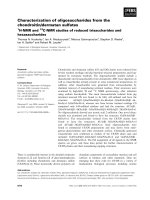
Figure 1: E. helvum in the zoological garden of Kumasi and the habitat range of E.
helvum. This species exist on the African continent only, and migrates over long
distances crossing country borders. The colony, studied in this thesis, resides
temporally in Kumasi (red star), Ghana. Foto: F.Gloza-Rausch. Map modified
according to [24].
4
and migration coincide with blossoming and fruiting of specific tree species [23].
During migration, colonies arrive at roosting areas when fruit abundance is increasing
and continue to migrate when fruit abundance is decreasing, following the seasonal
abundance of local food resources [22, 23]. As a result of deforestation and the
expansion of human settlements, E. helvum are increasingly roosting in urban areas
getting in closer contact with humans [25, 26]. Fruit bats have long lifespans and low
rates of reproduction. Mating occurs seasonally in April to July but gestation does not
begin until October. Females typically give birth in maternity colonies to one pup
(occasionally two) in February to late-March prior to the onset of rainfall season [18,
27-29]. Increased use of urban habitats often creates conflicts with humans. Residents
complain about noise and odour annoyance and depredation of crops. Hence E.
helvum is often hunted, but not only for reasons of nuisance but also as a source of
protein and income, if not used for self-consumption. In fact, E. helvum is one of the
most hunted bushmeat in Sub-Saharan Africa. In Ghana, a minimum of 128,000 E.
helvum bats are sold annually [26]. This is a serious concern, as fruit bats are essential
for seed dispersal, pollination and the genetic connectivity of plants among fragmented
patches of rainforest [22]. The resulting products of timber, fruit, fibres and tannins
contribute significant to world markets and local economies [22].
1.2.1. Viruses in E. helvum
There is increasing evidence that E. helvum harbour a variety of viruses from different
families. The first virus isolate from E. helvum was Lagos bat virus (LBV) from the
genus Lyssavirus [30]. Later, antibodies against LBV were detected in colonies from
Ghana [31, 32], Kenya [33] and Nigeria [34]. Antibodies against other members of the
genus Lyssavirus, Rabies virus (Nigeria) and Mokala virus (Kenya, Ghana), were also
detected [31, 33, 35]. In 2013, two related Rubulaviruses (Achimota 1 and 2) from the
family Paramyxoviridae were isolated from a straw-coloured fruit bat in Ghana. The
viruses are distantly related to the human pathogenic Mumps and Parainfluenza virus
2 and 4. Serum of E. helvum from Ghana and the islands São Tomé, Principe and
Annobón contained neutralizing antibodies against the two novel Rubulaviruses [36].
At least 20 other previously unknown Rubulaviruses circulate in E. helvum colonies
5
across Sub-Saharan Africa [16]. Henipaviruses have not yet been isolated from E.
helvum, but there is evidence of a high diversity of henipa viruses in these animals [16,
37], and serological cross-reaction and neutralization with Nipah virus and Hendra
virus were observed [16, 38, 39]. Apart from an Orbivirus (family Reoviridae), which
was isolated from a Nigerian straw-coloured fruit bat, there have been no other virus
isolate from E. helvum until now [40]. However, metagenomic analysis’s suggest the
presence of viruses from the families Reoviridae, Parvoviridae, Herpesviridae,
Papillomaviridae, Adenoviridae, Poxviridae and Picronaviridae [41-43]. It is therefore likely
that increased research effort will uncover higher diversity of viruses hosted by E.
helvum.
1.2.2. E. helvum colony in Kumasi
This study was conducted in a colony of approximately 300,000 individuals which
roosts temporally in Kumasi, Ghana. Their primary roosting side is the zoological
garden of Kumasi, located in central Kumasi, next to Kejetia market, the largest
market in Western Africa. “Animals were first observed in July 1992. In March 1993
individuals were recorded in a coconut tree and spread within four weeks on more
trees. In the following years, their number increased and roosting areas on prior
neglected trees were occupied. Since 1995, almost all trees in the zoological garden of
Kumasi were used as roosting areas” (pers. comm.). A second known roosting area, is
the Botanical garden on the campus of the Kwame Nkruma University, at the
outskirts of Kumasi. The colony visits Kumasi during its annual migration, typically
arriving in October with increasing numbers until December. Although the colony size
may fluctuate on a daily basis following available food resources, the roosting sites are
occupied until at least April. Parturition occurs in March, but a small population of
animals forms a resident population year-round. The colony has close contact with
humans, being within the zoological garden and in close proximity to Kejetia market,
and also on the university campus. Humans are exposed to urine and faeces of the
bats, particularly workers of the zoological garden who both live and work there.
Additionally, the animals are hunted for consumption and control reasons.
6
1.3. Paramyxoviridae
Paramyxoviruses are enveloped, negative-sense single strand RNA viruses that are
divided into two subfamilies, Paramyxovirinae and Pneumovirinae. The subfamily
Paramyxovirinae, comprises five genera, namely Respirovirus, Rubulavirus, Morbillivirus,
Henipavirus and Avulavirus. Prominent human pathogens within this subfamily, are
Human respiratory syncytial virus (genus Pneumovirus), Measles virus (genus
Morbillivirus) and Mumps virus (genus Rubulavirus). Viruses in the subfamily
Paramyxovirinae, have been associated with a number of emerging diseases in humans
and animals, in the past two decades [61-68]. In 1994, a novel paramyxovirus named
Hendra virus, associated with respiratory disease in horses and humans, caused two
outbreaks in Australia [69, 70]. In the second outbreak, a patient with contact to
horses, that had died of severe respiratory syndrome, died from relapsing encephalitis
[71, 72]. Hendra virus continues to cause re-emerging outbreaks in Australia. Nipah
virus, is another novel Paramyxovirus, which emerged in Malaysia in 1998, causing an
outbreak of febrile encephalitis among pig farmers. The outbreak was linked, later, to
cases of respiratory and neurological disease in domestic pigs [73, 74]. Since then,
Nipah virus has caused several outbreaks in Malaysia, Singapore, India and
Bangladesh causing lethal outcomes in many cases. These two viruses were assigned
to a novel genus, Henipavirus, within the Paramyxovirinae [75]. Until now, they are the
only assigned viruses in this genus. Although, human cases have been linked to
contacts with horses and pigs, Pteropus bats, commonly known as flying-foxes, are
suggested as wildlife reservoir for both viruses [64, 76]. Evidence of Henipavirus
infection, in flying-foxes of different species, was found in China [77], Thailand [78,
79], Cambodia [80], Papua New Guinea [81], Madagascar [82] and Ghana [37, 39]. In
Ghana, antibodies against henipaviruses were detected in domestic pigs [83].
However, none of the afore mentioned countries have reported Henipavirus outbreaks.
The transmission route of henipaviruses is hypothesized to be via urine and saliva.
Outbreaks are associated with Pteropus bats roosting in close proximity to horses and
piggeries. The viruses are transmitted via droppings or contaminated fruits, to horses
and pigs in which they are amplified and further transmitted to humans [84-86]. In
Bangladesh, Pteropus bats feed on date palm sap and transmission of Nipah virus to
7
humans, consuming contaminated date palm sap, occurred [87]. The only cases of
human-to-human transmission were reported from Bangladesh [88, 89]. Until now, no
cases of direct bat to human transmission of henipaviruses are known. Recently,
Cedar virus, a virus related to Hendra- and Nipah virus was isolated from an
Australian fruit bat [90]. Antibodies to Cedar virus cross-react with Hendra and Nipah
virus, but cross-neutralization was not observed. In experiments with ferrets and
guinea pigs, which are susceptible to Hendra and Nipah virus, no clinical signs
developed [90]. In the genus Rubulavirus seven novel, with fruit bat associated viruses,
were detected in the recent years. Menangle virus was originally isolated from stillborn
piglets in Australia [61]. The virus circulated briefly in piggeries before it was
eradicated in 1999 [91]. However, two infected humans developed severe influenzalike illness and rash [92]. Neutralizing antibodies were detected in Australian Pteropus
Figure 3: Global distribution of Henipaviruses. Outbreaks of, Hendra- or Nipahvirus,
were reported from Australia, Malaysia, Singapore, Bangladesh and India (red).
Serological evidence and/or viral RNA of henipaviruses, in flying foxes, were detected
in South-East Asia but also in Africa (brown). The distribution of Pteropus bats is
shaded in yellow. Picture modified according to [93].
8
bats and the virus was isolated from bats in 2012, linking Menangle virus to fruit bats
[91, 94, 95]. In Ghana, two Rubulaviruses, Achimota 1 and 2, were isolated from fruit
bats [36]. Achimota virus 1 and 2 neutralizing antibodies, were detected in several
fruit bat colonies across Sub-Saharan Africa. Although neutralizing antibodies were
detected in humans, no link to a disease was made [36]. Tioman virus, was isolated
from a fruit bat of Tioman island, a small island off the east coast of Malaysia [96],
and neutralizing antibodies in Pteropus were also detected in Madagascar [82, 96].
Tuhoko virus 1-3 from China, related to Menangle- and Tioman virus, have not yet
been isolated but antibodies have been detected in Leschenault's rousette bats [97].
None of the mentioned viruses caused clinical signs of illness is bats. In humans, only
infection with Hendra-, Nipah- or Menangle virus lead to the development of a
disease. In the past, the detection and characterisation of novel viruses on the base of
genetic information, was impossible. However, the development of deep sequencing
and enhanced tools for molecular biology, are expected to lead to a rapidly increase in
the detection of novel viruses.
9
1.4. Rhabdoviridae
The family Rhabdoviridae contains >250 known rhabdoviruses, currently classified in
six acknowledged genera (Lyssaviruses, Vesiculovirus, Ephemerovirus, Novirhabdovirus,
Nucleorhabdovirus and Cytorhabdovirus). According to the International Committee on
Taxonomy of Viruses (ICTV), three more genera are currently pending (Perhabdovirus,
Sigmavirus and Tibrovirus) and >100 rhabdoviruses are still unclassified [44].
Rhabdoviridae are enveloped viruses, with a negative-sense single-stranded RNA and a
typical bullet shape virion. The general genome structure is nucleocapsid (N) phosphoprotein (P) - matrixprotein (M) - glycoprotein (G) - large protein (L), however
a variety of rhabdoviruses contain genes between P - M, M - G and/or G - L. The
complexity of the genome is increased with overlapping reading frames (ORF) within
genes (e.g. P and G) or in novel ORFs, for some species [45]. All plant rhabdoviruses
Figure 2: Comparison of the genome structure of representatives of different
rhabdovirus genera. The reading frames for the conserved rhabdovirus genes N, P, M,
G and L are depicted as open arrows, additional genes are shown in grey. The size of
the genomes and the rhabdovirus genera are indicated. According to [46].
10
(Nucleorhabdovirus and Cytorhabdovirus) typically encode more than the usual five
genes. At least one, and a maximum of four genes, are inserted between the P and M
gene [47, 48]. Fish rhabdoviruses (some Vesiculorhabdoviruses and Novirhabdoviruses)
have an additional gene between G and L. Ephemeroviruses encode additional genes
between G and L [48]. Representatives of different rhabdovirus genera are shown in
(Fig. 2). Universal phylogenetic trees of the Rhabdoviridae, are traditionally generated
by using sequences of the N gene [49]. The degree of conservation decreases in the
order N > L > M > G > P [47]. Each of the five individual genes is flanked by
transcription initiation and termination/polyadenylation signals, which may be
conserved among members of the same genus [47]. Between each transcription unit
(gene and associated flanking signals) is a nontranscribed intergenic region that
usually contains a single or dinucleotide sequence [e.g. G or GG in Tupaia
rhabdovirus (TUPV)] [45]. Termini of rhabdoviruses are highly conserved with an
inverse complementary sequence of 15-20 nt, rich in A/U content, at both ends. These
regions contain the genomic and antigenomic promoters, essential for viral replication
and transcription [50]. In mammalian rhabdoviruses, the terminal nucleotides are
conserved as 5’-ACG/CGT-3’ [48, 50]. Rhabdoviruses have been shown to infect all
organisms, except bacteria (mammals, reptiles, fish, insects, fungi, and plants),
however, they are rarely associated with diseases in humans [51]. The majority have
two natural hosts: either insect and plants or insects and vertebrates, although never all
three [47]. Five of the six rhabdovirus genera contain viruses that are transmitted
and/or hosted by insects. Only fish rhabdoviruses and Lyssaviruses are not maintained
by insect hosts. It is therefore postulated that Rhabdoviridae evolved from an ancestral
insect virus. The supergroup dimarhabdovirus (dipteran-mammal associated
rhabdoviruses) summarise arthropod-transmitted animal rhabdoviruses. It comprises
the genera Ephemero- and Vesiculovirus and a variety of unassigned rhabdoviruses.
Included in this group are the viruses Bovine ephemeral fever virus (BEFV) [52],
Kontonkan virus (KOTV) [53] and Vesiculo Stomatitis virus (VSV) [52-54] which
cause severe disease in cattle. With the exception of Rabies virus, rhabdoviruses are
generally not associated with diseases in humans. However, three viruses from the
dimarhabdo supergroup cause fatal disease in humans. Chandipura virus (CHPV), has
caused outbreaks of encephalitis in India, and has also been detected in Africa [55]. Le
11
Dantec virus [56] and the recently described Bas-Congo virus (BASV) [57], have
caused individual cases of hemorrhagic fever in Africa. Three dimarhabdoviruses have
been isolated from bats: Oita virus (OIRV) [58], Mount Elgon bat virus (MEBV) [59]
which both originate from Kenya, and Kern Canyon (KCV) which was isolated from a
North American bat [59]. These viruses form a monophyletic clade and are probably
geographic variants, which are common for rhabdoviruses. In the genus Ephemerovirus,
the Australian viruses Kimberley- and Adelaide river virus are probably geographic
variants of the African Malakal- and Obodhiang virus [60]. So far, the role of bats in
the evolution and transmission of rhabdoviruses is still unclear.
12
1.5. Aim of the thesis
The focus on bats as reservoirs of potentially emerging diseases has increased in the
last decades. Most studies focus on the detection of viruses without exploring their
genetic diversity to lower taxonomic levels, for example, to genera and species within
bat colonies. Even less is known about the ecology and transmission patterns of these
viruses.
The aim of this thesis is to investigate bat virus diversity and dynamics in a
longitudinal approach. The 300,000 strong colony of E. helvum in highly populated
Kumasi, Ghana, provides a study site where bat-human interaction occurs on a daily
basis. The potential for zoonotic transmission is thus potentially high. Previous studies
have shown a high diversity of Paramyxoviridae genera Henipa- and Rubulavirus in fruit
bats. Therefore, investigation of the virus diversity in the E. helvum colony focused on
these genera.
For the study, an E. helvum organ collection was generated over a time frame of three
years. E. helvum organs were screened for the presence of novel and known
Paramyxoviridae, and virus sequences were compared to their abundance during the
sampling time, their relation to other fruit bat viruses and distribution in different
African countries.
I aimed to isolate viruses from E. helvum and characterise virus abundance in the
colony. Possible transmission pathways were investigated by testing for organ tropism.
For isolated viruses, serological assays were established to define the serological status
of the E. helvum colony and investigate potential cross-species transmission of bat
viruses to livestock and humans.
13
2. Materials and Methods
2.1. Materials
2.1.1. Chemicals
100 bp DNA ladder
2-Mercaptoethanol
(β-Mercaptoethanol)
ACCUGEN, RNAse free water
Acetic acid, 100%, Ph.Eur., reinst
Agarose Broad Range
Agarose GTQ
Ampuwa® (sterile, pyrogen-free water)
Beta propiolacton
Bovine Serum Albumin (BSA)
Bovine Serum Albumin
Bromphenol blue
Carrier RNA (10 mg/mL)
Chloric acid (HCl)
Coomasie PlusTM (Bradford solution)
Crystal Violet
DAPI ProLong Gold antifade reagent
Disodium hydrogen phosphate – dihydrate
(Na2HPO4-7H2O)
dNTP set (dATP, dTTP, dGTP, dCTP)
Ethanol ≥99.9%
Ethidium Bromide (10 mg/mL)
Ethylenediaminetetraacetic acid (EDTA)
EUROIMMUN sample buffer
Formaldehyde 37%
Glycerol
Ketamin 10%
LB-Agar (Lennox)
Magnesium chloride (PCR)
Methanol (99%)
Milk powder
Natriumhydrogencarbonat
Roti® -Histofix 4% (pH7)
Sacharose
Sodium hydroxide (NaOH)
Tris hydroxymethyl aminomethane (Tris)
Triton X-100
Tween 20
Xylene cyanol FF
Xyxlazin (Rompun®)
Life Technologies, Darmstadt, Germany
Carl Roth GmbH + Co. KG, Karlsruhe,
Germany
Lonza Cologne, Cologne, Germany
Carl Roth GmbH + Co. KG, Karlsruhe
Carl Roth GmbH + Co. KG, Karlsruhe
Carl Roth GmbH + Co. KG, Karlsruhe
Fresenius Kabi, Bad Homburg, Germany
Ferak Berlin, Berlin, Germany
New England Biolabs GmbH, Frankfurt,
Germany
Roche Diagnostics, Mannheim, Germany
Sigma-Aldrich Chemie GmbH, Munich,
Germany
QIAGEN, Hilden, Germany
Carl Roth GmbH + Co. KG, Karlsruhe
Thermo Scientific, Bonn, Germany
Carl Roth GmbH + Co. KG, Karlsruhe
Invitrogen, Karlsruhe, Germany
Merck KGaA, Darmstadt, Germany
Invitrogen, Karlsruhe
Carl Roth GmbH + Co. KG, Karlsruhe
Carl Roth GmbH + Co. KG, Karlsruhe
AppliChem, Darmstadt, Germany
EUROIMMUN AG, Lübeck. Germany
Carl Roth GmbH + Co. KG, Karlsruhe
Carl Roth GmbH + Co. KG, Karlsruhe
Medistar, Ascheberg, Germany
Carl Roth GmbH + Co. KG, Karlsruhe
Invitrogen, Karsruhe
Carl Roth GmbH + Co. KG, Karlsruhe
Carl Roth GmbH + Co. KG, Karlsruhe
Carl Roth GmbH + Co. KG, Karlsruhe
Carl Roth GmbH + Co. KG, Karlsruhe
Sigma-Aldrich Chemie GmbH, Munich
Carl Roth GmbH + Co. KG, Karlsruhe
Carl Roth GmbH + Co. KG, Karlsruhe
Sigma-Aldrich Chemie GmbH, Munich,
Sigma-Aldrich Chemie GmbH, Munich
Sigma-Aldrich Chemie GmbH, Munich
Bayer, Leverkusen, Germany
14
2.1.2. Buffers and Solutions
Name
6x Loading Dye
Crystal violet stock solution
Crystal violet working solution
PBS-Tween
Phosphate buffered saline (PBS) 10x, pH7.0
TBE 10x
Ingredients
40g Sacharose
0.25 g Bromphenol blue
0.223 g EDTA
in 100 mL deionized water
10 g Crystal violet
50 mL Formaldehyde (37%)
100 mL Ethanol (99.9%)
350 mL deionized water
100 mL Crystal violet stock solution
100 mL Formaldehyde (37%)
800 mL deionized water
0.1% TweenR 20
10% 10x PBS
in deionized water
80 g NaCl
2 g KCl
26.8 g Na2HPO4-7H2O
2.4 g KH2PO4
adjust pH with 37% HCl
add 1 L deionized water
autoclave
121 g Tris
61.8 g boric acid
186.12 g EDTA
in 1L deionized water
15
2.1.3. Consumables
12-well immunoslides 5mm
C-Chip, Disposable Neubauer improved
counting chamber
Cell culture flask with filter cap (25, 75,
175 cm2)
Cell culture plate (48-well)
Cell culture plates (6-well, 24-well)
Cell scraper
Centrifuge tubes (15, 50 mL)
Cryotubes
LightCyclerR Capillaries (20 XL)
LightCyclerR480 Multiwell Plate 96,
white
Master point Energie Cal 4,5 (.177)
Needles 21G
Nunc Maxi Sorp 96-well plates
PCR reaction tubes (0.2 XL)
Pipette Tips (10, 20, 200, 1000 XL)
Reaction tubes (1.5, 2 mL)
Scalpel (No 15, 11)
Serological pipettes (1, 2, 5, 10, 25 mL)
S-Monovette EDTA K2 (10 mL)
Stericup and Steritop Vacuum Filter Cups
(500 mL)
Syringe (1, 2, 5 mL)
Syringe Filter (0.2 μm)
UlltraClear tubes (15 mL, 50 mL)
Dunn Labortechnik GmbH, Asbach,
Germany
Biochrom AG, Berlin, Germany
SARSTEDT AG & Co., Numbrecht,
Germany
SARSTEDT AG & Co., Numbrecht
SARSTEDT AG & Co., Numbrecht
TPP Techno Plastic Products AG,
Trasadingen, Switzerland
SARSTEDT AG & Co., Numbrecht
SARSTEDT AG & Co., Numbrecht
Roche Diagnostics GmbH, Mannheim,
Germany
Roche Diagnostics GmbH,Mannheim
Industrias el Gamo, Barcelona, Spain
Servopax GmbH, Wesel, Germany
Thermo Fisher Scientific, Schwerte,
Germany
SARSTEDT AG & Co., Numbrecht
SARSTEDT AG & Co., Numbrecht
SARSTEDT AG & Co., Numbrecht
Feather Safety Razor Co., Osaka, Japan
SARSTEDT AG & Co., Numbrecht
SARSTEDT AG & Co., Numbrecht
Millipore GmbH, Schwalbach, Germany
BD, Heidelberg, Germany
Pall Corporation, Ann Aror, USA
Beckman Coulter, Krefeld, Germany
16
2.1.4. Technical Equipment
Equipment
454 sequencer
Type
GS Junior
Air rifle
Diana Panther 21
Autoclave
V120
Balance
SPO 61
Centrifuges
Centrifuge 5424
Source
Roche Diagnostics
GmbH,Mannheim
Mayer & Gummelsbacher
GmbH, Rastatt, Germany
Systec GmbH, Wettenberg,
Germany
Scaltec Instruments GmbH,
Göttingen, Germany
Eppendorf, Hamburg, Germany
Centrifuge 5810R
Eppendorf, Hamburg
Sorvall Evolution RC
Thermo Fisher Scientific,
Schwerte
BioTek, Bad Friedrichshall
Chemiluminescence
reader
SynergyTM 2
Spectramax 190
Dryshipper
Freezer
MVE SC 20/12 V
SC 4/2
XC 20/3 V
-20°C Liebherr premium
-80°C/Typ499
Liquid Nitrogen LS 750
Gel electrophoresis
PerfectBlue Gelsystem
MaxiS 200 mL
Gel electrophoresis
documentation
Heating block
Hood (Bioflow)
E-Box 3028, WL/26M
Incubators
HERAcellR 240
Thermomixer comfort
HeraSafe
HeraeusR B6126
Microscopes
TELAVAL31
PCR cycler
pH meter
IMAGER.M1
Mastercycler epgradient S
766 Calimatic
Molecular Device, Sunnyvale,
USA
German-cryo®GmbH, Jülich
Germany
Liebherr, Biberbach a. d. Ris,
Germany
Kaltis Europe GmbH,
Niederweningen, Switzerland
Taylor Wharton Germany
GmbH,Husum
PEQLAB Biotechnologie
GmbH,
Erlangen, Germany
Vilbert Lourmat, Marne-laVallee, France
Eppendorf, Hamburg
Thermo Fisher Scientific,
Schwerte
Thermo Fisher Scientific, St.
Leon-Roth, Germany
Thermo Fisher Scientific, St.
Leon-Roth
Carl Zeiss GmbH, Jena,
Germany
Carl Zeiss GmbH, Jena
Eppendorf, Hamburg
Knick Elektronische Messgeräte
17
Photometer
Pipette assistance
Pipettes
Power supply
Real-time PCR
cycler
NanoDrop 2000c
Biophotometer
Accu-jetR pro
Research, PhysioCare
(100-1000 μL, 20-200 μL,220 μL, 0.5-10 μL)
EV202
LightCyclerR 1.5
LightCyclerR 480
Rocking Block
Mini Rocker MR.1
Rotor
SW40 Ti, SW41 Ti
Tissue Lyser
Ultrazentrifuge
Vortexer
Qiagen
Optima L-80 XP
Vortex VF2
Water purification
system
Milli-QR Biocel
GmbH & Co. KG, Berlin,
Germany
PEQLAB Biotechnologie
GmbH,
Erlangen
Eppendorf, Hamburg
Brand, Wertheim, Germany
Eppendorf, Hamburg
Consort, Turnhout, Belgium
Roche Diagnostics GmbH,
Mannheim
Roche Diagnostics GmbH,
Mannheim
PEQLAB Biotechnologie
GmbH,
Erlangen
Beckman Coulter, Krefeld,
Germany
Retsch Inc., Newtown, USA
Beckman Coulter, Krefeld
IKAR-Werke GmbH & CO.
KG,
Staufen, Germany
Millipore GmbH, Schwalbach,
Germany
18
2.1.5. Cell culture media and supplements
Amino Acids Non Essential (100x, 50 mL)
Amphotericin B (250μg/mL)
Avicel RC581
CryoMaxx S (50 mL)
Dulbecco's Modified Eagles Medium (high
glucose, 4.5 g/L, 500 mL) (DMEM)
Dulbecco's PBS without Mg/Ca(1x, 500 mL)
Earl MEM (9.69 g/L)
Fetal Calf Serum (FCS) “Standard” (100
mL)
Imipinem/Cilastin (Zienam ®) (500 mg)
L-glutamine (20 mM, 50 mL)
OptiPROTM serum-free medium (1 L)
Penicillin/Streptomycin (100x, 50 mL)
Sodium pyruvat (100 mM, 50mL)
Trypsin EDTA (1x, 50 mL)
PAA Laboratories GmbH, Cölbe
PAA Laboratories GmbH, Cölbe
FCM BioPolymer, Brussels, Belgium
PAA Laboratories GmbH, Cölbe
PAA Laboratories GmbH, Cölbe
PAA Laboratories GmbH, Cölbe
Biochrom AG, Berlin, Germany
PAA Laboratories GmbH, Cölbe
MSD Sharp&Dohme GmbH, Haar,
Germany
PAA Laboratories GmbH, Cölbe
Life Technologies, Darmstadt,
Germany
PAA Laboratories GmbH, Cölbe
PAA Laboratories GmbH, Cölbe
PAA Laboratories GmbH, Cölbe
2.1.6. Cell lines
Name
Vero E6
Vero FM
MA104
A549
EidNi
EidLu
Source
Monkey kidney cell line (ATCC® CRL-1586)
Monkey kidney cell line (kind gift of Jindrich Cinatl, Universtiy of
Frankfurt)
Monkey kidney cell line (cell culture collection Bernhard Nocht-Institute
for Tropical Medicine, Hamburg)
Human lung carcinoma cells (ATCC®CCL-185)
Eidolon helvum kidney cell line (home made)
Eidlon helvum lung cell line (home made)
2.1.7. Antibodies
Donkey-anti-goat Cy2
Donkey-anti-sheep Alexa Fluor488
Goat-anti-bat antibody IgG
Goat-anti-bovine Alexa Fluor488
Goat-anti-human Cy2
Goat-anti-swine Alexa Fluor488
Goat-α-bat-HRP
Goat-α-human-HRP
Dianova, Hamburg, Germany
Dianova, Hamburg
Bethyl Laboratories, Montgomery, USA
Dianova, Hamburg
Dianova, Hamburg
Dianova, Hamburg
Dianova, Hamburg
Bethyl Laboratories, Montgomery
19
2.1.8. Oligonucleotides
Hemi-nested reverse transcription (RT) PCR
Paramyxoviridae
TCI TTC TTT AGA
RES-MOR-HEN-F1
GCC ATA TTT TGT
RES-MOR-HEN-F2
CTC ATT TTG TAI
RES-MOR-HEN-R
GGT TAT CCT CAT
AVU-RUB-F1
ACA CTC TAT GTI
AVU-RUB-F2
GCA ATT GCT TGA
AVU-RUB-R
GTG TAG GTA GIA
PNE-F1
ACT GAT CTI AGY
PNE-F2
GTC CCA CAA ITT
PNE-R
M13mod-F
M13mod-R
Real time RT PCR
ACI
GGA
GTC
TTI
GGI
TTI
TGT
AAR
TTG
TTY
ATA
ATY
TTY
GAI
TCI
TYG
TTY
RCA
GGN
ATH
TTN
GAR
CCN
CCY
CNA
AAY
CCA
CAY
ATH
GCR
TGG
TTY
TGN
TGC
CAR
NCC
CC
AAY
AA
ATH
AAY
AC
ARC
GC
YTC
GG
CA
CC
C
Colony PCR
GTAAAACGACGGCCAGTGAAT
CACACAGGAAACAGCTATGAC
BtRhabdoM17-rt F
BtRhabdoM17-rtP
BtRhabdoM17-rt R
Kumasi rhabdovirus
CTGACTATCGCGACATGCTGTAC
FAMa-ACACGGCGAAAGATCATGCCAAACA-BHQ1b
TCCATTGCTCTCTGGCTCAA
Spl6RMH-F
Spl3+6RMH-P
Spl6RMH-R
Spl3RMH-F
Spl3+6RMH-P
Spl3RMH-R
Spl2RMH-F
Spl2RMH-P
Spl2RMH-R
Spl33nRMH1-F
Spl33nRMH1-P
Spl33nRMH1-R
Spl28nRMH2-F
Spl28nRMH2-P
Spl28nRMH2-R
PVSpl43RMH-F
PVSpl43RMH-P
PVSpl43RMH-R
PV-Spl90-69RMH-F
PV-Spl90-69RMH-P
PV-Spl90-69RMH-R
PV-Spl67-51RMH-F
Henipa-like viruses
CGGGATAGACATGGAGGTGTGT
FAM-CCITCTTGTTTCCTTCCTGATCATGCATC-BHQ1
CCGTTCATCTTTTTGGATTTGAT
CGAGATAGACATGGAGGTGTATG
FAM-CCITCTTGTTTCCTTCCTGATCATGCATC-BHQ1
TTCTGCGCAATCCTCTATTGTCA
TTTACCCTTCCATCAACCTACGTT
FAM-CAACCCTCCTCAATCGTCCACTTCCA-BHQ1
TCTGTGTCCTTTAGATATTCTCCTGATATT
TGGTGTCTGGCCTCCTATGAA
FAM-TTCCCCAGGCATGTTTCAAATACCATCA-BBQc
CATATGTAAGTCTGTCTCCAGATGATTG
AGATAGACACGGAGGGATTTGG
FAM-TGCAAACTTCCAGATCATTGTTCACCTCA-BBQ
TCTCCGTTCATTTTTTTGCTTTT
TTGTGGCACCATAATAAATGGATT
FAM-ACTTGGCCTCCTTGCGAACTTCCTG-BHQ1
CTCTTAACCAGAGCAGAAGCATGA
GTTCAGAGACAGACATGGAGGTATGT
FAM-TGTGACCTCCCTCCACATTCTTCACCTC-BHQ1
TGGATAAGGACTCAGCATTAAGTTGT
TTTGTGGGACAATTATCAATGGAT
20