Chemical modifications of DNA activate the cGAS STING signaling pathway even in the presence of the cytosolic exonuclease TREX1
Bạn đang xem bản rút gọn của tài liệu. Xem và tải ngay bản đầy đủ của tài liệu tại đây (2.86 MB, 114 trang )
Chemical modifications of DNA
activate the cGAS/STING-signaling pathway
even in the presence of the cytosolic
exonuclease TREX1
Dissertation
zur
Erlangung des Doktorgrades (Dr. rer. nat.)
der
Mathematisch-Naturwissenschaftlichen Fakultät
der
Rheinischen Friedrich-Wilhelms-Universität Bonn
vorgelegt von
Christina Mertens
aus
Bergisch-Gladbach
Bonn, März 2015
!
Angefertigt mit der Genehmigung der Mathematisch-Naturwissenschaftlichen Fakultät
der Rheinischen Friedrich-Wilhelms-Universität Bonn.
1. Gutachter: Prof. Dr. Gunther Hartmann
2. Gutachter: Prof. Dr. Michael Hoch
Tag der Promotion: 12.08.2015
Erscheinungsjahr: 2015
!
!
Die vorliegende Arbeit wurde im Zeitraum von Mai 2011 bis März 2015 am Institut für Klinische
Chemie und Klinische Pharmakologie der Rheinischen Friedrich-Wilhelms-Universität Bonn
unter Leitung von Prof. Dr. Gunther Hartmann und Betreuung durch Prof. Dr. Winfried Barchet
angefertigt.
Hiermit erkläre ich an Eides statt,
-
dass ich die Arbeit ohne fremde Hilfe angefertigt und andere Hilfsmittel als die in der
Dissertation angegebenen nicht benutzt habe; insbesondere, dass wörtlich oder
sinngemäß aus Veröffentlichungen entnommene Stellen als solche kenntlich gemacht
worden sind,
-
dass ich mich bis zu diesem Tage noch keiner Doktorprüfung unterzogen habe. Ebenso
hat die von mir vorgelegte Dissertation noch keiner anderen Fakultät oder einem ihrer
Mitglieder vorgelegen,
-
dass ein Dienststraf- oder Ehrengerichtsverfahren gegen mich weder geschwebt hat
noch gegenwärtig schwebt.
Bonn, März 2015
(Christina Mertens)
!
!
Für meine Familie
!
!
Acknowledgement
I would like to express my gratitude to Prof. Dr. Gunther Hartmann and the Institute of Clinical
Chemistry and Clinical Pharmacology for giving me the possibility to complete this work.
Especially, I would like to thank Prof. Dr. Winfried Barchet for his support and supervision
throughout my research project. He was significantly involved in the success of my experiments.
I would like to thank my reviewers for their efforts reading and examining this work. I know that
they are very busy, and thus, I am even more grateful that they could spare a bit of their time for
my thesis and me.
I am also very grateful to the whole Barchet group, notably to Volker Böhnert, Dr. Nadine
Gehrke, Soheila Riemann, Malte Stasch and Dr. Thomas Zillinger, who were always willing
to give me any help I needed.
Moreover, I would like to thank Dr. Tobias Bald from the Institute of Dermatology for his help
with the ear injection experiments.
Additionally, I owe my friends a debt of gratitude for encouraging me to continue on with this
work and never give up. In particular, I am very much obliged to Christian Pipper who was of
great help in difficult times.
My family I would like to thank for support in all phases of life. This work is dedicated to you!
Last but not least, I would like to thank all who looked closely at the final version of this thesis.
Thank you!
!
!
Table of contents
Summary..................................................................................................................................... 1
1. Introduction ............................................................................................................................. 2
1.1. The Immune System ........................................................................................................ 2
1.2. Pattern Recognition Receptors ........................................................................................ 3
1.2.1. Immune Sensing Of Nucleic Acids............................................................................ 3
1.2.1.1. Endosomal Toll-like Receptors .......................................................................... 3
1.2.1.2. Cytosolic RNA Sensing By RIG-l-like Receptors ............................................... 5
1.2.1.3. Cytosolic DNA Sensing ...................................................................................... 7
1.3. Type I Interferon System................................................................................................ 11
1.4. UV Radiation .................................................................................................................. 12
1.4.1. UV- induced DNA Damage ..................................................................................... 13
1.4.2. Repair Of UV-induced DNA Damages .................................................................... 16
1.4.3. UV-induced Apoptosis............................................................................................. 16
1.5. Deoxyribonucleases....................................................................................................... 17
1.6. Lupus Erythematosus .................................................................................................... 17
1.7. Lupus And Neutrophil Extracellular Traps ..................................................................... 19
1.8. The MRL/lpr Mouse Model............................................................................................. 20
1.9. Aim ................................................................................................................................. 22
2. Material And Methods ........................................................................................................... 23
2.1. Materials ........................................................................................................................ 23
2.1.1. Equipment ............................................................................................................... 23
2.1.2. Expendable Materials.............................................................................................. 24
2.1.3. Chemicals ............................................................................................................... 24
2.1.4. ELISA ...................................................................................................................... 25
2.1.5. Transfection Reagents ............................................................................................ 26
2.1.6. Enzymes ................................................................................................................. 26
2.1.7. Western Blot And FACS Antibodies........................................................................ 26
2.1.8. Kits .......................................................................................................................... 26
2.1.9. MACS Beads From Miltenyi Biotec ......................................................................... 26
2.1.10. Oligonucleotides.................................................................................................... 26
2.1.11. Nucleic Acids......................................................................................................... 27
2.1.12. Media, Solutions, Substrates And Buffers............................................................. 27
2.1.13. Primary Cells And Cell Lines................................................................................. 29
2.1.14. Mice....................................................................................................................... 29
2. 2. Methods ........................................................................................................................ 30
2.2.1. Cell Culture ............................................................................................................. 30
2.2.1.1. General Preconditions...................................................................................... 30
2.2.1.2. Subculturing Of Cells ....................................................................................... 30
2.2.1.3. Determination Of The Cell Number.................................................................. 30
2.2.1.4. Freezing And Thawing Of Cells ....................................................................... 30
2.2.2. Isolation And Generation Of Cells........................................................................... 31
2.2.2.1. Preparation Of Murine Bone Marrow DCs ....................................................... 31
2.2.2.2. Isolation Of Murine Spleen Cells...................................................................... 31
2.2.2.3. Isolation Of Human Peripheral Blood Mononuclear Cells ................................ 31
2.2.2.4. Magnetic-activated Cell Sorting ....................................................................... 32
2.2.2.5. Isolation Of Human Neutrophils From Fresh Blood ......................................... 32
2.2.2.6. Isolation Of Murine Neutrophils From Bone Marrow ........................................ 33
2.2.3. Stimulation And Treatment Of Cells........................................................................ 33
2.2.3.1. Transfection Of Nucleic Acids .......................................................................... 33
2.2.3.2. UV Irradiation Of Cells And DNA ..................................................................... 33
2.2.3.3. HOCl/ H2O2-treatment Of Cells And DNA ........................................................ 33
2.2.3.4. Induction Of NETosis ....................................................................................... 34
2.2.3.5. Incubation Of DNA With Human LL37 Peptide ................................................ 34
!
!
2.2.4. Enzyme Linked Immunosorbent Assays ................................................................. 34
2.2.4.1. Murine IFN-! ELISA ......................................................................................... 34
2.2.4.2. Human IFN-! ELISA ........................................................................................ 35
2.2.4.3. 8-OHG EIA ELISA............................................................................................ 35
2.2.5. Molecular Methods.................................................................................................. 36
2.2.5.1. Polymerase Chain Reaction............................................................................. 36
2.2.5.2. Generation Of Biotinylated GFP Via PCR........................................................ 37
2.2.5.3. Incorporation Of 8-OHG Into DNA ................................................................... 37
2.2.5.4. Purification Of PCR Products........................................................................... 37
2.2.5.5. RNA-Isolation From Cells................................................................................. 37
2.2.5.6. cDNA Synthesis ............................................................................................... 38
2.2.5.7. Quantitative Real Time PCR ............................................................................ 38
2.2.5.8. In-vitro Transcription Of 3pRNA ....................................................................... 38
2.2.5.9. Isolation Of Genomic DNA ............................................................................... 39
2.2.5.10. Determining the Concentration of Nucleic Acids............................................ 39
2.2.6. Protein biochemistry................................................................................................ 39
2.2.6.1. Polyacrylamide Gel Electrophoresis ................................................................ 39
2.2.6.2. Bacterial Expression Of TREX1 And cGAS ..................................................... 40
2.2.6.3. Purification Of Proteins .................................................................................... 41
2.2.6.4. cGAS DNA Pulldown Assay............................................................................. 41
2.2.6.5. Western Blot..................................................................................................... 41
2.2.6.6. SybrGreen-based DNase I, II And III Activity Assay ........................................ 42
2.2.6.7. Fluorescence Activated Cell Sorting ................................................................ 42
2.2.6.8. Detection Of Cellular ROS And Superoxide Content In DNA .......................... 43
2.2.7. In Vivo Experiments ................................................................................................ 43
3. Results .................................................................................................................................. 44
3.1. Increased ROS Levels After UV-A/-B/-C Irradiation Correlate With Enhanced Immune
Stimulatory Properties Of DNA ............................................................................................. 44
3.2. UV Irradiation Only Enhances The Immunogenic Potential Of DNA ............................. 45
3.3. DNA Double-strand Breaks Are Not The Reason For The Increased Immunogenicity Of
Cell-free UV Irradiated DNA.................................................................................................. 46
3.4. UV Irradiated DNA Induces A Prolonged Upregulation Of Type I IFN ........................... 47
3.5. DNA Stimulus And UV Damage Signal Can Be Separated Temporally And Spatially .. 48
3.6. Using Inhibitors That Target Different Signal Transducers Or Regulators To Identify
Signal Pathways Involved In The Enhanced Recognition of UV-DNA .................................. 49
3.7. The Cytosolic DNA Receptor cGAS Recognizes Unmodified And UV Irradiated DNA In
Equal Measure ...................................................................................................................... 51
3.8. Oxidative Modifications Protect DNA From TREX1-mediated Degradation .................. 52
3.9. TREX1 Knockout Cells React To All Types Of DNA With High Amounts Of Type I IFN54
3.10. Ear Swelling Reactions Of Different Knockout Mice To UV-DNA ................................ 54
3.11. ROS Also Increase The Immune Response To Pathogenic DNA ............................... 55
3.12. Neutrophil Extracellular Trap - DNA Induces A Stronger Immune Response Than
Genomic Neutrophil DNA...................................................................................................... 56
3.13. High Amounts Of Oxidized DNA Alone Are Sufficient To Trigger A Type I IFN Response
In Human Monocytes ............................................................................................................ 58
3.14. NETing Neutrophils Induce A Type I IFN Response In Co-cultures With Myeloid Cells60
3.15. Effects Of DNA Modifications By Chemotherapeutic Agents ....................................... 60
3.16. Oxidized DNA Can Induce Lupus-like Skin Lesions .................................................... 63
3.17. Oxidized DNA Plays A Role In The Pathogenesis Of Lupus Erythematosus .............. 64
3.18. CD11b+ CD11c- Cells Constitute The Largest Fraction Of IFN-producing Cells
Demonstrating DNA Uptake.................................................................................................. 65
3.19. CD11b+Ly6ClowF4/80+ Cells Contribute To The Type I IFN Response To Oxidized DNA
In Vivo ................................................................................................................................... 68
!
!
4. Discussion............................................................................................................................. 70
4.1. UV Irradiation Causes ROS-dependent DNA Damage That Leads To Enhanced
Immunogenicity ..................................................................................................................... 70
4.2. UV Irradiated DNA Becomes Resistant To TREX1-mediated Degradation ................... 71
4.3. The Physiological Role Of Enhanced Immune Recognition Of Oxidized DNA .............. 74
4.4. Not Only DNA Oxidation Enhances The DNA-induced Immune Response .................. 81
4.5. The Role Of Oxidized DNA In The Pathogenesis Of Lupus Erythematosus ................. 82
4.6. Identification Of IFN Producing Cells In The MRL/lpr Mouse Model.............................. 83
4.7. Final Summary And Outlook .......................................................................................... 85
5. Literature............................................................................................................................... 86
6. Appendix ............................................................................................................................. 102
6.1. Abbreviations ............................................................................................................... 102
6.2. Figures and Tables ...................................................................................................... 105
!
Summary
Summary
To recognize pathogen threats, the innate immune system is equipped with pattern recognition
receptors (PRRs) that bind to and are activated by pathogen-associated molecular patterns
(PAMPs). Most PAMPs are conserved across species of microbes but at the same time not
present in the host, allowing for the efficient discrimination between endogenous and foreign
material. However, viruses rely on the host transcriptional and translational machinery to
produce every viral component, and therefore do not really contain foreign molecules. It has
become apparent that viruses instead are mainly detected via their nucleic acid genomes in the
endosomes or cytosol of the host cell. However, virus sensing based on their nucleic acids
comes at the risk of erroneous recognition of self-DNA - a process that leads to
autoinflammation and possibly autoimmune disease. In particular, the receptor cGAMP synthase
(cGAS) detects the mere presence of any DNA in the cytosol by binding its sugar phosphate
backbone, and thus shows no apparent preference for sequence or specific molecular
structures.
Within this work, evidence is provided that specific damage-associated DNA modifications
strongly enhance cGAS-dependent innate immune activation. DNA modifications occurring after
UV irradiation, incubation with cytostatic agents, ROS exposure or as a consequence of
neutrophil extracellular trap (NET) release were shown to potentiate the interferon (IFN) release
in response to cytosolic DNA. However, this differential immune response was not due to higher
affinity binding of the modified DNA to cGAS itself, but rather due to an impaired degradation by
the cytosolic exonuclease TREX1. Resistance to TREX1 promoted an accumulation of the
modified DNA in the cytosol, leading to a prolonged activation of the cGAS/STING-signaling
pathway and the release of type I IFN.
One well-known autoimmune disease driven by autoantibodies recognizing double-stranded
DNA is lupus erythematosus (LE). Using the lupus-prone mouse model MRL/lpr, UV-damaged
DNA (UV-DNA) was shown to be able to induce lupus-like lesions. Thus, UV-DNA could be a
potential cause for the phototoxicity often observed in LE patients. Moreover, intravenous
administration of UV-DNA induced a type I IFN response in MRL/lpr mice, which could be linked
to F4/80-positive monocytes/macrophages.
Together, these data show that under certain conditions self-DNA is transformed into a damageassociated molecular pattern (DAMP) that provides an additional layer of information to
distinguish danger and damage from healthy states
!
1!
Introduction
1. Introduction
1.1. The Immune System
The immune system (from Latin immunis = free or untouched) is the combination of various
defense systems that evolved to protect higher organisms against pathogens, foreign
substances and abnormal cells. In vertebrates, the immune system can be divided into innate
and adaptive immune system.
The innate immune system is of ancient origin and found in all organisms in some form. Its
features are germline encoded and recognize and respond to general molecular patterns that
are ideally essential to pathogens but foreign to the host. As such, the receptors and effectors of
the innate immune system are immediately available and can provide the first line of defense.
Cells of the innate immune system include natural killer (NK) cells, mast cells, neutrophils,
eosinophils, basophils, monocytes/ macrophages, and dendritic cells (DC). These cells are
responsible for the identification and removal of foreign substances, the recruitment of further
immune cells to the site of infection and finally the activation of the adaptive immune system for
a more specific immune response.
The adaptive immune system is antigen-specific, since it makes use of DNA recombination and
somatic hypermutation to generate a vast diversity of antigen-specific receptors (Brack et al.,
1978; Schatz et al., 1992). Exposure to pathogens bearing a particular antigen leads to the
selective expansion of cells which can recognize them. After initial exposure, it can take several
days until the adaptive immune system becomes protective. However, during this primary
immune response, memory cells are generated that remain inside the body and can initiate a
rapid secondary immune response if the body encounters the same threat again. Cells of the
adaptive immune system include B and T lymphocytes (B and T cells). The main function of B
cells involves the production of specific antibodies that can either neutralize their target directly
or tag the pathogen for attack by other immune cells. CD4-positive T helper (TH) cells assist
other immune cells by secreting cytokines that regulate or support immunologic processes, while
CD8-positive cytotoxic T (Tc) cells destroy host cells that are infected by viruses or have become
malignant.
One important link between innate and adaptive immune system are professional antigen
presenting cells (APCs) such as DCs or macrophages from the innate immune system. They
internalize pathogens and digest them into smaller fragments, which are then presented on
Major Histocompatibility Complex (MHC) class II molecules to TH cells from the adaptive
immune system. The interaction of the T cell receptor (TCR) with the antigen-MHC class II
complex then leads to the activation of the T cell, but only if also an additional co-stimulatory
signal is provided by the APC. To ensure that the adaptive immune system is only activated in
!
2!
Introduction
case of pathogen invasion or danger, APCs only upregulate the co-stimulatory signals if their
pattern recognition receptors (PRRs) have been activated by pathogen associated molecular
patterns (PAMPs).
1.2. Pattern Recognition Receptors
The innate recognition of foreign substances and structures is based on a limited number of
receptors encoded in the germline. The so-called pattern recognition receptors (PRRs)
recognize pathogen-associated molecular patterns (PAMPs) that are frequently found on
pathogens, but ideally absent on host molecules (Janeway, 1989a/b; Gordon, 2002; Janeway
and Medzhitov, 2002). There are different classes of PRRs. However, this work will focus on
signaling PRRs that recognize nucleic acids (NAs) within the cytosol and subsequently trigger
intracellular signaling cascades, which result in the expression of pro-inflammatory cytokines,
chemokines, antimicrobial proteins and antiviral molecules (Takeuchie and Akira, 2010).
1.2.1. Immune Sensing Of Nucleic Acids
The recognition of nucleic acids (NAs) is especially important for the detection of viral infections,
since viruses make us of the cellular host machinery for replication and have not many other
features suitable for identification. Safeguards such as specific NA modifications or the location
of the PRRs ensure that self-NAs do normally not cause an immune response. In the endosome,
toll-like receptors (TLRs) detect RNA and DNA species, while RIG-I-like receptors (RLRs) and
various DNA sensors detect NAs in the cytosol.
1.2.1.1. Endosomal Toll-like Receptors
The membrane bound toll-like receptors (TLRs) are the best-characterized PRR family (Gürtler
and Bowie, 2013). They contain an extracellular leucine rich repeats (LRRs) domain, which is
important for ligand recognition (Martin and Wesche, 2002), and an intracellular Toll/ interleukin1 receptor homology (TIR) domain, necessary for the recruitment of adapter proteins and
intracellular signaling. The TLR-family members TLR3, TLR7, TLR8 and TLR9 are located in the
endosomes of immune cells, where they detect different NA species. The compartmentalization
of these receptors appears to be a safeguard which better allows the distinction between ‘self’
and ‘non-self’ NAs, since endogenous NAs do normally not occur inside the endosomes (Barton
et al., 2006).
TLR3 was originally identified as a receptor that recognizes polyinosine-polycytidylic acid
(poly(I:C)), a synthetic analog of double stranded (ds) RNA. Later, it was shown that TLR3 also
detects naturally occurring dsRNA that is derived from protozoa and fungi (Aksoy et al., 2005;
Carvalho et al., 2012), present in the genome of dsRNA viruses, or generated during replication
or transcription of various single stranded (ss) RNA and DNA viruses (Alexopoulou et al., 2001;
Weber et al., 2006). TLR7 and TLR8 are structurally related and are both sensors for ssRNA
!
3!
Introduction
(Heil et al., 2004; Lund et al., 2004). In humans, TLR7 expression is restricted to plasmacytoid
DCs (pDCs) and B cells (Krug et al., 2001). In contrast, it is widely expressed in murine cells.
TLR8 is found in human monocytes, macrophages and myeloid DCs (mDC), where it seems to
complement the absence of TLR7 (Krug et al., 2001; Hornung et al., 2002). In mice, TLR8 is
found in splenic DC subsets and pDCs, yet its precise function there is not known (Jurk et al.,
2002; Alexopoulou et al., 2012).
TLR9 recognizes unmethylated CpG motifs in dsDNA that occur approximately once per 16
bases in bacteria. In vertebrates these motifs are much less frequent and usually highly
methylated (Bird, 1986). Stacey and colleagues showed that the methylation of CpG motifs
blocked the immune stimulatory properties of bacterial DNA (Stacey et al., 1996). In addition to
its endosomal localization, CpG methylation is a further reason why under normal circumstance
vertebrate DNA does not trigger a TLR9-dependent immune response. In mice, TLR9 is
expressed by B cells, monocytes, macrophages, pDCs and conventional DCs (cDCs), while, in
humans, it is only found in pDCs and B cells (He et al., 2013).
For the activation of TLR3, TLR7, TLR8 and TLR9, acidification of the endosome is required in
order to degrade pathogens and make their nucleic acids accessible. Upon recognition of their
respective foreign NA ligands, TLRs dimerize to build homo- or heterodimers (Kawai and Akira,
2007). For TLR7, TLR8 and TLR9 signal transduction is initiated by the recruitment of the
adapter protein myeloid differentiation factor 88 (MyD88) (Medzhitov et al., 1998; Kawai et al.,
1999), while TLR3 makes use of the TIR domain-containing adapter molecule (TRIF) (Kawai et
al., 2001; Hoshino et al., 2002; Yamamoto et al., 2003).
These adaptor molecules bind to the TIR domain of the TLRs (Figure 1), which in turn results in
the phosphorylation of interferon regulatory factors (IRF) 3 and 7 (Schoenemeyer et al., 2005;
Takaoka et al., 2005; Kawai et al., 2004). Once phosphorylated, IRFs translocate from the
cytoplasm to the nucleus, where they act as transcription factors and cause the induction of type
I IFNs and IFN-dependent genes (Fujita et al., 1989; Harada et al., 1996; Marié et al., 1998;
Sato et al., 2000). TLR signaling also results in an activation of the nuclear factor kappa-lightchain-enhancer of activated B-cells (NF"B)-dependent signaling pathway, which is important for
the induction of pro-inflammatory cytokines such as IL-1, IL-6 and tumor necrosis factor alpha
(TNF-!). Moreover, extracellular signal-regulated kinase (ERK), the mitogen activated protein
kinase (MAPK) p38 and Jun N-terminal kinase (JNK), are activated by the TLR signaling
pathway (Ninomiya-Tsuji et al., 1999; Sakurai, 2012). Together, ERK, p38 and JNK activate the
transcription factor activating protein-1 (AP-1), which then leads to the expression of proinflammatory cytokines.
!
4!
Introduction
()*+,-.)/&0.-1","/&2)-)".+,"/&%343+4+.*&*,55"
!"#$%&&&&&&&&&&&&&&&&&&""#$%&&&&&&&&&&&&&&&&&&&!"'$%
!"#$%%%%%%%%%%%%%%%%!"#&%%%%%!"#'%%%%%%%%%%%%%!"#(
6#78&&&&&&&&&&&9:';;&&&&&&&&9:';;&&&&&&&&&9:';;&
7#8<&)=!&7#8>&&&&&&&&&&&&$8?(&&&&&&&&&&&&&&&&&&&&&&7#8>
6:3,&7&78$&&&&&&&&&&&2-4.=@5)AA)+4-:&&&&&&&&&6:3,&7&7&&
&&&&&&&&&&&&&&&&&&&&&&&&&&&&&&&&*:+4?.=,"
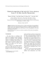
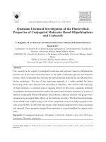
Figure 1: Endosomal Toll-like receptors (adapted from Krieg, 2010)
The endosomal Toll-like receptors TLR3, TLR7/8, and TLR9 detect dsRNA, ssRNA, or unmethylated CpG DNA,
respectively. Activation leads to the recruitment of the adaptor proteins TRIF (TLR3) or MyD88, which signal via IRFs
and NFkB transcription factors leading to the upregulation of type I IFNs and inflammatory cytokines.
1.2.1.2. Cytosolic RNA Sensing By RIG-l-like Receptors
Apart from endosomal sensing by TLRs, pathogenic RNA is also detected in the cytosol by RIGI-like receptors (RLRs). RLRs are ubiquitously expressed and include Retinoic acid inducible
gene-I (RIG-I), Melanoma differentiation- associated gene 5 (MDA5) and Laboratory of genetics
and physiology 2 (LGP2) (Yoneyama et al., 2004; Kang et al., 2002; Andrejeva et al., 2004).
Both, RIG-I and MDA5, contain a C-terminal RNA helicase domain with RNA-dependent ATPase
activity and two N-terminal caspase-recruitment domains (CARDs) required for downstream
signaling (Figure 2). LGP2 does not contain a CARD-domain and is thought to act as a primary
regulator of the RIG-I/ MDA5-inititated signaling pathway (Miyoshi et al., 2001; Yoneyama et al.,
2005). Signaling occurs through CARD interactions with the interferon promoter-stimulating
factor 1 (IPS-1) adaptor protein (also known as mitochondrial antiviral signaling protein (MAVS),
virus-induced signaling adaptor (VISA) or CARD adaptor inducing IFN-# (CARDif)), which
recruits RIG-I and MDA5 to the outer membrane of the mitochondria (Kawai et al., 2005; Meylan
et al., 2005; Seth et al., 2005; Xu et al., 2005). Receptor-adaptor interaction results in the
!
5!
Introduction
activation of IRF3 and IRF7, which then translocate to the nucleus where they induce the
transcription of type I IFN genes. IFNs then upregulate the expression of IRF3, IRF7, RIG-I and
MDA5 in a positive feedback loop. In addition, RLR signaling also leads to the activation of the
transcription factors ATF2, c-Jun (Yoshida et al., 2008) and NF"B, which then cause the
expression of pro-inflammatory cytokines.
RIG-I and MDA5 both detect viral RNA, but they have different ligand specificity. The synthetic
dsRNA poly(I:C) was the first ligand described for RIG-I (Yoneyama et al., 2004). Two years
later, it was shown that the 5$-triphosphate end of RNA (3pRNA) of in vitro transcribed RNA or
generated by viral polymerases is responsible for RIG-I–mediated detection of RNA molecules
(Hornung et al., 2006; Pichlmair et al., 2006). Consistent with this notion, RIG-I recognizes
ssRNA viruses that exhibit prior or during their replication phase a 5´- triphosphate in their RNA
genome, including hepaciviruses and members of the Paramyxoviridae, Rhabdoviridae, and
Orthomyxoviridae virus genera (Kato et al., 2006). In 2009, Schlee and colleagues specified the
ligand for RIG-I as a 5$-triphosphate short blunt end dsRNA structure (3pRNA) as contained in
the panhandle of negative strand viral genomes (Schlee et al., 2009).
The cytoplasmic presence of 3pRNA allows discrimination between self and viral RNA, because
free 5$-triphosphates are normally absent from self-RNA as a result of substantial
posttranscriptional modifications (Hornung et al., 2006; Pichlmair et al., 2006). However, some
dsRNAs lacking a 5´-triphosphate have also been proposed to act as RIG-I agonists (Takahasi
et al., 2008; Kato et al., 2008). Additionally, it was shown that small self-RNA produced by the
antiviral endoribonuclease RNase L could also activate the RIG-I signaling pathway (Malathi et
al., 2007). In contrast to RIG-I antagonists, little is known about MDA5 specific ligands. MDA5 is
associated with the recognition of Picornaviridae (Kato et al., 2006), murine norovirus
(McCartney et al., 2008) and murine hepatitis virus (Roth-Cross et al., 2008). A subset of
viruses, including dengue virus, West Nile virus and reovirus, was shown to be recognized by
both MDA5 and RIG-I (Loo et al., 2008). Comparison of RIG-I and MDA5 interaction with
poly(I:C) suggests that the length of dsRNA is important for differential recognition by RIG-I and
MDA5. Long poly(I:C) fragments are recognized by MDA5, whereas RIG-I shows a preference
for shorter RNA fragments (Kato et al., 2008).
!
6!
Introduction
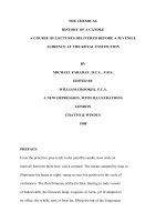
Figure 2: RLR signaling (Bruns and Hovarth, 2012)
Upon ligand binding, RIG-I and MDA5 recruit the adaptor protein IPS-1, which activates TANK-binding kinase 1
(TBK1) and inhibitor of nuclear factor kappa-B kinase (IKK). This results in the activation of NFkB and IRF
transcription factors, which translocate to the nucleus and induce antiviral genes.
1.2.1.3. Cytosolic DNA Sensing
Ten years ago, it was shown that cytosolic DNA could activate the immune system in a TLR9independent fashion (Okabe et al., 2005; Yasuda et al., 2005). Before any cytosolic DNA
receptor was discovered, it became clear that the signaling cascade comprises TBK1 and IRF3
(Ishii et al., 2006; Stetson and Medzhitov, 2006). Later, the endoplasmatic reticulum (ER)resident transmembrane protein STING (stimulator of interferon genes), also known as MITA
(mediator of IRF-3 activation), was identified as crucial adaptor upstream of TBK1 (Ishikawa and
Barber, 2008; Zhong et al., 2008; Sun et al., 2009; Ishikawa et al., 2009). STING functions as a
scaffold protein to specify and promote the phosphorylation of IRF3 by TBK1 (Tanaka and Chen,
2012). Over the years many cytosolic DNA receptors upstream of STING have been suggested
(Figure 3).
DNA-dependent activator of IRFs (DAI) was the first cytosolic DNA receptor candidate to be
discovered (Takaoka et al, 2007; Wang et al., 2008). It was shown to trigger type I IFN
expression in murine L929 fibroblasts upon dsDNA-binding and TBK1-IRF3 interaction.
!
7!
Introduction
Furthermore, the interaction of DAI with Receptor-interacting protein (RIP) 1 and 3 was
described to activate NF"B (Kaiser et al., 2008; Rebsamen et al., 2009). However, DAI was
dispensable for DNA-induced responses in many human cells and DAI-knockout mice
responded normally to DNA (Lippmann et al., 2008; Ishii et al., 2008). Thus, a restricted and
maybe cell-type specific role for DAI in DNA recognition was suggested.
RNA polymerase (Pol) III was the second cytosolic DNA receptor described. Prior to that, Pol III
was only known to transcribe transfer RNAs and other small non-coding RNA molecules. But in
2009, it was shown that Pol III is also able to transcribe AT-rich dsDNA into 3pRNA, which is
then recognized by RIG-I (Ablasser et al., 2009; Chiu et al., 2009). However, transfection of ATpoor dsDNA did not result in the production of type I IFNs, indicating a limited role of Pol III in the
recognition of cytosolic DNA.
In the same year, the PYHIN protein absent in melanoma 2 (AIM2) was identified by four
independent groups as a sensor of cytosolic DNA (Bürckstümmer et al., 2009; FernandesAlnemri et al., 2009; Hornung et al., 2009; Roberts et al., 2009). Upon DNA sensing, AIM2
recruits the adaptor protein ASC (apoptosis-associated speck-like protein containing a CARD)
as well as caspase-1 in order to form the AIM2 inflammasome, which then cleaves pro-IL-1# and
pro-IL-18 into their mature forms. Thus, AIM2 was shown to induce the release of IL-1ß and IL18 in response to DNA, but does not have a role in the induction of type I IFNs.
Another PYHIN protein called IFN-inducible protein 16 (IFI16) was also suggested as DNA
sensor, since it induced a STING-dependent IFN-ß response upon DNA binding (Unterholzner et
al., 2010). In accordance with that, knockdown of human IFI16 or its murine ortholog in mice,
p204, inhibited DNA and DNA-virus induced gene induction in a variety of cell types (Duan et al.,
2011; Conrady et al., 2012; Horan et al., 2013).
In 2011, a central kinase in the DNA damage response (DDR), DNA-dependent protein kinase
(DNA-PK), was described to result in the activation of NF"B and IRFs, and in the production of
IFNs (Brzostek-Racine et al. 2011). Thus, a link between viruses creating DNA breaks during
integration or lytic replication and the induction of an IFN response was made. DNA-PK consists
of a catalytic subunit and its binding partners Ku70 and Ku80. Together, they bind to DNA
breaks, promote cell cycle arrest, and thus, allow DNA damage repair. Zhang and colleagues
demonstrated via knockdown that Ku70 plays a role in the DNA-induced production of IFN-!1 in
HEK293 cells (Zhang et al. 2011a). In another study, Ferguson et al. showed that the DNA-PK
complex is required for the production of IFN-ß, ISGs and IL-6 in response to HSV-1 and
Modified Vaccinia Ankara infection (Ferguson et al., 2012). However, Ku70 had only an effect on
type III but not on type I IFNs, and additionally, the DNA-PK complex was shown to be
dispensable for the IFN response to intracellular DNA in murine bone marrow (bm)-derived
macrophages (Stetson and Medzhitov, 2006).
Mre11 (Meiotic recombination 11), another DNA damage factor has also been suggested as
STING-dependent cytosolic DNA sensor, since murine bmDCs and mouse embryonic fibroblasts
!
8!
Introduction
(MEFs) had defects in dsDNA-induced type I IFN production after Mre11 knockdown (Kondo et
al. 2013). However, Mre11 was dispensable for type I IFN production in response to pathogens
such as HSV-1 and Listeria monocytogenes (Kondo et al. 2013), posing in question the
physiological role of Mre11 during infection.
Members of the DExD/H box helicase protein family have also been implicated in the sensing of
cytosolic DNA. In 2010, Kim and colleagues described DHX9 and DHX36 as MyD88-dependent
DNA sensors in the cytosol of pDCs (Kim et al., 2010). DHX36 induced IRF7 nuclear
translocation and IFN-! production in response to CpG-A oligodeoxynucleotides (ODNs), while
DHX9 led to NF"B activation and TNF-!/ IL-6 production after stimulation with CpG-B ODNs.
Furthermore, pDCs in which DHX9 or DHX36 were knocked down showed a significantly
reduced cytokine response to the DNA virus HSV (Kim et al., 2010). However, DHX9 and
DHX36 were also described to sense RNA and induce MAVS- and TRIF- dependent signaling
(Zhang et al., 2011a/b). Thus, DHX9/ 36 might not be real DNA sensors, but rather act further
downstream in different NA recognition pathways (Paludan and Bowie, 2013).
In 2011, Zhang et al. demonstrated that the helicase DDX41 could bind both DNA and STING.
Furthermore, a reduction of DDX41 expression correlated with a reduced type I IFN response to
DNA and DNA viruses in mDCs and human monocytes (Zhang et al., 2011c). DDX41 has also
been shown to bind the bacterial cyclic dinucleotides (CDNs) cyclic di-AMP and cyclic di-GMP
(Parvatiyar et al. 2012). CDNs act as second messengers in bacteria and stimulate a STINGdependent immune response (Barker et al. 2013; Burdette et al. 2011; Jin et al. 2011; McWhirter
et al. 2009). Before DDX41 was identified, Burdette and colleagues demonstrated that STING
could sense CDNs directly (Burdette et al. 2011). Later, Parvatiyar et al. proposed that DDX41
works upstream of STING. They demonstrated a specific and direct interaction of cyclic di-GMP
and STING with immunoprecipitation and immunoblot analysis. Furthermore, knockdown of
DDX41 expression in mouse and human cell lines as well as in different primary cells resulted in
a reduced induction of IFN-# and TNF in response to CDNs or Listeria bacteria. Thus, they
suggested that DDX41 assists STING in binding to CDNs (Parvatiyar et al. 2012).
The binding of dinucleotides to STING has recently also been suggested as major step in the
sensing of cytosolic DNA. In 2013, the group of Zhijian J. Chen identified cyclic GMP-AMP
(cGAMP) as an endogenous second messenger that is produced in many different cell types
following DNA stimulation (Wu et al. 2013; Sun et al. 2013). It binds to and activates STING,
resulting in the phosphorylation and dimerisation of IRF3. With quantitative mass spectrometry
and classical protein purification strategies the human C6ORF150 and the mouse protein
E330016A19 were identified as enzyme that synthesizes cGAMP, and then renamed cyclic
GMP-AMP synthase (cGAS) (Sun et al. 2013). Overexpression of cGAS induced IFN-ß
expression, whereas knockdown of cGAS inhibited IFN-ß induction by DNA transfection or DNA
virus infection (Sun et al., 2013; Ablasser et al. 2013; Zhang et al. 2013). The binding of cGAS to
DNA and the production of cGAMP in a DNA-dependent manner have further been supported by
!
9!
Introduction
detailed structural analysis of the enzyme in the presence and absence of DNA (Civril et al.
2013; Diner et al. 2013; Gao et al. 2013a/b). It was shown that the binding of DNA to cGAS
results in conformational changes that make the catalytic pocket accessible to its substrate.
Thus, cGAS could be a DNA receptor in its own right.
As already mentioned, there were a lot of cytosolic DNA receptor candidates described over the
last years. Up to now, it remains unclear if “the one“ has already been found and how the
different candidates are linked together. Possible explanations for the high number of candidates
might be functional redundancy, as well as cell type or ligand specificity of certain receptors. It is
also conceivable that different receptors act over time, meaning that some may be more
important in the initial sensing of intracellular DNA, while others take over this function at a later
date. In addition, it might be that some proposed cytosolic DNA sensors are not real receptors,
since detailed molecular mechanisms of the signaling pathways are often missing. The only
proposed cytosolic DNA receptor that provides a clear molecular mechanism for signaling and
STING activation is the enzyme cGAS. Thus, further investigation in this field is absolutely
necessary (Unterholzer, 2013).
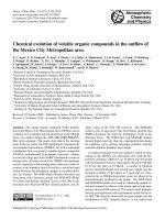
Figure 3: Possible cytosolic DNA receptors (Unterholzner, 2013)
Multiple cytosolic DNA sensors have been proposed to activate a STING-dependent signaling pathway that leads to
the activation of the transcription factors IRF3 and NF"B, and the induction of type I IFN. Of these candidates, only
RNA polymerase III initiates a STING-independent pathway involving the transcription of poly(dA:dT) to dsRNA, which
is then sensed by RIG-I.
!
10!
Introduction
1.3. Type I Interferon System
PRRs can induce the production of various proinflammatory cytokines, such as interleukin-1 (IL1), IL-6, IL-12 and TNF-". However, the focus of this study lies on interferons (IFNs), which
stimulate an “antiviral state” to block viral replication and to interfere with cellular and virus
processes. 60 years ago, IFNs were described as antiviral cytokines released by virus infected
cells and named after their ability to “interfere” with viral replication (Isaacs and Lindenmann,
1957).
Based on their receptor, IFNs are typically divided into three classes. Type I IFNs bind to the
IFN-alpha receptor (IFNAR) that is composed of IFNAR1 and IFNAR2 chains (de Weerd et al.,
2007). Human type I IFNs include IFN-alpha, -beta, -delta, -epsilon, -kappa and -omega
(Roberts et al., 1998; Liu, 2005; LaFleur et al., 2001; Adolf, 1995). Interferon-gamma is the only
type II IFN and binds to IFN-gamma receptor (IFNGR) consisting of IFNGR1 and IFNGR2,
whereas IFN-lambda is a type III IFN and signals through a receptor complex consisting of
IL10R2 and IFNLR1 (Kotenko et al., 2003; Sheppard et al., 2003).
All IFNs fight pathogens and tumor cells, however IFN-" and IFN-ß are the substantial
mediators of anti-viral immunity. The IFN-#!gene is only present as a single copy in humans and
mice (Weissmann and Weber, 1986). Transcriptional activation of IFN-ß requires activating
transcription factor 2 (ATF2)/c-Jun, NF"B, and the interferon regulatory factors (IRF) 3 and 7.
These transcription factors build up together an enhanceosome of regulatory elements
(Goodbourn and Maniatis, 1988; Leblanc et al., 1990; Du and Maniatis, 1992; Carey, 1998) that
recruits co-activators, chromatin- remodeling proteins and the transcriptional machinery to the
promoter region to initiate gene expression.
The IFN-" gene family consists of more than a dozen members. Their promoters include
sequences that are known to bind members of the IRF transcription factor family (Fujita et al.,
1988; Miyamoto et al., 1988). Particularly important for the transcription of IFN-" are IRF3 and 7
(Sato et al., 2001).
By interacting with IFNAR, type I IFNs initiate the Janus kinase (JAK)- signal transducer and
activator of transcription (STAT) signaling pathway (Platanias, 2005). JAK1 and Tyrosine kinase
2 (TYK2) bind to the activated IFNARs and phosphorylate both STAT1 and STAT2. As a result,
an IFN-stimulated gene factor 3 (ISGF3) complex forms that is made of STAT1, STAT2 and
IRF9. The ISGF3-complex translocates to the nucleus where it binds to IFN-stimulated response
elements (ISREs) within the promoters of IFN- stimulated genes (ISGs). The first ISGs were
discovered more than 30 years ago (Larner et al., 1984; Knight and Korant, 1979), and over
time, more than 300 ISGs were discovered by microarray studies. ISGs have antiviral,
antiproliferative, and immunomodulatory properties (de Veer et al., 2001). Protein kinase R
(PKR) is a classical ISG (Feng et al., 1992; Lee and Esteban, 1993). It phosphorylates the alpha
subunit of the eukaryotic translation initiation factor eIF2 (Hovanessian, 1989), which in turn
stops the initiation of protein synthesis and prevents viral replication (Hershey, 1989). Moreover,
!
11!
Introduction
PKR phosphorylates IkB that normally sequesters the transcription factor NFkB in the cytosol
(Zamanian-Daryoush et al., 2000). Upon phosphorylation, IkB releases NFkB, which can then
travel to the nucleus and becomes activated. In turn, NFkB upregulates the expression of IFNs
and with the help of this positive feedback loop the antiviral signal spreads further. Another IFNinduced enzyme is ribonuclease (RNase), which destroys all RNA within the cell. It thereby
reduces protein synthesis, and thus, causes apoptosis of the host cell (Dougherty et al., 1981).
Another function of IFNs is the upregulation of co-stimulatory molecules and the enhanced
expression of MHC class I and MHC class II molecules. In turn, more viral peptides are
presented to cytotoxic T cells, causing the enhanced killing of infected cells. Additionally, more
helper T cells are activated which then coordinate the activity of other immune cells (Zhou,
2009). But IFNs cannot only indirectly affect T cell responses by acting on APCs, they can also
directly promote T cell activation and keep the activated T cells alive (Conrad, 2003).
Additionally, all IFNs are capable of activating NK cells, and increasing their cytotoxicity through
the induction of TNF-related apoptosis-inducing ligand (TRAIL) (Sato et al., 2001).
To limit the extent and duration of type I IFN responses, regulatory molecules are also induced
by IFNs as part of a negative feedback loop. The suppressor of cytokine signaling (SOCS)
proteins 1 and 3 compete with STATs for binding to IFNAR and suppress JAK activity (Fenner et
al, 2006), whereas USP18, a type I IFN-inducible ubiquitin specific peptidase, binds to IFNAR2
and blocks the interaction between JAK1 and IFNAR (Malakhova et al., 2006). Another
mechanism that suppresses type I IFN-mediated responses is the downregulation of cell surface
IFNAR. Internalization of IFNAR is induced by various pro-inflammatory cytokine signaling
pathways such as IL-1, TLRs, immunoreceptor tyrosine-based activation motif (ITAM)associated receptors and oxidative or metabolic stress (Fuchs et al., 2013; Bhattacharya, et al.,
2013; Huynh et al., 2012; Huangfu et al., 2011). TLR stimulation and crosslinking of ITAMassociated receptors can also activate protein tyrosine phosphatases SHP-1, SHP-2 and PTP1B, which dephosphorylate JAK1, STAT1 and TYK2 (You et al., 1999; Myers et al., 2001). In
addition, specific miRNAs were shown to regulate the type I IFN response (Nazarov, et al., 2013;
Liu et al., 2009; Gracias et al., 2013).
1.4. UV Radiation
UV radiation (UVR) can be divided into UV-A (315-400 nm), UV-B (280-315 nm) and UV-C (100280 nm) light. Due to the absorption of short-wave UVR below 310 nm by atmospheric oxygen
and the blockage of mid-range UVR by the ozone layer, only UV-A (95 %) and 5-10 % of UV-B
reach the earth surface. UV-C is usually completely filtered off by the stratospheric ozone layer
(van der Leun, 2004).
Even though UV radiation constitutes only 10 % of the sunlight, it has a high energetic potential
and can ionize molecules and thereby induce chemical reactions (Maverakis et al., 2010). The
depth of penetration into the skin correlates with the wavelength of radiation. Long-wave UV-A
!
12!
Introduction
can penetrate deep into the dermis and affect fibroblasts and matrix metalloproteinases, as well
as DCs, T cells, mast cells and endothelial cells. UV-A is also the main cause of photoaging of
the skin, with irregular pigmentation, enlarged capillary vessels, hornification and reduced
elasticity of the connective tissue (Krutmann, 2003). Moreover, UV-A can induce an immediate
pigment darkening that involves oxidative modification of melanin (Beitner, 1988). Both UV-A
and UV-B are able to induce tanning, although UV-B is more efficient. UV-B reaches only the
epidermis and affects keratinocytes, Langerhans cells and melanocytes (Kindl and Raab, 1998).
However, it possesses much more energy than UV-A, and it is mainly UV-B that causes
sunburn. If keratinocytes are too long exposed to UV-B, they undergo apoptosis as a protective
mechanism against the carcinogenic effect of irreversibly and severely damaged DNA. Those
cells are known as sunburn cells (SBCs) (Kerr et al., 1972). Chronic UVR exposure however
cannot be compensated by SBCs. It is a strong environmental mutagen and can result in fatal
cancer (Brash et al., 1991). However, for the photo-isomerisation of 7-dehydrocholesteral and
ergosterol to previtamin D2 and D3, UV-B is indispensable (Norman, 1998; Okamura et al.,
1993).
UV-C provides the highest energy and has a very strong mutagenic potential. Due to strong
attenuation by the atmosphere, no significant UV-C radiation on earth results from natural
sources. However, studies with UV-C lamps have shown that UV-C can irritate the skin and
induce sunburn (Maverakis et al., 2010).
1.4.1. UV- induced DNA Damage
The skin is the organ that is mainly affected by solar UV radiation. One of the most important
chromophores is the epidermal DNA with its aromatic bases that absorb the radiation energy.
Since DNA has an absorption maximum at 260 nm, UV-C lamps are widely used to study UV
radiation induced DNA damage (Batista et al., 2009).
DNA damage is induced by the absorption of UV photons that generate lesions usually referred
to as photoproducts. The most common photoproducts are cyclobutane pyrimidine dimers
(CPDs) and (6-4) pyrimidine-pyrimidone photoproducts (PP) (Ravanat et al., 2001) (Figure 4).
CPDs are formed from the photo (2 + 2) cycloaddition of the 5,6-double bond of two adjacent
pyrimidine nucleotides (Torizawa et al., 2004). Thus, there are thymidine-thymidine (T-T),
thymidine-cytosine (T-C), cytosine-thymine (C-T) and cytosine-cytosine (C-C) CPDs, with T-T
dimers occurring most frequently (Setlow and Carrier, 1966). (6-4)-PPs arise from the linkage of
the C6 position of the 5´- pyrimidine to the C4 position of the 3´- pyrimidine in an adjacent pair
(Rosenstein and Mitchell, 1987). They can be further transformed to Dewar valence isomers by
photoisomerization (Taylor et al., 1988). Several studies showed that CPDs are at least three
times more often formed than (6-4)-PPS after UV-C irradiation, (Mitchell, 1988; Kao et al., 1993),
but the formation ratio varies and is related to the specific DNA sequence and UV wavelength.
After UV-A or UV-C radiation CPDs are especially formed at T-T sequences (Sage, 1993),
!
13!
Introduction
whereas T-C sequences are more susceptible to (6-4)-PPs and occur after UV-C radiation
(Lippke et al., 1981). Both, CPDs and (6-4)-PPs cause a conformational change of the DNA
double helix that results in the blockage of replication and transcription processes. The
replication arrest leads to the production of DNA double-strand breaks (DSBs) at the sites of
collapsed replication forks (Limoli et al., 2002; Batista et al., 2009). Furthermore, replication
stresses and free radicals may also cause DSBs by preventing the topoisomerase-mediated
DNA religation (Strumberg et al., 2000; Box et al., 2001; Ohnishi et al., 2009; Banáth and Olive,
2003).
DNA cannot only be damaged directly by UV irradiation, but also indirectly through the
generation of ROS. Upon UV exposure, the radiation energy is absorbed by photosensitizers like
porphyrins, bilirubin, melanin, flavins, pterins, vitamins, NAD(P)H, trans-urocanic acid and
tryptophan (Wondrak et al., 2005), which are promoted to an excited singlet state and undergo
intersystem crossing with oxygen to initially produce superoxide (O2-). Superoxide is biologically
toxic and known to denature enzymes, lipids and also DNA. Thus, an enzyme called superoxide
dismutase (SOD) rapidly converts superoxide into hydrogen peroxide (H2O2), which is then
further processed by an enzyme named catalse that catalyzes the decomposition of H2O2 to
water and oxygen. Hydrogen peroxide does not directly cause DNA damages but it can be
transformed into hydroxyl radicals (HO.), which are highly reactive (Halliwell and Aruoma, 1991).
Next to superoxide and hydrogen peroxide, ROS include also hypochlorous acid (HOCl), ozone
(O3) and singlet oxygen (1O2), which can be easily converted into radicals or have an oxidative
effect themselves.
The DNA damages by ROS comprise single and double strand breaks, DNA base modifications
or DNA-protein crosslinking, depending on whether DNA bases or deoxyribose are attacked
(Ward, et al., 1987; Hönigsmann and Dubertret, 1996). Particularly critical are base
modifications, since they result in mutations if not repaired immediately. Among the oxidatively
modified
bases,
8-hydroxy-2’-desoxy-guanosine
(8-OHG),
respectively
8-oxo-2’-desoxy-
guanosine (8-oxoG), are the most abundant and best investigated (Figure 5) (Kasai et al., 1984;
Floyd et al., 1988; Kasai, 1997). 8-OH-G is formed by many agents with different mechanisms
of action (Kohda et al., 1980; Kohda et al., 1990; Kasai et al., 1992; Epe et al., 1996). It base
pairs preferentially with adenine rather than cytosine and thus generates GC-TA transversion
mutations after replication (Grollman and Moriya, 1993; Kasai, 1997; Wood et al., 1990).
!
14!
Introduction
3
2
1
!
4
5
6+
"
@-'A
>?+
3
2
1
1
!
4
4
6+
5
2+
3+
"
5
6+
"
!
3
2
#$%&'()*+*,-./)(0+
3
2
1
!
6+
"
1
4
5
!
4
5
6+
"
>?+
76849+:-;/./$/)(8
:-;/./$<)(+:,<*<:;<$='*0++
3
2
1
!
6+
4
5
"
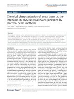
Figure 4: Cyclobutane Thymine Dimers and (6-4)-Pyrimidine-Pyrimidone Photoproducts
Upon UV irradiation two main classes of DNA photoproducts are formed: Cyclobutane pyrimidine dimers (CPDs) and
(6-4) pyrimidine-pyrimidone photoproducts ((6-4)-PPs). CPDs contain a four membered ring arising from the photo (2
+ 2) cycloaddition of the 5,6-double bond of two adjacent pyrimidines. (6-4)-PPs are formed by the linkage of the C6
position of a 5´- pyrimidine, to the C4 position of a 3´- pyrimidine.
!"
"
#"$
%&'()*+,
-./0)120(
3&4+'5)*+&%&'()*+,
-./0)120(,63&"!78
3&)*)&%&'()*+,
-./0)120(
Figure 5: Oxidation of guanosine (adapted from 8-OHG EIA ELISA kit manual)
8-hydroxy-2-deoxy guanosine (8-OHG) or 8-oxo-2-deoxy guanosine are produced by the oxidative damage of DNA by
reactive oxygen species (ROS) and serve as an established marker of oxidative stress.
!
15!
Introduction
1.4.2. Repair Of UV-induced DNA Damages
To prevent malignant transformation, cells have evolved a variety of mechanisms to detect and
repair DNA damages. The tumor suppressor gene p53 controls the integrity of the DNA during
the cell cycle and becomes activated in response to various stressors, one being UV irradiation
(Maltzman and Czyzyk, 1984). P53 can activate DNA repair proteins and arrest growth at the
G1/ S regulation point, so that the repair proteins have enough time to fix the DNA damage
(Kuerbitz et al., 1992). One mechanism to repair UV damage is called photoreactivation or "light
repair“. Photolyase enzymes specifically bind to CPDs or (6–4)-PPs and reverse the damage by
using the energy of light. Antenna molecules like methenyltetrahydrofolate or 8-hydroxy-7,8didemethyl-5-deazariboflavin transfer the absorbed energy to a deprotonated reduced flavin
adenine dinucleotide which then donates an electron to the pyrimidine dimer, resulting in the
splitting of the dimer into two monomeric units (Cook, 1970; Sutherland, 1974; Kim et al., 1992).
Photoproducts are also repaired through processes that do not relate on light and are much
more complex. Nucleotide excision repair (NER) and base excision repair (BER) pathways
handle DNA single strand breaks (Hedge et al., 2008), which can then be filled by different
polymerases and ligases. Double-strand breaks are either repaired through nonhomologous end
joining (NHEJ), which does not require a homologous template and can rejoin broken DNA ends
directly end-to-end, or through homologous recombination repair (HRR), which is dependent on
homology to guide repair (Moore and Haber, 1996, Pastwa and B%asiak, 2003). If DNA damage
cannot be repaired, the cellular DNA damage response (DDR) activates apoptotic pathways.
1.4.3. UV-induced Apoptosis
Overexposure to UV irradiation often results in keratinocytes undergoing apoptosis as a
protective mechanism against the carcinogenic effects of UV light (Daniels et al., 1961).
Apoptosis is also known as programmed cell death and characterized by a sequence of ordered
events leading to the elimination of cells without releasing harmful substances into the
surrounding area (Kerr et al., 1972). This is contrary to necrosis, which is an unordered and
accidental form of cell death caused by external factors and characterized by cellular swelling
and rupture, often leading to inflammation (Wyllie et al., 1980). Mechanisms of UV-induced
apoptosis include (i) UV-induced DNA damages followed by p53 activation and leakage of the
pro-apoptotic factor cytochrome c from mitochondria; (ii) UV-induced death receptor (DR)
activation, resulting in caspase cascades and the translocation of pro-apoptotic proteins of the
Bcl-2 family like Bax (bcl-2- associated x protein) to mitochondria, which causes the release of
cytochrome c; (ii) UV-induced overproduction of ROS, which then damage proteins and DNA,
and additionally increase the release of cytochrome c from impaired mitochondria (Lee et al.,
2013). A failure in the clearance of apoptotic cells can result in the exposure of self-antigens,
including self-DNA, which under certain circumstances might become immunogenic.
!
16!
Introduction
1.5. Deoxyribonucleases
Deoxyribonucleases (DNases) are enzymes that induce the degradation of DNA by catalyzing
the hydrolytic cleavage of phosphodiester linkages in the DNA backbone. DNases are essential
to maintain genome stability and to regulate immune responses by limiting the availability of NAreceptor ligands. They can be divided into endonucleases that cleave residues within the DNA
strand and exonucleases that only cut at the DNA ends.
The three main types found in metazoans are DNase I, DNase II and DNase III (also known as
three prime repair exonuclease 1, short TREX1). DNase I is an endonucleases that yields 5´phosphate-terminated polynucleotides with a free hydroxyl group at the 3´end. Its function is
waste management and the fragmentation of DNA during apoptosis (Samejima and Earnshaw,
2005).
DNase II is also an endonuclease and performs best at an acidic pH (Catchside and Holmes,
1947). Lysosomal localization and ubiquitous tissue distribution alluded to a role in the
degradation of exogenous DNA encountered by phagocytosis (Odaka and Mizuochi, 1999). In
1998, Krieser and Eastman reported that overexpression of DNase II was sufficient to induce
cell death in Chinese hamster ovary cells (Krieser and Eastman, 1998). However, loss-offunction analyses in C. elegans, Drosophila and mice have failed to demonstrate any
requirement for DNase II in the induction or procession of apoptosis (Evans and Aguilera, 2003).
DNase III/ TREX1 is the most prominent DNA 3'–5' exonuclease in mammalian cells and
especially found in the cytosol. Even though this enzyme was first purified in 1969 (Lindahl et al.
1969), its gene was not identified before 1999 (Höss et al., 1999; Mazur and Perrino, 1999).
TREX1 has a preference for ssDNA or mispaired 3' termini and generates 5' mono- or
dinucleotides (Höss et al., 1999; Mazur and Perrin, 1999; Bebenek et al., 2001). Based on
homology with known editing enzymes and its exonuclease function, TREX1 was suggested to
have a role in DNA replication or gap filling during DNA repair (Brucet et al., 2007). However,
TREX1 knockout mice did not confirm a role of TREX1 in DNA editing, since they did not show
higher numbers of spontaneous mutations. Instead, they displayed an autoimmune-like
inflammatory myocarditis and a dramatically reduced lifespan (Morita et al., 2004).
1.6. Lupus Erythematosus
Impaired clearance of apoptotic cells is strongly correlated with the progression of the
autoimmune disease systemic lupus erythematosus (SLE) (Franz et al., 2006). It is thought that
an inadequate clearance of apoptotic cells leads to a prolonged disposition of chromatin-protein
complexes in the extracellular space, which results in the generation of autoantibodies that are
especially directed against nuclear antigens (antinuclear antibodies = ANA) (Lahita, 1992; Tan,
1989). Since the immune system looses the ability to distinguish self from non-self, it starts to
attack the body’s own cells and tissues, resulting in inflammation and tissue damage. The name
lupus erythematosus (latin for wolf and redness) comes from two characteristic symptoms: a
!
17!