Báo cáo y học molecular approaches to the analysis of deformed wing virus replication and pathogenesis in the honey bee, apis mellifera
Bạn đang xem bản rút gọn của tài liệu. Xem và tải ngay bản đầy đủ của tài liệu tại đây (1.1 MB, 9 trang )
Virology Journal
BioMed Central
Open Access
Methodology
Molecular approaches to the analysis of deformed wing virus
replication and pathogenesis in the honey bee, Apis mellifera
Humberto F Boncristiani Jr1, Gennaro Di Prisco2, Jeffery S Pettis1,
Michele Hamilton and Yan Ping Chen*1
Address: 1USDA-ARS Bee Research Laboratory, Beltsville, MD 20705, USA and 2Dipartimento di Entomologia e Zoologia Agraria "Filippo Silvestri"
- Via Università n. °100, 80055 Portici, Napoli, Italy
Email: Humberto F Boncristiani - ; Gennaro Di Prisco - ;
Jeffery S Pettis - ; Michele Hamilton - ; Yan Ping Chen* -
* Corresponding author
Published: 11 December 2009
Virology Journal 2009, 6:221
doi:10.1186/1743-422X-6-221
Received: 24 August 2009
Accepted: 11 December 2009
This article is available from: />© 2009 Boncristiani et al; licensee BioMed Central Ltd.
This is an Open Access article distributed under the terms of the Creative Commons Attribution License ( />which permits unrestricted use, distribution, and reproduction in any medium, provided the original work is properly cited.
Abstract
Background: For years, the understanding of the pathogenetic mechanisms that underlie honey
bee viral diseases has been severely hindered because of the lack of a cell culture system for virus
propagation. As a result, it is very imperative to develop new methods that would permit the in
vitro pathogenesis study of honey bee viruses. The identification of virus replication is an important
step towards the understanding of the pathogenesis process of viruses in their respective hosts. In
the present study, we developed a strand-specific RT-PCR-based method for analysis of Deformed
Wing Virus (DWV) replication in honey bees and in honey bee parasitic mites, Varroa Destructor.
Results: The results shows that the method developed in our study allows reliable identification
of the virus replication and solves the problem of falsely-primed cDNA amplifications that
commonly exists in the current system. Using TaqMan real-time quantitative RT-PCR incorporated
with biotinylated primers and magnetic beads purification step, we characterized the replication
and tissue tropism of DWV infection in honey bees. We provide evidence for DWV replication in
the tissues of wings, head, thorax, legs, hemolymph, and gut of honey bees and also in Varroa mites.
Conclusion: The strategy reported in the present study forms a model system for studying bee
virus replication, pathogenesis and immunity. This study should be a significant contribution to the
goal of achieving a better understanding of virus pathogenesis in honey bees and to the design of
appropriate control measures for bee populations at risk to virus infections.
Background
The viruses pose a serious threat to the health and wellbeing of the honey bee, Apis mellifera, the most economically valuable pollinator of agricultural and horticultural
crops worldwide. In the U.S. alone, the honey bee has an
annual market value exceeding 14.6 billion dollars producing honey and other hive products [1]. So far, honey
bees have been reported to be attacked by at least 18
viruses, most of which are single-strand positive sense
RNA viruses [2,3]. Recently, honey bees have drawn significant attention to the scientific community and beekeeping industry due to the serious disease called Colony
Collapse Disorder (CCD), a malady that has killed billions of bees since 2006 across the U.S. and around the
Page 1 of 9
(page number not for citation purposes)
Virology Journal 2009, 6:221
/>
world [4-6]. A study using a metagenomic approach
found that Israeli Acute Paralysis Virus (IAPV), a species
that was originally identified in honey bees in Israel
showed that IAPV was detected in 25 of 30 (83%) CCDaffected honey bee colonies but in only one of 21 healthy
colonies (Cox-Foster et al., 2007). The observed tight correclation between the IAPV and CCD affected colonies in
the U.S. has raised serious concerns about risks of virus
infections in honey bees. Although significant progress
has been made in honey bee virus research in the last few
decades (Reviewed in Chen and Siede, 2007 [7], investigation into virus replication and pathogenicity has been
severely hindered because of the lack of a cell culture system for virus propagation. Therefore, observations of virus
cytopathic effect (CPE) in cultured cells, a standard
method used for unraveling the mechanisms of viral replication and the specific host responses to viral infections,
are not possible. As a result, it is to develop new methods
that would permit the study of virus replication in vitro.
matic infection). The replication of DWV in honey bee
parasitic mites (Varroa destructor), a potential vector of
DWV, was also investigated.
Recent advances in molecular technology have greatly
expanded our ability to detect and elucidate the molecular
events associated with virus infections and pathogenesis.
With the current molecular technology, complete
genomes of several honey bee viruses have been
sequenced and analyzed [8-14]. Using RT-PCR based
assays, the virus infections in honey bees can be detected
and quantified in a rapid and accurate manner [15,16]. As
with all single-strand positive sense RNA viruses, replication of honey bee viruses proceeds via the production of a
negative-strand intermediate and its presence is indicative
of active viral replication. Therefore, the detection of negative-strand RNA of viruses offers an excellent alternative
for studying virus replication and pathogenesis in naturally infected hosts [17]. Strand-specific RT-PCR was first
developed for detection of negative-strand RNAs of
viruses. However, the method has been reported to cause
falsely-primed amplification due to the self priming of
positive-strand RNA during reverse transcription or random priming by present contaminating cellular nucleic
acids as tRNA, challenging the accuracy previous methods
[18,19]. To overcome such occurrences, more effective
techniques including Tagged RT-PCR, rTth RT-PCR and
chemically blocking the free 3' ends of the RNA, have
been developed to reduce nonspecific priming events [1923]. In order to further improve the assay specificity, it was
developed a new sensitive assay incorporating TaqMan
quantitative RT-PCR with biotinylated primers and magnetic beads purification for detection of negative-strand
viral RNAs. Furthermore, using the method developed, we
analyzed replication and tissue tropism of Deformed
Wing Virus (DWV), a highly prevalent honey bee virus
that causes wing deformity and mortality in honey bees
worldwide, in both bees with wing deformity (symptomatic infection) and bees with normal wings (asympto-
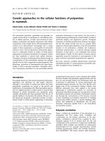
In this study, Tagged RT-PCR was evaluated for its specificity for amplification of both positive and negative-strand
RNA from bees with deformed wings using four combinations of primers. Tagged RT-PCR assay was based on the
generation of cDNAs by the primer containing a tag and
further amplification of cDNA by a tag-primer and a
primer complementary to the synthesized cDNA. The
result showed that cDNAs generated by either tag-forward
or tag-reverse primers were consistently amplified by subsequent PCR amplification, regardless of whether a pair of
primers or only a single primer was used for PCR amplification. As shown in Figure 2, when a tag-forward primer
was used to reverse transcribe the negative-strand of DWV
Results
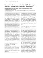
The strand specificity of conventional RT-PCR was evaluated. As shown in Figure 1, both negative and positivestrands of DWV RNA templates were detected from bees
with deformed wings using forward and reverse primers,
respectively, for initial reverse transcription followed by
amplification of the cDNA by PCR. The band intensity of
negative-strand DWV fragments was significantly stronger
than that of positive-strand DWV fragments. However, the
DWV specific fragments were also amplified by RT-PCR
without any primers for reverse transcription. Negative
signals were obtained for negative control reactions without template or reverse transcriptase, confirming that RTPCRs were not contaminated and were from the RNA templates.
RNA 1
Conventional
Figure
RT-PCR for strand-specific detection of DWV
Conventional RT-PCR for strand-specific detection of
DWV RNA. Total RNAs extracted from DWV-infected
bees. Both negative and positive-strands of DWV RNA were
specifically amplified by conventional RT-PCR using DWVspecific forward (line 2) and reverse primer (line 3), respectively, for initial reverse transcription. RT-PCR amplification,
was also conducted without inclusion of any primers for
reverse transcription (line 4). Negative controls containing
no template (line 5) and no reverse transcriptase (line 6)
yielded no products. The 100-bp ladder was loaded into lane
1. The arrow on the right indicates the expected 702 bp RTPCR products.
Page 2 of 9
(page number not for citation purposes)
Virology Journal 2009, 6:221
Figure RT-PCR
Tagged
2
for strand-specific detection of DWV RNA
Tagged RT-PCR for strand-specific detection of
DWV RNA. Total RNAs extracted from bees with
deformed wings. The negative and positive-strand cDNAs
that were generated by tag-forward primers or tag-reverse
primers in reverse transcription, respectively, were consistently amplified by PCR using a pair of tag-primer and reverse
primer (lane 2), a single reverse primer (lane 3), a pair of tagprimer and forward primer (lane 4), or a single forward
primer (lane 5). Water was used as a negative control (lane
6) and a plasmid with DWV fragment was used as a positive
control (lane 7). A 100-bp ladder was loaded into lane 1. The
arrow on the right indicates the expected 702 bp RT-PCR
products.
RNAs, the synthesized cDNA could be amplified by PCR
not only with the primer pair, tag-primer and reverse
primer, but also with a single reverse primer. Meanwhile,
when tag-reverse primer was used to reverse transcribe the
positive-strand of DWV RNAs, the synthesized cDNA
could be amplified by PCR with both primer pairs, tagprimer and forward primer, and with a single forward
primer. No amplification was detected in the negative
control (no template).
/>
In order to achieve highly strand-specific detection of
RNA for DWV, strand-specific RT-PCR was conducted
using a biotinylated primer for cDNA generation and
magnetic separation to purify synthesized cDNA prior to
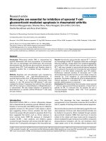
PCR amplification. As shown in Figure 3, without the
magnetic separation step, cDNA generated using biotinylated forward, reverse or lack of primers for reverse transcription were all amplified by subsequent PCR, just like
with conventional RT-PCR. However, the purification of
biotinylated cDNA using streptavidin-coated magnetic
beads excluded the non-specific amplification cDNAs that
were spontaneously formed without the addition of primers for reverse transcription, which occurred in both conventional RT-PCR and Tagged RT-PCR.
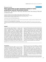
Further, the strand-specific TaqMan real-time qRT-PCR
incorporated with biotinylated primer and magnetic separation was carried out for quantification of DWV replication in host tissues and parasitic mites of honey bees. To
ensure an accurate quantification as well as the highest
sensitivity of the assay, a standard curve was first established by plotting seven 10-fold dilutions of DWV-specific
RNA in vitro transcribed from the pCR2.1 TA cloning vector against corresponding CT value. As shown in Figure 4A
and 4B, a linear progression of the RT-PCR amplification
was observed between the amount of input RNA ranging
from 103 pg to 1 fg and the corresponding CT values
within the concentration range. The detection limit of
positive and negative-strand DWV RNAs were the same.
The lowest limit of detection with qRT-PCR for DWV was
1 fg per reaction for both positive and negative-strand
Figure 3incorporated with biotinylated primer and purification of magnetic beads for strand-specific detection of DWV RNA
RT-PCR
RT-PCR incorporated with biotinylated primer and purification of magnetic beads for strand-specific detection of DWV RNA. Total RNAs extracted from bees with deformed wings. The negative and positive-strand cDNAs that
were generated by biotinylated forward (Lane 2 and 6) and reverse primers (Lane 3 and 7) in reverse transcription, respectively. The cDNA was generated by one step RT-PCR with both biotinylated forward and reverse primers (Lane 4 and 8).
Reverse transcription was conducted without the addition of primer (Lane 5 and 9). The biotinylated cDNAs were either
amplified directly by PCR (Lane 2-5) or subjected to magnetic bead purification before PCR amplification (Lane 6-9). Negative
control without template (Lane 10) and positive control with recombinant DWV plasmid DNA (Lane 11) were included in the
reaction. A 100-bp ladder was loaded into lane 1. The arrow on the right indicates the expected 702 bp RT-PCR products.
Page 3 of 9
(page number not for citation purposes)
Virology Journal 2009, 6:221
/>
Amplification
magnetic4 beads
Figure
plot
purification
and standard curve of TaqMan quantitative RT-PCR (qRT-PCR) incorporated with biotinylated primer and
Amplification plot and standard curve of TaqMan quantitative RT-PCR (qRT-PCR) incorporated with biotinylated primer and magnetic beads purification. In vitro transcribed DWV RNA was serially diluted and subjected to
RT-PCR to assess the sensitivity of the assay. A) Amplification plots were generated by using seven serial dilutions of the RNA,
ranging from 103 pg to 1 fg per reaction as the template for the qRT-PCR assays. The amplification plot shows the fluorescence
(dR) plotted against the cycle number for the standard dilution series of DWV. B) Standard curves were generated by plotting
the observed CT value (Y axis) against the initial quantities of 10-fold serial diluted RNA. CT values are the average of three repetitions.
RNA. The RNA concentration below 1 fg could not be
amplified in the reaction.
The absolute quantification of positive and negativestrand DWV RNA in tissues of bees with symptomatic or
asymptomatic infection and individual Varroa mites was
carried out using a developed strand-specific assay. In bees
with deformed wings, negative-strand DWV RNA was
detected in all tissues examined. The concentration of negative-strand DWV RNA varied significantly among different tissues of the deformed bees (p < 0.001) and
descended down in order of wings, hemolymph, legs, gut,
head, abdomen, and thorax (Figure 5A). Meanwhile,
except for the gut and abdomen, negative-strand DWV
RNA was also detected in tissues of bees with normal
wings. However, the average titer of negative-strand viral
RNA in bees with wing deformity was 14.6 times higher
than that in bees with normal wings in the hemolymph (p
value < 0.0001), 1.8 × 10 4 times higher in the wings (p <
0.0001), 27.8 times higher in the legs (p < 0.0001), 19.8
times higher in the head (p = 0.0015), 107.1 times higher
in the thorax (p = 0.0008) (Figure 5A).
Positive-strand DWV RNA showed predominant presence
and the average positive-strand DWV RNA being 3 × 10³
times more abundant than negative-strand DWV RNA in
the virus-infected bees (p = 0.007). Positive-strand DWV
RNA was detected in all tissues of bees with symptomatic
or asymptomatic infections even though the average concentration of positive-strand viral RNA in tissues of bees
with wing deformity was significantly higher than tissues
of bees with normal wings; 8 × 10² times more in the
hemolymph, 103 times more in the wings, 90 times more
in the legs, 29.8 times more in the head, 1.8 × 10³ times
more in the thorax, 15.2 times more in the gut (p <
0.0001) and 29.8 times more in the abdomen (p <
0.0001) (Figure 5B).
Using the same methodology, negative-strand RNA of
DWV was detected in 81% (17/21) while the positivestrand RNA of DWV was found in 95.2% (20/21) of the
Varroa mites tested. Quantification of positive and negative-strand in the Varroa mites showed no significant difference (p = 0.07) between the titers of the negative-strand
and positive-strand RNAs as seen in the honey bees (p <
0.05).
Discussion
Replication is a key step in successful virus infections. The
replication strategies of positive-strand RNA viruses share
remarkable similarities: all replicate and express their
genomes through negative-strand RNA intermediates that
are used as templates for the production of positive-strand
progeny RNAs packaged in new virion particles [24].
Therefore, the presence of negative-strand RNA intermedi-
Page 4 of 9
(page number not for citation purposes)
Virology Journal 2009, 6:221
/>
Figure
asymptomatic
TaqMan 5Real-Time
bees qRT-PCR for quantification of negative and positive-strand DWV RNA in tissues of symptomatic and
TaqMan Real-Time qRT-PCR for quantification of negative and positive-strand DWV RNA in tissues of symptomatic and asymptomatic bees. The hemolymph, gut, wings, legs, head, thorax and abdomen were individually dissected
out from symptomatic and asymptomatic bees and subjected to TaqMan real-time qRT-PCR coupled with biotinylated primer
and magnetic beads purification. The virus titer of negative-strand (A) and positive-strand (B) DWV of each sample was quantified with the standard curve and expressed as copy numbers (log).
ates should serve as a reliable marker for active virus replication in infected hosts.
In an attempt to develop an in vitro RNA replication assay
for honey bee viruses, we first evaluated the existing methods, including conventional RT-PCR and Tagged RT-PCR
for specificity of strand-specific detection of DWV RNA.
The results showed that DWV RNA could be amplified by
conventional RT-PCR without any primer for reverse transcription. The reason for nonspecific cDNA synthesis by
conventional RT-PCR could be attributed to different
events, including false-priming by antigenomic viral RNA
or cellular RNAs, as well as self primering due to the secondary structure at the 5'UTR of viral RNA during reverse
transcription, as earlier reports suggested [19,25-27].
Tagged RT-PCR was developed to resolve the problem of
PCR amplification of falsely-primed cDNA associated
with conventional RT-PCR [28,29]. However, the finding
that cDNAs generated by tag-forward or tag-reverse primers were amplified by subsequent PCR with a single
reverse primer or forward primer, respectively, suggested
that the residue of tagged-primer from reverse transcription possibly served in subsequent PCR and led to amplification of non-strand-specific products, making
necessary a purification step to assure total elimination of
remaining primer from RT products or primer concentration reduction in RT reaction [28]. The evidence that conventional RT-PCR and Tagged RT-PCR without
purification, failed to discriminate between the two
strands of viral RNA in this study, suggests that caution
should be taken with regards to the strand specificity of
such methods.
In order to circumvent the problems of false priming and
contamination of residue primers from RT, it is worthwhile to develop improved procedures for analysis of
virus replication. We report here the development of a
novel strand-specific RT-PCR coupled with a biotinylated
primer for reverse transcription and purification of biotinylated cDNA with magnetic beads. Using biotinylated
oligonucleotide primers during the reverse transcription
can lead to subsequent synthesis of biotinylated cDNAs,
which have a high binding affinity to the streptavidincoated magnetic beads. The purification step of streptavidin magnetic beads-cDNA complex, ensures the capture
of only biotinylated cDNAs. The disappearance of a positive signal for non-strand-specific amplification of magnetic-bead purified biotinylated cDNA in our study
suggests that the assay developed is a significant modification of conventional or Tagged RT-PCR. The purification
Page 5 of 9
(page number not for citation purposes)
Virology Journal 2009, 6:221
/>
of biotinylated cDNA with magnetic beads is a key step in
assuring the strand-specific detection and elimination of
the residues of RNA, non-incorporated RT primers as well
as enzymes and salts that would interfere with subsequent
PCR amplification.
detected in Varroa mites (data not shown) using the magnetic beads methodology. This finding is in agreement
with preliminary works [22,23] and supports the conclusion that the Varroa mite may be a biological vector of
DWV.
While quantitative detection of DWV infection and tissue
tropism of DWV in the host were previously reported
[30,31], there is lack of information on quantification of
virus replicative intermediates and differentiation of positive and negative-strand DWV RNA in the different tissues
of infected bees, which could provide important insight
into the complexity of virus replication strategies leading
to disease pathogenesis. To gain a better understanding of
active sites of DWV replication in infected bees, we
applied the method developed in our study to localize
and quantify the positive and negative-strand DWV RNA
in different host tissues. Our results showed that replication of DWV was spread throughout the body of bees with
symptomatic infection. The detection of replicative intermediates, negative-strand DWV RNA, in the hemolymph,
gut, wings, legs, head, thorax, and abdomen indicated
that active replication occurred in these sites.
In sum, strand-specific detection and quantification of
DWV RNA were achieved using qRT-PCR incorporated
with biotinylated primers and purification of biotinylated
cDNA with magnetic beads. The elucidation of DWV replication profiles in honey bees would have broad implications for future development of therapeutic strategies for
viral diseases. The assay developed in this study represents
a useful tool to study not only replication of honey bee
viruses but also other single-stranded RNA viruses.
The observation that different tissues showed distinct
kinetics of DWV replication, together with the fact that
differences in the titers of positive-strand DWV RNAs were
not significant among tissues examined, suggested that
DWV had a tropism to certain host tissues in the replication. Among seven tissues examined, the most abundant
amount of negative-strand DWV RNA was detected in the
wings, which suggests that the wings are likely the predominant tissue site of DWV replication. Our earlier studies [15,31] demonstrated that colony foods could act as a
vehicle for transmission of DWV and suggested that the
lining of the digestive tract was likely the primary site of
the DWV infection via food-borne transmission, with the
virus spreading to secondary sites from the digestive tract.
The presence of positive-strands, likely from food, and the
absence of replication (negative-strands) of DWV RNA
only in the gut and abdomen of asymptomatic bees, indicated that these sites are critical for the DWV pathogenesis
course, signifying the necessity of an unknown event at
these sites, such as a co-infection or a differential genetic
background, to generate the permissive environment for a
massive replication observed in symptomatic bees, likely
responsible for the symptoms observed.
Compared to the titer of positive-strand DWV RNA, the
relatively low amount of negative-strand DWV RNA
implies that a regulatory mechanism may exist to facilitate
the viral replication. These findings would stimulate further investigation to unveil the regulatory mechanisms
that honey bees use to control the pattern of replication.
Both positive and negative-strand RNAs of DWV were also
Conclusions
We conclude that qRT-PCR incorporated with biotinylated primers and purification of biotinylated cDNAs
with magnetic beads is a strong approach to specific detection and quantification of a virus genome and antigenome in vivo using the honey bee as a model. This permitted the specific identification of an important honey
bee virus (DWV) replication sites in the honey bee body
and it's quantification. The screening between symptomatic and asymptomatic bees using this technology had
permitted the identification of the digestive tract and
abdomen as a critical replication site in the course of
symptomatic infection.
Methods
Sample Preparation
Individual adult worker bees, with and without deformed
wings, and individual Varroa mites, were collected from
honey bee colonies that were left untreated for Varroa
mites and maintained in the USDA-ARS Bee Research Laboratory backyard apiary, Beltsville, MD. Bees intended for
tissue dissection were kept alive inside of containers with
the cap loosened to allow airflow before dissection. Otherwise, bees were immediately stored in -80°C freezer for
subsequent RNA extraction.
Tissue Dissection
Each live bee (N = 18) was fixed on the wax top of a dissecting dish with insect pins. Under a dissecting microscope, hemolymph was collected with a micropipette by
making a small hole at the joint area between the wing
and body with a needle to make it bleed. Following hemolymph collection, the gut was carefully pulled out with
forceps. The remaining body including wings, legs, head,
thorax and abdomen were cut apart with scissors and collected individually. To prevent possible contamination
with hemolymph, all tissues were thoroughly rinsed once
with 1× PBS and twice with nuclease-free water. All the tissue samples were subjected to RNA extraction.
Page 6 of 9
(page number not for citation purposes)
Virology Journal 2009, 6:221
RNA Extraction
Collected tissues from bees and Varroa mites (n = 21)
were individually homogenized in TRIzol Reagent, a solution of guanidine isothiocyanate and phenol, for RNA
extraction (Invitrogen, Carlsbad, CA). After the addition
of chloroform to remove proteins, lipids and DNA, the
upper aqueous phase containing RNA was removed to a
new microcentrifuge tube, precipitated with isopropanol,
and the resulting pellet was dissolved in diethyl-pyrocarbonate (DEPC)-treated water with the addition of 1 μl of
RNaseOUT a ribonuclease inhibitor (Invitrogen,
Carlsbad, CA). The concentration of total RNA was determined by measuring the absorption at 260 nm and the
purity of RNA was estimated by the absorbance ratio of
260 nm/280 nm using a spectrophotometer with a 50 μl
ultramicrovolume cell holder (Ultrospec 3300 pro, Amersham Biosciences). RNA samples were stored at -80°C
prior to molecular detection for viruses.
Conventional RT-PCR
The Access RT-PCR system (Promega, Madison, WI) was
used for RT-PCR following the manufacturer's instructions. Primers used in the study were a pair of DWV specific primers as reported before [30]. In order to
demonstrate the existence of falsely primed cDNAs amplification in the current detection system, the reverse transcriptions were conducted in the presence of 1 μM of
forward primer or reverse primer for negative-strand RNA
and positive-strand RNA, respectively. Reverse transcription without the addition of either forward or reverse
primer in the reaction mixture was performed as a control.
The reaction mixture contained: 1 × AMV/Tfl reaction
buffer, 0.2 mM each dNTP, 1 μM primer, 2 mM MgSO4,
0.1 unit AMV reverse transcriptase, 0.1 unit Tfl DNA
polymerase and 500 ng total RNA with a total volume of
25 μl. The reaction was conducted using the PTC-100
DNA Engine (MJ Research, Waltham, MA). The reverse
transcriptions were performed at 48°C for 45 minutes.
After the reverse transcription and inactivation of reverse
transcriptase at 95°C for 5 minutes, the thermal machine
was paused and the remaining primers (the reverse primer
in the reaction with a forward primer, the forward primer
in the reaction with a reverse primer, and both forward
and reverse primers in the reaction without any primers)
were added during the reverse transcription. The cDNAs
were then amplified by PCR in the following thermal
cycling profile: 40 cycles at 95°C for 30 sec, 55°C for 1
min, and 68°C for 2 min; 68°C for 7 min. Negative controls (water and a reaction without reverse transcriptase)
were included in each run of RT-PCR. Amplified products
were analyzed through determination of the size of PCR
products by electrophoresis through a 1% agarose gel containing 0.5 ug/ml ethidium bromide and then visualized
by UV transillumination. To prevent any potential con-
/>
tamination, pre-PCR set up and post-PCR analysis steps
were carried out in separate rooms.
Tagged RT-PCR
Tagged RT-PCRs were performed under the same thermal
cycling conditions as the conventional RT-PCR described
above, except that the primers used in the study were
modified by adding a 15-bp long sequence tag [22]. The
sequence of the tag was neither homologous nor complementary to the sequence of DWV. The reverse transcriptions were conducted with the addition of 1 μM of a tagforward primer or a tag-reverse primer for negative-strand
RNA and positive-strand RNA, respectively. After the
reverse transcription reactions and the inactivation of
reverse transcriptase, the tag and DWV-reverse primers or
only the DWV reverse primer was added to the PCR reaction with cDNA generated by a tag-forward primer. The
tag and DWV-forward primers or only the DWV forward
primer was added to the PCR reaction with cDNA generated by a tag-reverse primer.
Strand Specific RT-PCR
In order to increase strand specificity, the regular primers
or tagged-primers were replaced by biotinylated-primers
for reverse transcription. The biotinylated-primers were
synthesized by Invitrogen. After the reverse transcription
reaction, cDNAs generated by either biotinylated forward
or biotinylated reverse primers were magnetically purified
by the Dynabeads kilobaseBINDER M-280 kit (Invitrogen, Carlsbad, CA) following the manufacturer's recommendations. Magnetic beads coated with a monolayer of
streptavidin were added to the RT reaction mixture containing the biotinylated cDNAs. The reaction mixture was
incubated at room temperature for 3 hours in a roller
incubator to allow immobilization of the biotinylated
cDNA onto the magnetic beads. The Dynabeads/DNAcomplex were washed twice in washing solution and once
in distilled water and resuspended in PBS buffer. After
releasing immobilized Biotinylated cDNAs from magnetic
beads, PCR amplification was conducted for each sample
under the same conditions as described above. The cDNAs
that were generated by biotinylated primers and directly
subjected to PCR amplification without magnetic bead
purification were used as a control.
Strand-Specific real-time TaqMan quantitative RT-PCR
(qRT-PCR)
TaqMan real-time quantitative RT-PCR (qRT-PCR) incorporated with biotinylated-primers and magnetic bead
purification was performed for quantification of negative
and positive-strands of DWV using the Stratagene
Mx3000P spectrofluorometric thermal cycler operated by
MxPro qPCR software. The virus levels were quantified
based on the value of the cycle threshold (Ct), which represents the number of cycles needed to generate a fluores-
Page 7 of 9
(page number not for citation purposes)
Virology Journal 2009, 6:221
cent signal above a predefined threshold and is inversely
proportional to the concentration of the initial target that
has been amplified. The house keeping gene, β-actin, was
employed as an endogenous control for normalization of
the quantification. The sequence information of primers
and probes for both DWV and β-actin are the same as
described before [30]. The amplification reaction mixture
and conditions were the same as the strand-specific RTPCR, mentioned above, except that a 0.2 μM TaqMan
probe was incorporated in the PCR amplification. The
measurement of the strand-specific virus titer was conducted in bees with deformed wings and with apparently
normal wings.
Standard Curve Establishment
Purified DWV specific fragments were incorporated into a
pCR2.1 TA cloning vector (Invitrogen, Carlsbad, CA) following the manufacturer's protocol. Recombinant plasmid DNA, containing DWV fragment in both directions,
was purified using the Plasmid Mini Prep Kit (BIO-RAD,
Hercules, CA) and used as a template for in vitro transcription using the Megascript T7 kit (Applied Biosystems/
Ambion, Austin, TX) in order to generate positive and
negative DWV-specific ssRNAs and to construct a new
standard curve for sensitive analysis and absolute virus
quantification. Seven 10-fold serial dilutions of positive
or negative (103 pg, 102 pg, 101 pg, 100 pg, 10-1 pg, 10-2 pg,
and 10-3 pg) in vitro transcribed RNA were subjected to
reverse transcription with biotinylated primers followed
by magnetic bead purification and qPCR amplification.
Each sample was run in triplicate for statistical purposes.
The standard curve was established by plotting the initial
quantities of the 10-fold serial diluted RNA against the
corresponding threshold value (CT).
Sequencing
The specificity of the RT-PCR assay was confirmed by
sequencing analysis. RT-PCR bands were excised from a
low-melting-temperature agarose gel (Invitrogen,
Carlsbad, CA) and purified using the Wizard PCR Prep
DNA purification system (Promega, Madison, WI). The
nucleotide sequences of the RT-PCR fragments were determined from both forward and reverse directions to confirm the specificity of the DWV amplification. The
sequence data of each virus fragment were analyzed using
the BLAST server at the National Center for Biotechnology
Information, NIH.
/>
Abbreviations
DWV: Deformed Wing Virus; RT-PCR: Retro transcriptionPolymerase chain reaction; CCD; Colony Collapse Disorder; CPE: Cytopathic effect.
Competing interests
The authors declare that they have no competing interests.
Authors' contributions
HFB conceived the research, performed the experiments,
and wrote the manuscript. YPC developed the conceptual
aspects of the work and wrote and edited the manuscript.
All authors participated in data collection and read and
approved the final manuscript.
Disclaimer
Mention of trade names or commercial products in this
article is solely for the purpose of providing specific information and does not imply recommendation or endorsement by the U.S. Department of Agriculture.
Acknowledgements
We thank Dr. Jay Evans for helpful discussions and USDA-ARS Headquarters Postdoctoral Research Associate Program Grant.
References
1.
2.
3.
4.
5.
6.
7.
8.
9.
10.
11.
Statistical Analysis
The Fisher's Least Significant Difference (LSD) Comparison of Means Test was used to analyze for significant differences of positive and negative-strand DWV titers
among different tissues of the bees. The results are
expressed as mean ± SD. Differences were considered statistically significant if p value < 0.05.
12.
13.
Morse RA, Calderone M: The value of honey bee pollination in
the United States. Bee Culture 2000, 128:1-15.
Allen M, Ball B: The incidence and world distribution of honey
bee viruses. Bee World 1996, 77:141-162.
Ellis JD, Munn PA: The worldwide health status of honey bees.
Bee World 2005, 86:88-101.
Cox-Foster DL, Conlan S, Holmes EC, Palacios G, Evans JD, Moran
NA, Quan PL, Briese T, Hornig M, Geiser DM, et al.: A metagenomic survey of microbes in honey bee colony collapse disorder. Science 2007, 318:283-287.
Vanengelsdorp D, Underwood R, Caron D, Hayes J: An estimate of
managed colony losses in the winter of 2006-2007: A report
commissioned by the apiary inspectors of America. American
Bee Journal 2007, 147:599-603.
Vanengelsdorp D, Evans JD, Saegerman C, Mullin C, Haubruge E,
Nguyen BK, Frazier M, Frazier J, Cox-Foster D, Chen Y, et al.: Colony Collapse Disorder: A Descriptive Study. PLoS ONE 2009,
4:e6481.
Chen YP, Siede R: Honey bee viruses. Adv Virus Res 2007,
70:33-80.
Ghosh RC, Ball BV, Willcocks MM, Carter MJ: The nucleotide
sequence of sacbrood virus of the honey bee: an insect
picorna-like virus. J Gen Virol 1999, 80(Pt 6):1541-1549.
Govan VA, Leat N, Allsopp M, Davison S: Analysis of the complete
genome sequence of acute bee paralysis virus shows that it
belongs to the novel group of insect-infecting RNA viruses.
Virology 2000, 277:457-463.
Leat N, Ball B, Govan V, Davison S: Analysis of the complete
genome sequence of black queen-cell virus, a picorna-like
virus of honey bees. J Gen Virol 2000, 81:2111-2119.
Lanzi G, de Miranda JR, Boniotti MB, Cameron CE, Lavazza A, Capucci
L, Camazine SM, Rossi C: Molecular and biological characterization of deformed wing virus of honeybees (Apis mellifera L.).
J Virol 2006, 80:4998-5009.
Maori E, Lavi S, Mozes-Koch R, Gantman Y, Peretz Y, Edelbaum O,
Tanne E, Sela I: Isolation and characterization of Israeli acute
paralysis virus, a dicistrovirus affecting honeybees in Israel:
evidence for diversity due to intra- and inter-species recombination. J Gen Virol 2007, 88:3428-3438.
de Miranda JR, Drebot M, Tyler S, Shen M, Cameron CE, Stoltz DB,
Camazine SM: Complete nucleotide sequence of Kashmir bee
Page 8 of 9
(page number not for citation purposes)
Virology Journal 2009, 6:221
14.
15.
16.
17.
18.
19.
20.
21.
22.
23.
24.
25.
26.
27.
28.
29.
30.
31.
virus and comparison with acute bee paralysis virus. J Gen
Virol 2004, 85:2263-2270.
Olivier V, Blanchard P, Chaouch S, Lallemand P, Schurr F, Celle O,
Dubois E, Tordo N, Thiery R, Houlgatte R, Ribiere M: Molecular
characterisation and phylogenetic analysis of Chronic bee
paralysis virus, a honey bee virus. Virus Res 2008, 132:59-68.
Chen Y, Evans J, Feldlaufer M: Horizontal and vertical transmission of viruses in the honey bee, Apis mellifera. J Invertebr
Pathol 2006, 92:152-159.
Chen Y, Pettis JS, Feldlaufer MF: Detection of multiple viruses in
queens of the honey bee Apis mellifera L. J Invertebr Pathol
2005, 90:118-121.
Lanford RE, Sureau C, Jacob JR, White R, Fuerst TR: Demonstration of in vitro infection of chimpanzee hepatocytes with
hepatitis C virus using strand-specific RT/PCR. Virology 1994,
202:606-614.
Haddad F, Qin AX, Giger JM, Guo H, Baldwin KM: Potential pitfalls
in the accuracy of analysis of natural sense-antisense RNA
pairs by reverse transcription-PCR. BMC Biotechnol 2007, 7:21.
Boncristiani HF, Rossi RD, Criado MF, Furtado FM, Arruda E: Magnetic purification of biotinylated cDNA removes false-priming and ensures strandspecificity of RT-PCR for enteroviral
RNAs. Journal of Virological Methods 2009, 161:147-153.
Lanford RE, Sureau C, Jacob JR, White R, Fuerst TR: Demonstration of in-Vitro Infection of Chimpanzee Hepatocytes with
Hepatitis-C Virus Using Strand-Specific Rt/Pcr. Virology 1994,
202:606-614.
Gunji T, Kato N, Hijikata M, Hayashi K, Saitoh S, Shimotohno K: Specific detection of positive and negative-stranded hepatitis C
viral RNA using chemical RNA modification. Arch Virol 1994,
134:293-302.
Yue C, Genersch E: RT-PCR analysis of Deformed wing virus in
honeybees (Apis mellifera) and mites (Varroa destructor). J
Gen Virol 2005, 86:3419-3424.
Gisder S, Aumeier P, Genersch E: Deformed wing virus: replication and viral load in mites (Varroa destructor). J Gen Virol
2009, 90:463-467.
Racaniello V: Picornaviridae: the viruses and their replication.
Fields Virology. Philadelphia 4th edition. 2001, 01:685-722.
McGuinness PH, Bishop GA, McCaughan GW, Trowbridge R, Gowans EJ: False detection of negative-strand hepatitis C virus
RNA. Lancet 1994, 343:551-552.
Mellor J, Haydon G, Blair C, Livingstone W, Simmonds P: Low level
or absent in vivo replication of hepatitis C virus and hepatitis
G virus/GB virus C in peripheral blood mononuclear cells. J
Gen Virol 1998, 79(Pt 4):705-714.
Peyrefitte CN, Pastorino B, Bessaud M, Tolou HJ, Couissinier-Paris P:
Evidence for in vitro falsely-primed cDNAs that prevent specific detection of virus negative-strand RNAs in dengueinfected cells: improvement by Tagged RT-PCR. J Virol Methods 2003, 113:19-28.
Bessaud M, Autret A, Jegouic S, Balanant J, Joffret ML, Delpeyroux F:
Development of a Taqman RT-PCR assay for the detection
and quantification of negatively stranded RNA of human
enteroviruses: evidence for false-priming and improvement
by Tagged RT-PCR. J Virol Methods 2008, 153:182-189.
Craggs JK, Ball JK, Thomson BJ, Irving WL, Grabowska AM: Development of a strand-specific RT-PCR based assay to detect
the replicative form of hepatitis C virus RNA. J Virol Methods
2001, 94:111-120.
Chen YP, Higgins JA, Feldlaufer MF: Quantitative real-time
reverse transcription-PCR analysis of deformed wing virus
infection in the honeybee (Apis mellifera L.). Appl Environ
Microbiol 2005, 71:436-441.
Chen YP, Pettis JS, Collins A, Feldlaufer MF: Prevalence and transmission of honeybee viruses. Appl Environ Microbiol 2006,
72:606-611.
/>
Publish with Bio Med Central and every
scientist can read your work free of charge
"BioMed Central will be the most significant development for
disseminating the results of biomedical researc h in our lifetime."
Sir Paul Nurse, Cancer Research UK
Your research papers will be:
available free of charge to the entire biomedical community
peer reviewed and published immediately upon acceptance
cited in PubMed and archived on PubMed Central
yours — you keep the copyright
BioMedcentral
Submit your manuscript here:
/>
Page 9 of 9
(page number not for citation purposes)