Acute ischemic stroke imaging and intervention
Bạn đang xem bản rút gọn của tài liệu. Xem và tải ngay bản đầy đủ của tài liệu tại đây (9.08 MB, 273 trang )
ACUTE ISCHEMIC STROKE
R.G. Gonzalez, J.A. Hirsch,
W.J. Koroshetz, M.H. Lev,
P. Schaefer
(Eds.)
Acute
Ischemic
Stroke
Imaging and Intervention
With 107 Figures and 59 Tables
123
Library of Congress
Control Number 2005928382
ISBN-10 3-540-25264-9
Springer Berlin Heidelberg NewYork
ISBN-13 978-3-540-25264-9
Springer Berlin Heidelberg NewYork
R. Gilberto González
Neuroradiology Division
Massachusetts General Hospital
and Harvard Medical School
Boston, Mass., USA
Joshua A. Hirsch
Interventional Neuroradiology
and Endovascular Neurosugery Service
Massachusetts General Hospital
Harvard Medical School
Boston, Mass., USA
W.J. Koroshetz
Acute Stroke Service
Massachusetts General Hospital
Fruit Street, Boston, MA 02114, USA
Michael H. Lev
Neuroradiology Division
Massachusetts General Hospital
Harvard Medical School
Boston, Mass., USA
Pamela W. Schaefer
Neuroradiology
GRB 285, Fruit Street
Massachusetts General Hospital
Boston, MA 02114-2696
This work is subject to copyright. All rights are reserved,
whether the whole or part of the material is concerned, specifically the rights of translation, reprinting, reuse of illustrations,
recitation, broadcasting, reproduction on microfilm or in any
other way, and storage in data banks. Duplication of this publication or parts thereof is permitted only under the provisions
of the German Copyright Law of September 9, 1965, in its current version, and permission for use must always be obtained
from Springer-Verlag. Violations are liable for prosecution
under the German Copyright Law.
Springer is a part of Springer Science+Business Media
springeronline.com
© Springer-Verlag Berlin Heidelberg 2006
Printed in Germany
The use of general descriptive names, registered names, trademarks, etc. in this publication does not imply, even in the
absence of a specific statement, that such names are exempt
from the relevant protective laws and regulations and therefore
free for general use.
Product liability: The publishers cannot guarantee the accuracy of any information about dosage and application contained in this book. In every individual case the user must
check such information by consulting the relevant literature.
Medical Editor: Dr. Ute Heilmann, Heidelberg, Germany
Desk Editor: Wilma McHugh, Heidelberg, Germany
Cover design: Frido Steinen-Broo, Estudio Calamar, Spain
Layout: Bernd Wieland, Heidelberg, Germany
Production: LE-TEX Jelonek, Schmidt & Vöckler GbR,
Leipzig, Germany
Reproduction and typesetting: AM-productions GmbH,
Wiesloch, Germany
21/3151 – 5 4 3 2 1 0
Printed on acid-free paper
V
Preface
Acute ischemic stroke is treatable. Rapidly evolving
imaging technology is revolutionizing the management of the acute stroke patient, and the field of acute
stroke therapy is undergoing positive change. This
book is intended as a guide for a wide variety of clinicians who are involved in the care of acute stroke
patients, and is a compendium on how acute stroke
patients are imaged and managed at the Massachusetts General Hospital (MGH). The approaches delineated in this book derive from the published experiences of many groups, and the crucible of caring for
thousands of acute stroke patients at the MGH. It is
the result of the clinical experiences of the emergency
department physicians, neurologists, neuroradiologists, and interventional neuroradiologists that comprise the acute stroke team.
This book focuses on hyperacute ischemic stroke,
which we define operationally as that early period
after stroke onset when a significant portion of
threatened brain is potentially salvageable. The
time period this encompasses will depend on many
factors; it may only be a few minutes in some individuals or greater than 12 hours in others. In most
people, this hyperacute period will encompass
less than 6 hours when intervention is usually most
effective.
The authors believe that patients with acute ischemic stroke can benefit most from the earliest possible definitive diagnosis and rapid, appropriate
treatment. In the setting of hyperacute stroke, imaging plays a vital role in the assessment of patients.
The most recent advances in imaging can identify the
precise location of the occluded vessel, estimate the
age of the infarcted core, and estimate the area at risk
or the ‘ischemic penumbra’. This book will cover
these modern imaging modalities; advanced computed tomography and magnetic resonance methods
are considered in detail. These two modalities are
emphasized because of their widespread availability
and the rapid development of their capacities in the
diagnosis of stroke. Only brief mention is made of
other modalities because they are less widely available and less commonly used in the evaluation of hyperacute stroke patients.
Another major aspect of this book is the use of
standard and developing interventions that aim to
limit the size of a cerebral infarct and prevent its
growth. With the approval of intravenous therapy
using recombinant tissue plasminogen activator
(rt-PA), this treatment is now in use throughout the
United States, Canada, and Europe. Although this is
a major advance in the treatment of acute stroke, the
3-hour ‘window’ for rt-PA makes this therapy suitable
for only a minority of patients. Studies have indicated that intra-arterial thrombolysis is also effective
in patients in a wider window up to 6 hour. More
recently, phase II clinical studies have shown that
intravenous therapy with a new fibrinolytic agent
may be effective up to 9 hours after ischemic stroke
onset in patients selected using imaging criteria.
Thus, this approach is potentially available to many
more individuals. Finally, a wide variety of novel and
innovative new devices are being developed to mechanically recanalize the occluded vessel. It is likely
that these devices will come into clinical use in the
near future. The authors hope that their experiences
as summarized in these pages are of value to the
reader and, ultimately, the acute stroke patient.
R. Gilberto González
VII
Contents
PART I
Fundamentals
of Acute Ischemic Stroke
1
1.1
1.2
Ischemic Stroke:
Basic Pathophysiology
and Neuroprotective Strategies
Aneesh B. Singhal, Eng H. Lo,
Turgay Dalkara, Michael A. Moskowitz
Introduction . . . . . . . . . . . . . . . . . .
Mechanisms of Ischemic Cell Death . . . . .
1.2.1 Excitotoxicity and Ionic Imbalance . .
1.2.2 Oxidative and Nitrosative Stress . . .
1.2.3 Apoptosis . . . . . . . . . . . . . . . .
1.2.4 Inflammation . . . . . . . . . . . . . .
1.2.5 Peri-infarct Depolarizations . . . . . .
1.3 Grey Matter Versus White Matter Ischemia .
1.4 The Neurovascular Unit . . . . . . . . . . . .
1.5 Neuroprotection . . . . . . . . . . . . . . . .
1.6 Stroke Neuroprotective Clinical Trials:
Lessons from Past Failures . . . . . . . . . .
1.7 Identifying the Ischemic Penumbra . . . . .
1.8 Combination Neuroprotective Therapy . . .
1.9 Ischemic Pre-conditioning . . . . . . . . . .
1.10 Nonpharmaceutical Strategies
for Neuroprotection . . . . . . . . . . . . . .
1.10.1 Magnesium . . . . . . . . . . . . . . .
1.10.2 Albumin Infusion . . . . . . . . . . .
1.10.3 Hypothermia . . . . . . . . . . . . . .
1.10.4 Induced Hypertension . . . . . . . .
1.10.5 Hyperoxia . . . . . . . . . . . . . . . .
1.11 Prophylactic and Long-term Neuroprotection
1.12 Conclusion . . . . . . . . . . . . . . . . . . .
References . . . . . . . . . . . . . . . . . . . . . . .
.
.
.
.
.
.
.
.
.
.
. 1
. 1
. 3
. 3
. 4
. 6
. 7
. 8
. 8
. 10
.
.
.
.
.
.
.
.
.
.
.
.
.
.
.
.
.
.
.
.
10
12
13
13
14
14
14
14
15
15
16
. . 16
. . 17
2
Causes of Ischemic Stroke
W.J. Koroshetz, R.G. González
2.1
2.2
2.3
2.4
Introduction . . . . . . . . . . . . . . . . . . .
Key Concept: Core and Penumbra . . . . . . .
Risk Factors . . . . . . . . . . . . . . . . . . . .
Primary Lesions of the Cerebrovascular System
2.4.1 Carotid Stenosis . . . . . . . . . . . . . .
2.4.2 Plaque . . . . . . . . . . . . . . . . . . .
2.4.3 Atherosclerosis Leading to Stroke:
Two Pathways . . . . . . . . . . . . . . .
2.4.4 Collateral Pathways in the Event
of Carotid Stenosis or Occlusion . . . . .
2.4.5 Transient Neurological Deficits . . . . .
2.4.6 Intracranial Atherosclerosis . . . . . . .
2.4.7 Aortic Atherosclerosis . . . . . . . . . .
2.4.8 Risk Factors for Atherosclerosis . . . . .
2.4.9 Extra-cerebral Artery Dissection . . . . .
Primary Cardiac Abnormalities . . . . . . . . .
2.5.1 Atrial Fibrillation . . . . . . . . . . . . .
2.5.2 Myocardial Infarction . . . . . . . . . . .
2.5.3 Valvular Heart Disease . . . . . . . . . .
2.5.4 Patent Foramen Ovale . . . . . . . . . .
2.5.5 Cardiac Masses . . . . . . . . . . . . . .
Embolic Stroke . . . . . . . . . . . . . . . . . .
2.6.1 The Local Vascular Lesion . . . . . . . .
2.6.2 Microvascular Changes in Ischemic Brain
2.6.3 MCA Embolus . . . . . . . . . . . . . . .
2.6.4 Borderzone Versus Embolic Infarctions .
Lacunar Strokes . . . . . . . . . . . . . . . . .
Other Causes of Stroke . . . . . . . . . . . . .
2.8.1 Inflammatory Conditions . . . . . . . .
2.8.2 Venous Sinus Thrombosis . . . . . . . .
2.8.3 Vasospasm in the Setting
of Subarachnoid Hemorrhage . . . . . .
2.8.4 Migraine . . . . . . . . . . . . . . . . . .
2.8.5 Primary Hematologic Abnormalities . .
Conclusion . . . . . . . . . . . . . . . . . . . .
2.5
2.6
2.7
2.8
2.9
. 27
. 28
. 30
31
. 31
. 31
. 31
.
.
.
.
.
.
.
.
.
.
.
.
.
.
.
.
.
.
.
.
31
31
32
32
33
33
33
33
34
34
34
34
35
35
35
36
36
37
39
39
39
.
.
.
.
39
40
40
40
VIII
Contents
4.4
4.5
CTA Protocol for Acute Stroke . .
Accuracy and Clinical Utility of CTA
in Acute Stroke . . . . . . . . . . .
4.5.1 Optimal Image Review . . .
4.5.2 Role of CTA in Acute Stroke
4.6 Future Directions . . . . . . . . . .
4.7 Conclusion . . . . . . . . . . . . .
References . . . . . . . . . . . . . . . . .
PART II
Imaging of Acute Ischemic Stroke
3
Unenhanced Computed Tomography
Erica C.S. Camargo, Guido González,
R. Gilberto González, Michael H. Lev
3.1
3.2
3.3
3.4
Introduction . . . . . . . . . . . . . . . . . .
Technique . . . . . . . . . . . . . . . . . . . .
Physical Basis of Imaging Findings . . . . . .
Optimal Image Review . . . . . . . . . . . . .
3.4.1 Window-Width (W)
and Center-Level (L) CT Review Settings
3.4.2 Density Difference Analysis (DDA) . .
3.5 CT Early Ischemic Changes: Detection
and Prognostic Value . . . . . . . . . . . . .
3.5.1 Early Generation CT Scanners . . . . .
3.5.2 Early CT Findings in Hyperacute Stroke
3.5.3 Prognostic/Clinical Significance of EIC
3.6 ASPECTS . . . . . . . . . . . . . . . . . . . . .
3.6.1 Implications for Acute Stroke Triage .
3.6.2 Reading CT Scans . . . . . . . . . . . .
3.7 Conclusion . . . . . . . . . . . . . . . . . . .
References . . . . . . . . . . . . . . . . . . . . . . .
4
4.1
4.2
4.3
.
.
.
.
.
.
.
.
41
42
45
46
46
. . 48
.
.
.
.
.
.
.
.
.
.
.
.
.
.
.
.
.
.
48
48
48
49
50
51
52
54
54
Stroke CT Angiography (CTA)
Shams Sheikh, R. Gilberto González,
Michael H. Lev
Introduction . . . . . . . . . . . . . . . . . .
Background – General Principles of CTA . . .
4.2.1 Advantages and Disadvantages of CTA
4.2.1.1 Potential Advantages . . . . . . . . . .
4.2.1.2 Potential Disadvantages . . . . . . . . .
4.2.2 CTA Scanning Technique:
Pearls and Pitfalls . . . . . . . . . . . .
4.2.2.1 Single-slice Protocols . . . . . . . . . .
4.2.2.2 Multi-slice Protocols . . . . . . . . . . .
4.2.3 Radiation Dose Considerations . . . .
CTA Protocol for Acute Stroke . . . . . . . .
4.3.1 General Considerations . . . . . . . .
4.3.2 Contrast Considerations . . . . . . . .
4.3.2.1 Contrast Timing Strategies . . . . . . . .
4.3.3 Post-processing: Image Reconstruction
4.3.3.1 Image Review . . . . . . . . . . . . . .
4.3.3.2 Maximum Intensity Projection . . . . . .
4.3.3.3 Multiplanar Volume Reformat . . . . . .
4.3.3.4 Curved Reformat . . . . . . . . . . . . .
4.3.3.5 Shaded Surface Display . . . . . . . . .
4.3.3.6 Volume Rendering . . . . . . . . . . . .
.
.
.
.
.
.
.
.
.
.
57
59
59
59
59
.
.
.
.
.
.
.
.
.
.
.
.
.
.
.
.
.
.
.
.
.
.
.
61
63
63
63
64
64
65
66
70
70
71
72
73
73
73
.
.
.
.
.
.
. . . . . . . . 77
.
.
.
.
.
.
.
.
.
.
.
.
.
.
.
.
.
.
.
.
.
.
.
.
79
79
79
83
83
83
Introduction . . . . . . . . . . . . . . . . .
CTP Technical Considerations . . . . . . . .
Comparison with MR-PWI . . . . . . . . . .
5.3.1 Advantages . . . . . . . . . . . . . .
5.3.2 Disadvantages . . . . . . . . . . . .
5.4 CTP: General Principles . . . . . . . . . . .
5.5 CTP Theory and Modeling . . . . . . . . . .
5.6 CTP Post-Processing . . . . . . . . . . . . .
5.7 Clinical Applications of CTP . . . . . . . . .
5.8 CTP Interpretation: Infarct Detection
with CTA-SI . . . . . . . . . . . . . . . . . .
5.9 CTP Interpretation: Ischemic Penumbra
and Infarct Core . . . . . . . . . . . . . . .
5.10 Imaging Predictors of Clinical Outcome . .
5.11 Experimental Applications of CTP in Stroke
5.12 Conclusion . . . . . . . . . . . . . . . . . .
References . . . . . . . . . . . . . . . . . . . . . .
.
.
.
.
.
.
.
.
.
.
.
.
.
.
.
.
.
.
.
.
.
.
.
.
.
.
.
87
88
91
91
91
92
92
94
96
5
.
.
.
.
.
.
.
.
.
.
.
.
.
.
.
.
.
.
.
.
.
.
.
.
CT Perfusion (CTP)
Sanjay K. Shetty, Michael H. Lev
5.1
5.2
5.3
6
Conventional MRI
and MR Angiography of Stroke
David Vu, R. Gilberto González,
Pamela W. Schaefer
6.1
Conventional MRI and Stroke . . .
6.1.1 Hyperacute Infarct . . . . .
6.1.2 Acute Infarct . . . . . . . .
6.1.3 Subacute Infarct . . . . . .
6.1.4 Chronic Infarcts . . . . . . .
6.1.5 Hemorrhagic Transformation
6.1.6 Conclusion . . . . . . . . .
MR Angiogram and Stroke . . . .
6.2.1 Noncontrast MRA . . . . . .
6.2.1.1 TOF MRA . . . . . . . . . . .
6.2.1.2 Phase-Contrast MRA . . . . .
6.2.2 Contrast-Enhanced MRA . .
6.2.3 Image Processing . . . . . .
6.2.4 Extracranial Atherosclerosis
and Occlusions . . . . . . .
6.2.5 Intracranial Atherosclerosis
and Occlusions . . . . . . .
6.2
.
.
.
.
.
.
.
.
.
.
.
.
.
.
.
.
.
.
.
.
.
.
.
.
.
.
.
.
.
.
.
.
.
.
.
.
.
.
.
.
.
.
.
.
.
.
.
.
.
.
.
.
.
.
.
.
.
.
.
.
.
.
.
.
. . . 96
. . .
. . .
. .
. . .
. . .
101
107
107
108
108
.
.
.
.
.
.
.
.
.
.
.
.
.
115
115
117
117
118
119
120
121
122
122
124
126
126
.
.
.
.
.
.
.
.
.
.
.
.
.
.
.
.
.
.
.
.
.
.
.
.
.
.
. . . . . . . . 127
. . . . . . . . 130
Contents
6.2.6
6.2.7
Dissection . . . . . . .
Other Infarct Etiologies
6.2.7.1 Moya Moya . . . . . . .
6.2.7.2 Vasculitis . . . . . . . .
6.2.7.3 Fibromuscular Dysplasia
6.2.8 Venous Infarct . . . . .
6.2.9 Conclusion . . . . . .
References . . . . . . . . . . . . . .
7
7.1
7.2
.
.
.
.
.
.
.
.
.
.
.
.
.
.
.
.
.
.
.
.
.
.
.
.
.
.
.
.
.
.
.
.
.
.
.
.
.
.
.
.
.
.
.
.
.
.
.
.
.
.
.
.
.
.
.
.
.
.
.
.
.
.
.
.
.
.
.
.
.
.
.
.
.
.
.
.
.
.
.
.
.
.
.
.
.
.
.
.
131
132
132
133
133
134
134
135
Diffusion MR of Acute Stroke
Pamela W. Schaefer, A. Kiruluta,
R. Gilberto González
Introduction . . . . . . . . . . . . . . . . .
Basic Concepts/Physics of Diffusion MRI .
7.2.1 Diffusion Tensor Imaging (DTI) . . .
7.3 Diffusion MR Images for Acute Stroke . . .
7.4 Theory for Decreased Diffusion
in Acute Stroke . . . . . . . . . . . . . . . .
7.5 Time Course of Diffusion Lesion Evolution
in Acute Stroke . . . . . . . . . . . . . . . .
7.6 Reliability . . . . . . . . . . . . . . . . . . .
7.7 Reversibility of DWI Stroke Lesions . . . .
7.8 Prediction of Hemorrhagic Transformation
7.9 Diffusion Tensor Imaging . . . . . . . . . .
7.10 Correlation with Clinical Outcome . . . . .
7.11 Stroke Mimics . . . . . . . . . . . . . . . . .
7.12 Nonischemic Lesions
with No Acute Abnormality on Routine
or Diffusion-Weighted Images . . . . . . .
7.13 Syndromes with Reversible Clinical Deficits
that may have Decreased Diffusion . . . .
7.13.1 Transient Ischemic Attack . . . . . .
7.13.2 Transient Global Amnesia . . . . . .
7.14 Vasogenic Edema Syndromes . . . . . . .
7.14.1 Posterior Reversible Encephalopathy
Syndrome (PRES) . . . . . . . . . . .
7.14.2 Hyperperfusion Syndrome
Following Carotid Endarterectomy .
7.14.3 Other Syndromes . . . . . . . . . . .
7.15 Other Entities with Decreased Diffusion .
7.16 Venous Infarction . . . . . . . . . . . . . .
7.17 Conclusion . . . . . . . . . . . . . . . . . .
References . . . . . . . . . . . . . . . . . . . . . .
.
.
.
.
.
.
.
.
.
.
.
.
139
139
142
144
. . . 144
.
.
.
.
.
.
.
.
.
.
.
.
.
.
.
.
.
.
.
.
.
145
147
150
152
155
159
159
IX
8
Perfusion MRI of Acute Stroke
Pamela W. Schaefer, William A. Copen,
R. Gilberto González
8.1
8.2
8.3
8.4
Introduction . . . . . . . . . . . . . . . . . . . .
Dynamic Susceptibility Contrast Imaging . . . .
PWI Using Endogenous Contrast Agents . . . .
Post-Processing
of Dynamic Susceptibility Contrast Images . . .
8.5 Reliability . . . . . . . . . . . . . . . . . . . . . .
8.6 Diffusion in Combination with Perfusion MRI
in the Evaluation of Acute Stroke . . . . . . . . .
8.6.1 Diffusion and Perfusion MRI
in Predicting Tissue Viability . . . . . . . .
8.6.2 Perfusion MRI and Thrombolysis
in Acute Ischemic Stroke . . . . . . . . . .
8.6.3 Diffusion and Perfusion MRI
in Predicting Hemorrhagic Transformation
of Acute Stroke . . . . . . . . . . . . . . .
8.6.4 Correlation of Diffusion
and Perfusion MRI with Clinical Outcome
8.7 Conclusion . . . . . . . . . . . . . . . . . . . . .
References . . . . . . . . . . . . . . . . . . . . . . . . .
9
173
174
175
177
182
182
182
189
190
192
193
193
Acute Stroke Imaging
with SPECT, PET, Xenon-CT,
and MR Spectroscopy
Mark E. Mullins
. . . 159
.
.
.
.
.
.
.
.
.
.
.
.
159
159
160
161
. . . 161
.
.
.
.
.
.
.
.
.
.
.
.
.
.
.
.
.
.
162
164
164
165
166
167
9.1
9.2
Introduction . .
SPECT . . . . . .
9.2.1 Advantages
9.2.2 Liabilities
9.3 PET . . . . . . . .
9.3.1 Advantages
9.3.2 Liabilities
9.4 Xe-CT . . . . . . .
9.4.1 Advantages
9.4.2 Liabilities
9.5 MR Spectroscopy
9.5.1 Advantages
9.5.2 Liabilities
References . . . . . . .
. . .
. . .
. .
. . .
. . .
. .
. . .
. . .
. .
. . .
. . .
. .
. . .
. . .
.
.
.
.
.
.
.
.
.
.
.
.
.
.
.
.
.
.
.
.
.
.
.
.
.
.
.
.
.
.
.
.
.
.
.
.
.
.
.
.
.
.
.
.
.
.
.
.
.
.
.
.
.
.
.
.
.
.
.
.
.
.
.
.
.
.
.
.
.
.
.
.
.
.
.
.
.
.
.
.
.
.
.
.
.
.
.
.
.
.
.
.
.
.
.
.
.
.
.
.
.
.
.
.
.
.
.
.
.
.
.
.
.
.
.
.
.
.
.
.
.
.
.
.
.
.
.
.
.
.
.
.
.
.
.
.
.
.
.
.
.
.
.
.
.
.
.
.
.
.
.
.
.
.
.
.
.
.
.
.
.
.
.
.
.
.
.
.
.
.
.
.
.
.
.
.
.
.
.
.
.
.
.
.
.
.
.
.
.
.
.
.
.
.
.
.
.
.
.
.
.
.
.
.
.
.
.
.
.
.
199
199
201
201
202
203
204
204
205
205
205
207
207
207
X
Contents
PART III
12
Intervention in Acute Ischemic Stroke
10
10.1
10.2
10.3
10.4
10.5
Clinical Management
of Acute Stroke
W.J. Koroshetz, R.G. González
Introduction . . . . . . . . .
History of Stroke Onset . . .
Clinical Presentation . . . . .
Emergency Management . .
General Medical Support . .
10.5.1 ABCs of Emergency
Medical Management
10.6 Medical Evaluation . . . . . .
10.7 Neurologic Assessment . . .
10.8 Intervention and Treatment .
10.9 Conclusion . . . . . . . . . .
Suggested Reading . . . . . . . . .
11
11.1
11.2
11.3
11.4
11.5
.
.
.
.
.
.
.
.
.
.
.
.
.
.
.
.
.
.
.
.
.
.
.
.
.
.
.
.
.
.
.
.
.
.
.
.
.
.
.
.
.
.
.
.
.
.
.
.
.
.
.
.
.
.
.
209
209
210
211
211
.
.
.
.
.
.
.
.
.
.
.
.
.
.
.
.
.
.
.
.
.
.
.
.
.
.
.
.
.
.
.
.
.
.
.
.
.
.
.
.
.
.
.
.
.
.
.
.
.
.
.
.
.
.
.
.
.
.
.
.
.
.
.
.
.
.
211
215
215
219
219
220
Intravenous Thrombolysis
Lee H. Schwamm
Introduction . . . . . . . . . . . . .
Thrombosis and Fibrinolysis . . . .
Fibrinolytic Agents . . . . . . . . . .
Intravenous Fibrinolysis . . . . . . .
Evidence-Based Recommendations
for Acute Ischemic Stroke Treatment
with Intravenous Fibrinolysis . . . .
11.6 Acute Ischemic Stroke Treatment
with Intravenous t-PA . . . . . . . .
11.7 Conclusion . . . . . . . . . . . . . .
References . . . . . . . . . . . . . . . . . .
.
.
.
.
.
.
.
.
.
.
.
.
.
.
.
.
.
.
.
.
.
.
.
.
.
.
.
.
221
221
222
223
. . . . . . . 226
. . . . . . . 227
. . . . . . . 233
. . . . . . . 233
Endovascular Treatment
of Acute Stroke
Raul G. Nogueira, Johnny C. Pryor,
James D. Rabinov, Albert Yoo, Joshua A. Hirsch
12.1 Rationale . . . . . . . . . . . . . . . . . . . . .
12.2 Technical Aspects . . . . . . . . . . . . . . . .
12.2.1 Pre-procedure Evaluation
and Patient Monitoring . . . . . . . . .
12.2.2 Procedural Technique . . . . . . . . . .
12.2.2.1 Chemical Thrombolysis . . . . . . . . . .
12.2.2.2 Mechanical Thrombolysis . . . . . . . . .
12.2.2.3 New Mechanical Devices . . . . . . . . .
12.2.2.4 Thrombolytic Agents . . . . . . . . . . .
12.2.2.5 Adjunctive Therapy . . . . . . . . . . . .
12.3 Intra-arterial Thrombolysis Trials . . . . . . . .
12.3.1 Background . . . . . . . . . . . . . . .
12.3.2 Anterior Circulation Thrombolysis . . .
12.3.3 Posterior Circulation Thrombolysis . . .
12.3.4 Combined Intravenous
and Intra-arterial Thrombolysis . . . . .
12.4 Grading Systems . . . . . . . . . . . . . . . . .
12.5 Conclusion . . . . . . . . . . . . . . . . . . . .
Appendix: MGH Protocols
for Intra-arterial Thrombolytics
(Chemical and/or Mechanical) for Acute Stroke
Intra-arterial Inclusion Criteria . . . . . . . . . .
Absolute Exclusion Criteria . . . . . . . . . . . .
Relative Contraindications . . . . . . . . . . . .
Pre-Thrombolysis Work-up . . . . . . . . . . . .
Pre-Thrombolysis Management . . . . . . . . .
Peri-Thrombolysis Management . . . . . . . . .
Pre- and Post-Treatment Management . . . . .
Protocol for Blood Pressure Control
After Thrombolysis . . . . . . . . . . . . . . . .
Management of Symptomatic Hemorrhage
After Thrombolysis . . . . . . . . . . . . . . . .
References . . . . . . . . . . . . . . . . . . . . . . . .
. 237
. 238
.
.
.
.
.
.
.
.
.
.
.
241
243
243
244
247
247
250
251
251
251
252
. 253
. 255
. 255
.
.
.
.
.
.
.
256
256
256
257
257
257
258
258
. 258
. 259
. 259
Epilogue: CT versus MR
in Acute Ischemic Stroke
R. Gilberto González . . . . . . . . . . . . . . . . . . 263
Subject Index . . . . . . . . . . . . . . . . . . . . . . 265
XI
Contributors
Erica C.S. Camargo
Andrew Kiruluta
Neuroradiology Division
Massachusetts General Hospital
Harvard Medical School
Boston, Mass., USA
Neuroradiology Division
Massachusetts General Hospital
and Harvard Medical School
Boston, Mass., USA
William A. Copen
W.J. Koroshetz
Neuroradiology Division
Massachusetts General Hospital
and Harvard Medical School
Boston, Mass., USA
Acute Stroke Service
Massachusetts General Hospital
Fruit Street, Boston, MA 02114, USA
Michael H. Lev
Department of Neurology
Faculty of Medicine Hacettepe University
Ankara, Turkey
Neuroradiology Division
Massachusetts General Hospital
Harvard Medical School
Boston, Mass., USA
Guido González
Eng H. Lo
Neuroradiology Division
Massachusetts General Hospital
Harvard Medical School
Boston, Mass., USA
Neuroprotection Research Laboratory
Departments of Radiology and Neurology
Massachusetts General Hospital
Harvard Medical School
Charlestown, Mass., USA
Turgay Dalkara
R. Gilberto González
Neuroradiology Division
Massachusetts General Hospital
and Harvard Medical School
Boston, Mass., USA
Joshua A. Hirsch
Interventional Neuroradiology
and Endovascular Neurosugery Service
Massachusetts General Hospital
Harvard Medical School
Boston, Mass., USA
Michael A. Moskowitz
Stroke and Neurovascular Regulation Laboratory
Neuroscience Center
Departments of Radiology and Neurology
Massachusetts General Hospital
and Harvard Medical School
Charlestown, Mass., USA
XII
Chapter 2
Pediatric Cerebellar Astrocytomas
Contributors
Mark E. Mullins
Shams Sheikh
Neuroradiology Division
Massachusetts General Hospital
Harvard Medical School
Boston, Mass., USA
Neuroradiology Division
Massachusetts General Hospital
Harvard Medical School
Boston, Mass., USA
Raul G. Nogueira
Sanjay K. Shetty
Interventional Neuroradiology
and Endovascular Neurosugery Service
Massachusetts General Hospital
Harvard Medical School
Boston, Mass., USA
Neuroradiology Division
Massachusetts General Hospital
Harvard Medical School
Boston, Mass., USA
Aneesh B. Singhal
Johnny C. Pryor
Interventional Neuroradiology
and Endovascular Neurosugery Service
Massachusetts General Hospital
Harvard Medical School
Boston, Mass., USA
Stroke Service, Department of Neurology
and Neuroprotection Research Laboratory
Massachusetts General Hospital
Harvard Medical School
Boston, Mass., USA
David Vu
James D. Rabinov
Interventional Neuroradiology
and Endovascular Neurosugery Service
Massachusetts General Hospital
Harvard Medical School
Boston, Mass., USA
Pamela W. Schaefer
Neuroradiology
GRB 285, Fruit Street
Massachusetts General Hospital
Boston, MA 02114-2696
L.H. Schwamm
Acute Stroke Service
Massachusetts General Hospital
Harvard Medical School
Boston, Mass., USA
Neuroradiology Division
Massachusetts General Hospital
and Harvard Medical School
Boston, Mass., USA
Albert Yoo
Interventional Neuroradiology
and Endovascular Neurosugery Service
Massachusetts General Hospital
Harvard Medical School
Boston, Mass., USA
Chapter 1
PART I
Ischemic Stroke:
Basic Pathophysiology and
Neuroprotective Strategies
Fundamentals
of Acute Ischemic Stroke
Aneesh B. Singhal, Eng H. Lo, Turgay Dalkara,
Michael A. Moskowitz
Contents
1.1
1.2
Introduction . . . . . . . . . . . . . . . . . . . .
Mechanisms of Ischemic Cell Death . . . . . . .
1.2.1 Excitotoxicity and Ionic Imbalance . . .
1.2.2 Oxidative and Nitrative Stress . . . . . .
1.2.3 Apoptosis . . . . . . . . . . . . . . . . .
1.2.4 Inflammation . . . . . . . . . . . . . . .
1.2.5 Peri-infarct Depolarizations . . . . . . .
1.3 Grey Matter Versus White Matter Ischemia . . .
1.4 The Neurovascular Unit . . . . . . . . . . . . . .
1.5 Neuroprotection . . . . . . . . . . . . . . . . . .
1.6 Stroke Neuroprotective Clinical Trials:
Lessons from Past Failures . . . . . . . . . . . .
1.7 Identifying the Ischemic Penumbra . . . . . . .
1.8 Combination Neuroprotective Therapy . . . .
1.9 Ischemic Pre-conditioning . . . . . . . . . . . .
1.10 Nonpharmaceutical Strategies
for Neuroprotection . . . . . . . . . . . . . . . .
1.10.1 Magnesium . . . . . . . . . . . . . . . .
1.10.2 Albumin Infusion . . . . . . . . . . . . .
1.10.3 Hypothermia . . . . . . . . . . . . . . .
1.10.4 Induced Hypertension . . . . . . . . . .
1.10.5 Hyperoxia . . . . . . . . . . . . . . . . .
1.11 Prophylactic and Long-term Neuroprotection .
1.12 Conclusion . . . . . . . . . . . . . . . . . . . . .
References . . . . . . . . . . . . . . . . . . . . . . . . .
.
.
.
.
.
.
.
.
.
.
1
1
3
3
4
6
7
8
8
10
.
.
.
.
10
12
13
13
.
.
.
.
.
.
.
.
.
14
14
14
14
15
15
16
16
17
1.1 Introduction
Since the late 1980s, basic science research in the field
of stroke has elucidated multiple pathways of cellular
injury and repair after cerebral ischemia, resulting in
the identification of several promising targets for
neuroprotection. A large number of neuroprotective
agents have been shown to reduce stoke-related damage in animal models. To date, however, no single
agent has achieved success in clinical trials. Nevertheless, analysis of the reasons behind the failure of
recent drug trials, combined with the success of clotlysing drugs in improving clinical outcome, has revealed new potential therapeutic opportunities and
raised expectations that successful stroke treatment
will be achieved in the near future. In this chapter we
first highlight the major mechanisms of neuronal injury, emphasizing those that are promising targets for
stroke therapy. We then discuss the influence of these
pathways on white matter injury, and briefly review
the emerging concept of the neurovascular unit.
Finally, we review emerging strategies for treatment
of acute ischemic stroke.
1.2 Mechanisms of Ischemic Cell Death
Ischemic stroke compromises blood flow and energy
supply to the brain, which triggers at least five fundamental mechanisms that lead to cell death: excitotoxicity and ionic imbalance, oxidative/nitrative
stress, inflammation, apoptosis, and peri-infarct depolarization (Fig. 1.1). These pathophysiological
processes evolve in a series of complex spatial and
temporal events spread out over hours or even days
1
2
Chapter 1
A.B. Singhal · E.H. Lo · T. Dalkara · M.A. Moskowitz
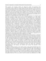
Figure 1.1
Major pathways implicated in ischemic cell death: excitotoxicity, ionic imbalance, oxidative and nitrative stresses, and
apoptotic-like mechanisms.There is extensive interaction and overlap between multiple mediators of cell injury and cell
death. After ischemic onset, loss of energy substrates leads to mitochondrial dysfunction and the generation of reactive
oxygen species (ROS) and reactive nitrogen species (RNS). Additionally, energy deficits lead to ionic imbalance, and excitotoxic glutamate efflux and build up of intracellular calcium. Downstream pathways ultimately include direct free radical damage to membrane lipids, cellular proteins, and DNA, as well as calcium-activated proteases, plus caspase cascades
that dismantle a wide range of homeostatic, reparative, and cytoskeletal proteins. (From Lo et al., Nat Rev Neurosci 2003,
4: 399–415)
Fig. 1.2
Putative cascade of damaging events in focal cerebral
ischemia. Very early after the onset of the focal perfusion deficit, excitotoxic mechanisms can damage neurons and glia lethally. In addition, excitotoxicity triggers
a number of events that can further contribute to the
demise of the tissue. Such events include peri-infarct
depolarizations and the more-delayed mechanisms of
inflammation and programmed cell death. The x-axis
reflects the evolution of the cascade over time, while
the y-axis aims to illustrate the impact of each element
of the cascade on the final outcome. (From Dirnagel
et al., Trends Neurosci 1999; 22: 391–397)
Ischemic Stroke
(Fig. 1.2), have overlapping and redundant features,
and mediate injury within neurons, glial cells, and
vascular elements [1]. The relative contribution of
each process to the net stroke-related injury is graphically depicted in Fig. 1.2. Within areas of severely reduced blood flow – the “core” of the ischemic territory – excitotoxic and necrotic cell death occurs within
minutes, and tissue undergoes irreversible damage in
the absence of prompt and adequate reperfusion.
However, cells in the peripheral zones are supported
by collateral circulation, and their fate is determined
by several factors including the degree of ischemia
and timing of reperfusion. In this peripheral region,
termed the “ischemic penumbra,” cell death occurs
relatively slowly via the active cell death mechanisms
noted above; targeting these mechanisms provides
promising therapeutic opportunities.
1.2.1 Excitotoxicity and Ionic Imbalance
Ischemic stroke results in impaired cellular energy
metabolism and failure of energy-dependent processes such as the sodium-potassium ATPase. Loss of
energy stores results in ionic imbalance, neurotransmitter release, and inhibition of the reuptake of excitatory neurotransmitters such as glutamate. Glutamate binding to ionotropic N-methyl-D-aspartate
(NMDA) and a-amino-3-hydroxy-5-methyl-4-isoxazolepropionic acid (AMPA) receptors promotes excessive calcium influx that triggers a wide array of
downstream phospholipases and proteases, which in
turn degrade membranes and proteins essential for
cellular integrity. In experimental models of stroke,
extracellular glutamate levels increase in the microdialysate [2, 3], and glutamate receptor blockade attenuates stroke lesion volumes. NMDA receptor antagonists prevent the expansion of stroke lesions in
part by blocking spontaneous and spreading depolarizations of neurons and glia (cortical spreading
depression) [4]. More recently, activation of the
metabotropic subfamily of receptors has been implicated in glutamate excitotoxicity [5].
Up- and downregulation of specific glutamate receptor subunits contribute to stroke pathophysiology
in different ways [6]. For example, after global cerebral ischemia, there is a relative reduction of calcium-
Chapter 1
impermeable GluR2 subunits in AMPA-type receptors, which makes these receptors more permeable to
deleterious calcium influx [7]. Antisense knockdown
of calcium-impermeable GluR2 subunits significantly increased hippocampal injury in a rat model of
transient global cerebral ischemia, confirming the
importance of these regulatory subunits in mediating
neuronal vulnerability [8]. Variations in NMDA receptor subunit composition can also have an impact
on tissue outcome. Knockout mice deficient in the
NR2A subunit show decreased cortical infarction
after focal stroke [9]. Medium spiny striatal neurons,
which are selectively vulnerable to ischemia and excitotoxicity, preferentially express NR2B subunits
[10]. Depending upon the subtype, metabotropic glutamate receptors can trigger either pro-survival or
pro-death signals in ischemic neurons [5]. Understanding how the expression of specific glutamate receptor subunits modifies cell survival should stimulate the search for stroke neuroprotective drugs that
selectively target specific subunits.
Ionotropic glutamate receptors also promote perturbations in ionic homeostasis that play a critical
role in cerebral ischemia. For example, L-, P/Q-, and
N-type calcium channel receptors mediate excessive
calcium influx, and calcium channel antagonists
reduce ischemic brain injury in preclinical studies
[11–13]. Zinc is stored in vesicles of excitatory
neurons and co-released upon depolarization after
focal cerebral ischemia, resulting in neuronal death
[14, 15]. Recently, imbalances in potassium have
also been implicated in ischemic cell death. Compounds that selectively modulate a class of calciumsensitive high-conductance potassium (maxi-K)
channels protect the brain against stroke in animal
models [16].
1.2.2 Oxidative and Nitrative Stress
Reactive oxygen species (ROS) such as superoxide
and hydroxyl radicals are known to mediate reperfusion-related tissue damage in several organ systems
including the brain, heart, and kidneys [17]. Oxygen
free radicals are normally produced by the mitochondria during electron transport, and, after ischemia, high levels of intracellular Ca2+, Na+, and
3
4
Chapter 1
ADP stimulate excessive mitochondrial oxygen radical production. Oxygen radical production may be
especially harmful to the injured brain because levels
of endogenous antioxidant enzymes [including superoxide dismutase (SOD), catalase, glutathione],
and antioxidant vitamins (e.g., alpha-tocopherol, and
ascorbic acid) are normally not high enough to
match excess radical formation. After ischemiareperfusion, enhanced production of ROS overwhelms endogenous scavenging mechanisms and
directly damages lipids, proteins, nucleic acids, and
carbohydrates. Importantly, oxygen radicals and oxidative stress facilitate mitochondrial transition pore
(MTP) formation, which dissipates the proton motive
force required for oxidative phosphorylation and
ATP generation [18]. As a result, mitochondria release apoptosis-related proteins and other constituents within the inner and outer mitochondrial
membranes [19]. Upon reperfusion and renewed tissue oxygenation, dysfunctional mitochondria may
generate oxidative stress and MTP formation. Oxygen radicals are also produced during enzymatic
conversions such as the cyclooxygenase-dependent
conversion of arachidonic acid to prostanoids and
degradation of hypoxanthine, especially upon reperfusion. Furthermore, free radicals are also generated
during the inflammatory response after ischemia
(see below). Not surprisingly then, oxidative stress,
excitotoxicity, energy failure, and ionic imbalances
are inextricably linked and contribute to ischemic
cell death.
Oxidative and nitrative stresses are modulated
by enzyme systems such as SOD and the nitric oxide
synthase (NOS) family. The important role of SOD in
cerebral ischemia is demonstrated in studies showing
that mice with enhanced SOD expression show reduced injury after cerebral ischemia whereas those
with a deficiency show increased injury [20–23]. Similarly, in the case of NOS, stroke-induced injury is attenuated in mice with deficient expression of the neuronal and inducible NOS isoforms [24, 25]. NOS activation during ischemia increases the generation of
NO production, which combines with superoxide to
produce peroxynitrite, a potent oxidant [26]. The
generation of NO and oxidative stress is also linked to
DNA damage and activation of poly(ADP-ribose)
A.B. Singhal · E.H. Lo · T. Dalkara · M.A. Moskowitz
polymerase-1 (PARP-1), a nuclear enzyme that facilitates DNA repair and regulates transcription [27].
PARP-1 catalyzes the transformation of b-nicotinamide adenine dinucleotide (NAD+) into nicotinamide and poly(ADP-ribose). In response to DNA
strand breaks, PARP-1 activity becomes excessive
and depletes the cell of NAD+ and possibly ATP.
Inhibiting PARP-1 activity or deleting the parp-1
gene reduces apoptotic and necrotic cell death [28,
29], pointing to the possible relevance of this enzyme
as a target for stroke therapy.
1.2.3 Apoptosis
Apoptosis, or programmed cell death [30], is characterized histologically by cells positive for terminaldeoxynucleotidyl-transferase-mediated dUTP nick
end labeling (TUNEL) that exhibit DNA laddering.
Necrotic cells, in contrast, show mitochondrial and
nuclear swelling, dissolution of organelles, nuclear
chromatin condensation, followed by rupture of nuclear and cytoplasmic membranes, and the degradation of DNA by random enzymatic cuts. Cell type, cell
age, and brain location render cells more or less resistant to apoptosis or necrosis. Mild ischemic injury
preferentially induces cell death via an apoptotic-like
process rather than necrosis, although “aponecrosis”
more accurately describes the pathology.
Apoptosis occurs via caspase-dependent as well as
caspase-independent mechanisms (Fig. 1.3). Caspases are protein-cleaving enzymes (zymogens) that belong to a family of cysteine aspartases constitutively
expressed in both adult and especially newborn brain
cells, particularly neurons. Since caspase-dependent
cell death requires energy in the form of ATP, apoptosis predominantly occurs in the ischemic penumbra
(which sustains milder injury) rather than in the
ischemic core, where ATP levels are rapidly depleted
[31]. The mechanisms of cleavage and activation
of caspases in human brain are believed to be similar
to those documented in experimental models of
stroke, trauma, and neurodegeneration [32]. Apoptogenic triggers [33] include oxygen free radicals [34],
Bcl2, death receptor ligation [35], DNA damage, and
possibly lysosomal protease activation [36]. Several
mediators facilitate cross communication between
Ischemic Stroke
Chapter 1
Figure 1.3
Cell death pathways relevant to an apoptotic-like mechanism in cerebral ischemia. Release of cytochrome c (Cyt. c) from
the mitochondria is modulated by pro- as well as anti-apoptotic Bcl2 family members. Cytochrome c release activates
downstream caspases through apoptosome formation (not shown) and caspase activation can be modulated by
secondary mitochondria-derived activator of caspase (Smac/Diablo) indirectly through suppressing protein inhibitors of
apoptosis (IAP). Effector caspases (caspases 3 and 7) target several substrates, which dismantle the cell by cleaving homeostatic, cytoskeletal, repair, metabolic, and cell signaling proteins. Caspases also activate caspase-activated deoxyribonuclease (CAD) by cleavage of an inhibitor protein (ICAD). Caspase-independent cell death may also be important. One
mechanism proposes that poly-ADP(ribose)polymerase activation (PARP) promotes the release of apoptosis-inducing
factor (AIF), which translocates to the nucleus, binds to DNA, and promotes cell death through a mechanism that awaits
clarification. (From Lo et al., Nat Rev Neurosci 2003, 4: 399–415)
cell death pathways [37, 38], including the calpains,
cathepsin B [39], nitric oxide [40, 41], and PARP
[42]. Ionic imbalances, and mechanisms such as
NMDA receptor-mediated K+ efflux, can also trigger
apoptotic-like cell death under certain conditions
[43, 44]. This inter-relationship between glutamate
excitotoxicity and apoptosis presents an opportunity
for combination stroke therapy targeting multiple
pathways.
The normal human brain expresses caspases-1, -3,
-8, and -9, apoptosis protease-activating factor 1
(APAF-1), death receptors, P53, and a number of Bcl2
family members, all of which are implicated in apoptosis. In addition, the tumor necrosis factor (TNF)
superfamily of death receptors powerfully regulates
upstream caspase processes. For example, ligation of
Fas induces apoptosis involving a series of caspases,
particularly procaspase-8 and caspase-3 [45]. Cas-
5
6
Chapter 1
pase-3 has a pivotal role in ischemic cell death. Caspase-3 cleavage occurs acutely in neurons and it appears in the ischemic core as well as penumbra early
during reperfusion [46]. A second wave of caspase
cleavage usually follows hours to days later, and
probably participates in delayed ischemic cell death.
Emerging data suggest that the nucleus – traditionally believed to be simply the target of apoptosis – is involved in releasing signals for apoptosis. However,
the mitochondrion plays a central role in mediating
apoptosis [47, 48]. Mitochondria possess membrane
recognition elements for upstream proapoptotic signaling molecules such as Bid, Bax, and Bad. Four mitochondrial molecules mediate downstream celldeath pathways: cytochrome c, secondary mitochondria-derived activator of caspase (Smac/Diablo),
apoptosis-inducing factor, and endonuclease G
[49]. Apoptosis-inducing factor and endonuclease G
mediate caspase-independent apoptosis, which is discussed below. Cytochrome c and Smac/Diablo mediate caspase-dependent apoptosis. Cytochrome c
binds to Apaf-1, which, together with procaspase-9,
forms the “apoptosome,” which activates caspase-9.
In turn, caspase-9 activates caspase-3. Smac/Diablo
binds to inhibitors of activated caspases and causes
further caspase activation. Upon activation, executioner caspases (caspase-3 and -7) target and degrade
numerous substrate proteins including gelsolin,
actin, PARP-1, caspase-activated deoxyribonuclease
inhibitor protein (ICAD), and other caspases, ultimately leading to DNA fragmentation and cell death
(Fig. 1.3).
Caspase-independent apoptosis was recently recognized to play an important role in cell death and
probably deserves careful scrutiny as a novel therapeutic target for stroke. NMDA receptor perturbations activate PARP-1, which promotes apoptosis-inducing factor (AIF) release from the mitochondria
[42]. AIF then relocates to the nucleus, binds DNA,
promotes chromatin condensation, and kills cells by
a complex series of events. Cell death by AIF appears
resistant to treatment with pan-caspase inhibitors
but can be suppressed by neutralizing AIF before its
nuclear translocation.
A number of experimental studies have shown
that caspase inhibition reduces ischemic injury [50].
A.B. Singhal · E.H. Lo · T. Dalkara · M.A. Moskowitz
Caspase-3 inhibitors [51], gene deletions of Bid or
caspase-3 [52], and the use of peptide inhibitors, viral
vector-mediated gene transfer, and antisense oligonucleotides that suppress the expression and activity
of apoptosis genes have all been found to be neuroprotective [50]. However, caspase inhibitors do not
reduce infarct size in all brain ischemia models,
perhaps related to the greater severity of ischemia,
limited potency or inability of the agent to cross the
blood–brain barrier, relatively minor impact of
apoptosis on stroke outcome, and upregulation of
caspase-independent or redundant cell death pathways. Ultimately, it may be necessary to combine caspase inhibitors and other inhibitors of apoptosis with
therapies directed towards other pathways, for successful neuroprotection.
1.2.4 Inflammation
Inflammation is intricately related to the onset of
stroke, and to subsequent stroke-related tissue damage. Inflammation within the arterial wall plays a
vital role in promoting atherosclerosis [53, 54]. Arterial thrombosis (usually associated with ulcerated
plaques) is triggered by multiple processes involving
endothelial activation, as well as pro-inflammatory
and pro-thrombotic interactions between the vessel
wall and circulating blood elements. Elevated stroke
risk has been linked to high levels of serologic markers of inflammation such as C-reactive protein [55],
erythrocyte sedimentation rate (ESR), interleukin-6,
TNF-a and soluble intercellular adhesion molecule
(sICAM) [56]. These events are promoted in part by
the binding of cell adhesion molecules from the
selectin and immunoglobulin gene families expressed on endothelial cells to glycoprotein receptors
expressed on the neutrophil surface. As evidence,
reduced ischemic infarction is observed in ICAM-1
knockout mice, and infarction volumes are increased
in mice that overexpress P-selectin [57, 58]. The proinflammatory molecule P-selectin is expressed on
vascular endothelium within 90 min after cerebral
ischemia, ICAM-1 by 4 h, and E-selectin by 24 h [59].
Inhibiting both selectin adhesion molecules and activation of complement reduces brain injury and suppresses neutrophil and platelet accumulation after
Ischemic Stroke
focal ischemia in mice [60]. In humans, neutrophil
and complement activation significantly worsened
outcomes in a clinical trial using humanized mouse
antibodies directed against ICAM (Enlimomab) [61].
Hence, the complexities of interactions between multiple pathways will have to be carefully considered for
optimal translation to the clinic.
Ischemic stroke-related brain injury itself triggers
inflammatory cascades within the parenchyma that
further amplify tissue damage [1, 59]. As reactive microglia, macrophages, and leukocytes are recruited
into ischemic brain, inflammatory mediators are
generated by these cells as well as by neurons and
astrocytes. Inducible nitric oxide synthase (iNOS),
cyclooxygenase-2 (COX-2), interleukin-1 (IL-1), and
monocyte chemoattractant protein-1 (MCP-1) are
key inflammatory mediators, as evidenced by attenuated ischemic injury in mutant mice with targeted
disruption of their genes [1, 62–65]. Initially after occlusion, there is a transient upregulation of immediate early genes encoding transcription factors (e.g.,
c-fos, c-jun) that occurs within minutes. This is followed by a second wave of heat shock genes (e.g.,
HSP70, HSP72) that increase within 1–2 h and then
decrease by 1–2 days. Approximately 12–24 h after a
stroke, a third wave comprised of chemokines and
cytokines is expressed (e.g., IL-1, IL-6, IL-8, TNF-a,
MCP-1, etc.). It is not known whether these three
waves are causally related. Nevertheless, therapies
that seek to target these pathways need to be carefully timed to match the complex temporal evolution of
tissue injury.
Inflammatory cascades stimulate both detrimental and potentially beneficial pathways after ischemia.
For example, administering TNF-a-neutralizing antibodies reduces brain injury after focal ischemia in
rats [66], whereas ischemic injury increases in TNF
receptor knockout mice [67]. In part, these contrasting results may reflect signal transduction cascades
activated by TNF-R1 and TNF-R2; with TNF-R1 augmenting cell death and TNF-R2 mediating neuroprotection [68]. Similarly, the peptide vascular endothelial growth factor (VEGF) exacerbates edema in the
acute phase of cerebral ischemia but promotes vascular remodeling during stroke recovery [69]. Ultimately, the net effect of these mediators depends upon the
Chapter 1
stage of tissue injury or the predominance of a single
signaling cascade among multiple divergent pathways.
1.2.5 Peri-infarct Depolarizations
Brain tissue depolarizations after ischemic stroke are
believed to play a vital role in recruiting adjacent
penumbral regions of reversible injury into the core
area of infarction. Cortical spreading depression
(CSD) is a self-propagating wave of electrochemical
activity that advances through neural tissues at a rate
of 2–5 mm/min, causing prolonged (1–5 min) cellular
depolarization, depressed neuro-electrical activity,
potassium and glutamate release into adjacent tissue
and reversible loss of membrane ionic gradients. CSD
is associated with a change in the levels of numerous
factors including immediate early genes, growth factors, and inflammatory mediators such as interleukin-1b and TNF-a [70]. CSD is a reversible phenomenon, and, while implicated in conditions such as
migraine, reportedly does not cause permanent tissue injury in humans. In severely ischemic regions,
energy failure is so profound that ionic disturbances
and simultaneous depolarizations become permanent, a process termed anoxic depolarization [71]. In
penumbral regions after stroke, where blood supply
is compromised, spreading depression exacerbates
tissue damage, perhaps due to the increased energy
requirements for reestablishing ionic equilibrium in
the metabolically compromised ischemic tissues. In
this context, spreading depression waves are referred
to as peri-infarct depolarizations (PIDs) [4], reflecting their pathogenic role and similarity to anoxic
depolarization.
PIDs have been demonstrated in mice, rat, and cat
stroke models [72, 73]; however, their relevance to
human stroke pathophysiology remains unclear. In
the initial 2–6 h after experimental stroke, PIDs result
in a step-wise increase in the region of core-infarcted
tissue into adjacent penumbral regions [74, 75], and
the incidence and total duration of spreading depression is shown to correlate with infarct size [76].
Recent evidence suggests that PIDs contribute to the
expansion of the infarct core throughout the period
of infarct maturation [77]. Inhibition of spreading
7
8
Chapter 1
depression using pharmaceutical agents such as
NMDA or glycine antagonists [77, 78], or physiological approaches such as hypothermia [79], could be an
important strategy to suppress the expansion of an
ischemic lesion.
1.3 Grey Matter Versus White Matter Ischemia
In addition to the size of the stroke, its location, and
the relative involvement of gray versus white matter
are key determinants of outcome. For example, small
white matter strokes often cause extensive neurologic deficits by interrupting the passage of large axonal
bundles such as those within the internal capsule.
Blood flow in white matter is lower than in gray matter, and white matter ischemia is typically severe,
with rapid cell swelling and tissue edema because
there is little collateral blood supply in deep white
matter. Moreover, cells within the gray and white
matter have different susceptibilities to ischemic injury. Amongst the neuronal population, well-defined
subsets (the CA1 hippocampal pyramidal neurons,
cortical projection neurons in layer 3, neurons in dorsolateral striatum, and cerebellar Purkinje cells) are
particularly susceptible and undergo selective death
after transient global cerebral ischemia [80]. The major cell types composing the neurovascular module
within white matter include the endothelial cell,
perinodal astrocyte, axon, oligodendrocyte, and
myelin. In general, oligodendrocytes are more vulnerable than astroglial or endothelial cells.
There are important differences in the pathophysiology of white matter ischemia as compared to that
of gray matter, which have implications for therapy
[81]. In the case of excitotoxicity, since the white
matter lacks synapses, neurotransmitter release from
vesicles does not occur despite energy depletion and
neurotransmitter accumulation. Instead, there is
reversal of Na+-dependent glutamate transport [82],
resulting in glutamate toxicity with subsequent
AMPA receptor activation, and excessive accumulation of calcium, which in turn activates calcium-dependent enzymes such as calpain, phospholipases,
and protein kinase C, resulting in irreversible injury.
The distinct lack of AMPA receptors expressing calci-
A.B. Singhal · E.H. Lo · T. Dalkara · M.A. Moskowitz
um-impermeable GluR2 subunits may make oligodendroglia particularly vulnerable to excitotoxic injury [83]. In the case of oxidative stress-induced
white matter injury, the severity of injury appears to
be greater in large axons as compared to small axons
[80], although the mechanisms underlying these differences need further study. Despite these differences
between gray and white matter injury, several common cascades of injury do exist. Damaged oligodendrocytes express death signals such as TNF and Fas
ligand, and recruit caspase-mediated apoptotic-like
pathways [84]. Degradation of myelin basic protein
by matrix metalloproteinases (MMPs) [85], and
upregulation of MMPs in autopsied samples from patients with vascular dementia [86] suggest that proteolytic pathways are also recruited in white matter.
These pathways might serve as common targets for
stroke therapy.
1.4 The Neurovascular Unit
In July 2001, the National Institutes of Neurological
Disorders and Stroke convened the Stroke Program
Review Group (SPRG) [87] to advise on directions for
basic and clinical stroke research for the following
decade. Although much progress had been made in
dissecting the molecular pathways of ischemic cell
death, focusing therapy to a single intracellular pathway or cell type had not yielded clinically effective
stroke treatment. Integrative approaches were felt to
be mandatory for successful stroke therapy. This
meeting emphasized the relevance of dynamic interactions between endothelial cells, vascular smooth
muscle, astro- and microglia, neurons, and associated
tissue matrix proteins, and gave rise to the concept of
the “neurovascular unit.” This modular concept emphasized the dynamics of vascular, cellular, and matrix signaling in maintaining the integrity of brain
tissue within both the gray and white matter, and its
importance to the pathophysiology of conditions
such as stroke, vascular dementia, migraine, trauma,
multiple sclerosis, and possibly the aging brain
(Fig. 1.4).
The neurovascular unit places stroke in the context of an integrative tissue response in which all cel-
Ischemic Stroke
Chapter 1
Figure 1.4
Schematic view of the neurovascular unit or module, and some of its components. Circulating blood elements, endothelial cells, astrocytes, extracellular matrix, basal lamina, adjacent neurons, and pericytes. After ischemia, perturbations in
neurovascular functional integrity initiate multiple cascades of injury. Upstream signals such as oxidative stress together
with neutrophil and/or platelet interactions with activated endothelium upregulate matrix metalloproteinases (MMPs),
plasminogen activators and other proteases which degrade matrix and lead to blood–brain barrier leakage. Inflammatory infiltrates through the damaged blood–brain barrier amplify brain tissue injury. Additionally, disruption of cellmatrix homeostasis may also trigger anoikis-like cell death in both vascular and parenchymal compartments. Overlaps
with excitotoxicity have also been documented via t-PA-mediated interactions with the NMDA receptor that augment
ionic imbalance and cell death. (t-PA Tissue plasminogen activator)
lular and matrix elements, not just neurons or blood
vessels, are players in the evolution of tissue injury.
For example, efficacy of the blood–brain barrier
is critically dependent upon endothelial–astrocyte–
matrix interactions [88]. Disruption of the neurovascular matrix, which includes basement membrane
components such as type IV collagen, heparan sulfate
proteoglycan, laminin, and fibronectin, upsets the
cell–matrix and cell–cell signaling that maintains
neurovascular homeostasis. Although many proteases including cathepsins and heparanases contribute
to extracellular matrix proteolysis, in the context of
stroke, plasminogen activator (PA) and MMP are
probably the two most important. This is because tissue plasminogen activator (t-PA) has been used successfully as a stroke therapy, and because emerging
data show important linkages between t-PA, MMPs,
edema, and hemorrhage after stroke.
9
10
Chapter 1
The MMPs are zinc endopeptidases produced by
all cell types of the neurovascular unit [89], that are
secreted as zymogens requiring cleavage for enzymatic activation. MMPs can be classified into gelatinases (MMP-2 and -9), collagenases (MMP-1, -8, -13),
stromelysins (MMP-3, -10, -11), membrane-type
MMPs (MMP-14, -15, -16, -17), and others (e.g.,
MMP-7 and -12) [90]. Together with the PA system,
MMPs play a central role in brain development and
plasticity as they modulate extracellular matrix to
allow neurite outgrowth and cell migration [91].
Upstream triggers of MMP include MAP kinase
pathways [92] and oxidative stress [93]. MMP signaling is intricately linked to other well-recognized
pathways after stroke, including oxidative and nitrative stress [94], caspase-mediated cell death [95], excitotoxicity, and neuro-inflammation [96, 97]. Several
experimental as well as human studies provide evidence for a major role of MMPs (particularly MMP9) in ischemic stroke, primary brain hemorrhage,
blood–brain barrier disruption and post-ischemic
or reperfusion hemorrhage [98–106]. For example,
MMP levels have been correlated with the extent of
stroke as measured by diffusion- and perfusionweighted MRI [107]. Unlike MMPs, however, there is
controversy surrounding the role of the PA axis (the
other major proteolytic system in mammalian brain,
comprising t-PA and urokinase PA, and their inhibitors plasminogen activator inhibitor-1 and neuroserpin) in stroke. Primary neuronal cultures genetically deficient in t-PA are resistant to oxygen-glucose
deprivation [108] and t-PA knockout mice are protected against excitotoxic injury [109]. In a mouse
focal ischemia model, treatment with neuroserpin
reduces infarction [110]. In contrast, the responses
are variable in t-PA knockouts, which are protected
against focal stroke in some [111] but not other studies [112]. In part, these inconsistencies may reflect
genetic differences and perhaps more importantly
the balance between the clot-lysing beneficial effects
of t-PA and its neurotoxic properties [113]. Emerging
data suggest that administered t-PA upregulates
MMP-9 via the low-density lipoprotein receptor-related protein (LRP), which avidly binds t-PA and
possesses signaling properties [114]. Targeting the
t-PA–LRP–MMP pathway may offer new therapeutic
A.B. Singhal · E.H. Lo · T. Dalkara · M.A. Moskowitz
approaches for improving the safety profile of t-PA in
patients with stroke.
1.5 Neuroprotection
Neuroprotection can be defined as the protection of
cell bodies and neuronal and glial processes by
strategies that impede the development of irreversible ischemic injury by effects on the cellular
processes involved. Neuroprotection can be achieved
using pharmaceutical or physiological therapies that
directly inhibit the biochemical, metabolic, and cellular consequences of ischemic injury, or by using indirect approaches such as t-PA and mechanical devices
to restore tissue perfusion. The complex and overlapping pathways involving excitotoxicity, ionic imbalance, oxidative and nitrative stress, and apoptoticlike mechanisms have been reviewed above. Each of
these pathways offers several potential therapeutic
targets, several of which have proved successful in reducing ischemic injury in animal models. However,
the successful translation of experimental results into
clinical practice remains elusive.
1.6 Stroke Neuroprotective Clinical Trials:
Lessons from Past Failures
Various classes of neuroprotective agents have been
tested in humans, with some showing promising
phase II results. However, with the exception of the
National Institute of Neurological Disorders and
Stroke (NINDS) rt-PA trial [115], none has been
proven efficacious on the basis of a positive phase III
trial. Notable failures include trials of the lipid peroxidation inhibitor tirilazad mesylate [116], the ICAM1 antibody enlimomab [61], the calcium channel
blocker nimodipine [117], the g-aminobutyric acid
(GABA) agonist clomethiazole [118, 119], the glutamate antagonist and sodium channel blocker lubeluzole [120], the competitive NMDA antagonist selfotel
[121], and several noncompetitive NMDA antagonists (dextrorphan, gavestinel, aptiganel and
eliprodil) [122–124]. The high financial costs of these
trials have raised questions about the commercial
Ischemic Stroke
viability of continued neuroprotective drug development. How can we explain this apparent discrepancy
between bench and bedside studies [125, 126]?
The lack of efficacy can be related to several factors,
some relating to the preclinical stage of drug development, and others to clinical trial design and
methodology.
In the preclinical stage, therapies are often tested
on healthy, young animals under rigorously controlled laboratory conditions, and, most often, the
treatment is not adequately tested (for example, by
multiple investigators in different stroke models) before it is brought to clinical trial. Whereas experimental animals are bred for genetic homogeneity,
genetic differences and factors such as advanced age
and co-morbidities (hypertension, diabetes) in patients may alter their therapeutic response. Moreover,
despite similarities in the basic pathophysiology of
stroke between species, there are important differences in brain structure, function, and vascular
anatomy. The human brain is gyrated, has greater
neuronal and glial densities, and is larger than the
rodent brain. Some rodents (gerbils) lack a complete
circle of Willis (gerbils), while others (rats) have
highly effective collaterals between large cerebral
vessels. As a result, there are important differences in
the size, spatial distribution, and temporal evolution
of the ischemic lesions between experimental models
and humans. This is important, because the infarct
volume is the standard outcome measure in animal
models, whereas success in clinical trials is typically
defined by clinical improvement. Finally, outcomes in
animal models are usually assessed within days to
weeks, whereas in humans, functional scores [National Institutes of Health Stroke Scale (NIHSS),
Barthel index, etc.] are typically assessed after
3–6 months.
In the clinical trial stage, major problems include
the relatively short therapeutic time window of most
drugs; the difficulties in transporting patients quickly to the hospital; the imprecise correlation between
symptom onset and the actual onset of cerebral
ischemia; the high cost of enrolling patients for an
adequately powered study; and the use of nonstandardized and relatively insensitive outcome measures. A recent review showed that of 88 stroke neuro-
Chapter 1
protective trials, the mean sample size was only 186
patients, and the median time window for recent
(1995–1999) neuroprotective trials was as late as 12 h
[127]. Another major factor accounting for past failures is that patients with different stroke pathophysiology and subtype are often combined in a trial,
whereas the drug being tested might be more effective in a certain stroke subtype (e.g., strokes with predominant gray matter involvement).
In addition to the above, delivery of the drug to
target ischemic tissues poses unique challenges
[128]. Pharmacokinetic properties of the drug, and
alterations in cerebral blood flow after stroke need to
be taken into account. Blood flow can drop to below
5–10% of normal levels in the infarct core, and to
30–40% of baseline in the surrounding penumbra
[129]. In addition, the blood–brain barrier restricts
direct exchange between the vascular compartment
and the cerebral parenchyma, and post-stroke edema
and raised intracranial pressure further impair efficient delivery. Strategies that have been explored to
penetrate the blood–brain barrier include intracerebral and intraventricular delivery, use of hyperosmolar substances (e.g., mannitol, arabinose) and pharmacological agents (bradykinin, mannitol, nitric oxide) to facilitate osmolar opening, and the development of carrier-mediated transport systems. These
strategies appear promising; however, they remain
limited by the prohibitively narrow time windows for
effective stroke treatment.
Given these past failures, the focus has shifted towards expanding the therapeutic time window, improved patient selection, the use of brain imaging as
a selection criterion, combination acute stroke drug
treatments, use of validated rating scales to assess
functional end points, and improved stroke trial
design and organization [127, 130]. A number of new
neuroprotection trials are currently underway or in
the planning stages. These include trials of the free
radical spin trap agent NXY-059 (now in phase III trials), intravenous magnesium, the antioxidant ebselen, the AMPA antagonist YM872, and the serotonin
antagonist repinotan [131–133]. With the insights
gained from prior neuroprotective trials, it is anticipated that one or more of the impending trials will
prove successful.
11
12
Chapter 1
1.7 Identifying the Ischemic Penumbra
As discussed above, although irreversible cell death
begins within minutes after stroke onset within regions of maximally reduced blood flow (the infarct
“core”), for several hours there exists a surrounding
“penumbra” of ischemic but noninfarcted tissue that
is potentially salvageable [134–137]. The concept of
an “ischemic penumbra” provides a rationale for the
use of neuroprotective drugs and reperfusion techniques to improve outcome after acute ischemic
stroke. However, the extent of penumbral tissue is
thought to diminish rapidly with time, hence the
therapeutic time window is narrow.With intravenous
t-PA [the only stroke therapy approved by the Food
and Drug Administration (FDA)] the window is 3 h,
which severely limits its use [138]; delayed therapy
increases the risk of hemorrhage [115]. Similarly, administering therapy outside of the therapeutic window is considered one of the most important factors
leading to the failure of neuroprotective drug trials.
Developing methods to rapidly and accurately identify the ischemic penumbra is therefore an important
area of current stroke research.
Imaging studies have validated the concept that
tissue viability is heterogeneous distal to an occluded
brain blood vessel. In animal models, the ischemic
penumbra can be visualized by autoradiographic
techniques that compare regions of reduced blood
flow to regions of actively metabolizing tissue (2-deoxyglucose), or larger regions of suppressed protein
synthesis to core areas with complete loss of ATP. In
humans, imaging and biochemical studies similarly
suggest that the window for efficacy may be prolonged in select individuals. Positron emission tomography (PET) [139, 140] can detect oxygen-utilizing tissue (oxygen extraction fraction) within regions of low blood flow, as well as locate 11C-flumazenil recognition sites on viable neurons within underperfused brain areas. While PET is arguably the most
accurate method, the greatest promise and experience appear to lie with multimodal magnetic resonance imaging (MRI) and multimodal CT because of
their widespread availability, lower cost, technical
ease, and shorter imaging times. With MRI, there is
A.B. Singhal · E.H. Lo · T. Dalkara · M.A. Moskowitz
often a volume mismatch between tissue showing
reduced water molecule diffusion (a signature for cell
swelling and ischemic tissue) and a larger area of
compromised tissue perfusion early after stroke onset – the so-called diffusion–perfusion mismatch.
The difference, at least for all practical purposes, is
believed to reflect the ischemic penumbra [129,
141–144]. Perfusion MRI currently affords a relative,
rather than absolute, quantitative measure of cerebral tissue perfusion. Recent studies indicate that
perfusion-CT can also be used to identify regions of
ischemic, noninfarcted tissue after stroke, and that
perfusion-CT may be comparable to MRI for this
purpose [145–147]. The main advantage of perfusion-CT is that it allows rapid data acquisition and
postprocessing, and can be performed in conjunction with CT angiography to complete the initial
evaluation of stroke [148]. Xenon-enhanced CT is a
more accurate technique than perfusion-CT and provides quantitative measurements of cerebral blood
flow within 10–15 min; however, it requires the use of
specialized equipment and at present its use is restricted to only a few centers [149]. Imaging methods
such as these can optimize the selection of candidates
for thrombolytic therapy or for adjunctive therapy
many hours after stroke onset. Importantly, imaging
may also provide quantitative surrogate endpoints
for clinical trials. Several clinical trials employing
imaging to select patients who might benefit from
delayed therapy are now in progress. The Desmoteplase in Acute Ischemic Stroke Trial (DIAS) is the
first published acute stroke thrombolysis trial using
MRI both for patient selection and as a primary efficacy endpoint [150]. In this trial, patients were selected on the basis of perfusion–diffusion mismatch on
the admission MRI and treated as late as 3–9 h after
stroke symptom onset with intravenous (i.v.) desmoteplase, a newer plasminogen activator with high
fibrin specificity. Desmoteplase-treated patients had
significantly higher rates of reperfusion, as defined
by MR-perfusion, and improved 90-day clinical
outcome. These results support the utility of MRI in
improving patient selection and as a surrogate outcome measure.
Ischemic Stroke
1.8 Combination Neuroprotective Therapy
Considering that several pathways leading to cell
death are activated in cerebral ischemia, effective
neuroprotection may require combining or adding
drugs in series that target distinct pathways during
the evolution of ischemic injury. Although seemingly
independent treatments may not always yield additive results [151], various neuroprotective combinations have been used with some success in animal
models. These include the co-administration of an
NMDA receptor antagonist with GABA receptor agonists [152], free radical scavengers [153], cytidine-5¢diphosphocholine (Citicholine®) [154], the protein
synthesis inhibitor cyclohexamide [155], caspase inhibitors [156] or growth factors such as basic fibroblast growth factor (bFGF) [157]. Synergy is also observed with 2 different antioxidants [158], and cytidine-5¢-diphosphocholine plus bFGF [159]. Caspase
inhibitors given with bFGF or an NMDA receptor
antagonist extend the therapeutic window and lower
effective doses [160].
Neuroprotective drugs may have a role in increasing the efficacy and safety of thrombolysis. Because
the risk of hemorrhage increases with time, treatment with intravenous t-PA is currently limited to 3 h
after vascular occlusion [161]. However, because ischemic but noninfarcted, potentially salvageable tissue exists for several hours after stroke in rats [162]
and probably also in humans [134–137], clot lysis
may be therapeutically useful at later times. Results
from the PROACT II study, in which recombinant
pro-urokinase was administered intra-arterially
until 8 h post-stroke to patients with middle cerebral
artery (MCA) occlusion, and a pooled analysis of the
ATLANTIS, ECASS, and NINDS rt-PA stroke trials
[163], support the contention that potential benefit
exists beyond the 3-h time window. However, the use
of thrombolysis must be weighed against the risk of
intracerebral hemorrhage and brain edema after 3 h.
Most preclinical observations suggest that treatment
is suboptimal without combining neuroprotective
therapy with clot-lysing drugs. This combination reduces reperfusion injury and inhibits downstream
targets in cell death cascades. Synergistic or additive
Chapter 1
effects have been reported when thrombolysis was
used with neuroprotectants such as oxygen radical
scavengers [164], AMPA [165] and NMDA [166] receptor antagonists, MMP inhibitors [103], cytidine5¢-diphosphocholine [167], topiramate [168], antileukocytic adhesion antibodies [169], and antithrombotics [170]. Combination therapies may decrease dosages for each agent, thereby reducing the
occurrence of adverse events. Two recent clinical trials have reported the feasibility and safety of treating
with intravenous t-PA followed by neuroprotectants,
clomethiazole [171] or lubeluzole [172]. Rational
therapy based on inhibiting multiple cell death
mechanisms may ultimately prove as useful for
stroke as for cancer chemotherapy.
1.9 Ischemic Pre-conditioning
Transient, nondamaging ischemic/hypoxic brain insults are known to protect against subsequent prolonged, potentially detrimental episodes by upregulating powerful endogenous pathways that increase
the resistance to injury [173]. The tolerance induced
by ischemic preconditioning can be acute (within
minutes), or delayed by several hours. Acute protective effects are short lasting and are mediated by
posttranslational protein modifications; delayed tolerance is sustained for days to weeks and results from
changes in gene expression and new protein synthesis [for example, of heat shock protein, Bcl2, hypoxiainducible factor, and mitogen-activated protein
(MAP) kinases] [174, 175]. Emerging human data indicate that preceding transient ischemic attacks
(TIAs) reduce the severity of subsequent stroke, perhaps from a preconditioning effect [176–178]. In a
study of 65 patients studied by diffusion and perfusion MRI, those with a prior history of TIA (n=16)
were found to have smaller initial diffusion lesions
and final infarct volumes, as well as milder clinical
deficits, despite a similar size and severity of the perfusion deficit [176]. Preconditioning may offer novel
insights into molecular mechanisms responsible for
endogenous neuroprotection, and thus provide new
strategies for making brain cells more resistant to
ischemic injury [179].
13
14
Chapter 1
A.B. Singhal · E.H. Lo · T. Dalkara · M.A. Moskowitz
1.10.3 Hypothermia
1.10 Nonpharmaceutical Strategies
for Neuroprotection
1.10.1 Magnesium
Magnesium is involved in multiple processes relevant
to cerebral ischemia, including inhibition of presynaptic glutamate release [180], NMDA receptor
blockade [181], calcium channel antagonism, and
maintenance of cerebral blood flow [182]. In animal
models of stroke, administration of intravenous
magnesium as late as 6 h after stroke onset, in doses
that double its physiological serum concentration,
was found to reduce infarct volumes [183, 184]. In
pilot clinical studies, magnesium was found to reduce
death and disability from stroke, raising expectations
that magnesium could be a safe and inexpensive
treatment [185]. However, in a large multicenter trial
involving 2589 patients, magnesium given within
12 h after acute stroke did not significantly reduce the
risk of death or disability, although some benefit was
documented in lacunar strokes [131]. Further studies
are ongoing to determine whether paramedic initiation of magnesium, by reducing the time to treatment, yields benefit in stroke patients [186].
1.10.2 Albumin Infusion
Albumin infusion enhances red cell perfusion and
suppresses thrombosis and leukocyte adhesion within the brain microcirculation, particularly during the
early reperfusion phase after experimental focal
ischemia [187]. Albumin also significantly lowers the
hematocrit and by so doing improves microcirculatory flow, viscosity of plasma and cell deformability, as
well as oxygen transport capacity. Albumin reduces
infarct size, improves neurological scores, and reduces cerebral edema in experimental animals [188].
These effects may reflect a combination of therapeutic properties including its antioxidant effects, antiapoptotic effects on the endothelium, and effects on
reducing blood stasis within the microcirculation.
Clinical trials to test the effects of albumin are now
being organized.
Nearly all ischemic events are modulated by temperature, and cerebroprotection from hypothermia is
believed to increase resistance against multiple deleterious pathways including oxidative stress and
inflammation [189–195]. Generally, most biological
processes exhibit a Q10 of approximately 2.5, which
means that a 1°C reduction in temperature reduces
the rate of cellular respiration, oxygen demand, and
carbon dioxide production by approximately 10%
[196]. Reduced temperature also slows the rate of
pathological processes such as lipid peroxidation, as
well as the activity of certain cysteine or serine proteases. However, detoxification and repair processes
are also slowed, so the net outcome may be complex.
Hence, hypothermia appears to be an attractive
therapy that targets multiple injury mechanisms.
Brain cooling can be achieved more rapidly (and
spontaneously) when blood flow to the entire brain
ceases following cardiac arrest, and thermoregulation may be abnormal due to hypothalamic dysfunction. If only a segment of brain is ischemic, noninjured brain remains a metabolically active heat
source. While moderate hypothermia (28–32 °C) is
technically difficult and fraught with complications,
recent experimental studies have shown that small
decreases in core temperature (from normothermia
to 33–36 °C) are sufficient to reduce neuronal death.
The consensus from preclinical data suggests that the
opportunity to treat does not extend beyond minutes
after reversible MCA occlusion when hypothermia is
maintained for a short duration (a few hours) [197].
In a global model of hippocampal ischemia, hypothermia is beneficial if begun 30 min before but
not 10 min after stroke onset [198]. However, if cooling is prolonged (12–48 h), protection against injury
is substantial following focal as well as global cerebral
ischemia [199, 200]. In humans, encouraging positive
results were recently reported in two randomized
clinical trials of mild hypothermia in survivors of
out-of-hospital cardiac arrest [201, 202]. Cooling significantly improved outcomes despite a relatively delayed interval (105 min) from ischemic onset until
the initiation of cooling. Based on these results, additional controlled trials are now underway to test the
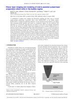
![academic skills problems [electronic resource] direct assessment and intervention](https://media.store123doc.com/images/document/14/y/va/medium_var1401465900.jpg)