Practice Guidelines for the Diagnosisand Management of Skin and Soft TissueInfections: 2014 Update by the InfectiousDiseases Society of America
Bạn đang xem bản rút gọn của tài liệu. Xem và tải ngay bản đầy đủ của tài liệu tại đây (1007.79 KB, 43 trang )
Clinical Infectious Diseases Advance Access published June 18, 2014
IDSA GUIDELINE
Practice Guidelines for the Diagnosis
and Management of Skin and Soft Tissue
Infections: 2014 Update by the Infectious
Diseases Society of America
Dennis L. Stevens,1 Alan L. Bisno,2 Henry F. Chambers,3 E. Patchen Dellinger,4 Ellie J. C. Goldstein,5 Sherwood L. Gorbach,6
Jan V. Hirschmann,7 Sheldon L. Kaplan,8 Jose G. Montoya,9 and James C. Wade10
1
Division of Infectious Diseases, Department of Veterans Affairs, Boise, Idaho; 2Medical Service, Miami Veterans Affairs Health Care System, Florida;
San Francisco General Hospital, University of California; 4Division of General Surgery, University of Washington, Seattle; 5University of California, Los
Angeles, School of Medicine, and R. M. Alden Research Laboratory, Santa Monica, California; 6Department of Community Health, Tufts University, Boston,
Massachusetts; 7Medical Service, Puget Sound Veterans Affairs Medical Center, Seattle, Washington; 8Department of Pediatrics, Baylor College of
Medicine, Houston, Texas; 9Department of Medicine, Stanford University, California; and 10Geisinger Health System, Geisinger Cancer Institute, Danville,
Pennsylvania
3
Downloaded from at IDSA member on June 30, 2015
A panel of national experts was convened by the Infectious Diseases Society of America (IDSA) to update the
2005 guidelines for the treatment of skin and soft tissue infections (SSTIs). The panel’s recommendations were
developed to be concordant with the recently published IDSA guidelines for the treatment of methicillinresistant Staphylococcus aureus infections. The focus of this guideline is the diagnosis and appropriate treatment
of diverse SSTIs ranging from minor superficial infections to life-threatening infections such as necrotizing fasciitis. In addition, because of an increasing number of immunocompromised hosts worldwide, the guideline
addresses the wide array of SSTIs that occur in this population. These guidelines emphasize the importance
of clinical skills in promptly diagnosing SSTIs, identifying the pathogen, and administering effective treatments
in a timely fashion.
EXECUTIVE SUMMARY
Summarized below are the recommendations made in
the new guidelines for skin and soft tissue infections
(SSTIs). Figure 1 was developed to simplify the management of localized purulent staphylococcal infections
such as skin abscesses, furuncles, and carbuncles in
Received 17 April 2014; accepted 21 April 2014.
It is important to realize that guidelines cannot always account for individual variation among patients. They are not intended to supplant physician judgment with
respect to particular patients or special clinical situations. IDSA considers adherence to these guidelines to be voluntary, with the ultimate determination regarding
their application to be made by the physician in the light of each patient’s individual
circumstances.
Correspondence: Dennis L. Stevens, PhD, MD, Infectious Diseases Section, VA
Medical Center, 500 W Fort St, Bldg 45, Boise, ID 83702 (dlsteven@mindspring.
com).
Clinical Infectious Diseases
© The Author 2014. Published by Oxford University Press on behalf of the Infectious
Diseases Society of America. All rights reserved. For Permissions, please e-mail:
DOI: 10.1093/cid/ciu296
the age of methicillin-resistant Staphylococcus aureus
(MRSA). In addition, Figure 2 is provided to simplify
the approach to patients with surgical site infections.
The panel followed a process used in the development
of other Infectious Diseases Society of America (IDSA)
guidelines, which included a systematic weighting of the
strength of recommendation and quality of evidence
using the GRADE (Grading of Recommendations
Assessment, Development, and Evaluation) system
(Table 1) [1–4]. A detailed description of the methods,
background, and evidence summaries that support each
of the recommendations can be found in the full text of
the guidelines.
I. What Is Appropriate for the Evaluation and Treatment
of Impetigo and Ecthyma?
Recommendations
1. Gram stain and culture of the pus or exudates
from skin lesions of impetigo and ecthyma are
IDSA Practice Guidelines for SSTIs
•
CID
•
1
recommended to help identify whether Staphylococcus aureus
and/or a β-hemolytic Streptococcus is the cause (strong, moderate), but treatment without these studies is reasonable in typical
cases (strong, moderate).
2. Bullous and nonbullous impetigo can be treated with
oral or topical antimicrobials, but oral therapy is recommended
for patients with numerous lesions or in outbreaks affecting several people to help decrease transmission of infection. Treatment for ecthyma should be an oral antimicrobial.
(a) Treatment of bullous and nonbullous impetigo should
be with either mupirocin or retapamulin twice daily (bid)
for 5 days (strong, high).
2
•
CID
•
Stevens et al
(b) Oral therapy for ecthyma or impetigo should be a 7-day
regimen with an agent active against S. aureus unless cultures
yield streptococci alone (when oral penicillin is the recommended agent) (strong, high). Because S. aureus isolates
from impetigo and ecthyma are usually methicillin susceptible, dicloxacillin or cephalexin is recommended. When
MRSA is suspected or confirmed, doxycycline, clindamycin,
or sulfamethoxazole-trimethoprim (SMX-TMP) is recommended (strong, moderate).
(c) Systemic antimicrobials should be used for infections
during outbreaks of poststreptococcal glomerulonephritis to
help eliminate nephritogenic strains of S. pyogenes from the
community (strong, moderate).
Downloaded from at IDSA member on June 30, 2015
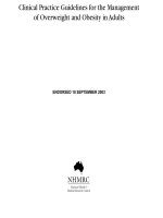
Figure 1. Purulent skin and soft tissue infections (SSTIs). Mild infection: for purulent SSTI, incision and drainage is indicated. Moderate infection: patients with purulent infection with systemic signs of infection. Severe infection: patients who have failed incision and drainage plus oral antibiotics or those
with systemic signs of infection such as temperature >38°C, tachycardia (heart rate >90 beats per minute), tachypnea (respiratory rate >24 breaths per
minute) or abnormal white blood cell count (<12 000 or <400 cells/µL), or immunocompromised patients. Nonpurulent SSTIs. Mild infection: typical cellulitis/erysipelas with no focus of purulence. Moderate infection: typical cellulitis/erysipelas with systemic signs of infection. Severe infection: patients who
have failed oral antibiotic treatment or those with systemic signs of infection (as defined above under purulent infection), or those who are immunocompromised, or those with clinical signs of deeper infection such as bullae, skin sloughing, hypotension, or evidence of organ dysfunction. Two newer agents,
tedizolid and dalbavancin, are also effective agents in SSTIs, including those caused by methicillin-resistant Staphylococcus aureus, and may be approved
for this indication by June 2014. Abbreviations: C & S, culture and sensitivity; I & D, incision and drainage; MRSA, methicillin-resistant Staphylococcus
aureus; MSSA, methicillin-susceptible Staphylococcus aureus; Rx, treatment; TMP/SMX, trimethoprim-sulfamethoxazole.
II. What Is the Appropriate Evaluation and Treatment for Purulent
SSTIs (Cutaneous Abscesses, Furuncles, Carbuncles, and
Inflamed Epidermoid Cysts)?
Recommendations
3. Gram stain and culture of pus from carbuncles and abscesses are recommended, but treatment without these studies
is reasonable in typical cases (strong, moderate).
4. Gram stain and culture of pus from inflamed epidermoid
cysts are not recommended (strong, moderate).
5. Incision and drainage is the recommended treatment for
inflamed epidermoid cysts, carbuncles, abscesses, and large furuncles, mild (Figure 1) (strong, high).
6. The decision to administer antibiotics directed against
S. aureus as an adjunct to incision and drainage should be
IDSA Practice Guidelines for SSTIs
•
CID
•
3
Downloaded from at IDSA member on June 30, 2015
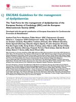
Figure 2. Algorithm for the management and treatment of surgical site infections (SSIs). *For patients with type 1 (anaphylaxis or hives) allergy to β-lactam
antibiotics. If Gram stain not available, open and debride if purulent drainage present. Where the rate of infection with methicillin-resistant Staphylococcus
aureus infection is high, consider vancomycin, daptomycin, or linezolid, pending results of culture and susceptibility tests. Adapted and modified with permission from Dellinger et al [96]. Abbreviations: GI, gastrointestinal; MRSA, methicillin-resistant Staphylococcus aureus ; WBC, white blood cell count.
Table 1. Strength of Recommendations and Quality of the Evidence
Strength of
Recommendation and
Quality of Evidence
Clarity of Balance Between
Desirable and Undesirable
Effects
Methodological Quality of
Supporting Evidence (Examples)
Implications
Desirable effects clearly
outweigh undesirable effects,
or vice versa
Consistent evidence from wellperformed RCTs or exceptionally
strong evidence from unbiased
observational studies
Recommendation can apply to most
patients in most circumstances.
Further research is unlikely to
change our confidence in the
estimate of effect
Strong recommendation,
moderate quality
evidence
Desirable effects clearly
outweigh undesirable effects,
or vice versa
Evidence from RCTs with important
limitations (inconsistent results,
methodological flaws, indirect, or
imprecise) or exceptionally strong
evidence from unbiased
observational studies
Recommendation can apply to most
patients in most circumstances.
Further research (if performed) is
likely to have an important impact on
our confidence in the estimate of
effect and may change the estimate
Strong recommendation,
low-quality quality
evidence
Desirable effects clearly
outweigh undesirable effects,
or vice versa
Evidence for at least 1 critical
outcome from observational
studies, RCTs with serious flaws
or indirect evidence
Recommendation may change when
higher-quality evidence becomes
available. Further research (if
performed) is likely to have an
important impact on our confidence
in the estimate of effect and is likely
to change the estimate
Strong recommendation,
very low-quality evidence
(very rarely applicable)
Desirable effects clearly
outweigh undesirable effects,
or vice versa
Evidence for at least 1 critical
outcome from unsystematic
clinical observations or very
indirect evidence
Recommendation may change when
higher-quality evidence becomes
available; any estimate of effect for
at least 1 critical outcome is very
uncertain.
Weak recommendation,
high-quality evidence
Desirable effects closely
balanced with undesirable
effects
Consistent evidence from wellperformed RCTs or exceptionally
strong evidence from unbiased
observational studies
Weak recommendation,
moderate-quality
evidence
Desirable effects closely
balanced with undesirable
effects
Evidence from RCTs with important
limitations (inconsistent results,
methodological flaws, indirect, or
imprecise) or exceptionally strong
evidence from unbiased
observational studies
The best action may differ depending
on circumstances or patient’s or
societal values. Further research is
unlikely to change our confidence in
the estimate of effect
Alternative approaches likely to be
better for some patients under
some circumstances. Further
research (if performed) is likely to
have an important impact on our
confidence in the estimate of effect
and may change the estimate
Weak recommendation,
low-quality evidence
Uncertainty in the estimates of
desirable effects, harms, and
burden; desirable effects,
harms, and burden may be
closely balanced
Evidence for at least 1 critical
outcome from observational
studies, from RCTs with serious
flaws or indirect evidence
Other alternatives may be equally
reasonable. Further research is very
likely to have an important impact on
our confidence in the estimate of
effect and is likely to change the
estimate
Weak recommendation,
very low-quality evidence
Major uncertainty in the
estimates of desirable effects,
harms, and burden; desirable
effects may or may not be
balanced with undesirable
effects
Evidence for at least 1 critical
outcome from unsystematic
clinical observations or very
indirect evidence
Other alternatives may be equally
reasonable. Any estimate of effect,
for at least 1 critical outcome, is very
uncertain
Abbreviation: RCT, randomized controlled trial.
made based upon presence or absence of systemic inflammatory response syndrome (SIRS), such as temperature >38°C or
<36°C, tachypnea >24 breaths per minute, tachycardia >90
beats per minute, or white blood cell count >12 000
or <400 cells/µL (moderate; Figure 1) (strong, low). An
4
•
CID
•
Stevens et al
antibiotic active against MRSA is recommended for patients
with carbuncles or abscesses who have failed initial antibiotic
treatment or have markedly impaired host defenses or in patients with SIRS and hypotension (severe; Figure 1 and
Table 2) (strong, low).
Downloaded from at IDSA member on June 30, 2015
Strong recommendation,
high-quality evidence
Table 2.
Antimicrobial Therapy for Staphylococcal and Streptococcal Skin and Soft Tissue Infections
Disease Entity
b
Dosage, Childrena
Dosage, Adults
Dicloxacillin
250 mg qid po
Comment
N/A
N/A
Cephalexin
250 mg qid po
25–50 mg/kg/d in 3–4 divided doses po
N/A
Erythromycin
250 mg qid poc
40 mg/kg/d in 3–4 divided doses po
Clindamycin
300–400 mg qid po
20 mg/kg/d in 3 divided doses po
Some strains of Staphylococcus aureus and Streptococcus
pyogenes may be resistant.
N/A
Amoxicillin-clavulanate
875/125 mg bid po
Retapamulin ointment
Apply to lesions bid
25 mg/kg/d of the amoxicillin component
in 2 divided doses po
Apply to lesions bid
For patients with limited number of lesions
Mupirocin ointment
Apply to lesions bid
Apply to lesions bid
For patients with limited number of lesions
Nafcillin or oxacillin
Cefazolin
1-2 g every 4 h IV
1 g every 8 h IV
100–150 mg/kg/d in 4 divided doses
50 mg/kg/d in 3 divided doses
Clindamycin
600 mg every 8 h IV
or
300–450 mg qid po
25–40 mg/kg/d in 3 divided doses IV or
25–30 mg/kg/d in 3 divided doses po
Parental drug of choice; inactive against MRSA
For penicillin-allergic patients except those with immediate
hypersensitivity reactions. More convenient than nafcillin
with less bone marrow suppression
Bacteriostatic; potential of cross-resistance and emergence
of resistance in erythromycin-resistant strains; inducible
resistance in MRSA
Dicloxacillin
500 mg qid po
25–50 mg/kg/d in 4 divided doses po
Oral agent of choice for methicillin-susceptible strains in
adults. Not used much in pediatrics
Cephalexin
500 mg qid po
25–50 mg/kg/d 4 divided doses po
Doxycycline,
minocycline
100 mg bid po
Not recommended for age <8 yd
For penicillin-allergic patients except those with immediate
hypersensitivity reactions. The availability of a suspension
and requirement for less frequent dosing
Bacteriostatic; limited recent clinical experience
Trimethoprimsulfamethoxazole
1–2 doublestrength tablets
bid po
8–12 mg/kg (based on trimethoprim
component) in either 4 divided doses IV
or 2 divided doses po
Bactericidal; efficacy poorly documented
Vancomycin
Linezolid
30 mg/kg/d in 2
divided doses IV
600 mg every 12 h
IV or 600 mg bid
po
600 mg every 8 h IV
or 300–450 mg
qid po
40 mg/kg/d in 4 divided doses IV
IDSA Practice Guidelines for SSTIs
For penicillin allergic patients; parenteral drug of choice for
treatment of infections caused by MRSA
Bacteriostatic; limited clinical experience; no crossresistance with other antibiotic classes; expensive
25–40 mg/kg/d in 3 divided doses IV or
30–40 mg/kg/d in 3 divided doses po
Bacteriostatic; potential of cross-resistance and emergence
of resistance in erythromycin-resistant strains; inducible
resistance in MRSA. Important option for children
Daptomycin
4 mg/kg every 24 h
IV
N/A
Bactericidal; possible myopathy
Ceftaroline
600 mg bid IV
N/A
Bactericidal
Doxycycline,
minocycline
100 mg bid po
Not recommended for age <8 yd
Bacteriostatic; limited recent clinical experience
•
Impetigo
(Staphylococcus and
Streptococcus)
Antibiotic
Trimethoprimsulfamethoxazole
1–2 doublestrength tablets
bid po
8–12 mg/kg/d (based on trimethoprim
component) in either 4 divided doses IV
or 2 divided doses po
Bactericidal; limited published efficacy data
MSSA SSTI
MRSA SSTI
Clindamycin
10 mg/kg every 12 h IV or po for children
<12 y
N/A
CID
•
5
Downloaded from at IDSA member on June 30, 2015
•
Stevens et al
See [246] for alternatives in children.
d
Infection due to Staphylococcus and Streptococcus species. Duration of therapy is 7 days, depending on the clinical response.
Adult dosage of erythromycin ethylsuccinate is 400 mg 4 times/d po.
Doses listed are not appropriate for neonates. Refer to the report by the Committee on Infectious Diseases, American Academy of Pediatrics [246], for neonatal doses.
IV. What Is Appropriate for the Evaluation and Treatment
of Erysipelas and Cellulitis?
c
b
a
Abbreviations: bid, twice daily; IV, intravenous; MRSA, methicillin-resistant Staphylococcus aureus; MSSA, methicillin-susceptible Staphylococcus aureus; N/A, not applicable; po, by mouth; qid, 4 times daily; SSTI, skin
and soft tissue infection; tid, 3 times daily.
N/A
Clindamycin, vancomycin, linezolid,
daptomycin, or telavancin. Clindamycin
resistance is <1% but may be increasing in
Asia
Penicillin 2–4 million
units every 4–6 h IV
Clindamycin 600–900
mg every 8 h IV
Nafcillin 1–2 g every 4–
6 h IV
Cefazolin 1 g every 8 h
IV
Penicillin VK 250–500
mg every 6 h po
Cephalexin 500 mg
every 6 h po
Streptococcal skin
infections
Penicillin 60–
100 000 units/kg/
dose every 6 h
10–13 mg/kg dose
every 8 h IV
50 mg/kg/dose
every 6 h
33 mg/kg/dose
every 8 h IV
Adult dosage
Non-purulent SSTI
(cellulitis)
Pediatric dosage
N/A
Comment
CID
antimicrobial agents for patients with severe
penicillin hypersensitivity
Dosage, Childrena
Dosage, Adults
Antibiotic
Disease Entity
Table 2 continued.
•
Recommendations
7. A recurrent abscess at a site of previous infection should
prompt a search for local causes such as a pilonidal cyst, hidradenitis suppurativa, or foreign material (strong, moderate).
8. Recurrent abscesses should be drained and cultured early
in the course of infection (strong, moderate).
9. After obtaining cultures of recurrent abscess, treat with a
5- to 10-day course of an antibiotic active against the pathogen
isolated (weak, low).
10. Consider a 5-day decolonization regimen twice daily of
intranasal mupirocin, daily chlorhexidine washes, and daily decontamination of personal items such as towels, sheets, and
clothes for recurrent S. aureus infection (weak, low).
11. Adult patients should be evaluated for neutrophil disorders if recurrent abscesses began in early childhood (strong,
moderate).
Recommendations
12. Cultures of blood or cutaneous aspirates, biopsies, or
swabs are not routinely recommended (strong, moderate).
13. Cultures of blood are recommended (strong, moderate),
and cultures and microscopic examination of cutaneous aspirates, biopsies, or swabs should be considered in patients with
malignancy on chemotherapy, neutropenia, severe cell-mediated immunodeficiency, immersion injuries, and animal bites
(weak, moderate).
14. Typical cases of cellulitis without systemic signs of infection should receive an antimicrobial agent that is active against
streptococci (mild; Figure 1) (strong, moderate). For cellulitis
with systemic signs of infection (moderate nonpurulent; Figure 1), systemic antibiotics are indicated. Many clinicians
could include coverage against methicillin-susceptible S. aureus
(MSSA) (weak, low). For patients whose cellulitis is associated
with penetrating trauma, evidence of MRSA infection elsewhere, nasal colonization with MRSA, injection drug use, or
SIRS (severe nonpurulent; Figure 1), vancomycin or another antimicrobial effective against both MRSA and streptococci is recommended (strong, moderate). In severely compromised
patients as defined in question 13 (severe nonpurulent;
Figure 1), broad-spectrum antimicrobial coverage may be considered (weak, moderate). Vancomycin plus either piperacillintazobactam or imipenem/meropenem is recommended as a
reasonable empiric regimen for severe infections (strong,
moderate).
15. The recommended duration of antimicrobial therapy is 5
days, but treatment should be extended if the infection has not
improved within this time period (strong, high).
Downloaded from at IDSA member on June 30, 2015
6
III. What Is the Appropriate Treatment for Recurrent Skin
Abscesses?
>38.5°C, heart rate >110 beats/minute, or white blood cell
(WBC) count >12 000/µL (weak, low).
24. A brief course of systemic antimicrobial therapy is indicated in patients with surgical site infections following clean operations on the trunk, head and neck, or extremities that also
have systemic signs of infection (strong, low).
25. A first-generation cephalosporin or an antistaphylococcal
penicillin for MSSA, or vancomycin, linezolid, daptomycin, telavancin, or ceftaroline where risk factors for MRSA are high (nasal
colonization, prior MRSA infection, recent hospitalization, recent
antibiotics), is recommended (strong, low). See also Tables 2 and 3.
26. Agents active against gram-negative bacteria and anaerobes, such as a cephalosporin or fluoroquinolone in combination with metronidazole, are recommended for infections
following operations on the axilla, gastrointestinal tract, perineum, or female genital tract (strong, low). See also Table 3.
V. Should Anti-inflammatory Agents Be Used to Complement
Antibiotic Treatment of Cellulitis?
VIII. What Is the Preferred Evaluation and Treatment
of Necrotizing Fasciitis, Including Fournier Gangrene?
Recommendation
19. Systemic corticosteroids (eg, prednisone 40 mg daily for
7 days) could be considered in nondiabetic adult patients with
cellulitis (weak, moderate).
Recommendations
27. Prompt surgical consultation is recommended for patients with aggressive infections associated with signs of systemic toxicity or suspicion of necrotizing fasciitis or gas gangrene
(severe nonpurulent; Figure 1) (strong, low).
28. Empiric antibiotic treatment should be broad (eg, vancomycin or linezolid plus piperacillin-tazobactam or a carbapenem; or plus ceftriaxone and metronidazole), as the etiology
can be polymicrobial (mixed aerobic–anaerobic microbes) or
monomicrobial (group A streptococci, community-acquired
MRSA) (strong, low). See also Table 4.
29. Penicillin plus clindamycin is recommended for treatment of documented group A streptococcal necrotizing fasciitis
(strong, low). See Figures 1, 2, and Table 4.
VI. What Is the Preferred Evaluation and Management of Patients
With Recurrent Cellulitis?
Recommendations
20. Identify and treat predisposing conditions such as
edema, obesity, eczema, venous insufficiency, and toe web abnormalities (strong, moderate). These practices should be performed as part of routine patient care and certainly during the
acute stage of cellulitis (strong, moderate).
21. Administration of prophylactic antibiotics, such as oral
penicillin or erythromycin bid for 4–52 weeks, or intramuscular
benzathine penicillin every 2–4 weeks, should be considered in
patients who have 3–4 episodes of cellulitis per year despite
attempts to treat or control predisposing factors (weak, moderate). This program should be continued so long as the predisposing factors persist (strong, moderate).
VII. What Is the Preferred Management of Surgical Site
Infections?
Recommendations
22. Suture removal plus incision and drainage should be performed for surgical site infections (strong, low).
23. Adjunctive systemic antimicrobial therapy is not routinely indicated, but in conjunction with incision and drainage may
be beneficial for surgical site infections associated with a significant systemic response (Figure 2), such as erythema and induration extending >5 cm from the wound edge, temperature
IX. What Is the Appropriate Approach to the Management of
Pyomyositis?
Recommendations
30. Magnetic resonance imaging (MRI) is the recommended
imaging modality for establishing the diagnosis of pyomyositis.
Computed tomography (CT) scan and ultrasound studies are
also useful (strong, moderate).
31. Cultures of blood and abscess material should be obtained (strong, moderate).
32. Vancomycin is recommended for initial empirical therapy. An agent active against enteric gram-negative bacilli should
be added for infection in immunocompromised patients or following open trauma to the muscles (strong, moderate).
33. Cefazolin or antistaphylococcal penicillin (eg, nafcillin or
oxacillin) is recommended for treatment of pyomyositis caused
by MSSA (strong, moderate). See Table 2.
IDSA Practice Guidelines for SSTIs
•
CID
•
7
Downloaded from at IDSA member on June 30, 2015
16. Elevation of the affected area and treatment of predisposing factors, such as edema or underlying cutaneous disorders,
are recommended (strong, moderate).
17. In lower-extremity cellulitis, clinicians should carefully
examine the interdigital toe spaces because treating fissuring,
scaling, or maceration may eradicate colonization with pathogens and reduce the incidence of recurrent infection (strong,
moderate).
18. Outpatient therapy is recommended for patients who do
not have SIRS, altered mental status, or hemodynamic instability (mild nonpurulent; Figure 1) (strong, moderate). Hospitalization is recommended if there is concern for a deeper or
necrotizing infection, for patients with poor adherence to therapy, for infection in a severely immunocompromised patient, or
if outpatient treatment is failing (moderate or severe nonpurulent; Figure 1) (strong, moderate).
34. Early drainage of purulent material should be performed
(strong, high).
35. Repeat imaging studies should be performed in the patient with persistent bacteremia to identify undrained foci of infection (strong, low).
36. Antibiotics should be administered intravenously initially, but once the patient is clinically improved, oral antibiotics
are appropriate for patients in whom bacteremia cleared
promptly and there is no evidence of endocarditis or metastatic
abscess. Two to 3 weeks of therapy is recommended (strong,
low).
X. What Is the Appropriate Approach to the Evaluation and
Treatment of Clostridial Gas Gangrene or Myonecrosis?
XI. What Is the Role of Preemptive Antimicrobial Therapy to
Prevent Infection for Dog or Cat Bites?
Recommendations
40. Preemptive early antimicrobial therapy for 3–5 days is
recommended for patients who (a) are immunocompromised;
(b) are asplenic; (c) have advanced liver disease; (d) have preexisting or resultant edema of the affected area; (e) have moderate
to severe injuries, especially to the hand or face; or (f ) have injuries that may have penetrated the periosteum or joint capsule
(strong, low).
41. Postexposure prophylaxis for rabies may be indicated; consultation with local health officials is recommended to determine if vaccination should be initiated (strong,
low).
XII. What Is the Treatment for Infected Animal Bite–Related
Wounds?
Recommendation
42. An antimicrobial agent or agents active against both aerobic and anaerobic bacteria such as amoxicillin-clavulanate
(Table 5) should be used (strong, moderate).
8
•
CID
•
Stevens et al
Recommendation
43. Tetanus toxoid should be administered to patients without toxoid vaccination within 10 years. Tetanus, diptheria, and
tetanus (Tdap) is preferred over Tetanus and diptheria (Td) if
the former has not been previously given (strong, low).
XIV. In Which Patients Is Primary Wound Closure Appropriate for
Animal Bite Wounds?
Recommendation
44. Primary wound closure is not recommended for wounds,
with the exception of those to the face, which should be managed with copious irrigation, cautious debridement, and
preemptive antibiotics (strong, low). Other wounds may be approximated (weak, low).
XV. What Is the Appropriate Treatment of Cutaneous Anthrax?
Recommendations
45. Oral penicillin V 500 mg 4 times daily (qid) for 7–10
days is the recommended treatment for naturally acquired cutaneous anthrax (strong, high).
46. Ciprofloxacin 500 mg by mouth ( po) bid or levofloxacin
500 mg intravenously (IV)/po every 24 hours × 60 days is recommended for bioterrorism cases because of presumed aerosol
exposure (strong, low).
XVI. What Is the Appropriate Approach for the Evaluation and
Treatment of Bacillary Angiomatosis and Cat Scratch Disease?
Recommendations
47. Azithromycin is recommended for cat scratch disease
(strong, moderate) according to the following dosing protocol:
(a) Patients >45 kg: 500 mg on day 1 followed by 250 mg
for 4 additional days (strong, moderate).
(b) Patients <45 kg: 10 mg/kg on day 1 and 5 mg/kg for 4
more days (strong, moderate).
48. Erythromycin 500 mg qid or doxycycline 100 mg bid for
2 weeks to 2 months is recommended for treatment of bacillary
angiomatosis (strong, moderate).
XVII. What Is the Preferred Treatment for Erysipeloid?
Recommendation
49. Penicillin (500 mg qid) or amoxicillin (500 mg 3 times
daily [tid]) for 7–10 days is recommended for treatment of erysipeloid (strong, high).
XVIII. What Is the Appropriate Treatment of Glanders?
Recommendation
50. Ceftazidime, gentamicin, imipenem, doxycycline, or ciprofloxacin is recommended based on in vitro susceptibility (strong, low).
Downloaded from at IDSA member on June 30, 2015
Recommendations
37. Urgent surgical exploration of the suspected gas gangrene
site and surgical debridement of involved tissue should be performed (severe nonpurulent; Figure 1) (strong, moderate).
38. In the absence of a definitive etiologic diagnosis, broadspectrum treatment with vancomycin plus either piperacillin/
tazobactam, ampicillin/sulbactam, or a carbapenem antimicrobial
is recommended (strong, low). Definitive antimicrobial therapy
with penicillin and clindamycin (Figure 1) is recommended
for treatment of clostridial myonecrosis (strong, low).
39. Hyperbaric oxygen (HBO) therapy is not recommended
because it has not been proven as a benefit to the patient and
may delay resuscitation and surgical debridement (strong,
low).
XIII. Should Tetanus Toxoid Be Administered for Animal Bite
Wounds?
XIX. What Is the Appropriate Diagnosis and Treatment of Bubonic
Plague?
Recommendation
51. Bubonic plague should be diagnosed by Gram stain and
culture of aspirated material from a suppurative lymph node
(strong, moderate). Streptomycin (15 mg/kg intramuscularly
[IM] every 12 hours) or doxycycline (100 mg bid po) is recommended for treatment of bubonic plague (strong, low). Gentamicin could be substituted for streptomycin (weak, low).
XX. What Is Appropriate for Diagnosis and Treatment for
Tularemia?
XXI. What Is the Appropriate Approach to Assess SSTIs in
Immunocompromised Patients?
Recommendations
56. In addition to infection, differential diagnosis of skin lesions should include drug eruption, cutaneous infiltration with
the underlying malignancy, chemotherapy- or radiation-induced reactions, Sweet syndrome, erythema multiforme, leukocytoclastic vasculitis, and graft-vs-host disease among
allogeneic transplant recipients (strong, high).
57. Differential diagnosis for infection of skin lesions should
include bacterial, fungal, viral, and parasitic agents (strong, high).
58. Biopsy or aspiration of the lesion to obtain material for
histological and microbiological evaluation should always be
implemented as an early diagnostic step (strong, high).
XXII. What Is the Appropriate Approach to Assess SSTIs in
Patients With Fever and Neutropenia?
Recommendations
59. Determine whether the current presentation of fever and
neutropenia is the patient’s initial episode of fever and neutropenia, or persistent unexplained fever of their initial episode
(after 4–7 days) or a subsequent episode of fever and neutropenia (recurrent) (strong, low).
60. Aggressively determine the etiology of the SSTI by aspiration and/or biopsy of skin and soft tissue lesions and submit
these for thorough cytological/histological assessments, microbial staining, and cultures (strong, low).
XXIII. What Is the Appropriate Antibiotic Therapy for Patients With
SSTIs During the Initial Episode of Fever and Neutropenia?
Recommendations
63. Hospitalization and empiric antibacterial therapy with
vancomycin plus antipseudomonal antibiotics such as cefepime,
a carbapenem (imipenem-cilastatin or meropenem or doripenem) or piperacillin-tazobactam is recommended (strong, high).
64. Documented clinical and microbiologic SSTIs should be
treated based on antimicrobial susceptibilities of isolated organisms (strong, high).
65. It is recommended that the treatment duration for most
bacterial SSTIs should be 7–14 days (strong, moderate).
66. Surgical intervention is recommended for drainage of
soft tissue abscess after marrow recovery or for a progressive
polymicrobial necrotizing fasciitis or myonecrosis (strong, low).
67. Adjunct colony-stimulating factor therapy (granulocyte
colony-stimulating factor [G-CSF], granulocyte macrophage
colony-stimulating factor [GM-CSF]) or granulocyte transfusions are not routinely recommended (weak, moderate).
68. Acyclovir should be administered to patients suspected
or confirmed to have cutaneous or disseminated varicella zoster
virus (herpes simplex virus [HSV] or varicella zoster virus
[VZV]) infection (strong, moderate).
XXIV. What Is the Appropriate Antimicrobial Therapy for Patients
With SSTIs During Persistent or Recurrent Episodes of Fever and
Neutropenia?
Recommendations
69. Yeasts and molds remain the primary cause of infectionassociated with persistent and recurrent fever and neutropenia;
therefore, empiric antifungal therapy (Table 6) should be added
to the antibacterial regimen (strong, high).
(a) Empiric administration of vancomycin or other agents
with gram-positive activity (linezolid, daptomycin, or ceftaroline,
Table 7) should be added if not already being administered
(strong, high).
IDSA Practice Guidelines for SSTIs
•
CID
•
9
Downloaded from at IDSA member on June 30, 2015
Recommendations
52. Serologic tests are the preferred method of diagnosing tularemia (weak, low).
53. Streptomycin (15 mg/kg every 12 hours IM) or gentamicin (1.5 mg/kg every 8 hours IV) is recommended for treatment
of severe cases of tularemia (strong, low).
54. Tetracycline (500 mg qid) or doxycycline (100 mg bid po) is
recommended for treatment of mild cases of tularemia (strong, low).
55. Notify the microbiology laboratory if tularemia is suspected (strong, high).
61. Risk-stratify patients with fever and neutropenia according to susceptibility to infection: high-risk patients are those
with anticipated prolonged (>7 days) and profound neutropenia
(absolute neutrophil count <100 cells/µL) or with a Multinational Association for Supportive Care (MASCC) score of
<21; low-risk patients are those with anticipated brief (<7
days) periods of neutropenia and few comorbidities (strong,
low) or with a MASCC score of ≥21 (strong, moderate).
62. Determine the extent of infection through a thorough
physical examination, blood cultures, chest radiograph, and additional imaging (including chest CT) as indicated by clinical
signs and symptoms (strong, low).
XXV. What Is the Appropriate Approach to Assess SSTIs in
Patients With Cellular Immunodeficiency?
Recommendations
73. Consider immediate consultation with a dermatologist
familiar with cutaneous manifestations of infection in patients
with cellular immune defects (eg, those with lymphoma, lymphocytic leukemia, recipients of organ transplants, or those receiving immunosuppressive drugs such as anti–tumor necrosis
factors or certain monoclonal antibodies) (weak, low).
74. Consider biopsy and surgical debridement early in the
management of these patients (weak, low).
75. Empiric antibiotics, antifungals, and/or antivirals should
be considered in life-threatening situations (weak, moderate).
The use of specific agents should be decided with the input of
10
•
CID
•
Stevens et al
the primary team, dermatology, infectious disease, and other
consulting teams (strong, moderate).
INTRODUCTION
This practice guideline provides recommendations for the diagnosis and management of skin and soft tissue infections (SSTIs)
in otherwise healthy hosts and compromised hosts of all age
groups. These recommendations take on new importance because of a dramatic increase in the frequency and severity of
infections and the emergence of resistance to many of the antimicrobial agents commonly used to treat SSTIs in the past. For
example, there was a 29% increase in the total hospital admissions for these infections between 2000 and 2004 [5]. In addition, 6.3 million physician’s office visits per year are attributable
to SSTIs [6]. Similarly, between 1993 and 2005, annual emergency department visits for SSTIs increased from 1.2 million
to 3.4 million patients [7]. Some of this increased frequency is
related to the emergence of community-associated methicillinresistant Staphylococcus aureus (MRSA) [5].
These infections have diverse etiologies that depend, in part,
on different epidemiological settings. As a result, obtaining a
careful history that includes information about the patient’s immune status, geographic locale, travel history, recent trauma or
surgery, previous antimicrobial therapy, lifestyle, hobbies, and
animal exposure or bites is essential when developing an adequate differential diagnosis and an appropriate index of suspicion for specific etiological agents. Recognition of the physical
examination findings and understanding the anatomical relationships of skin and soft tissue are crucial for establishing
the correct diagnosis. In some cases, this information is insufficient and biopsy or aspiration of tissue may be necessary. In addition, radiographic procedures may be critical in a small subset
of patients to determine the level of infection and the presence
of gas, abscess, or a necrotizing process. Last, surgical exploration or debridement is an important diagnostic, as well as therapeutic, procedure in patients with necrotizing infections or
myonecrosis and may be important for selected immunocompromised hosts.
Clinical evaluation of patients with SSTI aims to establish the
cause and severity of infection and must take into account pathogen-specific and local antibiotic resistance patterns. Many different microbes can cause soft tissue infections, and although
specific bacteria may cause a particular type of infection, considerable overlaps in clinical presentation occur. Clues to the diagnosis and algorithmic approaches to diagnosis are covered in
detail in the text to follow. Specific recommendations for therapy are given, each with a rating that indicates the strength of
and evidence for recommendations according to the Infectious
Diseases Society of America (IDSA)/US Public Health Service
grading system for rating recommendations in clinical
Downloaded from at IDSA member on June 30, 2015
(b) Candida species SSTIs should be treated with an echinocandin or, if Candida parapsilosis has been isolated, lipid
formulation amphotericin B (strong, high) with fluconazole
as an acceptable alternative (strong, moderate). Treatment
should be administered for 2 weeks after clearance of bloodstream infection or resolution of skin lesions (strong, moderate).
(c) Aspergillus SSTIs should be treated with voriconazole
(strong, high), or alternatively, lipid formulations of amphotericin B, posaconazole, or echinocandin for 6–12
weeks (strong, low). Mucor/Rhizopus infections should be
treated with lipid formulation amphotericin B (strong,
moderate) or posaconazole (strong, low) (Table 6). The addition of an echinocandin could be considered based on
synergy in murine models of mucormycosis, and observational clinical data (weak, low).
(d) Fusarium species infections should be treated with
high-dose IV voriconazole or posaconazole (strong, low).
(e) Begin treatment for antibiotic-resistant bacterial organisms (Table 7), in patients currently on antibiotics (strong,
moderate).
(f ) Intravenous acyclovir should be added to the patient’s
antimicrobial regimen for suspected or confirmed cutaneous
or disseminated HSV or VZV infections (strong, moderate).
70. Blood cultures should be obtained and skin lesions in this
population of patients should be aggressively evaluated by culture
aspiration, biopsy, or surgical excision, as they may be caused by
resistant microbes, yeast, or molds (strong, moderate).
71. The sensitivity of a single-serum fungal antigen test
(1,3-β-D-glucan or galactomannan tests) is low particularly in
patients receiving antifungal agents, and benefits from laboratory tests for fungal antigen or DNA detection remain inconsistent (strong, moderate).
72. Polymerase chain reaction (PCR) in peripheral blood for
HSV and VZV might be helpful in establishing a diagnosis of
disseminated infection in patients with unexplained skin lesions
(weak, moderate).
guidelines (Table 1) [2]. The following 24 clinical questions are
answered:
(XXV) What is the appropriate approach to assess SSTIs in
patients with cellular immunodeficiency?
(I) What is appropriate for the evaluation and treatment of
impetigo and ecthyma?
(II) What is the appropriate evaluation and treatment for cutaneous abscesses, furuncles, carbuncles, and inflamed epidermoid cysts?
(III) What is the appropriate treatment for recurrent skin
abscesses?
(IV) What is appropriate for the evaluation and treatment of
erysipelas and cellulitis?
(V) Should corticosteroids be used to complement antibiotic
treatment of cellulitis?
(VI) What is the preferred evaluation and management of
patients with recurrent cellulitis?
(VII) What is the preferred management of surgical site
infections?
(VIII) What is the preferred evaluation and treatment of
necrotizing fasciitis, including Fournier gangrene?
(IX) What is the appropriate approach to the management
of pyomyositis?
(X) What is the appropriate approach to the evaluation and
treatment of clostridial gas gangrene or myonecrosis?
(XI) What is the role of preemptive antimicrobial therapy to
prevent infection for dog or cat bites?
(XII) What is the treatment for infected animal bite–related
wounds?
(XIII) Should tetanus toxoid be administered for animal bite
wounds?
(XIV) In which patients is primary wound closure appropriate for animal bite wounds?
(XV) What is the appropriate treatment of cutaneous
anthrax?
(XVI) What is the appropriate approach for the evaluation
and treatment of bacillary angiomatosis and cat scratch disease?
(XVII) What is the preferred treatment for erysipeloid?
(XVIII) What is appropriate treatment of glanders?
(XIX) What is the appropriate diagnosis and treatment of
bubonic plague?
(XX) What is appropriate for diagnosis and treatment for
tularemia?
(XXI) What is the appropriate approach to assess SSTIs in
immunocompromised patients?
(XXII) What is the appropriate approach to assess SSTIs in
patients with fever and neutropenia?
(XXIII) What is the appropriate antibiotic therapy for patients
with SSTIs during the initial episode of fever and neutropenia?
(XXIV) What is the appropriate antimicrobial therapy for
patients with SSTIs during persistent or recurrent episodes of
fever and neutropenia?
PRACTICE GUIDELINES
“Practice guidelines are systematically developed statements to
assist practitioners and patients in making decisions about
appropriate health care for specific clinical circumstances” [8].
Attributes of high-quality guidelines include validity, reliability,
reproducibility, clinical applicability, clinical flexibility, clarity,
multidisciplinary process, review of evidence, and documentation [8].
METHODOLOGY
Panel Composition
Literature Review and Analysis
The recommendations in this guideline have been developed
following a review of studies published in English, although foreign-language articles were included in some of the Cochrane
reviews summarized in this guideline. Studies were identified
through Library of Congress, LISTA (EBSCO), and PubMed
searches with no date restrictions using subject headings. Examples of keywords used to conduct literature searches were as follows: skin abscess (recurrent and relapsing), dog bites, skin and
soft tissue infections, cellulitis, erysipelas, surgical site infections, wounds, staphylococcus, streptococcus, cat bites, tetanus,
bite wounds (care and closure), irrigation, amoxicillin, amoxicillin clavulanate, cefuroxime, levofloxacin, moxifloxacin, sulfamethoxazole-trimethoprim, erythromycin, azithromycin.
Process Overview
To evaluate evidence, the panel followed a process consistent
with other IDSA guidelines. The process for evaluating the
IDSA Practice Guidelines for SSTIs
•
CID
•
11
Downloaded from at IDSA member on June 30, 2015
A panel of 10 multidisciplinary experts in the management of
SSTIs in children and adults was convened in 2009. Efforts
were made to include representatives from diverse geographic
areas, pediatric and adult practitioners, and a wide breadth of
specialties. The panel consisted of 10 members of IDSA. Representation included 8 adult infectious disease physicians, 1 pediatric infectious disease physician, and 1 general surgeon. Panel
members were selected based on their clinical and research expertise on diverse SSTIs including infections in compromised
hosts, necrotizing fasciitis, gas gangrene, cellulitis, and cutaneous abscesses and infections following surgery and animal and
human bites. Finally, some members were selected on the basis
of their expertise for specific microbes such as staphylococci,
streptococci, Clostridium species, and anaerobes. Two members
were selected to provide congruency with the IDSA/MRSA
Guidelines Panel.
evidence was based on the IDSA Handbook on Clinical Practice
Guideline Development and involved a systematic weighting of
the quality of the evidence and the grade of recommendation
using the Grading of Recommendations Assessment, Development, and Evaluation (GRADE) system (Table 1) [1–4, 9, 10].
GRADE is a newly created system for grading the quality of evidence and strength of recommendations for healthcare [2, 11].
Panel members were divided into pairs, consisting of primary
and secondary authors. Each author was asked to review the literature, evaluate the evidence, and determine the strength of the
recommendations along with an evidence summary supporting
each recommendation. The panel reviewed all recommendations, their strength, and quality of evidence. Discrepancies
were discussed and resolved, and all panel members are in
agreement with the final recommendations.
Consensus Development Based on Evidence
Guidelines and Conflicts of Interest
The expert panel complied with the IDSA policy on conflicts of
interest, which requires disclosure of any financial or other interest that might be construed as constituting an actual, potential, or apparent conflict. Panel members were provided IDSA’s
conflicts of interest disclosure statement and were asked to identify ties to companies developing products that might be affected by promulgation of the guideline. Information was requested
regarding employment, consultancies, stock ownership, honoraria, research funding, expert testimony, and membership on
company advisory committees. Decisions were made on a
case-by-case basis as to whether an individual’s role should be
limited as a result of a conflict. Potential conflicts of interests are
listed in the Acknowledgments section.
Revision Dates
At annual intervals, the panel chair, the SPGC liaison advisor,
and the chair of the SPGC will determine the need for revisions
to the guideline based on an examination of current literature. If
necessary, the entire panel will reconvene to discuss potential
changes. When appropriate, the panel will recommend revision
of the guideline to the SPGC and IDSA board and other collaborating organizations for review and approval.
12
•
CID
•
Stevens et al
I. What Is Appropriate for the Evaluation and Treatment
of Impetigo and Ecthyma?
Recommendations
1. Gram stain and culture of the pus or exudates from skin
lesions of impetigo and ecthyma are recommended to help
identify whether Staphylococcus aureus and/or a β-hemolytic
Streptococcus is the cause (strong, moderate), but treatment
without these studies is reasonable in typical cases (strong,
moderate).
2. Bullous and nonbullous impetigo can be treated with oral
or topical antimicrobials, but oral therapy is recommended for
patients with numerous lesions or in outbreaks affecting several
people to help decrease transmission of infection. Treatment for
ecthyma should be an oral antimicrobial.
(a) Treatment of bullous and nonbullous impetigo should
be with either topical mupirocin or retapamulin twice daily
(bid) for 5 days (strong, high).
(b) Oral therapy for ecthyma or impetigo should be a 7-day
regimen with an agent active against S. aureus unless cultures
yield streptococci alone (when oral penicillin is the recommended agent) (strong, high). Because S. aureus isolates
from impetigo and ecthyma are usually methicillin susceptible, dicloxacillin or cephalexin is recommended. When
MRSA is suspected or confirmed, doxycycline, clindamycin,
or sulfamethoxazole-trimethoprim (SMX-TMP) is recommended (strong, moderate).
(c) Systemic antimicrobials should be used for infections
during outbreaks of poststreptococcal glomerulonephritis to
help eliminate nephritogenic strains of Streptococcus pyogenes from the community (strong, moderate).
Evidence Summary
Impetigo can be either bullous or nonbullous [12]. Bullous impetigo is caused by strains of S. aureus that produce a toxin that
cleaves the dermal-epidermal junction to form fragile, thinroofed vesicopustules. These lesions may rupture, creating
crusted, erythematous erosions, often surrounded by a collar
of the roof ’s remnants. Nonbullous impetigo can occur from infections with β-hemolytic streptococci or S. aureus, or both in
combination [12]. Impetigo begins as erythematous papules
that rapidly evolve into vesicles and pustules that rupture,
with the dried discharge forming honey-colored crusts on an erythematous base.
Ecthyma is a deeper infection than impetigo, and S. aureus
and/or streptococci may be the cause. Lesions begin as vesicles
that rupture, resulting in circular, erythematous ulcers with adherent crusts, often with surrounding erythematous edema. Unlike impetigo, ecthyma heals with scarring [12].
Downloaded from at IDSA member on June 30, 2015
The panel met twice for face-to-face meetings and conducted
teleconferences on 6 occasions to complete the work of the
guideline. The purpose of the teleconferences was to discuss
the clinical questions to be addressed, assign topics for review
and writing of the initial draft, and discuss recommendations.
The panel as a whole reviewed all individual sections. The
guideline was reviewed and approved by the IDSA Standards
and Practice Guidelines Committee (SPGC) and Board of Directors and endorsed by the Pediatric Infectious Diseases Society (PIDS).
RECOMMENDATIONS FOR IMPETIGO AND
ECTHYMA
Cultures of the vesicle fluid, pus, erosions, or ulcers establish
the cause. Unless cultures yield streptococci alone, antimicrobial
therapy should be active against both S. aureus and streptococci
[12]. Oral penicillinase–resistant penicillin or first-generation
cephalosporins are usually effective as most staphylococcal isolates from impetigo and ecthyma are methicillin susceptible
[13]. Alternatives for penicillin-allergic patients or infections
with MRSA include doxycycline, clindamycin, or SMX-TMP.
When streptococci alone are the cause, penicillin is the drug
of choice, with a macrolide or clindamycin as an alternative
for penicillin-allergic patients. Topical treatment with mupirocin [12] or retapamulin [14] is as effective as oral antimicrobials
for impetigo. Clinical experience suggests that systemic therapy
is preferred for patients with numerous lesions or in outbreaks
affecting several people, to help decrease transmission of infection [15] (Table 2).
II. What Is the Appropriate Evaluation and Treatment for Purulent
SSTIs (Cutaneous Abscesses, Furuncles, Carbuncles, and
Inflamed Epidermoid Cysts)? (Figure 1)
Recommendations
3. Gram stain and culture of pus from carbuncles and abscesses are recommended, but treatment without these studies
is reasonable in typical cases (strong, moderate).
4. Gram stain and culture of pus from inflamed epidermoid
cysts are not recommended (strong, moderate).
5. Incision and drainage is the recommended treatment for
inflamed epidermoid cysts, carbuncles, abscesses, and large furuncles (strong, high).
6. The decision to administer antibiotics directed against
S. aureus as an adjunct to incision and drainage should
be made based on the presence or absence of systemic inflammatory response syndrome (SIRS) such as temperature >38°C
or <36°C, tachypnea >24 breaths per minute, tachycardia >90
beats per minute, or white blood cell count >12 000 or <400
cells/µL (moderate; Figure 1) (strong, low). An antibiotic
active against MRSA is recommended for patients with carbuncles or abscesses who have markedly impaired host
defenses and in patients with SIRS (Figure 1, Table 2)
(strong, low).
Evidence Summary
Cutaneous Abscesses. Cutaneous abscesses are collections of
pus within the dermis and deeper skin tissues. They are usually
painful, tender, and fluctuant red nodules, often surmounted by
a pustule and encircled by a rim of erythematous swelling. Cutaneous abscesses can be polymicrobial, containing regional
skin flora or organisms from the adjacent mucous membranes,
IDSA Practice Guidelines for SSTIs
•
CID
•
13
Downloaded from at IDSA member on June 30, 2015
RECOMMENDATIONS FOR PURULENT SKIN
AND SOFT TISSUE INFECTIONS
but S. aureus alone causes a large percentage of skin abscesses,
with a substantial number due to MRSA strains [16–18].
Epidermoid (or epidermal inclusion) cysts, often erroneously
labeled sebaceous cysts, ordinarily contain skin flora in a cheesy
keratinous material. When inflammation and purulence occur,
they are a reaction to rupture of the cyst wall and extrusion of its
contents into the dermis, rather than an actual infectious process [19].
Incision, evacuation of pus and debris, and probing of the
cavity to break up loculations provides effective treatment of cutaneous abscesses and inflamed epidermoid cysts. A randomized trial comparing incision and drainage of cutaneous
abscesses to ultrasonographically guided needle aspiration of
the abscesses showed that aspiration was successful in only
25% of cases overall and <10% with MRSA infections [20]. Accordingly, this form of treatment is not recommended. Simply
covering the surgical site with a dry dressing is usually the easiest and most effective treatment of the wound [21, 22]. Some
clinicians close the wound with sutures or pack it with gauze
or other absorbent material. One small study, however, found
that packing caused more pain and did not improve healing
when compared to just covering the incision site with sterile
gauze [23].
The addition of systemic antibiotics to incision and drainage
of cutaneous abscesses does not improve cure rates [17, 21, 22,
24, 25], even in those due to MRSA, but did have a modest effect
on the time to recurrence of other abscesses [17, 25]. However,
systemic antibiotics should be given to patients with severely
impaired host defenses or signs or symptoms of systemic infection (Figure 1, Table 2). In addition, multiple abscesses, extremes of age, and lack of response to incision and drainage
alone are additional settings in which systemic antimicrobial
therapy should be considered.
Furuncles and Carbuncles. Furuncles (or “boils”) are infections of the hair follicle, usually caused by S. aureus, in
which suppuration extends through the dermis into the subcutaneous tissue, where a small abscess forms. They differ from
folliculitis, in which the inflammation is more superficial and
pus is limited to the epidermis. Clinically, furuncles are inflammatory nodules with overlying pustules through which
hair emerges. Infection involving several adjacent follicles produces a carbuncle, a coalescent inflammatory mass with pus
draining from multiple follicular orifices. Carbuncles develop
most commonly on the back of the neck, especially in individuals with diabetes. These are typically larger and deeper than
furuncles.
Furuncles often rupture and drain spontaneously or following treatment with moist heat. Most large furuncles and all carbuncles should be treated with incision and drainage. Systemic
antimicrobials are usually unnecessary, unless fever or other evidence of systemic infection is present (Figure 1).
RECOMMENDATIONS FOR RECURRENT SKIN
ABSCESSES
III. What Is the Appropriate Treatment for Recurrent Skin
Abscesses?
Evidence Summary
A recurrent abscess at a previous site of infection may be caused
by local factors such as foreign material, hidradenitis suppurativa, or pilonidal cyst [26, 27], eradication of which can be curative. Incision and drainage should be performed for recurrent
abscesses. The benefits of adjunctive antimicrobial therapy in
preventing recurrences are unknown. Older randomized trials
showed that twice-daily intranasal mupirocin for 5 days each
month [28] or a 3-month program of oral clindamycin 150
mg daily [29] reduced the rate of further infections. Whether
such regimens are effective in the current era of communityacquired MRSA is unclear [30]. In one randomized trial, twicedaily application of nasal mupirocin for 5 days among military
personnel who carried MRSA in the nose did not reduce the frequency of subsequent skin infections [30, 31]. Scrubbing the
body thrice weekly with chlorhexidine-impregnated cloths
after showering was also deemed ineffective [32]. A 5-day decolonization with twice-daily intranasal mupirocin and daily
bathing with chlorhexidine [32] or dilute bleach (1/4–1/2 cup
of bleach per full bath) for prevention of recurrences may be
considered, but data about efficacy are sparse. One uncontrolled
study reported termination of an epidemic of furunculosis in a
village by use of mupirocin, antibacterial hand cleanser, and
daily washing of towels, sheets, combs, and razors [33]. A recent
study in children found employing preventive measures for the
patient and the household contacts resulted in significantly
fewer recurrences in the patient than employing the measures
in the patient only [34]. Because patients with neutrophil dysfunction develop recurrent abscesses in early childhood,
14
•
CID
•
Stevens et al
RECOMMENDATIONS FOR ERYSIPELAS AND
CELLULITIS
IV. What Is Appropriate for the Evaluation and Treatment of
Erysipelas and Cellulitis?
Recommendations
12. Cultures of blood or cutaneous aspirates, biopsies, or
swabs are not routinely recommended (strong, moderate).
13. Cultures of blood are recommended (strong, moderate),
and cutaneous and microscopic examination of cutaneous aspirates, biopsies, or swabs should be considered in patients with
malignancy on chemotherapy, neutropenia, severe cell-mediated immunodeficiency, immersion injuries, and animal bites
(weak, moderate).
14. Typical cases of cellulitis without systemic signs of infection should receive an antimicrobial agent that is active against
streptococci (mild; Figure 1) (strong, moderate). For cellulitis
with systemic signs of infection (moderate nonpurulent SSTI;
Figure 1) systemic antibiotics are indicated. Many clinicians
could include coverage against MSSA (weak, low). For patients
whose cellulitis is associated with penetrating trauma, evidence
of MRSA infection elsewhere, nasal colonization with MRSA,
injection drug use, purulent drainage, or SIRS (severe nonpurulent), vancomycin or another antimicrobial effective against
both MRSA and streptococci is recommended (strong, moderate). In severely compromised patients (as defined in question
13), broad-spectrum antimicrobial coverage may be considered
(weak, moderate). Vancomycin plus either piperacillin-tazobactam or imipenem-meropenem is recommended as a reasonable
empiric regimen for severe infection (strong, moderate).
15. The recommended duration of antimicrobial therapy is 5
days, but treatment should be extended if the infection has not
improved within this time period (strong, high).
16. Elevation of the affected area and treatment of predisposing factors, such as edema or underlying cutaneous disorders,
are recommended (strong, moderate).
17. In lower extremity cellulitis, clinicians should carefully
examine the interdigital toe spaces because treating fissuring,
scaling, or maceration may eradicate colonization with pathogens and reduce the incidence of recurrent infection (strong,
moderate).
18. Outpatient therapy is recommended for patients who do
not have SIRS, altered mental status, or hemodynamic instability (mild nonpurulent; Figure 1) (strong, moderate). Hospitalization is recommended if there is concern for a deeper or
necrotizing infection, for patients with poor adherence to therapy, for infection in a severely immunocompromised patient, or
Downloaded from at IDSA member on June 30, 2015
Recommendations
7. A recurrent abscess at a site of previous infection should
prompt a search for local causes such as a pilonidal cyst, hidradenitis suppurativa, or foreign material (strong, moderate).
8. Recurrent abscesses should be drained and cultured early
in the course of infection (strong, moderate).
9. Culture recurrent abscess and treat with a 5- to 10-day
course of an antibiotic active against the pathogen isolated
(weak, low).
10. Consider a 5-day decolonization regimen twice daily of
intranasal mupirocin, daily chlorhexidine washes, and daily decontamination of personal items such as towels, sheets, and
clothes for recurrent S. aureus infection (weak, low).
11. Adult patients should be evaluated for neutrophil disorders if recurrent abscesses began in early childhood (strong,
moderate).
patients who develop abscesses during adulthood do not need
evaluation of neutrophil function.
if outpatient treatment is failing (moderate or severe nonpurulent; Figure 1) (strong, moderate).
IDSA Practice Guidelines for SSTIs
•
CID
•
15
Downloaded from at IDSA member on June 30, 2015
Evidence Summary
“Cellulitis” and “erysipelas” refer to diffuse, superficial, spreading skin infections. The term “cellulitis” is not appropriate for
cutaneous inflammation associated with collections of pus, such
as in septic bursitis, furuncles, or skin abscesses. For example,
when cutaneous redness, warmth, tenderness, and edema encircle a suppurative focus such as an infected bursa, the appropriate terminology is “septic bursitis with surrounding
inflammation,” rather than “septic bursitis with surrounding
cellulitis.” This distinction is clinically crucial, for the primary
treatment of cellulitis is antimicrobial therapy, whereas for purulent collections the major component of management is
drainage of the pus, with antimicrobial therapy either being unnecessary or having a subsidiary role (Figure 1 and Table 2).
The term “erysipelas” has 3 different meanings: (1) for some,
erysipelas is an infection limited to the upper dermis, including
the superficial lymphatics, whereas cellulitis involves the deeper
dermis and subcutaneous fat, and on examination erysipelas
putatively has more clearly delineated borders of inflammation
than cellulitis; (2) for many, erysipelas has been used to refer to
cellulitis involving the face only; and (3) for others, especially in
European countries, cellulitis and erysipelas are synonyms [35].
These infections cause rapidly spreading areas of erythema,
swelling, tenderness, and warmth, sometimes accompanied by
lymphangitis and inflammation of the regional lymph nodes.
The skin surface may resemble an orange peel ( peau d’orange)
due to superficial cutaneous edema surrounding hair follicles
and causing skin dimpling because the follicles remain tethered
to the underlying dermis. Vesicles, bullae, and cutaneous hemorrhage in the form of petechiae or ecchymoses may develop.
Systemic manifestations are usually mild, but fever, tachycardia,
confusion, hypotension, and leukocytosis are sometimes present and may occur hours before the skin abnormalities appear.
These infections arise when microbes breach the cutaneous
surface, especially in patients with fragile skin or diminished
local host defenses from such conditions as obesity, previous
cutaneous trauma (including surgery), prior episodes of cellulitis, and edema from venous insufficiency or lymphedema
[36, 37]. The origin of the disrupted skin surface may be obvious, such as trauma, ulceration, and preexisting cutaneous inflammation, but often the breaks in the skin are small and
clinically unapparent. These infections are most common on
the lower legs. Blood cultures are generally positive in ≤5% of
cases [38]. The yield of cultures of needle aspirations of the inflamed skin ranges from ≤5% to approximately 40% [39–46].
The differences in diagnostic sensitivity and specificity are
due to the variety of patient populations studied, the definitions
of cellulitis, the inclusion or exclusion of cases with associated
abscesses, and the determination of whether isolates are pathogens or contaminants.
Cultures of punch biopsy specimens yield an organism in
20%–30% of cases [39, 47], but the concentration of bacteria
in the tissues is usually quite low [47]. Combined data from
specimen cultures, serologic studies [41, 48–51], and other
methods (eg, immunohistochemical staining to detect antigens
in skin biopsies [51, 52]), suggests that the vast majority of these
infections arise from streptococci, often group A, but also from
other groups, such as B, C, F, or G. The source of these pathogens is frequently unclear, but in many cases of leg cellulitis, the
responsible streptococci reside in macerated, scaly, or fissured
interdigital toe spaces [53, 54]. This observation underscores
the importance of detecting and treating tinea pedis, erythrasma, and other causes of toe web abnormalities. Occasionally,
the reservoir of streptococci is the anal canal [55] or the vagina,
especially for group B streptococcal cellulitis in patients with
previous gynecologic cancer treated with surgery and radiation
therapy. Staphylococcus aureus less frequently causes cellulitis,
but cases due to this organism are typically associated with an
open wound or previous penetrating trauma, including sites of
illicit drug injection. Several other organisms can cause cellulitis, but usually only in special circumstances, such as animal
bites, freshwater or saltwater immersion injuries, neutropenia,
or severe cell-mediated immunodeficiency.
Cultures of blood, tissue aspirates, or skin biopsies are unnecessary for typical cases of cellulitis. Blood cultures should be obtained and cultures of skin biopsy or aspirate considered for
patients with malignancy, severe systemic features (such as
high fever and hypotension), and unusual predisposing factors,
such as immersion injury, animal bites, neutropenia, and severe
cell-mediated immunodeficiency [42].
Therapy for typical cases of cellulitis should include an antibiotic active against streptococci (Table 2). A large percentage of
patients can receive oral medications from the start for typical
cellulitis [56], and suitable antibiotics for most patients include
penicillin, amoxicillin, amoxicillin-clavulanate, dicloxacillin,
cephalexin, or clindamycin. In cases of uncomplicated cellulitis,
a 5-day course of antimicrobial therapy is as effective as a 10-day
course, if clinical improvement has occurred by 5 days [57]. In a
retrospective study of cellulitis and abscesses requiring hospitalization, the average duration of treatment was 2 weeks and only
about one-third of patients received specific treatment for grampositive pathogens [58]. Two-thirds received very-broad-spectrum treatment, and the failure rate of 12% was not different regardless of spectrum of treatment. In some patients, cutaneous
inflammation and systemic features worsen after initiating therapy, probably because sudden destruction of the pathogens releases potent enzymes that increase local inflammation.
MRSA is an unusual cause of typical cellulitis. A prospective
study of patients with cellulitis in a medical center with a high
V. Should Anti-inflammatory Agents Be Used to Complement
Antibiotic Treatment of Cellulitis?
Recommendation
19. Systemic corticosteroids (eg, prednisone 40 mg daily for
7 days) could be considered in nondiabetic adult patients with
cellulitis (weak, moderate).
Evidence Summary
Treating the inflammation in these infections by combining antimicrobial therapy with either a nonsteroidal anti-inflammatory
agent (ibuprofen 400 mg 4 times daily [qid] for 5 days) or systemic corticosteroids significantly hastens clinical improvement compared with antimicrobial therapy alone [60, 61]. A randomized,
double-blind, placebo-controlled trial involving 108 adult nondiabetic patients, demonstrated that an 8-day course of oral corticosteroids in combination with antimicrobial therapy led to a
significantly more rapid clinical resolution of cellulitis (primarily
of the legs) than antimicrobial therapy alone [61, 62]. Long-term
follow-up of these patients showed no difference in relapse or recurrence [61, 62]. The benefits of systemic corticosteroids in this
situation are consistent with their efficacy and safety as adjunctive
treatment in other infections [63]. The clinician must ensure that
a deeper infection such as necrotizing fasciitis is not present.
Most patients can receive treatment without hospitalization
[63, 64]. Hospitalization is indicated for suspicion of necrotizing
infection or for patients with severe systemic features, such as
16
•
CID
•
Stevens et al
fever, delirium, or hypotension. Other indications include
poor response to outpatient therapy, severe immunocompromise, and problems with a patient’s adherence to treatment.
RECOMMENDATIONS FOR PATIENTS WITH
RECURRENT CELLULITIS
VI. What Is the Preferred Evaluation and Management of Patients
with Recurrent Cellulitis?
Recommendations
20. Identify and treat predisposing conditions such as
edema, obesity, eczema, venous insufficiency, and toe web abnormalities (strong, moderate). These practices should be performed as part of routine patient care and certainly during the
acute stage of cellulitis (strong, moderate).
21. Administration of prophylactic antibiotics, such as oral
penicillin or erythromycin bid for 4–52 weeks, or intramuscular
benzathine penicillin every 2–4 weeks, should be considered in
patients who have 3–4 episodes of cellulitis per year despite attempts to treat or control predisposing factors (weak, moderate). This program should be continued so long as the
predisposing factors persist (strong, moderate).
Evidence Summary
Patients with a previous attack of cellulitis, especially involving
the legs, have annual recurrences rates of about 8%–20% [65–
67]. The infection usually occurs in the same area as the previous episode. Edema, especially lymphedema and other local risk
factors such as venous insufficiency, prior trauma (including
surgery) to the area, and tinea pedis or other toe web abnormalities [65–71], increase the frequency of recurrences. Other predisposing conditions include obesity, tobacco use, a history of
cancer, and homelessness [66, 67, 71]. Addressing these factors
might decrease the frequency of recurrences, but evidence for
any such a benefit is sparse. For patients with recurrences despite such efforts, antimicrobial prophylaxis may reduce the
frequency of future episodes. Two randomized trials using
twice-daily oral penicillin or erythromycin demonstrated a substantial reduction in recurrences among the antibiotic recipients
compared to controls [72, 73]. An observational trial of monthly
intramuscular injections of 1.2 million units of benzathine penicillin found that this regimen was beneficial only in the subgroup of patients who had no identifiable predisposing factors
for recurrence [74]. In a study of patients with recurrent cellulitis involving arm lymphedema caused by breast cancer treatment, 2.4 million units of biweekly intramuscular benzathine
penicillin seemed to reduce the frequency of episodes, but
there was no control group [75]. The duration of therapy is indefinite, and infections may recur once prophylaxis is discontinued. For example, a recent double-blind comparative trial
demonstrated that phenoxymethyl-penicillin given as 250 mg
Downloaded from at IDSA member on June 30, 2015
incidence of other MRSA-related SSTIs demonstrated that
treatment with β-lactams, such as cefazolin or oxacillin, was
successful in 96% of patients, suggesting that cellulitis due to
MRSA is uncommon and treatment for that organism is usually
unnecessary [50]. However, coverage for MRSA may be prudent
in cellulitis associated with penetrating trauma, especially from
illicit drug use, purulent drainage, or with concurrent evidence
of MRSA infection elsewhere. Options for treatment of MRSA
in those circumstances (Table 2) include intravenous drugs
(vancomycin, daptomycin, linezolid, or telavancin) or oral therapy with doxycycline, clindamycin, or SMX-TMP. If coverage
for both streptococci and MRSA is desired for oral therapy, options include clindamycin alone or the combination of either
SMX-TMP or doxycycline with a β-lactam (eg, penicillin, cephalexin, or amoxicillin). The activity of doxycycline and SMXTMP against β-hemolytic streptococci is not known, and in
the absence of abscess, ulcer, or purulent drainage, β-lactam
monotherapy is recommended. This is further substantiated
by a recent double-blind study showing that a combination of
SMX-TMP plus cephalexin was no more efficacious than cephalexin alone in pure cellulitis [59]. Elevation of the affected area
hastens improvement by promoting gravity drainage of edema
and inflammatory substances. Patients should also receive therapy for any predisposing conditions, such as tinea pedis, trauma, or venous eczema (“stasis dermatitis”).
twice daily for 12 months increased the time to recurrence to
626 days compared with 532 days in the control group and decreased the frequency of recurrence from 37% to 22% [76].
RECOMMENDATIONS FOR SURGICAL SITE
INFECTIONS
VII. What Is the Preferred Management of Surgical Site
Infections?
Evidence Summary
Wound infections, or surgical site infections (SSIs) are the most
common adverse event affecting hospitalized surgical patients
[77]. Data from the National Nosocomial Infection Surveillance
System (NNIS) show an average incidence of SSI of 2.6%, accounting for 38% of nosocomial infections in surgical patients
[78]. The frequency of SSI is clearly related to the category of
operation, with clean and low-risk operations (by NNIS classification) having the lowest incidence, and contaminated and
high-risk operations having higher infection rates [79]. Unfortunately, there are no studies that have objectively compared
treatments for SSI.
SSIs are divided into the categories of superficial incisional
SSI, deep incisional SSI, and organ/space SSI [78]. Superficial
incisional SSIs involve only the subcutaneous space, between
the skin and underlying muscular fascia, occur within 30 days
IDSA Practice Guidelines for SSTIs
•
CID
•
17
Downloaded from at IDSA member on June 30, 2015
Recommendations
22. Suture removal plus incision and drainage should be performed for surgical site infections (strong, low).
23. Adjunctive systemic antimicrobial therapy is not routinely indicated, but in conjunction with incision and drainage may
be beneficial for surgical site infections associated with a significant systemic response (Figure 2) such as erythema and induration extending >5 cm from the wound edge, temperature >38.5°
C, heart rate >110 beats/minute, or white blood cell (WBC)
count >12 000/µL (weak, low).
24. A brief course of systemic antimicrobial therapy is indicated in patients with surgical site infections following clean operations on the trunk, head and neck, or extremities that also
have systemic signs of infection (strong, low).
25. A first-generation cephalosporin or an antistaphylococcal penicillin for methicillin-susceptible Staphylococcus aureus
(MSSA) or vancomycin, linezolid, daptomycin, telavancin, or
ceftaroline where risk factors for MRSA are high (nasal colonization, prior MRSA infection, recent hospitalization, recent antibiotics) is recommended (strong, low).
26. Agents active against gram-negative bacteria and anaerobes, such as a cephalosporin or fluoroquinolone in combination with metronidazole are recommended for infections
following operations on the axilla, gastrointestinal (GI) tract,
perineum, or female genital tract (Table 2) (strong, low).
of the surgery, and are documented with at least 1 of the following: (1) purulent incisional drainage, (2) positive culture of
aseptically obtained fluid or tissue from the superficial
wound, (3) local signs and symptoms of pain or tenderness,
swelling, and erythema after the incision is opened by the surgeon (unless culture negative), or (4) diagnosis of SSI by the attending surgeon or physician based on their experience and
expert opinion. A deep incisional infection involves the deeper
soft tissue (eg, fascia and muscle), and occurs within 30 days of
the operation or within 1 year if a prosthesis was inserted and
has the same findings as described for a superficial incisional
SSI. An organ/space SSI has the same time constraints and evidence for infection as a deep incisional SSI, and it may involve
any part of the anatomy (organs or spaces) other than the original surgical incision [78]. Examples would include postoperative peritonitis, empyema, or joint space infection. Any deep SSI
that does not resolve in the expected manner following treatment should be investigated as a possible superficial manifestation of a deeper organ/space infection. Diagnosis and treatment
of organ space infections in the abdomen are discussed in other
guidelines. Tedizolid and dalbavancin are also effective treatments of SSTI including those caused by MRSA and may be approved by the US Food and Drug Administration (FDA) in June
2014.
Local signs of pain, swelling, erythema, and purulent drainage provide the most reliable information in diagnosing an SSI.
In morbidly obese patients or in those with deep, multilayer
wounds such as after thoracotomy, external signs of SSI may
be delayed. While many patients with a SSI will develop fever,
it usually does not occur immediately postoperatively, and in
fact, most postoperative fevers are not associated with an SSI
[80]. Flat, erythematous skin changes can occur around or
near a surgical incision during the first week without swelling
or wound drainage. Most resolve without any treatment. The
cause is unknown but may relate to tape sensitivity or other
local tissue insult not involving bacteria. Numerous experimental studies and clinical trials demonstrate that antibiotics begun
immediately postoperatively or continued for long periods after
the procedure do not prevent or cure this inflammation or infection [81–88]. Therefore, the suspicion of possible SSI does
not justify use of antibiotics without a definitive diagnosis
and the institution of other therapeutic measures such as opening the wound (Figure 2).
SSIs rarely occur during the first 48 hours after surgery, and
fever during that period usually arises from noninfectious or
unknown causes. SSIs that do occur in this time frame are almost always due to S. pyogenes or Clostridium species. After
48 hours, SSI is a more common source of fever, and careful inspection of the wound is indicated; by 4 days after surgery, a
fever is equally likely to be caused by an SSI or by another infection or other unknown sources [80]. Later infections are less
18
•
CID
•
Stevens et al
intestinal or genital tracts, S. aureus and streptococcal species
are the most common organisms. If the institution in which
the operation was performed has a significant proportion of infections with MRSA or the patient has had prior MRSA infection, nasal colonization or was previously on antibiotics, the
initial antibiotic should include vancomycin, linezolid, daptomycin, telavancin, or ceftaroline for MRSA coverage as well as
one of the following for gram-negative and anaerobic coverage:
(1) piperacillin-tazobactam, (2) a carbapenem, or (3) ceftriaxone and metronidazole (Table 3).
Infections following surgical operations on the axilla also
have a significant recovery of gram-negative organisms, and
those in the perineum have a higher incidence of gram-negative
organisms and anaerobes [100, 103, 104]; antibiotic selections
should provide coverage for these organisms (Table 3). Figure 2
presents a schematic algorithm to approach patients with suspected SSIs and includes specific antibiotic recommendations
[105]. Infections developing after surgical procedures involving
nonsterile areas such as colonic, vaginal, biliary, or respiratory
mucosa may be caused by a combination of aerobic and anaerobic bacteria [18, 87, 88, 101]. These infections can rapidly progress and involve deeper structures than just the skin, such as
fascia, fat, or muscle (Tables 3 and 4).
RECOMMENDATIONS FOR EVALUATION AND
TREATMENT OF NECROTIZING FASCIITIS
VIII. What Is the Preferred Evaluation and Treatment of
Necrotizing Fasciitis, Including Fournier Gangrene?
Recommendations
27. Prompt surgical consultation is recommended for patients with aggressive infections associated with signs of systemic toxicity or suspicion of necrotizing fasciitis or gas gangrene
(severe nonpurulent; Figure 1) (strong, low).
28. Empiric antibiotic treatment should be broad (eg, vancomycin or linezolid plus piperacillin-tazobactam or plus a carbapenem, or plus ceftriaxone and metronidazole), as the etiology
can be polymicrobial (mixed aerobic-anaerobic microbes) or
monomicrobial (group A Streptococcus, community-acquired
MRSA) (strong, low).
29. Penicillin plus clindamycin is recommended for treatment of documented group A streptococcal necrotizing fasciitis
(strong, low).
Evidence Summary
Necrotizing SSTIs differ from the milder, superficial infections
by clinical presentation, coexisting systemic manifestations, and
treatment strategies (Table 4). These deep infections involve the
fascial and/or muscle compartments and are potentially devastating due to major tissue destruction and death. They usually
develop from an initial break in the skin related to trauma or
Downloaded from at IDSA member on June 30, 2015
likely, but surveillance standards mandate 30 days of follow-up
for operations without placement of prosthetic material and for
1 year for operations where a prosthesis was inserted.
Accordingly, fever or systemic signs during the first several
postoperative days should be followed by direct examination
of the wound to rule out signs suggestive of streptococcal or
clostridial infection (see section on necrotizing soft tissue infections and clostridial myonecrosis), but should not otherwise
cause further manipulation of the wound. Patients with an
early infection due to streptococci or clostridia have wound
drainage with the responsible organisms present on Gram
stain (Figure 2). White blood cells may not be evident in the
drainage in most clostridial and some early streptococcal infections. Another rare cause of early fever and systemic signs
following operation is staphylococcal wound toxic shock syndrome [89, 90]. In these cases the wound is often deceptively
benign in appearance. Erythroderma occurs early and desquamation occurs late. Fever, hypotension, abnormal hepatic and
renal blood studies, and diarrhea are early findings. Appropriate
treatment is to open the incision, perform culture, and begin
antistaphylococcal treatment.
The most important therapy for an SSI is to open the incision, evacuate the infected material, and continue dressing
changes until the wound heals by secondary intention. Most
textbooks of surgery, infectious diseases, or even surgical infectious diseases extensively discuss the epidemiology, prevention,
and surveillance of SSIs, but not their treatment [91–97]. Two
contain simple, unreferenced, recommendations to open an infected wound without using antibiotics [96, 98]. Thus, if there is
<5 cm of erythema and induration, and if the patient has minimal systemic signs of infection (temperature <38.5°C, WBC
count <12 000 cells/µL, and pulse <100 beats/minute), antibiotics are unnecessary [99]. Studies of subcutaneous abscesses
found little or no benefit for antibiotics when combined with
drainage [18, 21, 100, 101]. The single published trial of antibiotic administration for SSI specifically found no clinical benefit
[99]. Incision and drainage of superficial abscesses rarely causes
bacteremia [102], and thus prophylactic antibiotics are not
recommended.
Patients with temperature >38.5°C or heart rate >110 beats/
minute or erythema extending beyond the wound margins for
>5 cm may require a short course (eg, 24–48 hours) of antibiotics, as well as opening of the suture line (Figure 2). The antibiotic choice is usually empiric but can be supported by Gram
stain, culture of the wound contents (Table 2), and the site of
surgery. For example, an SSI following an operation on the intestinal tract or female genitalia has a high probability of a
mixed gram-positive and gram-negative flora with both facultative and anaerobic organisms. Antibiotics considered suitable
for treatment of intra-abdominal infection are appropriate. If
the operation was a clean procedure that did not enter the
muscular fascia or aponeurosis is involved, but in fact it is the
superficial fascia that is most commonly involved.
Surgery of Intestinal or Genitourinary Tract
Clinical Features
Extension from a skin lesion is seen in most cases. The initial
lesion can be trivial, such as a minor abrasion, insect bite, injection site (as in drug addicts), or boil, and a small minority of
patients have no visible skin lesion. The initial presentation is
that of cellulitis, which can advance rapidly or slowly. As it progresses, there is systemic toxicity, often including high temperatures, disorientation, and lethargy. Examination of the local
site typically reveals cutaneous inflammation, edema, and discoloration or gangrene and anesthesia. A distinguishing clinical
feature is the wooden-hard induration of the subcutaneous tissues. In cellulitis, the subcutaneous tissues are palpable and
yielding; in fasciitis the underlying tissues are firm, and the fascial planes and muscle groups cannot be discerned by palpation.
A broad erythematous tract is sometimes evident along the
route of the infection, as it advances proximally in an extremity.
If there is an open wound, probing the edges with a blunt instrument permits ready dissection of the superficial fascial
planes well beyond the wound margins.
Single-drug regimens
Ticarcillin-clavulanate 3.1 g every 6 h IV
Piperacillin-tazobactam 3.375 g every 6 h or 4.5 g every 8 h IV
Imipenem-cilastatin 500 mg every 6 h IV
Meropenem 1 g every 8 h IV
Ertapenem 1 g every 24 h IV
Combination regimens
Ceftriaxone 1 g every 24 h + metronidazole 500 mg every 8 h IV
Ciprofloxacin 400 mg IV every 12 h or 750 mg po every
12 h + metronidazole 500 mg every 8 h IV
Levofloxacin 750 mg IV every 24 h + metronidazole 500 mg every
8 h IV
Ampicillin-sulbactam 3 g every 6 h + gentamicin or tobramycin
5 mg/kg every 24 h IV
Surgery of trunk or extremity away from axilla or perineum
Oxacillin or nafcillin 2 g every 6 h IV
Cefazolin 0.5–1 g every 8 h IV
Cephalexin 500 mg every 6 h po
SMX-TMP 160–800 mg po every 6 h
Vancomycin 15 mg/kg every 12 h IV
Surgery of axilla or perineuma
Metronidazole 500 mg every 8 h IV
plus
Ciprofloxacin 400 mg IV every 12 h or 750 mg po every 12 h IV po
Levofloxacin 750 mg every 24 h IV po
Ceftriaxone 1 g every 24 h
Abbreviations: IV, intravenous; po, by mouth; SMX-TMP, sulfamethoxazoletrimethoprim.
a
May also need to cover for methicillin-resistant Staphylococcus aureus with
vancomycin 15 mg/kg every 12 h.
surgery. They can be monomicrobial, usually from streptococci
or less commonly community-acquired MRSA, Aeromonas hydrophila, or Vibrio vulnificus, or polymicrobial, involving a
mixed aerobe–anaerobe bacterial flora. Although many specific
variations of necrotizing soft tissue infections have been described based on etiology, microbiology, and specific anatomic
location of the infection, the initial approach to diagnosis, antimicrobial treatment, and surgical intervention is similar for all
forms and is more important than determining the specific variant. Early in the course, distinguishing between a cellulitis that
should respond to antimicrobial treatment alone and a necrotizing infection that requires operative intervention is critical but
may be difficult.
Necrotizing Fasciitis
Necrotizing fasciitis is an aggressive subcutaneous infection that
tracks along the superficial fascia, which comprises all the tissue
between the skin and underlying muscles [106, 107]. The term
“fasciitis” sometimes leads to the mistaken impression that the
Bacteriology
In the monomicrobial form, the usual pathogens are S. pyogenes, S. aureus, V. vulnificus, A. hydrophila, and anaerobic
streptococci (Peptostreptococcus). Infection with staphylococci
and hemolytic streptococci can occur simultaneously. Most infections are community acquired and present in the limbs, with
approximately two-thirds in the lower extremities. There is
often a predisposing condition, such as diabetes, arteriosclerotic
vascular disease, venous insufficiency with edema, venous stasis
or vascular insufficiency, ulcer, or injection drug use. Cases
of necrotizing fasciitis that arise after varicella or trivial injuries,
such as minor scratches or insect bites, are usually due to
S. pyogenes or, far less commonly, community-acquired MRSA
[108]. The mortality in patients with group A streptococcal necrotizing fasciitis, hypotension, and organ failure is high, ranging
from 30% to 70% [109, 110]. Nearly 50% of patients with necrotizing fasciitis caused by S. pyogenes have no portal of entry
but develop deep infection at the exact site of nonpenetrating
trauma such as a bruise or muscle strain. Some cases are associated with child delivery and involve the uterus or episiotomy
site. Severe pain may be the initial clinical symptom with little
cutaneous evidence due to the deep infection.
Polymicrobial infection is most commonly associated with 4
clinical settings: (1) perianal abscesses, penetrating abdominal
trauma, or surgical procedures involving the bowel; (2) decubitus
ulcers; (3) injection sites in illicit drug users; and (4) spread from a
genital site such as Bartholin abscess, episiotomy wound, or a
minor vulvovaginal infection. In the polymicrobial form,
IDSA Practice Guidelines for SSTIs
•
CID
•
19
Downloaded from at IDSA member on June 30, 2015
Table 3. Antibiotics for Treatment of Incisional Surgical Site
Infections
20
CID
Treatment of Necrotizing Infections of the Skin, Fascia, and Muscle
Adult Dosage
Piperacillin-tazobactam
plus
vancomycin
3.37 g every 6–8 h IV
30 mg/kg/d in 2
divided doses
60–75 mg/kg/dose of the piperacillin
component every 6 h IV
10–13 mg/kg/dose every 8 h IV
Clindamycin or metronidazolea with an aminoglycoside or
fluoroquinolone
Imipenem-cilastatin
1 g every 6–8 h IV
N/A
N/A
Meropenem
Ertapenem
1 g every 8 h IV
1 g daily IV
20 mg/kg/dose every 8 h IV
15 mg/kg/dose every 12 h IV for children
3 mo-12 y
Cefotaxime
plus
metronidazole
or
clindamycin
Penicillin
plus
clindamycin
2 g every 6 h IV
500 mg every 6 h IV
600–900 mg every
8 h IV
50 mg/kg/dose every 6 h IV
7.5 mg/kg/dose every 6 h IV
10–13 mg/kg/dose every 8 h IV
N/A
2–4 million units
every 4–6 h IV
(adult)
600–900 mg every
8 h IV
60 000–100 000 units/kg/dose every 6 h IV
10–13 mg/kg/dose every 8 h IV
Vancomycin, linezolid, quinupristin/dalfopristin, daptomycin
Nafcillin
1–2 g every 4 h IV
50 mg/kg/dose every 6 h IV
Vancomycin, linezolid, quinupristin/dalfopristin, daptomycin
Oxacillin
Cefazolin
1–2 g every 4 h IV
1 g every 8 h IV
50 mg/kg/dose every 6 h IV
33 mg/kg/dose every 8 h IV
Vancomycin (for
resistant strains)
Clindamycin
30 mg/kg/d in 2
divided doses IV
600–900 mg every
8 h IV
15 mg/kg/dose every 6 h IV
10–13 mg/kg/dose every 8 h IV
Bacteriostatic; potential cross-resistance and emergence of resistance
in erythromycin-resistant strains; inducible resistance in MRSAb
Clostridium
species
Clindamycin
plus penicillin
10–13 mg/kg/dose every 8 h IV
60 000–100 00 units/kg/dose every 6 h IV
N/A
Aeromonas
hydrophila
Doxycycline
plus
ciprofloxacin
or
ceftriaxone
600–900 mg every
8 h IV
2–4 million units
every 4–6 h IV
(adult)
100 mg every 12 h
IV
500 mg every 12 h
IV
1 to 2 g every 24 h IV
Not recommended for children but may need
to use in life-threatening situations
N/A
Vibrio vulnificus
Doxycycline
plus
ceftriaxone
or
cefotaxime
100 mg every 12 h
IV
1 g qid IV
2 g tid IV
Not recommended for children but may need
to use in life-threatening situations
N/A
•
First-line Antimicrobial
Agent
Stevens et al
Type of Infection
Mixed infections
Streptococcus
Staphylococcus
aureus
Antimicrobial Agent for Patients With Severe Penicillin
Hypersensitivity
Pediatric Dosage Beyond the Neonatal Period
Abbreviations: IV, intravenous; MRSA, methicillin-resistant Staphylococcus aureus; N/A, not applicable; qid, 4 times daily; tid, 3 times daily.
a
If staphylococcus present or suspected, add an appropriate agent.
b
If MRSA is present or suspected, add vancomycin not to exceed the maximum adult daily dose.
Downloaded from at IDSA member on June 30, 2015
•
Table 4.
patient. Biopsy for frozen section analysis may also be used to
make the diagnosis, but, if enough suspicion exists to do a biopsy,
the diagnosis is usually evident on gross inspection without histologic confirmation. In addition, sampling errors of biopsy
alone may produce a false-negative result.
Diagnosis
The diagnosis of fasciitis may not be apparent upon first seeing
the patient. Overlying cutaneous inflammation may resemble
cellulitis. However, features that suggest involvement of deeper
tissues include (1) severe pain that seems disproportional to the
clinical findings; (2) failure to respond to initial antibiotic therapy; (3) the hard, wooden feel of the subcutaneous tissue, extending beyond the area of apparent skin involvement; (4)
systemic toxicity, often with altered mental status; (5) edema
or tenderness extending beyond the cutaneous erythema; (6)
crepitus, indicating gas in the tissues; (7) bullous lesions; and
(8) skin necrosis or ecchymoses.
Computed tomography (CT) or magnetic resonance imaging
(MRI) may show edema extending along the fascial plane, although the sensitivity and specificity of these imaging studies
are ill defined. CT or MRI also may delay definitive diagnosis
and treatment. In practice, clinical judgment is the most important element in diagnosis. The most important diagnostic feature
of necrotizing fasciitis is the appearance of the subcutaneous tissues or fascial planes at operation. The fascia at the time of direct
visual examination is swollen and dull gray in appearance with
stringy areas of necrosis; a thin, brownish exudate may be present. Even after deep dissection, there is typically no true pus detected. Extensive undermining of surrounding tissues is usually
present, and the tissue planes can be readily dissected with a
gloved finger or a blunt instrument. Several clinical scoring systems have been proposed, but all of these are more useful for excluding necrotizing soft tissue infections than identifying them.
A high index of suspicion remains paramount [111].
A definitive bacteriologic diagnosis is best established by culture and Gram stain of deep tissue obtained at operation or by
positive blood cultures. Cultures of the superficial wound may
be misleading because results may not reflect organisms in the
deep tissue infection. Direct needle aspiration of an area of cutaneous inflammation may yield fluid for Gram stain and culture. In suspected cases a small, exploratory incision made in
the area of maximum suspicion can be useful for excluding or
confirming the diagnosis. Gram stains of the exudate will
demonstrate the pathogens and provide an early guide to antimicrobial therapy. Gram-positive cocci in chains suggest Streptococcus (either group A or anaerobic). Large gram-positive
cocci in clusters suggest S. aureus. If a necrotizing infection is
present, it will be obvious from the findings described above.
If there is no necrosis on exploratory incision, the procedure
can be terminated with very little risk or morbidity to the
Treatment
Surgical intervention is the primary therapeutic modality in
cases of necrotizing fasciitis and is indicated when this infection
is confirmed or suspected. Features suggestive of necrotizing
fasciitis include (1) the clinical findings described above; (2)
failure of apparently uncomplicated cellulitis to respond to antibiotics after a reasonable trial; (3) profound toxicity; fever, hypotension, or advancement of the SSTI during antibiotic
therapy; (4) skin necrosis with easy dissection along the fascia
by a blunt instrument; or (5) presence of gas in the soft tissues.
Most patients with necrotizing fasciitis should return to the
operating room 24–36 hours after the first debridement and
daily thereafter until the surgical team finds no further need
for debridement. Although discrete pus is usually absent,
these wounds can discharge copious amounts of tissue fluid,
and aggressive fluid administration is a necessary adjunct.
In the absence of definitive clinical trials, antimicrobial therapy should be administered until further debridement is no longer necessary, the patient has improved clinically, and fever has
been absent for 48–72 hours. Empiric treatment of polymicrobial necrotizing fasciitis should include agents effective against
both aerobes, including MRSA, and anaerobes (Table 4).
Among the many choices is vancomycin, linezolid, or daptomycin
combined with one of the following options: (1) piperacillintazobactam, (2) a carbapenem (imipenem-cilastatin, meropenem,
and ertapenem), (3) ceftriaxone plus metronidazole, or (4) a fluoroquinolone plus metronidazole (Table 4). Once the microbial
etiology has been determined, the antibiotic coverage should be
appropriately modified.
Necrotizing fasciitis and/or streptococcal toxic shock syndrome caused by group A streptococci should be treated with
both clindamycin and penicillin. Clindamycin suppresses streptococcal toxin and cytokine production. Clindamycin was
found to be superior to penicillin in animal models, and 2 observational studies show greater efficacy for clindamycin than βlactam antibiotics [112, 113]. Penicillin should be added because
of potential resistance of group A streptococci to clindamycin.
Macrolide resistance in the United States is <5.0% among group
A streptococci [114], but in Germany macrolide resistance is
8.2%, and in Spain 18.3% [115, 116]. Some of these strains are
also clindamycin resistant. Interestingly, in the United States, no
resistance to clindamycin was found from invasive strains of
group A streptococci in Chicago [117].
The efficacy of intravenous immunoglobulin (IVIG) in
treating streptococcal toxic shock syndrome has not been
IDSA Practice Guidelines for SSTIs
•
CID
•
21
Downloaded from at IDSA member on June 30, 2015
numerous different anaerobic and aerobic organisms can be cultured from the involved fascial plane, with an average of 5 pathogens in each wound. Most of the organisms originate from the
bowel or genitourinary flora (eg, coliforms and anaerobic
bacteria).
Fournier Gangrene
This variant of necrotizing soft tissue infection involves the
scrotum and penis or vulva [121, 122]. The average age at
onset is 50–60 years. Eighty percent of patients have significant
underlying diseases, particularly diabetes mellitus.
Clinical Features
Fournier gangrene usually occurs from a perianal or retroperitoneal infection that has spread along fascial planes to the genitalia;
a urinary tract infection, most commonly secondary to a urethral
stricture, that involves the periurethral glands and extends into
the penis and scrotum; or previous trauma to the genital area,
providing access of organisms to the subcutaneous tissues.
Bacteriology
The pace of infection can begin insidiously or abruptly with fever
and pain, erythema, and swelling in the genitalia [121, 122]. As the
disease progresses, cutaneous necrosis and crepitus, indicating gas
in the soft tissue, may develop. The gangrene is usually limited to
skin and subcutaneous tissue. The testes, glans penis, and spermatic cord are typically spared, as they have a separate blood supply. The infection may extend to the perineum and the anterior
abdominal wall. Most cases are caused by mixed aerobic and anaerobic flora. Staphylococcus aureus and Pseudomonas species are
sometimes present, usually in mixed culture. Staphylococcus aureus is known to cause this infection as the sole pathogen.
Treatment
As with other necrotizing infections, prompt, aggressive surgical debridement is necessary to remove all necrotic tissue, sparing the deeper structures when possible.
22
•
CID
•
Stevens et al
PYOMYOSITIS
IX. What Is the Appropriate Approach to the Management of
Pyomyositis?
Recommendations
30. MRI is the recommended imaging modality for establishing the diagnosis of pyomyositis. CT scan and ultrasound
studies are also useful (strong, moderate).
31. Cultures of blood and abscess material should be obtained (strong, moderate).
32. Vancomycin is recommended for initial empirical therapy. An agent active against enteric gram-negative bacilli should
be added for infection in immunocompromised patients or following open trauma to the muscles (strong, moderate).
33. Cefazolin or antistaphylococcal penicillin (eg, nafcillin or
oxacillin) is recommended for treatment of pyomyositis caused
by MSSA (strong, moderate).
34. Early drainage of purulent material should be performed
(strong, high).
35. Repeat imaging studies should be performed in the patient with persistent bacteremia to identify undrained foci of infection (strong, low).
36. Antibiotics should be administered intravenously initially,
but once the patient is clinically improved, oral antibiotics are appropriate for patients whose bacteremia cleared promptly and
those with no evidence of endocarditis or metastatic abscess.
Two to 3 weeks of therapy is recommended (strong, low).
Evidence Summary
Pyomyositis is the presence of pus within individual muscle
groups, caused mainly by S. aureus. Due to geographical distribution, this condition is often called tropical pyomyositis, but
cases can occur in temperate climates, especially in patients
with human immunodeficiency virus (HIV) infection or diabetes mellitus [123]. Presenting findings are localized pain in a
single muscle group, muscle tenderness, and fever. The disease
typically occurs in an extremity, but any muscle group can be
involved, including the psoas or trunk muscles. Initially, it
may not be possible to palpate a discrete fluctuance because
the infection is deep within the muscle, but the area may have
a firm, “woody” feel, along with pain and tenderness. In more
advanced cases, a bulging abscess may become clinically apparent. Local trauma or vigorous use of muscles may precede this
infection.
Staphylococcus aureus accounts for about 90% of pathogens
causing pyomyositis; community-acquired MRSA isolates in
this infection have been reported in many nontropical communities [124–126]. Group A streptococci, Streptococcus pneumoniae, and gram-negative enteric bacteria are other possible
etiologic agents [127]. Blood cultures are positive in 5%–30%
of patients. Serum creatine kinase concentrations are typically
Downloaded from at IDSA member on June 30, 2015
definitively established. As extracellular streptococcal toxins
have a role in organ failure, shock, and tissue destruction, neutralization of these toxins theoretically could be beneficial. Because a standardized antitoxin is unavailable, IVIG has been
studied. However, there is considerable batch-to-batch variation
of IVIG in terms of the quantity of neutralizing antibodies, and
clinical data of efficacy are lacking [118]. One observational
study demonstrated better outcomes in patients receiving
IVIG, but this report was confounded because IVIG recipients
were more likely to have had surgery and to have received clindamycin than the historical controls [119]. A double-blind, placebo-controlled trial from Northern Europe in which both
groups were similar in terms of surgery and clindamycin treatment showed no statistically significant improvement in survival and a statistically nonsignificant reduction in the median
time to no further progression of necrotizing fasciitis or cellulitis (20 hours for the IVIG group vs 24 hours for the placebo
group) [120]. Additional studies of the efficacy of IVIG are necessary before a recommendation can be made supporting its use
in this setting.
RECOMMENDATIONS FOR EVALUATION AND
TREATMENT OF CLOSTRIDIAL GAS GANGRENE
AND MYONECROSIS
X. What Is the Appropriate Approach to the Evaluation and
Treatment of Clostridial Gas Gangrene or Myonecrosis?
Recommendations
37. Urgent surgical exploration of the suspected gas gangrene
site and surgical debridement of involved tissue should be performed (severe nonpurulent; Figure 1) (strong, moderate).
38. In the absence of a definitive etiologic diagnosis, broadspectrum treatment with vancomycin plus either piperacillintazobactam, ampicillin-sulbactam, or a carbapenem antimicrobial is recommended (strong, low). Definitive antimicrobial
therapy along with penicillin and clindamycin is recommended
for treatment of clostridial myonecrosis (strong, low).
39. Hyperbaric oxygen (HBO) therapy is not recommended
because it has not been proven as a benefit to the patient and
may delay resuscitation and surgical debridement (strong, low).
Evidence Summary
Clostridial gas gangrene or myonecrosis is most commonly caused
by Clostridium perfringens, Clostridium novyi, Clostridium
histolyticum, or Clostridium septicum. Clostridium perfringens
is the most frequent cause of trauma-associated gas gangrene
[136]. Increasingly severe pain beginning within 24 hours at
the injury site is the first reliable clinical symptom. The skin
may initially appear pale, but quickly changes to bronze, then
purplish-red. The infected region becomes tense and tender,
and bullae filled with reddish-blue fluid appear. Gas in the tissue, detected as crepitus or by imaging, is usually present by this
late stage. Signs of systemic toxicity, including tachycardia,
fever, and diaphoresis, develop rapidly, followed by shock and
multiple organ failure.
Spontaneous gangrene, in contrast to trauma-associated gangrene, is principally associated with the more aerotolerant C.
septicum and occurs predominantly in patients with neutropenia or gastrointestinal malignancy. It develops in normal soft
tissue in the absence of trauma as a result of hematogenous
spread from a colonic lesion, usually cancer. A rather innocuous
early lesion evolves over the course of 24 hours into an infection
with all of the cardinal manifestations of gas gangrene. The diagnosis is frequently not considered until gas is detected in
tissue or systemic signs of toxicity appear. Early surgical inspection and debridement are necessary, and tissue Gram stain
shows large, gram-positive or gram-variable rods at the site of
infection [136].
Clostridial gas gangrene is a fulminant infection that requires
meticulous intensive care, supportive measures, emergent surgical debridement, and appropriate antibiotics. Because bacteria
other than clostridia produce tissue gas, initial coverage should
be broad as for necrotizing fasciitis until the diagnosis is established by culture or Gram stain. Treatment of experimental gas
gangrene has demonstrated that tetracycline, clindamycin, and
chloramphenicol are more effective than penicillin [137, 138].
Because 5% of strains of C. perfringens are clindamycin resistant, the combination of penicillin plus clindamycin is the recommended antibiotic treatment [137, 138].
The value of adjunctive HBO treatment for gas gangrene is
controversial [139]. HBO is advocated on the basis of laboratory
studies showing that it suppressed log-phase growth of C. perfringens, but not the more aerotolerant C. septicum [140, 141].
Studies in animal models demonstrate little efficacy of HBO
when used alone, whereas antibiotics alone, especially those
that inhibit bacterial protein synthesis, have marked benefit
[139].
Clinical data for a role of HBO are very poor quality and are
entirely based on uncontrolled, observational case series [142].
The absence of criteria to identify patients who may benefit
from HBO therapy, the appropriate time to initiate therapy,
and its association with serious adverse events are additional
concerns [142, 143].
Emergent and aggressive surgical debridement and administration of systemic antimicrobials are the cornerstones of
IDSA Practice Guidelines for SSTIs
•
CID
•
23
Downloaded from at IDSA member on June 30, 2015
normal in patients with a single area of pyomyositis related to
hematogenous seeding of muscle [124].
MRI is the imaging modality that demonstrates pyomyositis
most effectively [128, 129]. Muscle inflammation and abscess formation are readily noted; other sites of infection such as osteomyelitis or septic arthritis may also be observed or a venous
thrombosis detected [130, 131]. In patients with disseminated S.
aureus infection, multiple small areas of pyomyositis may become
apparent. If an MRI cannot be performed, a CT scan can be useful, but it lacks the detail seen with MRI. Ultrasound is helpful if
the infected muscle groups are superficial. Plain radiographs are
sometimes used, but may demonstrate only soft tissue swelling.
In most cases of abscess, drainage is critical for optimal therapy [132]. Given the prevalence of community-acquired MRSA
in the United States [124, 132], vancomycin is recommended
for initial empirical therapy. Other agents active against MRSA
(eg, linezolid, daptomycin, telavancin, or ceftaroline; clindamycin
for susceptible isolates) may also be effective; however, clinical
data are lacking because pyomyositis was an exclusion in randomized trials comparing these agents to vancomycin in treating
complicated SSTIs [133–135]. Cefazolin or antistaphylococcal
penicillin is recommended for definitive therapy of pyomyositis
caused by MSSA. A broader spectrum of organisms causes
pyomyositis in patients with underlying conditions [126], and
empirical coverage with vancomycin plus 1 of the following
is recommended: (a) piperacillin-tazobactam, (b) ampicillinsulbactam, or (c) a carbapenem antimicrobial.
effective therapy and crucial to ensure survival [144–146]. Unnecessary delay because of ancillary procedures such as CT
scans or MRI should be avoided. Some trauma centers associated with HBO units may have greater expertise in managing
these aggressive infections, but proximity and speed of transfer
should be carefully considered before transporting the patient to
HBO units, which may delay potentially life-saving surgical
intervention.
RECOMMENDATIONS FOR ANIMAL AND
HUMAN BITE WOUNDS PREVENTION AND
TREATMENT
XI. What Is the Role of Preemptive Antimicrobial Therapy to
Prevent Infection for Dog or Cat Bites?
Evidence Summary
Numerous studies highly variable in quality and employing diverse and nonstandardized approaches to basic wound care and
a variety of antimicrobial agents, have failed to definitively determine who should receive early, preemptive therapy for bite
wounds. Consequently, the decision to give “prophylactic” antibiotics should be based on wound severity and host immune
competence [147, 148].
Prophylactic or early preemptive therapy seems to provide
marginal benefit to wound care for patients with dog bites
who present within 12–24 hours after injury, particularly in
low-risk wounds—that is, those that are not associated with
puncture wounds; those in patients with no history of an immunocompromising disorder or use of immunosuppressive drugs;
or wounds not involving the face, hand, or foot [149–152]. A
meta-analysis of 8 randomized trials of dog bite wounds
found a cumulative incidence of infection of 16%, with a relative
risk of infection in patients treated with antibiotics compared
with controls to be 0.56 [153]. The authors concluded that antibiotics reduced the risk of infection in dog bite wounds but
suggested limiting this to “high risk” wounds. Amoxicillin-clavulanate administered in one study for a variety of full-skin
thickness animal bites in patients presenting >9 hours after
the bite resulted in a lower infection rate [144]. A Cochrane
24
•
CID
•
Stevens et al
XII. What Is the Treatment for Infected Animal Bite–Related
Wounds?
Recommendation
42. An antimicrobial agent or agents active against both aerobic and anaerobic bacteria such as amoxicillin-clavulanate
(Table 5) should be used (strong, moderate).
Evidence Summary
Purulent bite wounds and abscess are more likely to be polymicrobial (mixed aerobes and anaerobes), whereas nonpurulent
wounds commonly yield staphylococci and streptococci [156,
157]. Pasteurella species are commonly isolated from both nonpurulent wounds with or without lymphangitis and from abscesses. Additionally, nonpurulent wound infections may also
be polymicrobial [156].
Based on this bacteriology, amoxicillin-clavulanate is appropriate oral therapy that covers the most likely aerobes and anaerobes found in bite wounds. Alternative therapies could
include second-generation cephalosporins (intravenously [IV]
or by mouth [ po]) (eg, cefuroxime, other second- or third-generation cephalosporins), plus anaerobic coverage (clindamycin
or metronidazole) if required (Table 5). A carbapenem, moxifloxacin, or doxycycline is also appropriate. If SMX-TMP or levofloxacin is used, anaerobic coverage with either clindamycin or
metronidazole should be added (Table 5). Unless no alternative
agents are available, macrolides should be avoided due to variable activity against Pasteurella multocida and fusobacteria.
Pregnancy is a relative contraindication for use of tetracyclines
and fluoroquinolones, whereas SMX-TMP may be safely prescribed except in the third trimester of pregnancy [140, 141,
143, 156–160].
Human bites may occur from accidental injuries, purposeful
biting, or closed-fist injuries. The bacteriologic characteristics of
these wounds are complex, but include aerobic bacteria, such as
Downloaded from at IDSA member on June 30, 2015
Recommendations
40. Preemptive early antimicrobial therapy for 3–5 days is
recommended for patients who (a) are immunocompromised,
(b) are asplenic, (c) have advanced liver disease, (d) have preexisting or resultant edema of the affected area, (e) have moderate
to severe injuries, especially to the hand or face, or (f ) have injuries that may have penetrated the periosteum or joint capsule
(strong, low).
41. Postexposure prophylaxis for rabies may be indicated;
consultation with local health officials is recommended to determine if vaccination should be initiated (strong, low).
review supported a recommendation to limit prophylactic antibiotics in mammalian bites to only those with hand injuries and
human bites [145, 154, 155]. However, the 8 studies analyzed in
the review had serious limitations including small numbers of
patients (range, 12–190), inappropriate choices of empiric antibiotics, failure to perform intention-to-treat analysis (4 of 8
studies), and unspecified method of randomization (4 of 8 studies) [155]. Proper selection of patients benefiting from prophylaxis could reduce the incidence of infection and save drug costs
and diminish side effects. Unfortunately, some patients who
may benefit from therapy may not receive it in a timely fashion
and become infected. Research with controlled aspects of
wound care and standard definitions for inclusion would help
further define the role of wound care compared with antimicrobial agents for prevention of infection. The need for rabies prophylaxis and/or therapy should be addressed.
Table 5. Recommended Therapy for Infections Following Animal or Human Bites
Therapy Type
Antimicrobial Agent by
Type of Bite
Animal bite
Amoxicillin-clavulanate
Oral
875/125 mg bid
Intravenous
...
Comments
Some gram-negative rods are resistant; misses MRSA
Ampicillin-sulbactam
...
1.5–3.0 g every 6–8 h
Some gram-negative rods are resistant; misses MRSA
Piperacillin-tazobactam
Carbapenems
...
3.37 g every 6–8 h
See individual info.
Misses MRSA
Misses MRSA
100 mg every 12 h
Excellent activity against Pasteurella multocida; some
streptococci are resistant
Doxycycline
100 mg bid
Penicillin plus
dicloxacillin
500 mg qid/500 mg qid
SMX-TMP
160–800 mg bid
...
250–500 mg tid
Clindamycin
300 mg tid
600 mg every 6–8 h
500 mg bid
...
1 g every 12 h
1 g every 6–8 h
Second-generation
cephalosporin
Cefuroxime
Cefoxitin
Good activity against aerobes; poor activity against
anaerobes
Good activity against anaerobes; no activity against aerobes
Good activity against staphylococci, streptococci, and
anaerobes; misses P. multocida
Good activity against P. multocida; misses anaerobes
Third-generation cephalosporin
Ceftriaxone
Cefotaxime
...
...
1 g every 12 h
1–2 g every 6–8 h
Fluoroquinolones
Good activity against P. multocida; misses MRSA and some
anaerobes
Ciprofloxacin
500–750 mg bid
400 mg every 12 h
Levofloxacin
750 mg daily
750 mg daily
Moxifloxacin
Human bite
400 mg daily
400 mg daily
Amoxicillin-clavulanate
Ampicillin-sulbactam
Carbapenems
Doxycycline
875/125 mg bid
...
100 mg bid
Monotherapy; good for anaerobes also
...
Some gram-negative rods are resistant; misses MRSA
1.5–3.0 g every 6 h
...
Some gram-negative rods are resistant; misses MRSA
Misses MRSA
Good activity against Eikenella species, staphylococci, and
anaerobes; some streptococci are resistant
Abbreviations: bid, twice daily; MRSA, methicillin-resistant Staphylococcus aureus; qid, 4 times daily; SMX-TMP, sulfamethoxazole-trimethoprim; tid, 3 times daily.
streptococci, S. aureus, and Eikenella corrodens, as well as with
multiple anaerobic organisms, including Fusobacterium, Peptostreptococcus, Prevotella, and Porphyromonas species. Eikenella corrodens is resistant to first-generation cephalosporins,
macrolides, clindamycin, and aminoglycosides (Table 5).
Therefore, treatment with amoxicillin-clavulanate, ampicillinsulbactam, or ertapenem is recommended; if there is history
of hypersensitivity to β-lactams, a fluoroquinolone, such as ciprofloxacin or levofloxacin plus metronidazole, or moxifloxacin
as a single agent is recommended. Broader empirical coverage
for abscesses might yield better therapeutic results. Additionally, a more focused therapy for nonpurulent infected wounds
could allow narrower therapy. Cultures are often not done on
wounds, and empirical therapy might miss pathogens. The bacteriology of these wounds can differentiate the number of
isolates per wound and whether additional coverage for anaerobes is required.
XIII. Should Tetanus Toxoid Be Administered for Animal Bite
Wounds?
Recommendation
43. Tetanus toxoid should be administered to patients without toxoid vaccination within 10 years. Tetanus, diptheria, and
pertussis (Tdap) is preferred over Tetanus and diptheria (Td) if
the former has not been previously given (strong, low).
Evidence Summary
Tetanus is a severe and often fatal disease preventable through
routine vaccination (ie, primary series and decennial boosters).
The incidence of tetanus in the United States has declined
IDSA Practice Guidelines for SSTIs
•
CID
•
25
Downloaded from at IDSA member on June 30, 2015
Metronidazole
5–10 mg/kg/day of TMP
component
500 mg every 8 h