Cardiovascular magnetic resonance imaging in the assesment of myocardial blood flow, viability, and diffuse fibrosis in congenital and acquired heart disease
Bạn đang xem bản rút gọn của tài liệu. Xem và tải ngay bản đầy đủ của tài liệu tại đây (2.28 MB, 103 trang )
From the Department of Congenital Heart Disease and Pediatric Cardiology
Director: Prof. Dr. med. Hans-Heiner Kramer
of the Christian-Albrechts-University, Kiel
Cardiovascular Magnetic Resonance Imaging in the
Assessment of Myocardial Blood Flow, Viability,
and Diffuse Fibrosis in Congenital and
Acquired Heart Disease
Dissertation to obtain doctoral honor
from the Medical Faculty
of Christian-Albrechts-University, Kiel
Written by
Hoang Minh Pham
Can Tho, Vietnam
Kiel 2014
1. Berichterstatter: Prof. Dr. med. Carsten Rickers
2. Berichterstatter: Prof. Dr. med. Norbert Frey
Zum Druck genehmigt, Kiel, den 11.Juni 2014
Tag der mündlichen Prüfung: 23.Juni 2014
gez.: Priv.Doz. Dr.med. Inga Voges
(Vorsitzender der Prüfungskommission)
Page | I
Table of Contents
Table of Contents ......................................................................................................................... II
List of Figures .............................................................................................................................. V
List of Tables ............................................................................................................................ VIII
1. INTRODUCTION ................................................................................................................ 1
1.1 The Importance of Myocardial Perfusion in Congenital and Acquired Heart
Diseases .......................................................................................................................... 1
1.1.1 Transposition of the Great Arteries (TGA) ........................................................ 1
1.1.2 Congenital Anomalies of the Coronary Arteries ................................................ 4
1.1.3 Ross Operation ..................................................................................................... 7
1.1.4 Heart Transplantation .......................................................................................... 7
1.1.5 Kawasaki Syndrome ............................................................................................ 7
1.2 Non-Invasive Diagnostic Imaging for Detection of Myocardial Ischemia ................ 8
1.2.1 Nuclear Medicine ................................................................................................. 8
1.2.2 Other Cardiac Stress Test .................................................................................. 10
1.2.3 Cardiovascular Magnetic Resonance Imaging (CMR) .................................... 10
1.3 Previous Studies .......................................................................................................... 14
1.4 The Aim of This Study ................................................................................................ 15
2. METHODS .......................................................................................................................... 17
2.1 Patients ......................................................................................................................... 17
2.2 Image Acquisition ....................................................................................................... 21
2.3 Image Analysis ............................................................................................................ 24
2.3.1 Segmentation of the Left Ventricle ................................................................... 24
2.3.2 Quantitative Analysis of Global LV and LA Volumes and Function............. 25
2.3.3 Quantitative Analysis of LA Volume and Function ........................................ 26
2.3.4 First-Pass Perfusion Analysis ............................................................................ 27
2.3.5 LGE ..................................................................................................................... 30
2.3.6 T1 Mapping Analysis......................................................................................... 30
2.3.7 Functional Analysis of the Aorta ...................................................................... 32
2.4 Statistical Analysis ...................................................................................................... 34
Page | II
3. RESULTS ............................................................................................................................. 36
3.1 Patient Findings ........................................................................................................... 36
3.2 Cardiac MRI ................................................................................................................ 39
3.2.1 Left Ventricular Volumes and Function ........................................................... 39
3.2.2 Myocardial Perfusion ......................................................................................... 43
3.2.3 Late Gadolinium Enhancement ......................................................................... 51
3.2.4 T1 Mapping ........................................................................................................ 51
3.2.5 Aortic Function in TGA Patients after One-Stage ASO .................................. 54
4. DISCUSSION ...................................................................................................................... 57
4.1 Myocardial Perfusion ................................................................................................. 57
4.1.1 Myocardial Perfusion after Coronary Reimplantation in Patient after ASO
and Ross Operation ............................................................................................ 57
4.1.2 BWG ................................................................................................................... 60
4.1.3 Kawasaki Disease............................................................................................... 62
4.1.4 Other Patients ..................................................................................................... 62
4.1.5 The Importance of Absolute Quantification of Myocardial Perfusion by CMR
............................................................................................................................. 64
4.2 Late Gadolinium Enhancement ................................................................................. 65
4.3 CMR for Assessment Myocardial Fibrosis ................................................................ 65
4.4 Left Ventricular Function............................................................................................ 67
4.5 Comparison of CMR with Other Diagnostic Imaging Techniques .......................... 68
4.6 Aortic Function in TGA Patients after the ASO........................................................ 68
4.7 Study Limitations ........................................................................................................ 70
5. SUMMARY ......................................................................................................................... 71
6. LIST OF REFERENCES .................................................................................................. 73
7. ACKNOWLEDGEMENTS............................................................................................... 90
8. CURRICULUM VITAE .................................................................................................... 91
Page | III
List of Abbreviations
ALCAPA
Anomalous origin of the left coronary artery from the pulmonary artery
AR
Aortic regurgitation
ASO
Arterial switch operation
BWG
Bland White Garland Syndrome
CAD
CHD
Coronary artery disease
Congenital heart disease
CI
Cardiac index
CMR
Cardiovascular magnetic resonance
CO
Cardiac output
DORV
Double-outlet right ventricle
DMF
ECG
Diffuse myocardial fibrosis
Electrocardiography
ECV
Extracellular volume
EDV
End diastolic volume
EF
Ejection fraction
ESV
End systolic volume
KD
LAD
Kawasaki disease
Left anterior descending artery
LAV
Left atrium volume
LCA
Left coronary artery
LV
Left ventricle
LA
Left ventricle
MBF
MF
Myocardial blood flow
Myocardial fibrosis
MIDCAP
Minimally invasive direct coronary artery bypass
MPR
Myocardial perfusion reserve
MRI
Magnetic resonance imaging
PET
Positron emission tomography
PWV
RCA
Pulse wave velocity
Right coronary artery
RV
Right ventricle
SI
Signal intensity
SPECT
Single photon emission computed tomography
SV
Stroke volume
TGA
Transposition of the great arteries
Page | IV
List of Figures
Figure 1.
TGA with ventricular septal defect, coronary artery abnormalities, coarctation
of the aorta as well as tricuspid and mitral valve abnormalities (Kimball 2010).
.................................................................................................................................. 2
Figure 2.
Classification of coronary arterial patterns in TGA by Yacoub & RadleySmith, 1978. A: Left coronary artery (LCA) takes origin from the left sinus and
right coronary artery (RCA) from the right sinus. B: Single coronary artery,
LCA and RCA arise from a single ostium. C: Two para-commissural ostia with
or without intramural course. D: RCA and circumflex arise from the right
ostium, left anterior descending (LAD) alone takes origin from the left ostium.
E: RCA and LAD originate from the left from the left posterior sinus,
circumflex alone takes origin from the right ostium (Yacoub & Radley-Smith.,
1978). ....................................................................................................................... 3
Figure 3.
Normal anatomy of the left and right coronary arteries. Based on an illustration
in (Driscoll, 2006). .................................................................................................. 4
Figure 4.
Aberrant main LCA. Main LCA and RCA arise from anterior sinus of
Valsalva. The LCA passes obliquely between the aorta and the pulmonary
artery; R. Cor: right coronary artery; L. Circ: left coronary artery; LAD: left
anterior descending artery; P.A: pulmonary artery. Based on an illustration in
(Cheitlin et al., Circulation 1974). ......................................................................... 6
Figure 5.
Schematic drawing shows coronary artery aneurysms of KD (Based on an
illustration in Sridharan et al., 2010)...................................................................... 8
Figure 6.
The Leiden classification for coronary pattern in TGA (Gittenberger-de Groot
et al., 1983). ........................................................................................................... 21
Figure 7.
Perfusion imaging was planned from the 4 chamber views (A) and 4 chamber
views in end-systolic. Three slices were acquired every beat heart in at basal
(b), mid-cavity (m), and apical (a). ...................................................................... 23
Figure 8.
The left ventricle was divided into 17 segments (Cerqueira 2002). .................. 25
Figure 9.
Endo and epicardial borders were defined from the short axis view at enddiastolic (d) and end-systolic (s) phases in the left ventricular. ......................... 26
Page | V
Figure 10. LA contours were defined from the axial images in a patient after Ross
operation. A: LAVmax; B: LAVbac; C: LAVmin .................................................... 27
Figure 11. An example of mid and basal ventricular perfusion imaging with a perfusion
defect in the anterior and anterolateral wall. ....................................................... 28
Figure 12. The LV was divided into 16 segments according to the AHA model for
myocardial perfusion analysis (Cerqueira 2002). Six segments for the basal and
mid-cavity portions, four segment for the apical portion. .................................. 29
Figure 13. Look-Locker imaging was analyzed by using QMass® MR software.
Endocardial and epicardial contours were defined in the LV. The LV wall was
divided into 6 standard segments. ........................................................................ 31
Figure 14. Aortic area measurements. Aortic area was assessed from axial MR images
acquired with a gradient echo cine sequence at three different locations of the
thoracic aorta: aortic root (1), ascending aorta (2), descending aorta at the
aortic isthmus (3), descending aorta above the diaphragm (4). Aortic area
measurements were used for distensibility estimation........................................ 33
Figure 15. Coronary pattern in 2 subgroups of TGA patients. .............................................. 37
Figure 16. Visual analysis and semiquantitative analysis of myocardial perfusion CMR in
a TGA patient with an aberrant of LCA. Pre-operation, visual analysis firstpass perfusion CMR showed a region of myocardial perfusion defect in
anteroseptal (1), semiquantitative showed that SI was slightly increased after at
peak of contrast agent in anteroseptal (2-3).
Post-MIDCAP operation,
qualitative analysis showed no regional myocardial ischemia in this area (4),
and SI was significantly increased in this area (5-6). ......................................... 45
Figure 17. MPR values in each myocardial segment in a patient with aberrant LCA preand post - MIDCAP operation. MPR values increased post-operation in all
myocardial segments. ............................................................................................ 46
Figure 18. Comparison of mean MBF at rest between patients and controls. ..................... 47
Figure 19. Comparison of mean MBF at stress between TGA – coronary problems
andmatched normal controls (p-value < 0.01; Mann-Whitney-U test). ............. 47
Figure 20. Comparison of mean MBF at stress between TGA – open coronaries and
matched normal controls (p-value < 0.01; Mann-Whitney-U test). ................... 48
Page | VI
Figure 21. Mean MPR in TGA patients after ASO with coronary problems versus
meanMPR in normal subjects (p-value = 0.0001; Mann-Whitney-U test)........ 48
Figure 22. Mean MPR in TGA – open coronaries versus mean MPR in normal controls
(p- value = 0.02; Mann-Whitney-U test). ............................................................ 49
Figure 23. Mean MPR in Ross patients versus mean MPR in matched normal controls (pvalue = 0.6; Mann-Whitney-U test). .................................................................... 49
Figure 24. LGE was identified anterior, anterolateral, and anteroseptal in the LV in a
BWG patient. LV: left ventricle; RV: Right ventricle. ....................................... 51
Figure 25. An example of T1 measurement in a patient after Ross operation. (A)
Derivation of the partition coefficient by calculating the slope of the linear
relationship between R1 for myocardium versus R1 for the blood pool from all
R1 measurements. (B) Bull's eye maps for the ECV results in each myocardial
wall segment. ......................................................................................................... 52
Figure 26. Comparison of mean extracellular volume fraction (ECV) between TGAcoronary problems and matched normal controls. Mean ECV increased in
patients as compared to controls (p=0.014); (Mann-Whitney-U test). .............. 52
Figure 27. Comparison of mean extracellular volume fraction (ECV) between TGA-open
coronaries and matched normal controls. Mean ECV increased in patients as
compared to controls (p=0.028); (Mann-Whitney-U test).................................. 53
Figure 28. Comparison of mean extracellular volume fraction (ECV) between Ross
patients and matched normal controls. Mean ECV increased in patients as
compared to controls (p=0.017); (Mann-Whitney-U test).................................. 53
Figure 29. Three-dimensional volume rendered gadolinium-enhanced MR-angiography in
a patient with TGA showing the bifurcation of the pulmonary arteries in front
of the aorta after ASO with Lecompte procedure. Note the steep course of the
aortic arch. ............................................................................................................. 55
Figure 30. Pre- and post-operative coronary angiography, patient with ALCAPA. A: Preop injection into the dilated RCA and retrograde staining of the LCA and
MPA. B: Post-Op injection into the LCA from the left coronary sinus. ........... 61
Page | VII
List of Tables
Table 1.
Patient characteristics vs. matched normal controls for myocardial perfusion
study ....................................................................................................................... 19
Table 2.
Patient characteristics vs. matched normal controls for ECV study .................. 19
Table 3.
Clinical characteristics of TGA patients and control subjects in aortic function
study ....................................................................................................................... 20
Table 4.
Cardiac medications during the follow-up in each subgroup ............................. 36
Table 5.
Coronary artery problems in 13 TGA patients in TGA-coronary problems ..... 38
Table 6.
Left ventricular volumes and function in TGA-coronary problems. ................. 40
Table 7.
Left ventricular volumes and function in TGA-open coronaries. ...................... 40
Table 8.
Left ventricular volumes and function in patients after Ross procedure. .......... 41
Table 9.
Left ventricular volumes and function in patients with BWG. .......................... 41
Table 10. Left ventricular volumes and function in with a history of KD. ........................ 42
Table 11. Left ventricular volumes and function in other patients. .................................... 42
Table 12. Hemodynamic parameters perfusion imaging ..................................................... 43
Table 13. Presence of visual perfusion defect assessed by qualitative myocardial
perfusion analysis. ................................................................................................. 44
Table 14. Absolute quantification of myocardial perfusion. ............................................... 50
Table 15. Comparison of CMR measurements in TGA patients and controls................... 56
Page | VIII
1. INTRODUCTION
Congenital heart diseases (CHD) are characterized by abnormalities of the heart or great
vessel structures that occur before birth. The prevalence of CHD in live newborns varies
from 4/1000 to 50/1000 (Hoffman & Kaplan 2002). In patients after surgical correction of
CHD involving the coronary arteries, and in patients with CHD including coronary artery
anomalies (Angelini 2007; Hauser et al., 2001; Maiers & Hurwitz, 2008; Vogel et al., 1991),
or in acquired coronary artery disease, such as Kawasaki syndrome (Daniels et al., 2012),
myocardial ischemia, infarction, and sudden cardiac death can occur. Therefore, assessment
of myocardial perfusion and viability is important for the long-term follow-up in these
patients.
Diagnostic imaging tools play an important role in the detection of myocardial ischemia.
Noninvasive methods which can evaluate myocardial perfusion and viability are stress
electro- and echocardiography (Krahwinkel et al., 1997; Mulvagh 2004), single photon
emission computed tomography (SPECT) and positron emission tomography (PET). More
recently, cardiac magnetic resonance (CMR) imaging has emerged as a promising
diagnostic tool for the evaluation of myocardial ischemia (Berman et al., 2006; Salerno &
Beller, 2009).
CMR imaging has become a clinically useful modality for diagnosis and management of
congenital and acquired heart diseases in children. Advanced techniques in both, data
acquisition and image analysis, allow reducing scan time, to improve image quality, and to
evaluate cardiac morphology including the coronary arteries, cardiac function, myocardial
tissue characteristics, and myocardial perfusion. Therefore, CMR has been become a routine
method in the clinical practice of pediatric cardiology.
In this thesis, we evaluated myocardial perfusion, viability diffuse fibrosis using CMR in a
population of patients with congenital and acquired heart disease.
1.1 The Importance of Myocardial Perfusion in Congenital and Acquired
Heart Diseases
1.1.1 Transposition of the Great Arteries (TGA)
TGA is one of the most common cyanotic CHD’s occurring in approximately 3 per 10,000
births or in 5% to 7% of all congenital heart defects (Samánek et al., 1989). Males are more
Page | 1
commonly affected than females, with a male-to-female ratio of 2 to 2.3:1 (Bianca et al.,
2001; Samánek, 1994). In TGA, the aorta arises from the right ventricle, and the pulmonary
artery originates from the left ventricle (Figure 1) and is commonly associated with other
defects such as ventricular septal defect, left ventricular outflow tract obstruction, abnormal
coronary artery patterns, aortic coarctation or interrupted aortic arch (Kimball 2010).
Various origins and distributions of the coronary circulation have been observed (Martins &
Castela., 2008; Sim et al., 1994). Unusual coronary artery origins and courses were
described and classified by Yacoub et al in 1978 (Figure 2).
Figure 1. TGA with ventricular septal defect, coronary artery abnormalities, coarctation of
the aorta as well as tricuspid and mitral valve abnormalities (Kimball 2010).
Classification of the variations in coronary artery pattern is important for the arterial switch
operation (ASO), which has become a common surgical procedure for the anatomical repair
of TGA and some forms of double-outlet right ventricle (DORV) (Losay et al., 2001;
Pasquali et al., 2002). Transfer of the coronary arteries is one of the most difficult processes
during ASO, particularly in cases of various origins and distributions of the coronary artery
circulation (Kirklin et al., 1992; Lalezari et al., 2011). Data from several sources have
identified coronary events after ASO in TGA patients (Bonhoeffer et al., 1997; Pasquali et
al., 2002; Legendre et al., 2003; Raja et al., 2005). Therefore, assessment of myocardial
perfusion is important during the follow-up in patients after ASO.
Page | 2
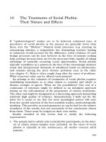
Figure 2. Classification of coronary arterial patterns in TGA by Yacoub & Radley-Smith,
1978. A: Left coronary artery (LCA) takes origin from the left sinus and right coronary
artery (RCA) from the right sinus. B: Single coronary artery, LCA and RCA arise from a
single ostium. C: Two para-commissural ostia with or without intramural course. D: RCA
and circumflex arise from the right ostium, left anterior descending (LAD) alone takes
origin from the left ostium. E: RCA and LAD originate from the left from the left posterior
sinus, circumflex alone takes origin from the right ostium (Yacoub & Radley-Smith., 1978).
In addition, the successful of the ASO in TGA depends on the elastic function of the
transposed aorta. However, several studies have shown evidence, that even after successful
anatomical repair, patients may be prone to long term problems. The fate of the aorta and
aortic valve has been assessed in previous studies (Losay et al., 2006; Kramer et al., 2003;
Langer et al., 2008). The majority of patients show non-progressive dilatation of the aortic
root, but only few cases suffer from aortic insufficiency (Görler et al., 2011). In addition,
reduced proximal aortic elasticity, structural abnormalities of the arterial walls, and
increased carotid artery stiffness have been reported in TGA patients (Niwa et al., 2001;
Grotenhuis et al., 2008; Mersich et al., 2006; Murakami et al., 2000). However, data are
lacking about the functional status of the entire length of the thoracic aorta as well as its
potential change with age after surgical repair, and the impact on left ventricular (LV)
function.
Page | 3
1.1.2 Congenital Anomalies of the Coronary Arteries
Normal coronary artery anatomy includes the left and right main coronaries (LCA and
RCA). The LCA originates from the left valsava sinus and branches into the LAD and the
circumflex artery (CFX). The LAD divides into three branches, such as the left conus, the
septal, and the diagonal artery (Figure 3). The RCA arises from the right sinus of valsava
and divides into many branches including the sinus node artery, the conal branch, an atrial
branch, the right ventricular muscular branches, the posterior descending artery, the
atrioventricular node artery, and septal branches (Figure 3) (Driscoll 2006).
Figure 3. Normal anatomy of the left and right coronary arteries. Based on an illustration in
(Driscoll, 2006).
The term congenital anomalies of the coronaries is defined as anomalies of the origin,
course, or structure of epicardial coronary arteries (Angelini 2002). They are rare CHD
diseases occurring in approximately 0.2-1.4% of the population (Davis et al., 2001). The
classification of coronary artery anomalies depends on anatomy and origin of the coronary
arteries and has been discussed extensively in the literature (Ogden 1970; Angelini 2002;
Fratz et al., 2006; Jacobs & Mavroudis., 2010). Anomalies of the coronary arteries are a
high risk factor for myocardial ischemia, the leading cause of myocardial infarction and
sudden cardiac death (Alexander & Griffith., 1956). In this thesis, we evaluated patients
with different coronary arteries anomalies, such as Bland-White-Garland syndrome,
congenital coronary artery fistula, and aberrant main left coronary artery.
Page | 4
a) Bland-White-Garland-Syndrome
Anomalous origin of the LCA from the pulmonary artery (ALCAPA), also known as BlandWhite-Garland syndrome (BWG) was described in 1933 by Bland, White, and Garland
(Bland et al., 1933). It is a rare congenital coronary artery abnormality and is associated
with early infant mortality and also sudden death in adulthood. The incidence of ALCAPA
is approximately 1 in 300,000 live births (Davis et al., 2001) and 0.26% of CHD undergoing
cardiac catheterization (Askenazi & Nadas., 1975). Patients live into adulthood without
treatment in approximately 15% of the reported cases (Perloff 2003). These patients may
present with myocardial ischemia, left ventricular dysfunction, myocardial infarction, and as
well as sudden cardiac death. In ALCAPA patients, coronary blood flow is supplied mainly
by the RCA and coronary collateral vessels from the RCA to the LCA. Patients with a poor
collateral circulation may develop myocardial ischemia and infarction. In patients with a
well developed coronary collateral system, symptoms may appear later in life (DodgeKhatami et al., 2002; Wesselhoeft et al., 1968).
Most patients with ALCAPA will undergo surgical treatment early in life. The aim of
surgical therapy is to preserve as much myocardium as possible. There are several methods
for surgical correction depending on the coronary artery anatomy, such as direct reimplantation, the Tackeuchi procedure, and coronary artery bypass grafting (Perloff 2003).
Direct surgical re-implantation of the LCA into the aorta is the most common surgical
procedure nowadays. However, there is a high risk of stenosis or occlusion of the LCA after
surgical treatment (Kazmierczak et al., 2013; Ramírez et al., 2011). Therefore, it is most
important to assess myocardial ischemia in ALCAPA patients before and after surgical
treatment.
b) Coronary Fistula
Coronary fistulas are also known as coronary arteriovenous fistula, and were first described
by Krause in 1865 (Krause 1865). They are rare anomalies and occur in 0.2 to 0.4% of all
CHD (Driscoll 2006) or in 0.3% to 0.87% of patients who undergo coronary angiography
(Angelini 2007). In this anatomical condition, the coronary arteries are abnormally
connected to the heart chambers or great vessels. In 90% the fistula drains into the cavum of
the right ventricle (Perloff 2003). The main therapeutic methods for correction of coronary
fistulas are surgical or interventional ligations, which are safe and have good long term
results (Urrutia-S et al., 1983). However, several reports showed that myocardial infarction
Page | 5
and cardiac death can occur in the long-term follow up (Valente et al., 2010; Canga et al.,
2012).
c) Aberrant LCA
An aberrant LCA is a rare congenital coronary artery anomaly with a reported prevalence of
0.017% - 0.03% (Tuo et al., 2013; Yamanaka & Hobbs, 1990). In this anomaly, the LCA
and RCA arise from the same or different anterior sinus of Valsalva (Figure 4). The
anomalous LCA from the right sinus was classified into 4 types including the following: A:
origin at left main trunk from right sinus or right coronary artery; B: origin of LAD and
CFX from the right coronary sinus; C: origin of LAD from right sinus of Valsalva or RCA;
D: origin of CFX from right sinus or right coronary artery (Roberts et al., 1992). A high
incidence of sudden death typically occurs in these patients during or immediately following
physical exercise (Cheitlin et al., 2009; Yamanaka & Hobbs, 1990). Particularly in the
presence of an inter-arterial course of LCA between the aorta and pulmonary artery, the risk
of sudden death is higher. Most patients with such anomalies were treated by surgical
therapies, such as bypass, reimplantation, and unroofing. However, myocardial ischemia
and sudden death can occur due to development of stenosis or closure of LCA after surgical
treatment (Krasuski et al., 2011).
Figure 4. Aberrant main LCA. Main LCA and RCA arise from anterior sinus of Valsalva.
The LCA passes obliquely between the aorta and the pulmonary artery; R. Cor: right
coronary artery; L. Circ: left coronary artery; LAD: left anterior descending artery; P.A:
pulmonary artery. Based on an illustration in (Cheitlin et al., Circulation 1974).
Page | 6
1.1.3 Ross Operation
The Ross procedure is a surgical method which uses the autologous pulmonary valve for
replacement of a diseased aortic valve and was first described by Donald Ross in the United
Kingdom in 1967 (Ross 1967). It has become a surgical treatment option also in CHD to
avoid the use of long-term anticoagulation (Svensson et al., 2003). Other possible
advantages of the Ross operation are the following: low risk of endocarditis and
thromboembolism, long-term durability, potential growth ability in children, and excellent
hemodynamic performance (Charitos et al., 2012).
Coronary artery reimplantation is a part of the operation and can lead to coronary artery
stenosis with the risk of myocardial infarction and sudden cardiac death (Somerville et al.,
1979). The assessment of myocardial perfusion and ischemia is therefore important during
postoperative care.
1.1.4 Heart Transplantation
Heart transplantation was first performed in 1967 by Christian Barnard in South Africa
(Barnard et al., 1967). Since then, this technique has been developed and become the
treatment of choice for the management of end-stage heart failure in children and adults
(Herrington & Tsirka., 2004). The number of cardiac transplantation has been increased, in
2011 more than 100,000 cardiac transplantations were performed worldwide according to
the registry of the International Society of Heart and Lung Transplantation (Stehlik et al.,
2011). However, there are many factors that have an effect on the results after cardiac
transplantation. Coronary allograft vasculopathy is the main factor, limiting the long-term
success of the operation and is a recognized cause of myocardial ischemia and sudden
cardiac death (Roussel et al., 2008; Nickel et al., 2011).
1.1.5 Kawasaki Syndrome
Kawasaki disease (KD), also known as Kawasaki syndrome or mucocutaneous lymph node
syndrome, is an acute systemic vasculitis of unknown etiology and the most important cause
of acquired heart disease in childhood in the developed countries. KD was first described in
Japan by Tomisaku Kawasaki in 1967 (Kawasaki 1967). Since then, many cases in different
countries have been reported. KD occurs most frequently in Japan with an incidence of
approximately 188.1 per 100,000 children younger than four years of age, and a male-tofemale ratio of 1.5:1 (Kato 2010). KD is associated with the development of coronary artery
Page | 7
aneurysms and stenosis (Figure 5). Acute myocardial infarction can occur in KD patients
due to thrombosis of aneurysms or due to developing stenosis of the coronary artery (Dajani
et al., 1993). In 1996, Kato et al. reported a follow-up study of 594 KD patients and found
coronary artery aneurysms in 25%, myocardial ischemia in 4.7%, myocardial infarction in
1.9% and death due to myocardial infarction in 0.8% of all cases (Kato et al., 1996). Due to
this data, assessment of myocardial perfusion and ischemia is of importance in the longterm follow-up of these patients.
Figure 5. Schematic drawing shows coronary artery aneurysms of KD (Based on an
illustration in Sridharan et al., 2010).
1.2 Non-Invasive Diagnostic Imaging for Detection of Myocardial
Ischemia
1.2.1 Nuclear Medicine
Nuclear cardiac imaging is the branch of cardiovascular diagnostic imaging that uses
radioactive tracers to perform functional images of the heart. SPECT and PET are two types
of nuclear imaging which are commonly used in clinical practice. They allow to evaluate
cardiac morphology, function, myocardial blood flow and viability (Auerbach et al., 1999;
Ghosh et al., 2010; Weindling et al., 1994).
Page | 8
SPECT myocardial perfusion scintigraphy (MPS) is a useful technique for evaluation of
ischemic heart disease. Diagnosis of CAD uses a scintillation camera and intravenously
injected radiopharmaceuticals, such as thallium-201 and technetium-99m sestamibi, and
technetium-99m tetrofosmin, whose distribution in the myocardium is dependent on, and
reflects the level of myocardial perfusion. SPECT MPS is normally performed during rest
and pharmacological stress. Besides other indications, SPECT MPS is a useful modality for
detection of CAD in children with congenital and acquired heart diseases (Sundaram et al.,
2009). However, there are some disadvantages: a normal SPECT MPS protocol usually
takes 3-4 hours, and uses ionising radiation. In addition, SPECT does not allow exact
quantification of myocardial perfusion and perfusion reserve (Bateman 2012; Jadvar et al.,
1999).
In contrast, PET provides the ability to quantify absolute myocardial perfusion blood flow
and is therefore considered a promising method for the examination of myocardial ischemia.
Typical radionuclides used for a PET study are Rubidium-82, Nitrogen-13 (in ammonia)
and Oxygen-15 (in water). Blood flow is quantified in units of ml/min/g. The sensitivity and
specificity for detection of myocardial ischemia are 87% to 97% and 78% to 100%,
respectively (Sampson et al., 2007; Bateman et al., 2006; Grover-McKay et al., 1992).
Furthermore, F18-FDG PET allows to differentiate between hibernating or stunned
myocardium and to assess myocardial viability in post myocardial infarction patients who
benefit significantly from revascularization. However, PET uses ionizing radiation and is
expensive (Bateman 2012).
Evaluation of myocardial perfusion and viability by PET offers several potential
advantages. Previous studies have demonstrated that PET is superior to SPECT for the
detection of myocardial ischemia, because it offers images with a higher resolution and
contrast, a better attenuation correction, less scatter, and has the ability to quantify absolute
myocardial perfusion (Bateman 2012; Ghosh et al., 2010). For the detection of myocardial
ischemia, PET perfusion imaging offers a higher sensitivity and specificity than SPECT
(Jaarsma et al., 2012). But there are only few studies using PET and SPECT for the
detection of ischemic heart disease in pediatric patients (Sundaram et al., 2009; Singh et al.,
2003; Hernandez-Pampaloni et al., 2002; Rickers et al., 2000). Other non-invasive methods
for assessing myocardial ischemia without ionizing radiation are often preferred.
Page | 9
1.2.2 Other Cardiac Stress Test
Myocardial contrast echocardiography (MCE) is a diagnostic imaging tool for the
assessment of the myocardial microcirculation using microscopic gas-filled bubbles, which
can burst by insonation in a myocardial region of interest. Replenishment of this same
region with gas-filled bubbles (i.e. myocardial opacification) will provide a measure of
myocardial blood flow (Wei et al., 1998; Porter et al., 2001; Kutty et al., 2012) .It can be
used to assess myocardial perfusion and viability for detection of myocardial ischemia
(Gaibazzi et al., 2012; Kaufmann et al., 2007; Mulvagh, 2004). MCE is non-invasive, does
not use ionizing radiation and is easy to perform. However, image quality depends on the
acoustic windows.
Exercise stress testing is the most commonly used method to evaluate patients with
suspected myocardial ischemia. Treadmill and bicycle ergometer protocols are the most
popular stress tests (Rhodes et al., 2010; Morrison et al., 2013). Electrocardiography (ECG)
exercise testing can be used for evaluation of cardiac perfusion and function with high yield
of diagnostic, prognostic, and functional information (Kashyap et al., 2011). However, in
small children ECG exercise testing is difficult to perform.
1.2.3 Cardiovascular Magnetic Resonance Imaging (CMR)
a) History of Magnetic Resonance Imaging and the Development of CMR
In 1946, Felix Bloch and Edward Purcell discovered the nuclear magnetic resonance
phenomenon that was a foundation for the development of magnetic resonance imaging
(MRI). In 1971, Raymond Damadian could show different tissue MR relaxation times in
rats, and the differences of the tissue relaxation times are the basis for good soft tissue
contrast in MRI. Peter Mansfield made another fundamental contribution to the
development of MRI in 1976 by developing the fast imaging technique known as echoplanar imaging. In 1977, Damadian obtained the first magnetic resonance images of the
human (Geva 2006).
The first publication regarding CMR in CHD dates back to 1982 and reported the diagnosis
of a ventricular septal defect in lamb hearts (Heneghan et al., 1982). In pediatric cardiology,
MRI was applied in the late 1980’s by using ECG-triggered spin echo techniques for
assessment of cardiac function and blood flow in patients with CHD (Higgins et al., 1988;
Chung et al., 1988). Gadolinium-enhanced MRI was first applied in clinical studies in 1984.
Page | 10
Myocardial viability imaging using late gadolinium enhancement was first mentioned in
1988 (Schaefer et al., 1988) and first-pass perfusion imaging is used since 1990 for the
detection of myocardial ischemia (Atkinson et al., 1990). Viability assessment by MRI has
since then evolved into a “gold-standard” based on the work of Kim et al. (Kim et al., 2000).
Since its beginnings data acquisition and image analysis have continuously improved (Earls
et al., 2002).
b) Advantages of CMR
CMR imaging has emerged as a promising diagnostic tool for the evaluation of CAD in
children. Advantages of cardiac MRI include absence of ionizing radiation, the high spatial
resolution, and the ability to assess in one exam morphology, global and regional function,
viability, myocardial perfusion, and coronary artery anatomy and patency. A number of
studies showed that first-pass perfusion CMR at rest and during pharmacologic stress allows
to assess myocardial ischemia, and that LGE can detect scar tissue (Klein et al., 2002;
Prakash et al., 2004). First pass perfusion MRI can be analyzed qualitatively, by semiquantitative analysis, and by absolute quantification of myocardial blood flow (MBF)
(Jerosch-Herold et al., 2002).
X-ray coronary angiography is known as the reference standard for detection of CAD
(White et al., 1984; Scanlon et al., 1999). However, especially in pediatric patients its
invasive nature, and the use of ionizing radiation are important limitations. It has been
shown that PET and cardiac MRI have the highest diagnostic accuracy for detection
myocardial perfusion abnormalities (Greenwood et al., 2012; Morton et al., 2012).
However, CMR has a higher resolution than PET (Jaarsma et al., 2012). The majority of
available CMR studies, were performed in adult patients and there are only few
examinations in children, in part due to the lack of expertise, access to CMR scanners in
pediatric cardiology departments, perhaps also due to need to sedate young patients.
c) CMR Imaging For Detection of Ischemia Heart Disease
First-Pass Perfusion CMR Imaging
First-pass myocardial perfusion MRI is used to monitor the changes in myocardial signal
intensity after intravenous injection of a contrast agent by using T1-weighted imaging. In
CMR perfusion imaging, the myocardial wash-in of contrast during the first pass of a
contrast bolus forms the basis for assessing myocardial perfusion. The T1-weighted signal
Page | 11
intensity is directly related to the concentration of the contrast agent, and its temporal
variation in the myocardium can be used to assess regional myocardial perfusion. In
ischemic regions the supply of blood and thus contrast enhancement is decreased. As a
consequence the signal intensity change is lower in ischemic regions, relative to normally
perfused myocardium. First-pass perfusion imaging is generally performed at rest and
pharmacologic stress (Al-Saadi et al., 2000), to assess the myocardial perfusion reserve
(Wilke et al., 1999).
CMR Adenosine Stress Perfusion
Adenosine is an endogenous nucleotide that promotes vasodilatation by activation of the α2
receptors in the vessels. In the field of CMR imaging, adenosine is most commonly used for
stress perfusion imaging for the detection of CAD with an iv dosage of 140 μg/kg/min body
weight per minute. The peak effect of adenosine occurs 2-3 min after start of the iv infusion,
with an increase of the heart rate. After stopping the iv. infusion of adenosine, the heart rate
returns to normal levels after 1-2 minutes (Pennell 2004). In CMR perfusion studies,
adenosine stress testing is used to increase the differentiation in the first-pass delivery of the
contrast agent between myocardial regions perfused by normal and abnormal coronary
arteries. Under resting conditions differences in perfusion can only be seen for 90% or
higher luminal narrowing of a coronary artery, and assuming there is no collateral supply.
CMR adenosine stress perfusion is safe, and the occurrence of AV-block is very rare,
occurring in < 1% of 9256 cases, and it has a very short half-life (< 10 seconds) (Al-Saadi
and Bogaert J, 2004; Pennell 2004; Cerqueira et al., 1994).
Contrast Media
Gadolinium chelates (Gd) are commonly used as paramagnetic contrast agents for
myocardial perfusion and late gadolinium enhancement (LGE) CMR imaging. Gd is an
extracellular paramagnetic contrast agent of low molecular weight (e.g. molecular weight of
938 for gadopentetate dimeglumine). After intravenous injection, it is carried to the right
ventricular cavity, then to the left ventricular blood pool. Then it diffuses rapidly from the
intravascular space into the myocardial extracellular space (Al-Saadi and Bogaert J, 2004).
Gd cause shortening of the T1 relaxation times.
The Gd passage through the myocardium is usually monitored by T1-weighted imaging.
Depending on the concentration and the time of wash-in and washout of the contrast agent
in the extracellular space, the myocardial tissue appears bright with high Gd content and
Page | 12
dark with low Gd content. Therefore, myocardial perfusion imaging shows dark areas with
low SI and bright areas with high SI by using contrast agents (Al-Saadi and Bogaert, 2004).
Late Gadolinium Enhancement
LGE imaging was developed by Kim and Judd in 1996 (Kim & Judd, 1996) and is an
excellent tool for assessment of tissue viability, e.g. in the diagnosis of CAD (Kim et al.,
2000; Bruder et al., 2009; West et al., 2010; Grover et al., 2011). Today, LGE-CMR is an
important imaging tool in both, congenital and acquired heart diseases, for detection of
necrosis and scar tissue (Harris et al., 2007; Desai et al., 2004; Babu-Narayan et al., 2010).
The basic principle of LGE rests on the differences in distribution volume between viable
and non-viable myocardial tissue. After intravenous administration of Gd, its distribution in
the myocardium is determined by cell-membrane integrity, and the loss of cell-membrane
integrity is a key step in the loss of myocardial viability. The use of Gd in conjunction with
T1 weighted inversion recovery imaging can be used to maximimize the contrast between
normal and injured myocardium. Using this technique, normal myocardium is made to
appear dark, and regions of myocardial infarction or scar appear bright (Kim et al., 2000).
CMR for Assessment of Myocardial Fibrosis
Diffuse myocardial fibrosis (DMF) is an important marker in heart diseases. Increased DMF
has been demonstrated to correlate with diastolic and systolic dysfunction, arrhythmia, and
sudden cardiac death (Martin et al., 1980; Villari et al., 1993). Previous studies showed
evidence for DMF in congenital and acquired heart disease (Broberg et al., 2010). The gold
standard method in evaluations of DMF marker of heart diseases is endomyocardial biopsy,
which is an invasive method and has several disadvantages, including risk of the hazard,
sampling error, and high cost (Becker et al., 1991; Holzmann et al., 2008).
CMR T1 mapping is a non-invasive method that can differentiate between diffuse fibrosis
and normal myocardium by using a Gd extracellular contrast agents. For cardiac
applications, T1 mapping within a breathhold can be performed with an ECG-gated LookLocker type of technique, where image data are read-out continuously after an initial
inversion pulse, to reconstruct images for 10-20 times after inversion (TI’s). More recently,
an ECG-gated single-shot Modified Look and Locker Inversion-recovery (MOLLI)
sequence, was described by Messroghli et al., which provides a high resolution T1 map of
the myocardium (Messroghli et al., 2004). The MOLLI sequence acquires multiple single
shot steady-state free precession images in the same slice and during the same cardiac
Page | 13
phase. The acquisition extends over ~10 heart beats, and the TI’s are varied by shifting the
time for application of the inversion pulse relative to the (diastolic) single-shot image
acquisition. A disadvantage of MOLLI compared to the Look-Locker technique is that in
general only a 5-9 images, corresponding to different TI’s are acquired, compared to ~20
TI’s that can be sampled with the Look-Locker technique. In addition, T1 mapping can be
used to quantify the changes of concentration of Gd in myocardium and in the blood pool
before and after Gd administration. This information can be used to differentiate between
normal and abnormal myocardium, and further allows absolute quantification of the
extracellular volume (ECV) (Sado et al., 2012; Messroghli et al., 2011). It has been shown
for multiple pathologies (e.g. aortic stenosis, hypertrophic cardiomyopathy, dilated
cardiomyopathy), that an expansion of the ECV is a marker of increased collagen and
connective tissue accumulation in the interstitial space (Jerosch-Herold et al., 2008).
Therefore, the T1 maping CMR technique is emerging as a method for quantitative
assessment of myocardial fibrosis in ischemic heart disease.
1.3 Previous Studies
In 2004, Prakash et al. performed a study using CMR to examine the feasibility and
potential clinical utility of CMR for the evaluation of ischemic heart disease in congenital
and acquired heart disease. They applied first-pass perfusion and LGE in 30 patients (age:
0.3 to 40 years) and could show that CMR can evaluate myocardial perfusion and viability.
However, absolute myocardial blood flow was not analyzed in this study (Prakash et al.,
2004).
Mavrogeni et al. used CMR to visualize the coronary arteries, to evaluate cardiac function
and to show scar tissue in 20 patients with KD, aged 7-12 years. They found aneurysms of
the coronary arteries in 7 patients, scar tissue in 4 patients, and left ventricular dysfunction
in 2 patients. First-pass perfusion imaging was not performed (Mavrogeni et al., 2006).
In 2009, Buechel et al. published a CMR perfusion study in pediatric patients. First-pass
perfusion with adenosine was performed in 47 patients (age: 1 month - 18 years). Perfusion
CMR showed a sensitivity of 87% and a specificity of 95% for the detection of myocardial
ischemia. This study demonstrated the feasibility of perfusion CMR in children (Buechel et
al., 2009).
A study using CMR during follow-up of 63 patients (median age: 14.6 years) with KD was
published in 2011. The CMR protocol included rest and stress perfusion imaging with
Page | 14
adenosine, LGE imaging and magnetic resonance coronary angiography. CMR findings
were compared with echocardiographic data. Aneurysms of the coronary arteries were
identified in 15 patients. CMR imaging detected LGE in 5 patients, myocardial ischemia in
4 patients, and thrombus formation in 4 patients. In summary the authors concluded that
CMR is a promising diagnostic tool during the long-term follow-up in KD (Tacke et al.,
2011).
In another study rest and stress perfusion with adenosine, LGE and 3D whole-heart imaging
were performed for assessment of myocardial ischemia in ALCPA patients (Secinaro et al.,
2011). This study showed the role of CMR for the follow-up of ALCAPA patients after
surgical repair.
Broberg et al. performed a study for detection and quantification of DMF in patients with
TGA, repaired tetralogy of Fallot, or Eisenmenger syndrome. They found the evidence of
increased diffuse fibrosis in this population, compared to normal controls, and a correlation
of the fibrosis index with end-diastolic function, and also with LV-EF (Broberg et al.,
2010).
There are only few CMR studies, which focus on ischemic heart disease in children. So far,
the published studies have demonstrated the promising role of MRI for the detection of
myocardial ischemia in children. But most CMR studies only used qualitative and/or semiquantitative analysis of myocardial perfusion in children, and the data on T1 mapping in
children with congenital heart disease is very sparse at the present time.
1.4 The Aim of This Study
Myocardial ischemia is a leading cause of myocardial infarction and sudden cardiac death.
In children, it may occur after surgery for CHD involving the coronary arteries, in
congenital coronary artery anomalies, and in patients with inflammatory disease of the
coronary arteries such as KD. Therefore, assessment of myocardial ischemia is important in
this population during the long-term follow up, but current diagnostic imaging methods,
such as electrocardiography, stress echocardiography, SPECT and PET, are often limited in
the pediatric population.
There are only few CMR studies which analyzed markers of myocardial ischemia in
children. In order to avoid ionizing radiation, an inherent burden of nuclear imaging (PET
and SPECT), this study used CMR imaging for the evaluation of ischemic heart disease in
children. We utilized advanced CMR methods to assess myocardial blood flow, viability,
Page | 15
function and diffuse fibrosis, to guide further therapy, and in order to get a better
understanding of the myocardial microcirculation in congenital and acquired heart disease.
We further asked if CMR predicts the functional recovery after treatment therapy of these
patients.
Page | 16