Role of neutrophil NADPH oxidase derived reactive oxygen species (ROS) in innate immune responses
Bạn đang xem bản rút gọn của tài liệu. Xem và tải ngay bản đầy đủ của tài liệu tại đây (2.39 MB, 82 trang )
Role of neutrophil NADPH oxidase
derived reactive oxygen species (ROS)
in innate immune responses
Inauguraldissertation
zur
Erlangung des akademischen Grades
doctor rerum nuturalium (Dr. rer. nat.)
and der Mathematisch-Naturwissenschaftlichen Fakultät
der
Ernst-Moritz-Arndt-Universität Greifswald
vorgelegt von
TRAN Bich Thu
geboren am 06.12.1980
in Ho Chi Minh Stadt, Viet Nam
Greifswald, den 29 März 2012
Dekan:
Prof. Dr. Klaus Fesser
1.
Gutachter:
Prof. Dr. Reinhard Walther
2.
Gutachter:
Prof. Fritz Ulrich Schade
Tag der Promotion: Greifswald, July 20th 2012
Table of contents
SUMMARY ................................................................................................................................. 1
INTRODUCTION ....................................................................................................................... 2
INNATE IMMUNE SURVEILLANCE ..................................................................................... 2
1.1
Sentinel systems of innate immunity ........................................................................ 2
1.2
Innate system effector cells ...................................................................................... 3
2
PRODUCTION OF ROS OF CELLS OF THE INNATE IMMUNE SYSTEM .................................. 8
2.1
What are ROS? ........................................................................................................ 8
2.2
Source in mitochondria ........................................................................................... 9
2.3
NADPH oxidase .................................................................................................... 10
3
FUNCTIONS OF ROS OF CELLS OF THE INNATE IMMUNE SYSTEM .................................. 12
3.1
Role(s) in killing bacteria ...................................................................................... 12
3.2
Detection of biologically relevant ROS ................................................................. 14
3.3
ROS as a signalling component ............................................................................. 14
4
OBJECTIVES OF THIS WORK ............................................................................................ 16
4.1
Bacterial killing ..................................................................................................... 16
4.2
ROS as signalling elements ................................................................................... 17
1
MATERIALS AND METHODS.............................................................................................. 18
1
MATERIALS .................................................................................................................... 18
1.1
Instruments ............................................................................................................ 18
1.2
Laboratory equipment ........................................................................................... 18
1.3
Reagents ................................................................................................................ 19
1.4
Buffers and solutions ............................................................................................. 20
1.5
Antibodies .............................................................................................................. 21
1.6
Software ................................................................................................................. 22
1.7
Mice ....................................................................................................................... 22
2
METHODS ....................................................................................................................... 22
2.1
Gold preparation ................................................................................................... 22
2.2
Anaesthetics ........................................................................................................... 22
2.3
Gold implantation.................................................................................................. 23
2.4
Organ sampling in mice ........................................................................................ 23
2.5
Inflammatory models ............................................................................................. 24
2.6
Effect of chemokines on the recruitment of neutrophils into the peritoneal cavity 24
2.7
Sample preparation for flow cytometry analysis ................................................... 24
2.8
Isolation and subsequent analysis of murine mononuclear cells attaching to gold
implants .............................................................................................................................. 27
RESULTS................................................................................................................................... 29
1
DETECTION OF ROS IN PHAGOCYTES ............................................................................. 29
1.1
Detection of extracellular ROS in vivo .................................................................. 29
1.2
Detection of intracellular ROS ex vivo .................................................................. 29
2
EXTRACELLULAR ROS ................................................................................................... 31
2.1
The effect of extracellular phagocyte derived ROS on the surface of gold implants
in vivo 31
3
CELL POPULATIONS IN THE PERITONEAL CAVITY AND THEIR ASSOCIATION WITH THE
IMPLANT ......................................................................................................................... 32
3.1
Cell populations in the peritoneal cavity of untreated mice .................................. 32
3.2
Implant associated phagocyte populations............................................................ 33
3.3
Peritoneal wash phagocyte population after implantation ................................... 35
4
3.4
Implants as inducers of peritoneal inflammation .................................................. 38
ROS AND MECHANISMS OF LEUKOCYTE EXTRAVASATION AFTER INFLAMMATORY
STIMULI .......................................................................................................................... 41
4.1
Expression of cell adhesion molecules on neutrophils .......................................... 41
4.2
Chemokines ........................................................................................................... 45
4.3
ROS production and the ability to extravasate ...................................................... 46
4.4
In vivo competiton assay for gp91phox-deficient and wild type neutrophil
recruitment to the peritoneum ............................................................................... 48
4.5
gp91phox-deficient and wild type neutrophil recruitment in peritoneal inflammation
............................................................................................................................... 52
4.6
gp91phox deficient and wild type neutrophil recruitment: chemokine application . 53
4.7
gp91phox deficient and wild type neutrophil recruitment in sterile peritoneal
inflammation .......................................................................................................... 54
DISCUSSION ............................................................................................................................ 57
REFFERENCES ....................................................................................................................... 62
ERKLÄRUNG ........................................................................................................................... 68
CURRICULUM VITAE ............................................ ERROR! BOOKMARK NOT DEFINED.
PUBLICATIONS ...................................................................................................................... 71
ACKNOWLEDGMENTS ........................................................................................................ 72
Doctoral dissertation
List of Abbreviations
ABS:
Anti-lock braking system
AFM:
Atomic force microscopy
APC:
Allophycocyanin
ATP:
Adenosine triphosphate
BPI:
Bactericidal/permeability-increasing protein
BSA:
Bovine serum albumin
CC:
Ceacal contents
CD:
Cluster of differentiation
CG:
Cathepsin G
CGD:
Chronic granulomatous disease
CRMP-2:
Collapsin response mediator protein 2
DAPI:
4',6-diamidino-2-phenylindole
DHR:
Dihydrorhodamine 123
DNA:
Deoxyribonucleic acid
DMSO:
Dimethyl sulfoxide
Duox:
Dual oxidase
EDTA:
Ethylene diamine tetra acetic acid
EGF:
Epidermal growth factor
ESAM:
Endothelial cell selective adhesion molecule
FACS:
Fluorescent activated cell sorting
FcR:
Fc receptor
FCS:
Foetal calf serum
FITC:
Fluorescein isothiocyanate
fMLP:
N-formyl-methionine-leucine-phenylalanine
FP:
Fenton polished
FPR:
Formyl methionyl peptide receptor
GAG:
Glucose amino glycans
GPx:
Glutathione peroxidase
HBSS:
Hanks' balanced salt solution
ICAM:
Intercellular adhesion molecule
IL:
Interleukin
IMP:
Implantation
JAM:
Junctional adhesion molecule
List of abbreviations
Doctoral dissertation
List of abbreviations
KC:
Keratinocyte chemo-attractant
K/o:
Knock out
LDL:
Low density lipoprotein
LFA-1:
Lymphocyte-associated functional antigen-1
LIX:
LPS-induced CXC chemokine
LPS:
Lipopolysaccharide
MCP-1:
Monocyte chemotactic protein-1
MIP-2:
Macrophage inflammatory protein-2
MP:
Mechanically polished
MPO:
Myeloperoxidase
NALP3:
NACHT, LRR and PYD domains-containing protein-3
NE:
Neutrophil elastase
NET:
Neutrophil extracellular traps
NGAL:
Neutrophil gelatinase–asssociated lipocalin
PAF:
Platelet activating factor
PBS:
Phosphate buffered saline
PCR:
Polymerase chain reaction
PDGF:
Platelet derived growth factor
PE:
Phycoerythrin
PECAM-1:
Platelet/endothelial cell adhesion molecule-1
PerCp:
Peridinin chlorphyll protein
PMA:
Phorbol-12-myristate-13-acetate
Phox:
Phagocyte oxidase
PR3:
Proteinase 3
PRR:
Pathogen Recognition Receptor
PSGL-1:
P-selectin glycoprotein ligand-1
Rh-123:
Rhodamine-123
ROS:
Reactive oxygen species
SDS:
Sodium dodecyl sulphate
SOD:
Superoxide dismutase
TG:
Thioglycollate
TNF:
Tumour necrosis factor
TLR:
Toll like receptor
VCAM:
Vascular cell adhesion molecule
VLA:
Very late antigen
Doctoral dissertation
List of figures
List of figures
Figure 1
The tripartite area code
6
Figure 2
The electron transport chain in the mitochondrion
9
Figure 3
Regulation of the phagocyte NADPH oxidase complex
11
Figure 4
Atomic force micrographs of a polished gold surface
29
Figure 5
Rhodamine generation by blood neutrophils
30
Figure 6
The effect of extracellular phagocyte derived ROS on the surface of gold
implants in vivo.
32
Untreated C57BL6 peritoneal cell wash contains various immune cell
populations
33
Figure 8
Cell populations attaching to the gold surfaces
34
Figure 9
Cell populations in the peritoneal wash fraction 14 days after implantation
35
Figure 10
Comparison of cell populations in the peritoneal cell wash and on the
mechanically polished gold surfaces
36
Ex vivo ROS production of phagocyte populations in the peritoneal cell
wash after implantation
37
Comparison of cell populations in the peritoneal cell wash of wild type
mice at 6 hours and 14 days after implantation
38
Figure 13
Quantitation of granulocyte populations early in inflammation
39
Figure 14
Kinetics of granulocyte population change in inflammation
40
Figure 15
Peritoneal cell wash of wild-type BALB/c and of congenic eosinophil
ablated mice 18h after surgery
40
Figure 16
Adhesion molecule expression on blood neutrophils from untreated mice
42
Figure 17
Expression of α7 and β2 integrins on neutrophils from wild type and
gp91phox knock-out mice after being challenged with commensal flora for 3
hours
43
Expression of α7 and β2 integrins on neutrophils from wild type and
gp91phox knock-out mice after being challenged with thioglycollate for 3
hours
44
MFI of α7 and β2 integrins of blood and peritoneal neutrophils from wild
type and gp91phox knock-out 3 hours after treatment
45
Figure 20
KC, LIX or MIP-2 induce neutrophil recruitment into the peritoneal cavity
46
Figure 21
Ex vivo ROS production of neutrophils in blood of untreated and in the
blood and peritoneal cell wash 3h after caecal content induced peritonitis
47
(A) C57BL6 peritoneal cell wash 6h after implantation (IMP) and caeceal
content induced peritonitis (CC); (B) Ex vivo ROS production of peritoneal
neutrophils 6h after implantation and caecal content induced peritonitis.
47
Comparison of cell populations in the peritoneal wash of wild-type and
gp91phox knock-out mice 3 hours after caecal content induced peritonitis.
48
Figure 7
Figure 11
Figure 12
Figure 18
Figure 19
Figure 22
Figure 23
Doctoral dissertation
List of figures
Figure 24
Assay for the identification of mosaic neutrophil subpopulations in gp91+/ko
heterozygous mice.
Figure 25
+/ko
Figure 26
Figure 27
Figure 28
Figure 29
Figure 30
Figure 31
Flow cytomectric analysis of peripheral blood and bone marrow of gp91
heterozygous mice
49
50
phox
The ratio of wild type to gp91
deficient neutrophil subpopulations of
+/ko
one gp91 heterozygous female mouse determined on day 1, 3 and 5
51
phox
Distribution of wild type and gp91 -deficient blood neutrophil
subpopulations in gp91+/ko heterozygous female mice
51
Wild type neutrophils have an advantage in entering the peritoneal cavity
early after induction of inflammation
52
phox
Wild type and gp91
deficient neutrophils have the same ability to enter
the peritoneum after being treated with chemokines
54
phox
Wild type and gp91
deficient neutrophils have the same ability to enter
the peritoneum in a sterile peritoneal inflammation (thioglycollate model)
55
(A) MFI of α7 and β2 integrins of peritoneal neutrophils from wild type
and gp91 knock-out mice 3 hours after treatment. (B) In the gp91+/ko
heterozygous female mice, wild type neutrophils have an advantage in
entering the peritoneal cavity in the CC but not in the TG inflammatory
models
56
Doctoral dissertation
List of tables
List of tables
Table 1
List of antibodies used for flow cytometry analysis
20
Doctoral dissertation
Summary
SUMMARY
I have investigated the role played by reactive oxygen species (ROS) generated by the
phagocyte NADPH oxidase system in the innate immune response. I first looked at
effector functions by asking whether ROS released from phagocytes might be effective
in the killing of extracellular bacteria. Since bacteria can be killed in many other ways –
for example by proteases or by cationic peptides – I made use of the recently
demonstrated capacity of ROS to remove discontinuities from the surface of gold as the
basis of an in vivo assay for extracellular ROS. Unlike bacterial killing, this readout
system is not affected by enzymes, cationic peptides or other biological anti-bacterial
agents. By this means I was able to use wild type mice and a congenic strain which
lacks the gene coding for the gp91 subunit of the phagocyte NADPH oxidase to
demonstrate that ROS generated by the NADPH oxidase system are indeed found
outside the cells during an inflammation in vivo and that their principle source is
neutrophil granulocytes rather than tissue macrophages. Since ROS released by these
cells will be non-specific in its action it is to be expected that the releasing cell will
itself suffer considerable damage. This fits well to the known short life of activated
neutrophils and may explain the established fact that their death is dependent on the
NADPH oxidase system. The long lived macrophages, in contrast, restrict their
production of extracellular ROS.
ROS are increasingly being found to be involved in both intra and intercellular
signalling processes I looked for an involvement of NADPH oxidase derived ROS in
the recruitment of neutrophils to sites of inflammation in vivo. Since the gene coding for
the gp91 subunit of the NADPH oxidase is on the X chromosome I made use of a
mosaic expression strategy based on X chromosomal inactivation. The results show that
indeed ROS serves as a component of the neutrophil recruitment process in the critical
early stages of an infection. Possible mechanisms are explored.
1
Doctoral dissertation
Introduction
INTRODUCTION
1
Innate immune surveillance
The innate immune system provides a first line of active defence against infection and is
centrally involved in initiating tissue repair after sterile injury. To achieve these ends
innate immunity must be in a position to detect deviations from normal tissue
homeostasis resulting from infection or injury, and to re-instate the status quo ante. This
requires that the system have sensors for infection and injury coupled to effector
mechanisms which are able to respond appropriately.
1.1
Sentinel systems of innate immunity
The sentinel systems of innate immunity are in part made up of soluble components of
the serum such as the complement system (Frank and Fries 1991) or acute phase
proteins (Bopst, Haas et al. 1998) though the major contribution is made by cell bound
receptors – often referred to as “Pathogen Recognition Receptors” (PRR) (Medzhitov,
Preston-Hurlburt et al. 1997). PRR are expressed on or in many cells including the
mononuclear phagocytes (monocytes, macrophages and dendritic cells) which act as
immune sentinels. The most intensively investigated receptors of this type are the “Toll
Like Receptors” (TLR) and the first of these to be characterised was TLR4 (Medzhitov
and Janeway 1997; Beutler, Jiang et al. 2006). This receptor is expressed on the cell
surface where it interacts with the secreted protein MD-2 which has the ability to bind
lipopolysaccharide of Gram negative bacteria with very high affinity (Viriyakosol,
Tobias et al. 2001). When the TLR4–MD2 complex binds LPS (Park, Song et al. 2009),
a signal transduction pathway is activated which causes the sentinel cell to produce and
release pro-inflammatory mediators (Akira, Uematsu et al. 2006).
These sentinel cells also carry receptors capable of detecting endogenous danger signals
and indeed some of the PRR do double duty as detectors of tissue injury. Thus TLR4
forms a complex with CD36 which detects oxidised low density lipoprotein (LDL)
(Stewart, Stuart et al. 2009), TLR2 detects lipid break down products released from
necrotic cells (Schaefer, Babelova et al. 2005) and the formyl methionyl peptide
2
Doctoral dissertation
Introduction
receptor (FPR) detects not only peptides released from invading bacteria but also those
released from mitochondria in areas of cell necrosis (Zhang, Raoof et al. 2010).
The tissue macrophages and other phagocytic cells of the innate immune system such as
granulocytes also have the ability to detect sterile particles ranging from uric acid
crystals (Kono, Chen et al. 2010), which are readily formed from the breakdown of
nucleic acid released from necrotic cells, to asbestos fibres, hyaluronan (Taylor,
Yamasaki et al. 2007) and silica particles (Dostert, Petrilli et al. 2008). The means of
sentinel cell activation in these cases does not involve specific cell surface receptors but
results from the attempts of the phagocyte to ingest a particle which is many times
larger than itself. This results in "NACHT, LRR and PYD domains-containing protein3" (NALP3) activation by “frustrated phagocytosis” (Martinon, Mayor et al. 2009).
In these ways the sentinel cells are activated and this activation quickly results in their
producing and releasing inflammatory mediators which are centrally involved in
recruiting innate system effector cells – principally the neutrophil granulocytes – to the
site of the inflammatory disturbance.
1.2
1.2.1
Innate system effector cells
Origin of the innate system cells
The principle phagocytes of innate immunity are the macrophages and neutrophil
granulocytes. Circulating neutrophils emerge from the post-mitotic pool of neutrophils
in bone marrow (Pillay, den Braber et al. 2010) as functionally dormant cells which can
be quickly activated to leave the circulation and enter tissues at sites where the
inflammatory mediators are being produced by the sentinel cells (Svanborg, Godaly et
al. 1999). Macrophages also originate in the bone marrow. However, because different
tissues have macrophages with very different properties, the cells produced in the bone
marrow are in an incompletely differentiated form known as monocytes. These
monocytes leave the bone marrow and spread via the circulation to the different tissues
where their final differentiation into end stage macrophage takes place in response to
tissue specific signals (Auffray, Sieweke et al. 2009). Like neutrophils they can also be
recruited to sites of inflammation (Auffray, Fogg et al. 2007).
3
Doctoral dissertation
1.2.2
Introduction
Neutrophils
Neutrophils are the most abundant innate immune cell type and they are the principle
phagocytic cells of the body. Once these cells have entered the tissue at a site of
inflammation they are activated – and activated neutrophils are extremely destructive.
Because of this they may cause problems not only for invading bacteria but also for our
own cells and tissues. For this reason activated neutrophils have an extremely short life
expectancy which is not much more than two hours (Brinkmann, Reichard et al. 2004;
Ermert, Urban et al. 2009). During this time they may kill many bacteria by
phagocytosis (Potter and Harding 2001) and, when they die, they extrude their genome
as decorated chromatin to form so-called “Neutrophil extra cellular traps” (NETs)
which can trap and kill yet more micro-organisms (Brinkmann and Zychlinsky 2007).
Since these activated neutrophils have a short half life, sites of inflammation rapidly
acquire a large population of dead and dying neutrophils.
1.2.3
Macrophages
Macrophages act as innate system sentinel cells in tissues throughout the body.
Different tissues have different requirements of their sentinels and hence a bewildering
array of macrophage phenotypes is found. Many of the macrophage populations seem to
be maintained under steady state conditions by influx of circulating monocytes
(Auffray, Sieweke et al. 2009), while others such as Langerhans cells of the skin are
normally renewed from a self replicating peripheral pool (Merad, Ginhoux et al. 2008).
During an inflammatory response many extra so-called “inflammatory macrophages”
are recruited from the pool of circulating monocytes. The principle function of these
macrophages is to remove the dead neutrophils (Savill, Wyllie et al. 1989) and damaged
cells and tissues (Gordon and Martinez 2010). They are, however, also themselves
capable of ingesting bacteria by phagocytosis and killing them within their phagocytic
vesicles (Green, Meltzer et al. 1990).
4
Doctoral dissertation
1.2.4
Introduction
Trafficking of innate system cells in response to inflammatory
signal
Tissue macrophages are activated when tissue homeostasis is disturbed and they
respond by releasing pro-inflammatory mediators which initiate the recruitment of
innate cells into the tissue by inducing the expression of cell trafficking signals on the
surface of the endothelial cells (Beutler, Jiang et al. 2006).
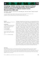
The emigration of leukocytes from the vasculature is regulated by three major molecular
signals: These involve interactions between (a) selectins and selectin ligands, (b)
chemokines and chemokine receptors and (c) integrin and integrin ligands (Springer
1994). These three sets of interactions form the basis of a cellular “area code” or
“addressing” system and this tripartite area code hypothesis is supported by the fact that
inhibition of any one of these steps inhibits emigration. This system provides great
combinatorial diversity for regulating the selectivity of leukocyte localization in vivo
because at each step a ligand on one cell must meet the appropriate receptor on the
other.
The first step involves selectin – selectin ligand interactions. The endothelial cells lining
the blood vessels interact with pro-inflammatory mediators, such as TNFα (Tumour
necrosis factor α), which are released from the tissue macrophages at sites of infection
or injury. The endothelial cells respond with the rapid externalization of preformed Pselectin (CD62P) from their Weibel-Palade body granules. The P-selectin can thus be
expressed on the surface of local endothelial cells within minutes of the release of
TNFα by macrophages. E-selectin is also expressed on the surface of endothelial cells
but this takes longer because de novo mRNA and protein synthesis are required. Both of
these selectins interact with the sulfated-sialyl-Lewisx of mucin-like proteins such as Pselectin glycoprotein ligand-1 (PSGL-1), which is present on the surface of neutrophils.
This selectin – selectin ligand interaction has a very fast “on” rate and a fast “off” rate
and thus acts like an anti-lock braking system (ABS) to slow up the rapidly flowing
leukocyte without the risk that their membrane be torn apart. The leukocyte now rolls
across the surface of the endothelium at an ever slower pace (Springer 1995).
5
Doctoral dissertation
Introduction
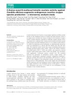
Figure 1: The tripartite area code: the combinatorial specificity of the leukocyte
adhesion cascade. Adapted from Timothy A. Springer 1994 and Klaus Ley et. al. 2006.
6
Doctoral dissertation
Introduction
The second step involves a chemokine – chemokine receptor interaction between
leukocyte and endothelium. The activated endothelial cells up-regulate the expression of
chemokines on their surface over which the leukocyte is rolling. The chemokines
secreted by the endothelial cells bind to glucose amino glycans (GAGs) on the
endothelial cell surface (Handel, Johnson et al. 2005; Massena, Christoffersson et al.
2010) and can thus interact with those leukocytes which express the appropriate
receptor. Since chemokines and their receptors are by far the most diverse group of
trafficking molecules, they provide the greatest number of molecular choices and hence
the greatest cellular specificity in the extravasation process.
The third step involves integrin activation on the leukocyte surface. Chemokine
receptors are G protein coupled signal transducers which mediate the conformational
activation of the integrins expressed on the leukocyte surface. Integrins are noncovalently associated hetero-dimeric cell surface adhesion molecules. 18 α subunits and
8 β subunits are encoded in the genome making for 144 potential αβ integrin
heterodimers of which 24 have been shown to be expressed on cells in vivo. The
diversity in subunit composition contributes to diversity in ligand recognition and to the
coupling to downstream signalling pathways. The β2 and β7 integrins are exclusively
expressed on leukocytes, whereas the β1 integrins are expressed on a wide variety of
cells throughout the body. All hetero-dimeric integrins have a large extracellular
ectodomain supported on two “legs”. The globular extracellular domain mediates
subunit association and ligand binding while the two C-terminal α- and β-“legs” cross
the plasma membrane and terminate in short cytoplasmic domains. Circulating
leukocytes generally maintain their integrins in a non-adhesive state in which the head
lies close to the cell membrane and in this state has little access to appropriate ligands
expressed on other cells. However binding of chemokine to the chemokine receptor
leads to rapid separation of the integrin cytoplasmic tails and this causes the ectodomain
of the receptor to take on an extended conformation in which it now has high-affinity
for its ligands (Campbell and Humphries 2011). The integrin – integrin ligand
interaction leads to firm leukocyte arrest on the endothelium after which the arrested
cell may transmigrate across the endothelial cell barrier.
Transmigration through venular walls is the final step in the process of leukocyte
emigration into inflamed tissues and has to occur with minimal disruption to the
7
Doctoral dissertation
Introduction
structure of the vessel walls. Leukocyte migration through the endothelial cell barrier
can be rapid (2 – 5 minutes), but penetrating the endothelial cell basement membrane
can take much longer (5 – 15 minutes) (Ley, Laudanna et al. 2007). Both in in vivo and
in in vitro models neutrophils have the ability to transmigrate either directly through the
endothelial cells or at the junctions between them. The junctional pathway is strongly
preferred and inflamed endothelial cells redistribute junctional molecules in a way that
favours cell migration there. Junctional molecules which form homophilic interactions
such as platelet/endothelial cell adhesion molecule-1 (PECAM-1), CD99 and junctional
adhesion molecules (JAM) are expressed both at endothelial cell junctions and on the
leukocytes and this allows for a “zipper” mechanism of transmigration (Weber,
Fraemohs et al. 2007; Woodfin, Voisin et al. 2011) in which the leukocyte slips between
the endothelial cells without disturbing their barrier function. As the leukocyte passes
through the endothelial junction reseals.
Having penetrated the endothelial cell barrier, leukocytes pass through the basement
membrane, a protein mesh consisting largely of laminins and collagen type IV, a
process which, at least in part is facilitated by leukocyte proteases (Khandoga, Kessler
et al. 2006).
2
2.1
Production of ROS of cells of the innate immune system
What are ROS?
ROS include a number of chemically reactive partially reduced forms of oxygen
including O2−, H2O2, and HO•. These ROS are substantially more reactive than
molecular oxygen which is itself a di-radical. To reduce O2 to two molecules of water,
four electrons and four protons are needed. Since the electrons are added one at a time
intermediates are formed during this reduction process.
8
Doctoral dissertation
Introduction
It is these intermediates that are primarily responsible for the toxicity of O2, and defense
against that toxicity involves minimizing their production and eliminating those whose
production cannot be avoided.
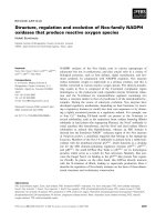
2.2
Source in mitochondria
Figure 2: The electron transport chain in the mitochondrion is the site of oxidative
phosphorylation in eukaryotes. Adapted from Frasconcellos 2007.
O2 consumption by respiring cells is largely restricted to the mitochondria and, on that
basis alone, one would expect these organelles to produce substantial amounts of the
partial reduction products of O2. During mitochondrial respiration, electrons are
extracted from reduced substrates and are transferred to molecular oxygen (O2) through
a chain of enzymatic complexes (I to IV). This electron flux through the respiratory
chain polarises the inner mitochondrial membrane, and this polarisation is used as an
9
Doctoral dissertation
Introduction
energy source for the synthesis of ATP (Adenosine triphosphate). In the final step of the
electron transport chain, cytochrome c oxidase (complex IV) carries out the fourelectron reduction of O2 to 2H2O and thus ensures the complete reduction of O2 to water
without the formation of ROS. However, partial reduction of O2, which results in the
generation of ROS, can occur if O2 interacts with the electron transport chain upstream
of complex IV. Some electrons can escape from the mitochondrial electron transport
chain and react with O2 to form O2− which is subsequently converted to H2O2.
In mitochondria the extent of electron leakage varies depending on cell type and
respiratory status but will generally represent around 0.1% of total electron flux.
Mitochondria are well endowed with defences against O2− in the form of superoxide
dismutases (SODs). The abundance of MnSOD in the matrix and of Cu, ZnSOD in the
inter-membrane space suggests the need for elimination of O2− on both sides of the
inner membrane and that in turn suggests that O2− is formed on both faces of that
membrane. The severe pathology and perinatal death of mice lacking MnSOD (Li,
Huang et al. 1995; Lebovitz, Zhang et al. 1996) is a clear indication that
mitochondrial O2− is capable of causing great damage when not scavenged by the
MnSOD. However, Cu, ZnSOD null mice do not display such obvious pathology
(Sentman, Granstrom et al. 2006), indicating that there may be back-up mechanisms for
elimination of O2− in the cytosol and inter-membrane space. In the inter-membrane
space of mitochondria such a back up may be provided by cytochrome c, since it can be
reduced by O2− and re-oxidized by complex IV (cytochrome c oxidase). Mitochondria
also contain glutathione peroxidase (GPx) which will remove hydrogen peroxide (H2O2)
that is formed. Nevertheless, since hydrogen peroxide is much more stable than
superoxide, some of it may escape the organelle.
2.3
NADPH oxidase
Professional phagocytes are indispensable for the rapid eradication of pathogenic
microbes. These phagocytes are equipped with a number of antimicrobial systems,
including the NOX2-containing NADPH oxidase complex (NADPH oxidase II), which
reduces molecular oxygen to reactive oxygen species (ROS). The enzyme complex is
also commonly referred to as the phagocyte oxidase (phox) and it can lead to the
10
Doctoral dissertation
Introduction
generation of substantially higher levels of ROS than are produced by other cellular
oxidases.
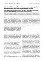
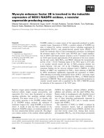
Figure 3: Regulation of the phagocyte NADPH oxidase complex. Translocation and
assembly of the cytosolic components of the NADPH oxidase complex (p40phox,
p47phox, and p67phox, p21rac) with the membrane associated cytochrome b558 (p22phox and
gp91phox) and further mutiple phosphorylation events lead to the activation of NADPH
oxidase which in turn catalyses the reduction of molecular oxygen O2 into O2−. Adapted
from genkyotex.
The phagocyte oxidase is a multi-component, electron transfer complex. Two of the
subunits, p22phox and gp91phox (the subunit also known as NOX2), form a membranebound, hetero-dimeric flavohemoprotein referred to as cytochrome b (cytochrome
b558). Cytochrome b constitutes the catalytic, electron transferring part of the NADPH
oxidase. In the absence of cellular activation, the cytosolic components of the NADPH
oxidase, namely p40phox, p47phox, and p67phox are not associated with cytochrome b and
the oxidase is dormant. Upon cellular activation, the cytosolic components translocate
to the membrane and associate with cytochrome b to form a functional NADPH
oxidase.
Assembly of NADPH oxidase II at the plasma membrane results in the release of ROS
into the extracellular milieu, whereas NADPH oxidase II assembly on a phagosomal
membrane would result in ROS being pumped into the phagosome. In neutrophils
around 95% of the cytochrome b is present in phagocyte granule membranes and some
11
Doctoral dissertation
Introduction
5% is associated with the plasmalemma (Borregaard, Heiple et al. 1983; Borregaard and
Tauber 1984). In these cells the phagocyte granule membranes are thus the major sites
for NADPH oxidase assembly and activation.
The active NADPH oxidase transfers electrons from NADPH in the cytoplasm across
the membrane where they interact with molecular oxygen (O2) to form superoxide (O2−)
that dismutates spontaneously to hydrogen peroxide (H2O2). These primary ROS can be
further processed to generate other reactive metabolites including hypochlorous acid
(HOCl) which is a highly microbicidal product formed by the myeloperoxidase present
in the azurophil granules of neutrophils (Chapman, Hampton et al. 2002; Rosen,
Crowley et al. 2002; Hirche, Gaut et al. 2005; Rosen, Klebanoff et al. 2009).
3
3.1
Functions of ROS of cells of the innate immune system
Role(s) in killing bacteria
Neutrophil defense against infecting micro-organisms largely depends on phagocytosis
which is initiated by invagination of the plasma membrane and results in a membraneenclosed vesicle called the phagosome. The phagosome is further processed through
heterotypic fusion of granules (gelatinase, specific, and azurophil granules) forming the
mature phagolysosome. The fusion of these granules with the phagosomal vacuole is
completed roughly 20 seconds after the uptake of micro-organisms. These granules
contain large amounts of anti-microbial protein which together are present at a
concentration of about 500 mg/ml. There are three fundamental types of granules in
neutrophils. Azurophilic granules (also known as peroxidase-positive or primary
granules) are the largest, measuring approximately 0.3 µM in diameter, and contain
myeloperoxidase (MPO), an enzyme critical in the oxidative burst. Other cargo of this
granule class include the defensins, lysozyme, bactericidal/permeability-increasing
protein (BPI), and a number of serine proteases: neutrophil elastase (NE), proteinase 3
(PR3), and cathepsin G (CG). The second class of granules, the specific (or secondary)
granules, are smaller (0.1 µM diameter), do not contain myeloperoxidase (MPO), and
are characterized by the presence of the glycoprotein lactoferrin. These granules also
contain a wide range of anti-microbial compounds including neutrophil gelatinaseassociated lipocalin (NGAL), hCAP-18, and lysozyme. The third class, the gelatinase
12
Doctoral dissertation
Introduction
(tertiary) granules, are also MPO-negative, are smaller than specific granules, and
contain few anti-microbials, but they serve as a storage location for a number of
metalloproteases, such as gelatinase and leukolysin (Amulic, Cazalet et al. 2012).
Around 0.2 fmols of O2 are estimated to be consumed when a particle the size of a
bacterium is engulfed and this would correspond to the generation of a concentration of
O2− of 1–4 M within the vacuole. Experiments with the MPO-H2O2-halide system
demonstrate that the HOCl produced by this enzyme can kill bacteria in the test tube
(Albrich, Gilbaugh et al. 1986). However, these experiments were conducted under nonphysiological conditions in the absence of the high levels of proteins (approximately
500 mg/ml) which are present in the vacuole. When bacteria were exposed to 100 mM
H2O2 or 1 mM HOCl in the presence of 25 mg/ml granule proteins, killing was almost
completely abolished (Reeves, Nagl et al. 2003).
The NADPH oxidase transfers electrons unaccompanied by protons across the vacuolar
membrane where they are used to reduce molecular oxygen to water. Each molecule of
oxygen requires four protons for its reduction and this loss of protons elevates the pH
within the vacuole to a level optimal for the activation of neutral proteases, a process
which is supported by K+ counter ions driven into the vacuole to compensate the
membrane polarisation. The hypertonic K+ solubilises the cationic granule proteases and
peptides by displacing them from the anionic sulphated proteoglycan granule matrix.
These proteases play a major role in bacterial killing and their activation may be the
mechanism by which ROS mediate bacterial killing (Segal 2005). Of further interest,
mice deficient in both NE and CG are highly susceptible to fungal infections as was
observed in patients with chronic granulomatous disease (CGD) and in the
corresponding mouse models. Despite a completely normal oxidative burst, these
proteases deficient mice were shown to be unable to control fungal infections.
Therefore, the activation of both phagocyte NADPH oxidase and neutral serine
proteases are required for full protection against fungi in vivo (Tkalcevic, Novelli et al.
2000).
Thus, although ROS produced by the functional NADPH oxidase II complex are
essential for the effective killing of microbes as is demonstrated by the fact that the
deficiency of the complex leads to chronic granulomatous disease (CGD), there remains
13
Doctoral dissertation
Introduction
the unresolved question of whether they directly react with and kill microbes to any
significant extent or whether they act indirectly by the activation of phagocyte
proteases.
3.2
Detection of biologically relevant ROS
To determine whether ROS are able to kill bacteria in a biologically relevant
environment one might make use of the observation that neutrophils generate
considerable quantities of extracellular ROS (Scholz, Lopez de Lara Gonzalez et al.
2007). However, even in this situation killing of extracellular bacteria may be mediated
by many other cellular components. A possible contribution of direct bacterial killing by
ROS to innate immune defence requires some readout which is not dependent on
measuring microbial survival. For this purpose the recent demonstration that
mechanically polished gold surfaces can be smoothed by ROS generated by Fenton
chemistry provides a potential solution. The surface of mechanically polished gold
which appears to be flat on a millimetre scale contains a density of micrometre scale
asperities. The surface is characterised by “rough” areas in which, for some atoms,
neighbours are missing. These gold atoms are not stably packed and integrated into a
crystal structure and hence are chemically more reactive than their fully bonded
neighbours. Such atoms can be mobilised and removed by the action of highly reactive
radicals (Nowicka, Hasse et al. 2010; Nowicka, Hasse et al. 2010). The resulting
smoothing of the surface can be shown by atomic force microscopy scanning of the
surface before and after Fenton treatment. This provides the basis for demonstrating a
potential direct role of ROS at least in extracellular bacterial killing.
3.3
ROS as a signalling component
Environmental insults, such as ionizing radiation and xenobiotics like polycyclic
aromatic hydrocarbons or phorbol esters, increase ROS production, and this led to the
idea that ROS play a negative role in physiology by contributing to increased cell
proliferation and to malignant transformation (Klaunig and Kamendulis 2004). Later on,
it became evident that intracellular H2O2 production is directly implicated in the signal
transduction events triggered by growth factor. Indeed the intracellular level of H2O2 is
regulated by various growth factors − such as epidermal growth factor (EGF), platelet-
14
Doctoral dissertation
Introduction
derived growth factor (PDGF) and insulin − and H2O2 itself inhibits crucial
phosphatases that are involved in the attenuation of signal propagation from the
activated growth factor receptors (Giorgio, Trinei et al. 2007).
H2O2 functions as a signalling molecule by virtue of its ability to oxidise certain
cysteine residues in proteins. The cysteine thiol moiety is only a good target for the
oxidising action of H2O2 if it exists in the form of a cysteine thiolate anion (-S-) and
most cysteine residues in proteins are not in this form at physiological pH values. Only
in a few proteins, including certain phosphatases involved in signal transduction
cascades, has a cysteine located in a local environment in which it can function as a
target for H2O2 (Reth 2002).
Recently, it was shown that the embryonic brain development depends on the enzymatic
activity of glutaredoxin 2 and zebrafish with silenced expression of glutaredoxin 2 lost
virtually all types of neurons by apoptotic cell death and the inability to develop an
axonal scaffold. As demonstrated in zebrafish, glutaredoxin 2 controls axonal outgrowth
via thiol redox regulation of collapsin response mediator protein-2 (CRMP-2), a central
component of the semaphorin pathway. This study provides an example of a specific
thiol redox regulation (Brautigam, Schutte et al. 2011). Though ROS induced
modifications have been shown to be functionally relevant and reversible for only a few
proteins, the involvement of ROS in numerous signal transduction cascades has been
demonstrated and this suggests that redox modifications can be tightly regulated in
cells.
Not all ROS have half lives in biological milieus which would restrict their participation
in signal transduction cascades to intracellular functions. In particular, H2O2 is both
sufficiently long lived, readily diffusible and membrane permeable so that it can take
part in intercellular signalling over a distance of many cell diameters (Winterbourn
2008). Indeed, H2O2 has been shown to serve as an early inflammatory response
mediator after injury in the zebra fish. A gradient of H2O2 initiates at the wound margin,
extends approximately 100 – 200 µm from the wound − far enough to reach the nearest
blood vessels. The H2O2 gradient is established within 5 minutes of wounding and
peaks at around 20 minutes just prior to the movement of the first neutrophils from the
circulation towards the wound. Using a combination of genetic and pharmacological
15
Doctoral dissertation
Introduction
experiments, dual oxidase (Duox), a specific NADPH oxidase, was identified to be the
source of the H2O2 signal (Niethammer, Grabher et al. 2009). The H2O2 is detected by
the neutrophils which use the protein tyrosine kinase Lyn as a redox sensor. Inhibition
of this neutrophil Src family kinase reduces neutrophil recruitment in zebra fish, and is
associated with the oxidation of a single cysteine thiol at position 466. SFKs inhibition
also disrupted H2O2-mediated chemotaxis of primary human neutrophils (Yoo, Starnes
et al. 2011).
4
Objectives of this work
Biologically generated free radicals are believed to be involved in two crucial areas of
innate immunity – the killing of bacteria and the transfer of information between cells
needed to integrate an immune response. We have used mouse peritoneal inflammation
models to examine aspects of both areas.
4.1
Bacterial killing
The importance of free radicals generated by the phagocyte NADPH oxidase system in
the killing of bacteria is underscored by the phenotype of individuals carrying genetic
lesions affecting elements of the system. These so-called “chronic granulomatous
patients” suffer from recurrent bacterial infections which cannot be adequately
controlled. However, as discussed above, there is considerable controversy as to how
the ROS generated by this system may perform their killing function in vivo.
Determining whether the free radicals are able to act per se on bacteria, or whether they
act only indirectly by activating proteases is not easy to test. Bacteria themselves cannot
be used as the readout because they are killed both ways. However, as our colleagues in
the Institute of Biochemistry here in the University of Greifswald have recently shown,
irregular gold surfaces can be efficiently smoothed by the action of free radicals. These
surfaces are impervious to the action of lytic enzymes.
We have therefore used the phagocyte NADPH oxidase dependent smoothing of gold
surfaces as a detector of direct radical action taking place outside of phagocytes. We
show that ROS generated by this system can indeed smooth gold surfaces in vivo and
16