Bản chất của hình ảnh y sinh học (Phần 8)
Bạn đang xem bản rút gọn của tài liệu. Xem và tải ngay bản đầy đủ của tài liệu tại đây (4.21 MB, 158 trang )
8
Analysis of Oriented Patterns
Many images are composed of piecewise linear objects. Linear or oriented
objects possess directional coherence that can be quanti ed and examined to
assess the underlying pattern. An area that is closely related to directional image processing is texture identi cation and segmentation. For example, given
an image of a human face, a method for texture segmentation would attempt
to separate the region consisting of hair from the region with skin, as well as
other regions such as the eyes that have a texture that is di erent from that
of either the skin or hair. In texture segmentation, a common approach for
identifying the di ering regions is via nding the dominant orientation of the
di erent texture elements, and then segmenting the image using this information. The subject matter of this chapter is more focused, and concerned with
issues of whether there is coherent structure in regions such as the hair or skin.
To put it simply, the question is whether the hair is combed or not, and if it is
not, the degree of disorder is of interest, which we shall attempt to quantify.
Directional analysis is useful in the e ective identi cation, segmentation, and
characterization of oriented (or weakly ordered) texture 432].
8.1 Oriented Patterns in Images
In most cases of natural materials, strength is derived from highly coherent,
oriented bers an example of such structure is found in ligaments 35, 36].
Normal, healthy ligaments are composed of bundles of collagen brils that
are coherently oriented along the long axis of the ligament see Figure 1.8 (a).
Injured and healing ligaments, on the other hand, contain scabs of scar material that are not aligned. Thus, the determination of the relative disorder
of collagen brils could provide a direct indicator of the health, strength, and
functional integrity (or lack thereof) of a ligament 35, 36, 37, 531] similar
patterns exist in other biological tissues such as bones, muscle bers, and
blood vessels in ligaments as well 414, 415, 532, 533, 534, 535, 536, 537, 538,
539, 540, 541, 542, 543].
Examples of oriented patterns in biomedical images include the following:
Fibers in muscles and ligaments see Figure 8.22.
© 2005 by CRC Press LLC
639
640
Biomedical Image Analysis
Fibroglandular tissue, ligaments, and ducts in the breast see Figures
7.2 and 8.66.
Vascular networks in ligaments, lungs, and the heart see Figures 9.20
and 8.27.
Bronchial trees in the lungs see Figure 7.1.
Several more examples are presented in the sections to follow.
In man-made materials such as paper and textiles, strength usually relies
upon the individual bers uniformly knotting together. Thus, the strength
of the material is directly related to the organization of the individual bril
strands 544, 545, 546, 547, 548, 549].
Oriented patterns have been found to bear signi cant information in several
other applications of imaging and image processing. In geophysics, the accurate interpretation of seismic soundings or \stacks" is dependent upon the
elimination of selected linear segments from the stacks, primarily the \ground
roll" or low-frequency component of a seismic sounding 550, 551, 552]. Thorarinsson et al. 553] used directional analysis to discover linear anomalies in
magnetic maps that represent tectonic features.
In robotics and computer vision, the detection of the objects in the vicinity
and the determination of their orientation relative to the robot are important
in order for the machine to function in a nonstandard environment 554, 555,
556]. By using visual cues in images, such as the dominant orientation of a
scene, robots may be enabled to identify basic directions such as up and down.
Information related to orientation has been used in remote sensing to analyze satellite maps for the detection of anomalies in map data 557, 558, 559,
560, 561, 562]. Underlying structures of the earth are commonly identi ed by
directional patterns in satellite images for example, ancient river beds 557].
Identifying directional patterns in remotely sensed images helps geologists to
understand the underlying processes in the earth that are in action 553, 562].
Because man-made structures also tend to have strong linear segments, directional features can help in the identi cation of buildings, roads, and urban
features 561].
Images commonly have sharp edges that make them nonstationary. Edges
render image coding and compression techniques such as LP coding and
DPCM (see Chapter 11) less e cient. By dividing the frequency space into directional bands that contain the directional image components in each band,
and then coding the bands separately, higher rates of compression may be
obtained 563, 564, 565, 566, 567, 568, 569]. In this manner, directional ltering can be useful in other applications of image processing, such as data
compression.
© 2005 by CRC Press LLC
Analysis of Directional Patterns
641
8.2 Measures of Directional Distribution
Mardia 570] pointed out that the statistical measures that are commonly used
for the analysis of data points in rectangular coordinate systems may lead to
improper results if applied to circular or directional data. Because we do not
usually consider directional components in images to be directed elements (or
vectors), there should be no need to di erentiate between components that
are at angles and
180o therefore, we could limit our analysis to the
o
semicircular space of 0 180o ] or ;90o 90o ].
8.2.1 The rose diagram
The rose diagram is a graphical representation of directional data. Corresponding to each angular interval or bin, a sector (a petal of the rose) is
plotted with its apex at the origin. In common practice, the radius of the
sector is made proportional to the area of the image components directed in
the corresponding angle band.
The area of each sector in a rose diagram as above varies in proportion
to the square of the directional data. In order to make the areas of the
sectors directly proportional to the orientation data, the square roots of the
data elements could be related to the radii of the sectors. Linear histograms
conserve areas and are comparatively simple to construct however, they lack
the strong visual association with directionality that is obtained through the
use of rose diagrams. Several examples of rose diagrams are provided in the
sections to follow.
8.2.2 The principal axis
The spatial moments of an image may be used to determine its principal axis,
which could be helpful in nding the dominant angle of directional alignment.
The moment of inertia of an image f (x y) is at its minimum when the moment
is taken about the centroid (x y) of the image. The moment of inertia of the
image about the line (y ; y ) cos = (x ; x) sin passing through (x y) and
having the slope tan is given by
m =
Z Z
x y
(x ; x) sin
; (y ; y ) cos ]2 f (x
y) dx dy:
(8.1)
In order to make m independent of the choice of the coordinates, the
centroid of the image could be used as the origin. Then, x = 0 and y = 0,
and Equation 8.1 becomes
m =
Z Z
x y
(x sin
© 2005 by CRC Press LLC
; y cos
)2 f (x y) dx dy
642
Biomedical Image Analysis
= m20 sin2 ; 2 m11 sin cos + m02 cos2
where mpq is the (p q)th moment of the image, given by
mpq =
Z Z
x y
xp yq f (x y) dx dy:
(8.2)
(8.3)
By de nition, the moment of inertia about the principal axis is at its minimum.
Di erentiating Equation 8.2 with respect to and equating the result to zero
gives
(8.4)
m20 sin 2 ; 2 m11 cos 2 ; m02 sin 2 = 0
or
11
:
(8.5)
tan 2 = (m 2 m
;m )
20
02
By solving this equation, we can nd the slope or the direction of the principal
axis of the given image 11].
If the input image consists of directional components along an angle only,
then
. If there are a number of directional components at di erent
angles, then represents their weighted average direction. Evidently, this
method cannot detect the existence of components in various angle bands,
and is thus inapplicable for the analysis of multiple directional components.
Also, this method cannot quantify the directional components in various angle
bands.
8.2.3 Angular moments
The angular moment Mk of order k of an angular distribution is de ned as
Mk =
N
X
n=1
k (n) p(n)
(8.6)
where (n) represents the center of the nth angle band in degrees, p(n) represents the normalized weight or probability of the data in the nth band, and N
is the number of angle bands. If we are interested in determining the dispersion of the angular data about their principal axis, the moments may be taken
with respect to the centroidal angle = M1 of the distribution. Because the
second-order moment is at its minimum when taken about the centroid, we
could choose k = 2 for statistical analysis of angular distributions. Hence, the
second central moment M2 may be de ned as
M2 =
N
X
n=1
(n) ; ]2 p(n):
(8.7)
The use of M2 as a measure of angular dispersion has a drawback: because
the moment is calculated using the product of the square of the angular distance and the weight of the distribution, even a small component at a large
© 2005 by CRC Press LLC
Analysis of Directional Patterns
643
angular distance from the centroidal angle could result in a high value for M2 .
(See also Section 6.2.2.)
8.2.4 Distance measures
The directional distribution obtained by a particular method for an image
may be represented by a vector p1 = p1 (1) p1 (2)
p1 (N )]T , where p1 (n)
th
represents the distribution in the n angle band. The true distribution of the
image, if known, may be represented by another vector p0 . Then, the Euclidean distance between the distribution obtained by the directional analysis
method p1 and the true distribution of the image p0 is given as
v
u
N
uX
p1 (n) ; p0 (n)]2:
kp1 ; p0 k = t
n=1
(8.8)
This distance measure may be used to compare the accuracies of di erent
methods of directional analysis.
Another distance measure that is commonly used is the Manhattan distance, de ned as
jp1 ; p0 j =
N
X
n=1
jp1 (n) ; p0 (n)j:
(8.9)
The distance measures de ned above may also be used to compare the
directional distribution of one image with that of another.
8.2.5 Entropy
The concept of entropy from information theory 127] (see Section 2.8) can be
e ectively applied to directional data. If we take p(n) as the directional PDF
of an image in the nth angle band, the entropy H of the distribution is given
by
H =;
N
X
n=1
p(n) log2 p(n)]:
(8.10)
Entropy provides a useful measure of the scatter of the directional elements
in an image. If the image is composed of directional elements with a uniform
distribution (maximal scatter), the entropy is at its maximum if, however,
the image is composed of directional elements oriented at a single angle or in
a narrow angle band, the entropy is (close to) zero. Thus, entropy, while not
giving the angle band of primary orientation or the principal axis, could give
a good indication of the directional spread or scatter of an image 35, 36, 414,
415]. (See Figure 8.24.)
Other approaches that have been followed by researchers for the characterization of directional distributions are: numerical and statistical characterization of directional strength 535], morphological operations using a rotating
© 2005 by CRC Press LLC
644
Biomedical Image Analysis
structural element 541], laser small-angle light scattering 538, 539, 549], and
optical di raction and Fourier analysis 532, 548, 558, 560].
8.3 Directional Filtering
Methods based upon the Fourier transform have dominated the area of directional image processing 36, 532, 550, 551, 552]. The Fourier transform of an
oriented linear segment is a sinc function oriented in the direction orthogonal
to that of the original segment in the spatial domain see Figure 8.1. Based
upon this property, we can design lters to select linear components at speci c
angles. However, a di culty in using the Fourier domain for directional ltering lies in the development of high-quality lters that are able to select linear
components without the undesirable e ects of ringing in the spatial domain.
Schiller et al. 571] showed that the human eye contains orientation-selective
structures. This motivated research on human vision by Marr 282], who
showed that the orientation of linear segments, primarily edges, is important in forming the primal sketch. Several researchers, including Kass and
Witkin 572], Zucker 573], and Low and Coggins 574] used oriented bandpass lters in an e ort to simulate the human visual system's ability to identify
oriented structures in images. Allen et al. 575] developed a very-large-scale
integrated (VLSI) circuit implementation of an orientation-speci c \retina".
Several researchers 36, 572, 573, 574] have used many types of simple lters
with wide passbands at various angles to obtain a redundant decomposition
or representation of the given image. Such representations were used to derive directional properties of the image. For example, Kass and Witkin 572]
formed a map of ow lines in the given image, and under conformal mapping,
obtained a transformation to regularize the ow lines onto a grid. The resulting transformation was used as a parameter representing the texture of
the image. In this manner, various types of texture could be recognized or
generated by using the conformal map speci c to the texture.
Chaudhuri et al. 36] used a set of bandpass lters to obtain directional components in SEM images of ligaments however, the lter used was relatively
simple (see Sections 8.3.1 and 8.7.1). Generating highly selective lters in 2D
is not trivial, and considerable research has been directed toward nding general rules for the formation of 2D lters. Bigun et al. 576] developed rules for
the generation of least-squares optimal beam lters in multiple dimensions.
Bruton et al. 577] developed a method for designing high-quality fan lters
using methods from circuit theory. This method results in 2D recursive lters
that have high directional selectivity and good roll-o characteristics, and is
described in Section 8.3.3.
© 2005 by CRC Press LLC
Analysis of Directional Patterns
645
(a)
(b)
(c)
(d)
FIGURE 8.1
(a) A test image with a linear feature. (b) Log-magnitude Fourier spectrum
of the test image in (a). (c) Another test image with a linear feature at a
di erent angle. (d) Log-magnitude Fourier spectrum of the test image in (b).
See also Figure 2.30.
© 2005 by CRC Press LLC
646
Biomedical Image Analysis
8.3.1 Sector ltering in the Fourier domain
Fourier-domain techniques are popular methods for directional quanti cation
of images 36, 532, 547, 550, 551, 552, 553, 557, 558, 559, 560, 562, 564,
565, 566, 567, 568, 569, 577, 578, 579, 580, 581, 582, 583, 584, 585, 586,
587, 588, 589]. The results of research on biological visual systems provide
a biological base for directional analysis of images using lter-based methods
389, 563, 571, 572, 573, 575].
The Fourier transform is the most straightforward method for identifying
linear components. The Fourier transform of a line segment is a sinc function
oriented at =2 radians with respect to the direction of the line segment in
the spatial domain see Figure 8.1. This fact allows the selective ltering of
line segments at a speci c orientation by ltering the transformed image with
a bandpass lter.
Consider a line segment of orientation (slope) a and y-axis intercept b in
the (x y) plane, with the spatial limits ;X X ] and ;Y Y ]. In order to
obtain the Fourier transform of the image, we could evaluate a line integral
in 2D along the line y = ax + b. To simplify the procedure, let us assume
that the integration occurs over a square region with X = Y . Because the
function f (x y) is a constant along the line, the term f (x y) in the Fourier
integral can be normalized to unity, giving the equation f (x y) = 1 along the
line y = ax + b. Making the substitution x = (y ; b)=a, we have the Fourier
transform of the line image given by
F (u v) = ja1j
ZY ZY
;Y ;Y
exp
;j 2
u (y ;a b) + v y
h u
i
sinc
+
v
Y
:
= 2jaYj exp j 2 bu
a
a
dy dy
(8.11)
From the result above, we can see that, for the image of a line, the Fourier
transform is a sinc function with an argument that is a linear combination
of the two frequency variables (u v), and with a slope that is the negative
reciprocal of the slope of the original line. The intercept is translated into
a phase shift of b=a in the u variable. Thus, the Fourier transform of the
line is a sinc function oriented at 90o to the original line, centered about the
origin in the frequency domain regardless of the intercept of the original line.
This allows us to form lters to select lines solely on the basis of orientation
and regardless of the location in the space domain. Spatial components in
a certain angle band may thus be obtained by applying a bandpass lter in
an angle band perpendicular to the band of interest and applying the inverse
transform. If we include a spatial o set in the above calculation, it would
only result in a phase shift the magnitude spectrum would remain the same.
Figure 8.2 illustrates the ideal form of the \fan" lter that may used to select
oriented segments in the Fourier domain.
© 2005 by CRC Press LLC
Analysis of Directional Patterns
647
FIGURE 8.2
Ideal fan lter in the Fourier domain to select linear components oriented
between +10o and ;10o in the image plane. Black represents the stopband
and white represents the passband. The origin (u v) = (0 0) is at the center
of the gure.
Prior to the availability of high-speed digital processing systems, attempts
at directional ltering used optical processing in the Fourier domain. Arsenault et al. 558] used optical bandpass lters to selectively lter contour lines in aeromagnetic maps. Using optical technology, Duvernoy and
Chalasinska-Macukow 560] developed a directional sampling method to analyze images the method involved integrating along an angle band of the
Fourier-transformed image to obtain the directional content. This method
was used by Dziedzic-Goclawska et al. 532] to identify directional content in
bone tissue images. The need for specialized equipment and precise instrumentation limits the applicability of optical processing. The essential idea of
ltering selected angle bands, however, remains valid as a processing tool, and
is the basis of Fourier-domain techniques.
The main problem with Fourier-domain techniques is that the lters do
not behave well with occluded components or at junctions of linear components smearing of the line segments occurs, leading to inaccurate results when
inverse transformed to the space domain. Another problem lies in the truncation artifacts and spectral leakage that can exist when ltering digitally,
which leads to ringing in the inverse-transformed image. Ringing artifacts
may be avoided by e ective lter design, but this, in turn, could limit the
spatial angle band to be ltered.
Considerable research has been reported in the eld of multidimensional
signal processing to optimize direction-selective lters 569, 576, 577, 580,
583, 585, 590]. Bigun et al. 576] addressed the problem of detection of orientation in a least-squares sense with FIR bandpass lters. This method has
the added bene t of being easily implementable in the space domain. Bruton
© 2005 by CRC Press LLC
648
Biomedical Image Analysis
et al. 577] proposed guidelines for the design of stable IIR fan lters. Hou
and Vogel 569] developed a novel method of using the DCT for directional
ltering: this method uses the fact that the DCT divides the spectrum into
an upper band and a lower band. In 2D, such band-splitting divides the
frequency plane into directional lter bands. By selecting coe cients of the
DCT, the desired spectral components can be obtained. Because the DCT
has excellent spectral reconstruction qualities, this results in high-quality, directionally selective lters. A limitation of this technique is that the method
only detects the bounding edges of the directional components, because the
band-splitting in the DCT domain does not include the DC component of the
directional elements.
In the method developed by Chaudhuri et al. 36], a simple decomposition
of the spectral domain into 12 equal angle bands was employed, at 15o per
angle band. Each sector lter in this design is a combination of an ideal
fan lter, a Butterworth bandpass lter, a ramp-shaped lowpass lter, and a
raised cosine window as follows:
H (fr ) =
(1 ; fr )
2p
1 + ffLr
2q
1 + ffHr
1=2
cos
; o
B
(8.12)
where
= 0p:7
= u2 + v2
=6
=4
= 0:5
= 0:02
= angle of the Fourier transform sample = atan(v=u)
o = central angle of the desired angle band
B = angular bandwidth, and
= weighting factor
= 0:5:
= slope of the weighting function
fr = normalized radial frequency
p = order of the highpass lter
q = order of the lowpass lter
fH = upper cuto frequency (normalized)
fL = lower cuto frequency (normalized)
The combined lter with = 135o and B = 15o is illustrated in Figure 8.3.
Filtering an image with sector lters as above results in 12 component
images. Each component image contains the linear components of the original
image in the corresponding angle band.
Although the directional lter was designed to minimize spectral leakage,
some ringing artifacts were observed in the results. To minimize the artifacts,
a thresholding method was applied to accentuate the linear features in the
image. Otsu's thresholding algorithm 591] (see Section 8.3.2) was applied in
the study of collagen ber images by Chaudhuri et al. 36].
© 2005 by CRC Press LLC
Analysis of Directional Patterns
649
FIGURE 8.3
Directional (sector) lter in the Fourier domain. The brightness is proportional to the gain 36]. Figure courtesy of W.A. Rolston 542].
8.3.2 Thresholding of the component images
Many methods are available for thresholding images with an optimal threshold for a given application 589, 591, 592]. The component images that result
from the sector lters as described in Section 8.3.1 possess histograms that
are smeared mainly due to the strong DC component that is present in most
images. Even with high-quality lters, the DC component appears as a constant in all of the component images due to its isotropic nature. This could
pose problems in obtaining an e ective threshold to select linear image features from the component images. On the other hand, the removal of the DC
component would lead to the detection of edges, and the loss of information
related to the thickness of the oriented patterns.
Otsu's method of threshold selection 591] is based upon discriminant measures derived from the gray-level PDF of the given image. Discriminant criteria are designed so as to maximize the separation of two classes of pixels into
a foreground (the desired objects) and a background.
Consider the gray-level PDF p(l) of an image with L gray levels, l =
0 1 2 : : : L ; 1. If the PDF is divided into two classes C0 and C1 separated by a threshold k, then the probability of occurrence !i of the class Ci ,
i = f0 1g is given by
!0 (k) = P (C0) =
!1 (k) = P (C1) =
© 2005 by CRC Press LLC
k
X
l=0
LX
;1
l=k+1
p(l) = !(k)
p(l) = 1 ; !(k)
(8.13)
(8.14)
650
Biomedical Image Analysis
and the class mean levels i for Ci , i = f0 1g are given by
0 (k ) =
and
1 (k) =
LX
;1
l=k+1
k
X
k
X
l P (ljC0 ) =
l=0
l P (ljC1) =
l=k+1
(k) =
l !p((lk)) = 1T;;!((kk))
1
(8.16)
k
X
!(k) =
and
(8.15)
l=0
LX
;1
where
l !p((lk)) = !((kk))
l=0
k
X
l=0
0
p(l)
(8.17)
l p(l)
(8.18)
are the cumulative probability and rst-order moment of the PDF p(l) up to
the threshold level k, and
T=
LX
;1
l p(l)
l=0
(8.19)
is the average gray level of the image.
The class variances are given by
X
X
2
l ; 0 (k)]2 P (ljC0) =
0 (k) =
l=0
l=0
k
and
2
1 (k) =
LX
;1
l=k+1
k
l ; 1 (k)]2 P (ljC1 ) =
l ; 0 (k)]2 !p((lk))
0
LX
;1
l=k+1
l ; 1 (k)]2 !p((lk)) :
1
(8.20)
(8.21)
Using the discriminant criterion
2
= B (2k)
T
where
2
2
2
B (k) = !0 (k) 0 (k) ; T ] + !1 (k) 1 (k) ; T ]
and
2
T=
© 2005 by CRC Press LLC
LX
;1
l=0
(l ; T )2 p(l)
(8.22)
(8.23)
(8.24)
Analysis of Directional Patterns
651
Otsu's algorithm aims to nd the threshold level k that maximizes the discriminant criterion given in Equation 8.22. Maximizing reduces to maximizing B2 , because the value T2 does not vary with the threshold value k.
The optimal threshold value k is given as
k = arg
max 2 (k)
0 k L;1 B
:
(8.25)
Otsu's method of thresholding performs well in binarizing a large class of
images.
Example: Chaudhuri et al. 36] applied the directional ltering procedure
described above to a test pattern with line segments of various length, width,
and gray level at four di erent angles, namely 0o , 45o , 90o , and 135o , as
shown in Figure 8.4 (a). Signi cant overlap was included in order to test
the performance of the ltering procedures under nonideal conditions. The
log-magnitude Fourier spectrum of the test image is shown in part (b) of the
gure directional concentrations of energy are evident in the spectrum. The
component image obtained using the ltering procedure for the angle band
125o ; 140o in the Fourier domain is shown in Figure 8.4 (c). It is evident
that only those lines oriented at 45o in the image plane have been passed,
along with some artifacts. The corresponding binarized image, using the
threshold value given by Otsu's method described above, is shown in part (d)
of the gure. Parts (e) and (f) of the gure show the binarized components
extracted from the test image for the angle bands 80o ; 95o and 125o ; 140o
in the image plane.
A close inspection of the component images in Figure 8.4 indicates that
regions of overlap of lines oriented at di erent directions contribute to each
direction. The 135o component image in Figure 8.4 (f) has the largest error
due to its low gray level and the large extent of overlap with the other lines.
The areas of the line segments extracted by the ltering procedure had errors,
with respect to the known areas in the original test image, of 3:0%, ;4:3%,
;3:0%, and ;28:6% for the 0o , 45o , 90o , and 135o components, respectively.
The results of application of methods as above for the directional analysis of
collagen bers in ligaments are described in Section 8.7.1.
8.3.3 Design of fan lters
Fan lters are peculiar to 2D ltering: there is no direct 1D analog of this
type of ltering. This fact presents some di culties in designing fan lters:
because we cannot easily extend the well-established concepts that are used to
design 1D lters. The main problem in the design of fan lters lies in forming
the lter at the origin (u v) = (0 0) or the DC point in the Fourier domain.
At the DC point, the ideal fan lter structure has a knife edge, which makes
the lter nonanalytic that is, if one were to approach the origin in the Fourier
domain from any point within the passband of the fan, the limit would ideally
© 2005 by CRC Press LLC
652
Biomedical Image Analysis
(a)
(b)
(c)
(d)
Figure 8.4 (e)
(f)
© 2005 by CRC Press LLC
Analysis of Directional Patterns
653
FIGURE 8.4
(a) A test image with overlapping directional components at 0o 45o 90o and
135o . (b) Log-magnitude Fourier spectrum of the test image. Results of
directional ltering (with the angle bands speci ed in the image domain):
(c) 35o ;50o . (d) Result in (c) after thresholding and binarization. (e) 80o ;95o
(binarized). (f) 125o ; 140o (binarized). Reproduced with permission from
S. Chaudhuri, H. Nguyen, R.M. Rangayyan, S. Walsh, and C.B. Frank, \A
Fourier domain directional ltering method for analysis of collagen alignment
in ligaments", IEEE Transactions on Biomedical Engineering, 34(7): 509 {
518, 1987. c IEEE.
be unity. On the other hand, the limit as one approaches the origin from a
point within the stopband should be zero.
Various methods have been applied to overcome the problem stated above,
such as the use of spectrum-shaping smoothing functions with the ideal fan
lter for example, Chaudhuri et al. 36] used the Butterworth and raised
cosine functions, as described in Section 8.3.1. However, the performance
of even the best spectrum-shaping function is limited by the tight spectral
constraints imposed by the nonanalytic point at the origin. To obtain better
spectral shaping, as in 1D, high-order FIR lters or low-order IIR lters may
be used.
The discontinuity at the origin (u v) = (0 0) is the main problem in the
design of recursive fan lters. With nonrecursive or FIR lters, this problem
does not result in instability. With IIR lters, instability can occur if the lters
are not properly designed. Stability of lters is usually de ned as boundedinput { bounded-output (BIBO) stability 577]. Filters that are BIBO stable
ensure that all inputs that are not in nite in magnitude will result in outputs
that are bounded.
2D lters are commonly derived from real, rational, continuous functions
of the form
P 2 PN2 q sm sn
(s1 s2 ) = M
m=0 Pn=0 mn 1 2
T (s1 s2 ) = Q
(8.26)
N1
1
m n
P (s1 s2 ) PM
m=0 n=0 pmn s1 s2
where s1 and s2 are the Laplace variables the function T (s1 s2 ) is the Laplacetransformed version of the 2D partial di erential equation that is related to
the required lter response Q(s1 s2 ) is the numerator polynomial resulting
from the Laplace transform of the forward di erential forms expressed as a
sum of products in s1 and s2 with the coe cients qmn M2 and N2 represent
the order of the polynomial Q in m and n, respectively P (s1 s2 ) is the denominator polynomial obtained from the Laplace transform of the backward
di erential forms expressed as a sum of products in s1 and s2 with the coefcients pmn and M1 and N1 represent the order of the polynomial P in m
and n, respectively. The corresponding frequency response function T (u v) is
obtained by the substitution of s1 = j 2 u and s2 = j 2 v.
© 2005 by CRC Press LLC
654
Biomedical Image Analysis
The discontinuous requirement in the continuous prototype lter at the
origin results in the lter transfer function T (s1 s2 ) having a nonessential
singularity of the second kind at the origin. A nonessential singularity of the
second kind occurs when the numerator and the denominator polynomials,
P (s1 s2 ) and Q(s1 s2 ) in Equation 8.26, approach zero at the same frequency
location (a1 a2 ), resulting in T (a1 a2 ) = 00 .
The discrete form of the function in Equation 8.26 is obtained through the
2D version of the bilinear transform in 1D, given as
; 1)
si = ((zzi +
for i = 1 2
(8.27)
i 1)
to obtain the following discrete version of the lter:
PM2 PN2 b z;m z;n
n=0 mn 1
2
H (z1 z2 ) = BA((zz1 zz2)) = PMm=0
(8.28)
P
N
;
m
;n
1
1
1 2
m=0 n=0 amn z1 z2
where the orders of the polynomials M1 , N1 , M2 , and N2 are di erent from
the corresponding limits of the continuous-domain lter in Equation 8.26 due
to the bilinear transform.
Filter design using nonessential singularities: The 2D lter design
method of Bruton and Bartley 587] views the nonessential singularity inherent to fan lters not as an obstacle in the design process, but as being
necessary in order to generate useful magnitude responses. The method relies
on classical electrical circuit theory, and views the input image as a surface
of electrical potential. The surface of electrical potential is then acted upon
by a 2D network of electrical components such as capacitors, inductors, and
resistors the components act as integrators, di erentiators, and dissipators,
respectively. The central idea is to construct a network of components that
will not add energy to the input that is, to make a completely passive circuit. The passiveness of the resulting circuit will result in no energy being
added to the system (that is, the lter is \nonenergic"), and thus will ensure
that the lter is stable. Bruton and Bartley 587] showed that the necessary
condition for a lter to be stable is that the admittance matrix that links
the current and voltage surfaces must have negative Toeplitz symmetry with
reactive elements supplied by inductive elements that satisfy the nonenergic
constraint.
The nonenergic condition ensures that the lter is stable, because it implies
that the lter is not adding any energy to the system. The maximum amount
of energy that is output from the lter is the maximum amount put into the
system by the input image. The derivation given by Bruton and Bartley 587]
is the starting point of a numerical method for designing stable, recursive,
2D lters. The derivation shows that recursive lters can be built reliably, as
long as the condition above on the admittance matrix is met.
Bruton and Bartley 587] provided the design and coe cients of a narrow,
15o fan-stop lter, obtained using a numerical optimization method with the
© 2005 by CRC Press LLC
Analysis of Directional Patterns
655
TABLE 8.1
Coe cients of the Discrete-domain Fan Filter with a 15o Fan Stopband 542].
bmn
n=0
n=1
n=2
m=0
m=1
m=2
m=3
m=4
m=5
0.02983439380935332
-0.1469615281783627
0.2998008459584214
-0.3165448124171246
0.1724438585800683
-0.03857214742977072
-0.6855181788590949
3.397745073546105
-6.767662643767763
6.771378027945815
-3.403226865621513
0.6872844383634052
amn
n=0
n=1
m = 0 1.000000000000000 -0.82545044546957
m = 1 -4.476280705843249 3.791276128445935
m = 2 8.03143251366382 -7.00124160940265
m = 3 -7.220029589516617 6.499290024154175
m = 4 3.252431250257176 -3.03268003600527
m = 5 -0.5875259501210567 0.5687740686107076
0.7027763362367445
-3.629041657524303
7.49061181619684
-7.725572280971142
3.981678690012933
-0.82045337027416
n=2
0.03722700706807863
-0.161179724642936
0.2870351311929377
-0.2623441075303727
0.122960282645262
-0.0236904653803231
Data courtesy of N.R. Bartley 587].
condition described above added. The method results in a recursive lter
of small order with remarkable characteristics. A lter of fth order in z1
and second order in z2 was designed using this method. The corresponding
coe cients of the discrete function H (z1 z2 ) as in Equation 8.28 are listed
in Table 8.1. The coe cients in the numerator and denominator each add
up to zero at z1 = 1 and z2 = 1, con rming that the lter conforms to the
requirement of the knife-edge discontinuity.
Rotation of the lter and image: The fan lter design algorithm of
Bruton and Bartley 587] provides lters only for a speci c angle band | in
the above case, for a 15o bandstop lter centered at 0o in the Fourier domain.
In order to obtain lters with di erent central orientations, it is necessary to
perform a rotation of the prototype lter. This may be achieved by using the
following substitution in the analog prototype lter transfer function 542]:
s1 ( s1 cos + s2 sin
s2 ( s2
© 2005 by CRC Press LLC
(8.29)
656
Biomedical Image Analysis
where is the amount of rotation desired. The discrete version of the lter
is obtained through the bilinear transform. The rotation step above is not
the usual rotational transformation for lters, but it is necessary to use this
transformation in order to ensure that the lter is stable. If the normal
rotational transformation were to be used, s2 would also be rotated as
s2 ( ;s1 sin + s2 cos :
(8.30)
Then, values of s2 could turn out to be negative: this would indicate that there
would be energy added to the system, which would make the lter unstable.]
Suppose that the prototype lter of the form shown in Equation 8.26, given
by T0 (s1 s2 ) and with the corresponding frequency response function given
by T0 (u v), is bounded by the straight lines L; and L+ passing through the
origin at angles of ; p and + p with the central line of the lter CL = 0o
where u = 0, as shown in Figure 8.5 (a). The lines L; and L+ are given by
u cos p ; v sin p = 0 : L;
u cos p + v sin p = 0 : L+ :
(8.31)
As a result of the transformation in Equation 8.29, the center of the passband
of the rotated frequency response Tr (u v) is given as T0 (u0 v0 ) = T0 (u cos c +
v sin c v). Similarly, the straight lines L; and L+ are rotated to the straight
lines given by
u cos p cos c + v (sin c cos p ; sin
u cos p cos c + v (sin c cos p + sin
p ) = 0 : L;
p ) = 0 : L+
(8.32)
see Figure 8.5 (b)].
A limitation to lter rotation as above is that rotating the lter by more
that 45o would result in a loss of symmetry about the central line of the lter.
The rotational warping e ect may be compensated for in the prototype lter
T0 (s1 s2 ). In the work of Rolston 542], the prototype lter was rotated by
45o in either direction to obtain lters covering an angle band of 90o (0o ; 45o
and 135o ; 180o in the Fourier domain). Filtering in the range 45o ; 135o
was achieved by rotating the image by 90o before passing it through the same
lters as above.
The fan lter as above has unit gain, which has some drawbacks as well
as some advantages. An advantage is that features in the ltered component
images have no more intensity than the corresponding features in the original
image, so that image components that exist in the original image in a particular angle band will not be attenuated at all or will be attenuated only slightly.
This also limits the number of iterations necessary to attenuate out-of-band
components for a certain class of images. The unit gain is an advantage with
images that have only a small depth of eld, because a global threshold can be
used for all component images to obtain a representation of the components
that exist in each speci c angle band.
© 2005 by CRC Press LLC
Analysis of Directional Patterns
v
657
v
−θ
+θ
p
L-
θc
CL
u
p
L+
u
LCL
L+
FIGURE 8.5
(a)
(b)
(a) Original fan lter. (b) The fan lter after rotation by the transformation
given in Equation 8.29. Figure courtesy of W.A. Rolston 542].
Example: A test image including rectangular patches of varying thickness
and length at 0o 45o 90o and 135o , with signi cant overlap, is shown in
Figure 8.6 (a). The fan lter as described above, for the central angle of
90o , was applied to the test image. Because the fan lter is a fan-stop lter,
the output of the lter was subtracted from the original image to obtain
the fan-pass response. As evident in Figure 8.6 (b), the other directional
components are still present in the result after one pass of the lter due to
the nonideal attenuation characteristics of the lter. The lter, however, can
provide improved results with multiple passes or iterations. The result of
ltering the test image nine times is shown in Figure 8.6 (c). The result
possesses good contrast between the desired objects and the background, and
may be thresholded to reject the remaining artifacts.
8.4 Gabor Filters
Most of the directional, fan, and sector lters that have been used in the
Fourier domain to extract directional elements are not analytic functions.
This implies that lter design methods in 1D are not applicable in 2D. Such
lters tend to possess poor spectral response, and yield images with not only
the desired directional elements but also artifacts.
One of the fundamental problems with Fourier methods of directional ltering is the di culty in resolving directional content at the DC point (the origin
in the Fourier domain) 587]. The design of high-quality fan lters requires
© 2005 by CRC Press LLC
658
Biomedical Image Analysis
(a)
(b)
FIGURE 8.6
(c)
(a) A test image with overlapping directional components at 0o 45o 90o and
135o . Results of fan ltering at 90o after (b) one pass, (c) nine passes. Figure
courtesy of W.A. Rolston 542].
© 2005 by CRC Press LLC
Analysis of Directional Patterns
659
con icting constraints at the DC point: approaching the DC point from any
location within the passband requires the lter gain to converge to unity however, approaching the same point from any location in the stopband requires
the lter gain to approach zero. In terms of complex analysis, this means
that the lter is not analytic, or that it does not satisfy the Cauchy-Riemann
equations. This prevents extending results in 1D to problems in 2D.
The Gabor lter provides a solution to the problem mentioned above by
increasing the resolution at the DC point. Gabor lters are complex, sinusoidally modulated, Gaussian functions that have optimal localization in both
the frequency and space domains 389]. Gabor lters have been used for texture segmentation and discrimination 381, 495, 542, 543, 593, 594, 595], and
may yield better results than simple Fourier methods for directional ltering.
Time-limited or space-limited functions have Fourier spectra of unlimited
extent. For example, the time-limited rectangular pulse function transforms
into the in nitely long sinc function. On the other hand, the time-unlimited
sine function transforms into a delta function with in nite resolution in the
Fourier domain. In nitely long functions cannot be represented in nite calculating machinery. Gabor 596] suggested the use of time-limited functions
as the kernels of a transform instead of the unlimited sine and cosine functions
that are the kernel functions of the Fourier transform. The functional nature
of the Fourier transform implies that there exists an \uncertainty principle",
similar, but not identical, to the well-known Heisenberg uncertainty principle
of quantum mechanics. Gabor showed that complex, sinusoidally modulated,
Gaussian basis functions satisfy the lower bound on the fundamental uncertainty principle that governs the resolution in time and frequency, given by
t f 41
(8.33)
where t and f are time and frequency resolution, respectively. The uncertainty principle implies that there is a resolution limit between the spatial
and the Fourier domains. Gabor proved that there are functions that can
form the kernel of a transform that exactly satisfy the uncertainty relationship. The functions named after Gabor are Gaussian-windowed sine and cosine functions. By limiting the kernel functions of the Fourier transform with
a Gaussian windowing function, it becomes possible to achieve the optimal
resolution limit in both the frequency and time domains. The size of the
Gaussian window function needs to be used as a new parameter in addition
to the frequency of the sine and cosine functions.
Gabor functions provide optimal joint resolution in both the Fourier and
time domains in 1D, and form a complete basis set through phase shift and
scaling or dilation of the original (mother) basis function. The set of functions
forms a multiresolution basis that is commonly referred to as a wavelet basis
(formalized by Mallat 386]).
Daugman 389] extended Gabor functions to 2D as 2D sinusoidal plane
waves of some frequency and orientation within a 2D Gaussian envelope. Ga© 2005 by CRC Press LLC
660
Biomedical Image Analysis
bor functions have also been found to provide good models for the receptive
elds of simple cells in the striate cortex 571, 389] for this reason, there has
been a signi cant amount of research conducted on using the functions for
texture segmentation, analysis, and discrimination 381, 495, 542, 543, 593,
594, 595].
The extension of the principle above to 2D leads to space-limited plane
waves or complex exponentials. Such an analysis was performed by Daugman 389]. The uncertainty relationship in 2D is given by
1
x y u v
(8.34)
16 2
where x and y represent the spatial resolution, and u and v represent
the frequency resolution. The 2D Gabor functions are given as
h(x y) = g(x0 y0 ) exp ;j 2 (Ux + V y)]
(x0 y0 ) = (x cos + y sin ;x sin + y cos )
(8.35)
where (x0 y0 ) are the (x y) coordinates rotated by an arbitrary angle ,
(x= )2 + y2
(8.36)
2 2
is a Gaussian shaping window with the aspect ratio , and U and V are the
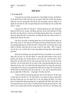
center frequencies in the (u v) frequency plane. An example of the real part
of a Gabor kernel function is given in Figure 8.7 with = 0:5 = 0:6 U =
1 V = 0 and = 0 (with reference to Equations 8.35 and 8.36). Another
Gabor kernel function is shown in gray scale in Figure 8.8.
The imaginary component of the Gabor function is the Hilbert transform
of its real component. The Hilbert transform shifts the phase of the original
function by 90o , resulting in an odd version of the function.
The \Gabor transform" is not a transform as such that is, there is usually
no transform domain into which the image is transformed. The frequency
domain is usually divided into a symmetric set of slightly overlapping regions
at octave intervals. Examples of the ranges related to a few Gabor functions
are shown in Figure 8.9 see also Figures 5.69, 8.57, and 8.68. It is evident
that Gabor functions act as bandpass lters with directional selectivity.
g(x y) = 2 1
2
exp
;
8.4.1 Multiresolution signal decomposition
Multiresolution signal analysis is performed using a single prototype function
called a wavelet. Fine temporal or spatial analysis is performed with contracted versions of the wavelet on the other hand, ne frequency analysis is
performed with dilated versions. The de nition of a wavelet is exible, and
requires only that the function have a bandpass transform thus, a wavelet at
a particular resolution acts as a bandpass lter. The bandpass lters must
© 2005 by CRC Press LLC
Analysis of Directional Patterns
661
0.4
Magnitude
0.2
0
-0.2
-0.4
40
40
30
30
20
20
10
rows
10
0
0
columns
FIGURE 8.7
An example of the Gabor kernel with = 0:5 = 0:6 U = 1 V = 0 and
= 0 (with reference to Equations 8.35 and 8.36). Figure courtesy of W.A.
Rolston 542].
FIGURE 8.8
An example of a Gabor kernel, displayed as an image. Figure courtesy of
W.A. Rolston 542].
© 2005 by CRC Press LLC
662
Biomedical Image Analysis
v
u
FIGURE 8.9
Division of the frequency domain by Gabor lters. Two sets of oval regions
are shown in black, corresponding to the passbands of three lters in each
set, oriented at 0o and 90o . In each case, the three regions correspond to
three scales of the Gabor wavelets. There is a 90o shift between the angles
of corresponding lter functions in the space and frequency domains. Figure
courtesy of W.A. Rolston 542].
have constant relative bandwidth or constant quality factor. The importance
of constant relative bandwidth of perceptual processes such as the auditory
and visual systems has long been recognized 571]. Multiresolution analysis
has also been used in computer vision for tasks such as segmentation and
object recognition 284, 285, 288, 487]. The analysis of nonstationary signals
often involves a compromise between how well transitions or discontinuities
can be located, and how nely long-term behavior can be identi ed. This
is re ected in the above-mentioned uncertainty principle, as established by
Gabor.
Gabor originally suggested his kernel function to be used over band-limited,
equally spaced areas of the frequency domain, or equivalently, with constant
window functions. This is commonly referred to as the short-time Fourier
transform (STFT) for short-time analysis of nonstationary signals 176, 31].
The 2D equivalent of the STFT is given by
FS (x0 y0 u v) =
Z1
Z1
f (x y) w(x ; x0 y ; y0 )
x=;1 y=;1
exp ;j 2 (ux + vy)] dx dy
(8.37)
where w is a windowing function and f is the signal (image) to be analyzed.
The advantage of short-time (or moving-window) analysis is that if the energy
of the signal is localized in a particular part of the signal, it is also localized
to a part of the resultant 4D space (x0 y0 u v). The disadvantage of this
method is that the same window is used at all frequencies, and hence, the
© 2005 by CRC Press LLC
Analysis of Directional Patterns
663
resolution is the same at all locations in the resultant space. The uncertainty
principle does not allow for arbitrary resolution in both of the space and
frequency domains thus, with this method of analysis, if the window function
is small, the large-scale behavior of the signal is lost, whereas if the window
is large, rapid discontinuities are washed out. In order to identify the ne or
small-scale discontinuities in signals, one would need to use basis functions
that are small in spatial extent, whereas functions of large spatial extent
would be required to obtain ne frequency analysis. By varying the window
function, one will be able to identify both the discontinuous and stationary
characteristics of a signal. The notion of scale is introduced when the size
of the window is increased by an order of magnitude. Such a multiresolution
or multiscale view of signal analysis is the essence of the wavelet transform.
Wavelet decomposition, in comparison to STFT analysis, is performed over
regions in the frequency domain of constant relative bandwidth as opposed to
a constant bandwidth.
In the problem of determining the directional nature of an image, we have
the discontinuity in the frequency domain at the origin, or DC, to overcome.
Wavelet analysis is usually applied to identify discontinuities in the spatial
domain however, there is a duality in wavelet analysis, provided by the uncertainty principle, that allows discontinuity analysis in the frequency domain
as well. In order to analyze the discontinuity at DC, large-scale or dilated
versions of the wavelet need to be used. This is the dual of using contracted
versions of the wavelet to analyze spatial discontinuities.
The wavelet basis is given by
hx0 y0
1
2
(x y) = p 1
1 2
0
0
h x;x y;y
1
2
(8.38)
where x0 y0 1 and 2 are real numbers, and h is the basic or mother wavelet.
For large values of 1 and 2 , the basis function becomes a stretched or expanded version of the prototype wavelet or a low-frequency function, whereas
for small 1 and 2 , the basis function becomes a contracted wavelet, that is,
a short, high-frequency function.
The wavelet transform is then de ned as
FW (x0 y0 1 2 ) =
p
Z1
1
1 2
Z1
x=;1 y=;1
f (x y)
0
0
h x ; x y ; y dx dy:
1
2
(8.39)
From this de nition, we can see that wavelet analysis of a signal consists of
the contraction, dilation, and translation of the basic mother wavelet, and
computing the projections of the resulting wavelets on to the given signal.
© 2005 by CRC Press LLC