Vữa xơ động mạch GS huỳnh văn minh
Bạn đang xem bản rút gọn của tài liệu. Xem và tải ngay bản đầy đủ của tài liệu tại đây (1.29 MB, 33 trang )
VỮA XƠ ĐỘNG MẠCH
PGS.TS. HUỲNH VĂN MINH
BỘ MÔN NỘI
ĐẠI HỌC Y HUẾ
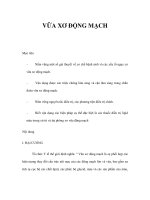
Risk Factors for Cardiovascular
Disease
Non-modifiable
Modifiable
Smoking
Dyslipidaemia
Raised LDL-C
Low HDL-C
Raised triglycerides
Raised blood pressure
Diabetes mellitus
Obesity
Dietary factors
Thrombogenic factors
Lack of exercise
Excess alcohol consumption
Non-modifiable
Personal history
of CVD
Family history
of CVD
Age
Gender
Pyörälä K et al. Eur Heart J 1994;15:1300–1331.
Levels of Risk Associated with
Smoking, Hypertension and
Hypertension
(SBP 195 mmHg)
Hypercholesterolaemia
x3
x9
x4.5
x16
Smoking
x1.6
x6
x4
Serum cholesterol level
(8.5 mmol/L, 330 mg/dL)
Poulter N et al., 1993
Historical Model of
Atherogenesis
Threshold
Decades
Years-Months
healthy
subclinical
Months-Days
symptomatic
Intima
Media
Lumen
Plaque
•
•
•
•
•
Stable angina
Stable plaques with narrowing
Simple diagnostic (ECG, angiography)
Rare MI
Easy to treat
Antischkow N. Beitr Path Anat Allg Path 1913;56:379-404.
New Paradigm
Threshold
Decades
Years-Months
healthy
subclinical
Months-Days
symptomatic
Thrombus
Intima
Media
Lumen
Plaque
•
•
•
•
•
Unstable angina
Unstable plaque no narrowing
Difficult to diagnose (IVUS, MRI)
Frequent MI with sudden death
Easy to prevent
The Vulnerable Atherosclerotic
Plaque
SMC – smooth muscle cell
HDL-DR – transplantation antigen indicating ‘activation’ of SMCs
Libby P. Circulation 1995;91:2844-2850.
CHẨN ĐOÁN
Chẩn đoán dựa vào nhiều dấu chứng và kết
quả thăm dò cận lâm sàng không có một tiêu
chuẩn rõ rệt. Có thể:
Các rối loạn cơ năng do thiếu máu cục bộ cơ
quan hoặc ngoại biên.
Sự hiện diện của những yếu tố nguy cơ.
Tình trạng động mạch ngoại biên.
Kết quả xét nghiệm: soi đáy mắt, xét nghiệm
bilan lipid, chụp động mạch cản quang, siêu
âm doppler.
Imaging Techniques Used to
Assess Atherosclerosis
Invasive techniques
Intravascular ultrasound (IVUS)
Coronary angiography
Non-invasive techniques
Magnetic resonance imaging (MRI)
Computed tomography (CT)
Ultrasound (B-mode)
Clinical Manifestations of
Atherosclerosis
Coronary heart disease
Cerebrovascular disease
Angina pectoris, myocardial infarction, sudden
cardiac death, congestive heart failure (CHF), and
arrhythmias
Transient ischaemic attack, stroke
Peripheral vascular disease
Intermittent claudication, gangrene, cold feet,
painful feet, impotence
ĐIỀU TRỊ
1. Nguyên tắc điều trị
Có 3 mục đích chính
Điều trị các yếu tố nguy cơ chính: rối loạn lipid, đái tháo
đưòng, tăng huyết áp, thuốc lá trước khi có các triệu
chứng (phòng bệnh sơ cấp) hoặc sau khi có các triệu
chứng (phòng bệnh thứ cấp).
Điều trị các biến chứng vữa xơ động mạch (VXĐM) sơ
cấp và thứ cấp (điều trị chống ngưng kết tiểu cầu)
Điều trị đặc hiệu tổn thương
Một số nguy cơ phối hợp song song cần điều trị như:
điều trị thay thế hocmon trong tiền mãn kinh, chế độ ăn
kiêng trong béo phì, tăng hoạt động thể lực
2. Điều trị cụ thể
2.1. Thay đổi các yếu tố nguy cơ (YTNC)
Một số YTNC của VXĐM có thể tác động
nhằm ngăn chặn sự tiến triển hoặc làm giảm
dần XVĐM: ngừng hút thuốc, kiểm soát HA,
ổn định đường máu, tránh dùng rượu quá
nhiều, tập thể dục đều đặn nhất là kiểm soát sự
rối loạn lipid máu.
2.2. Thuốc ức chế men chuyển và thuốc chẹn bêta
Thuốc ức chế men chuyển giảm từ 14 đến 28%
biến cố tim mạch;
Thuốc chẹn bêta giảm tỉ lệ tử vong sau nhồi
máu cơ tim 20%, giảm tái phát nhồi máu 25%
và giảm đột tử 30%.
2.3. Điều trị tăng lipid máu
Điều trị vữa xơ động mạch là một điều trị toàn
diện
Tuy vậy một trong những mục tiêu cơ bản vẫn
là thoái triển mãng vữa xơ có sự lắng đọng
lipid, giảm đi các thành phần lipoprotein máu
có hại.
Classification of Dyslipidaemias:
Fredrickson (WHO)
Classification
Phenotype
Lipoprotein
elevated
Serum
cholesterol
I
Chylomicrons
mean to
IIa
LDL
IIb
Atherogenicity
Prevalence
None seen
Rare
+++
Common
LDL and VLDL
+++
Common
III
IDL
+++
Intermediate
IV
VLDL
mean to
+
Common
VLDL and
mean to
chylomicrons
+
Rare
V
Serum
TG
mean
LDL – low-density lipoprotein; IDL – intermediate-density lipoprotein; VLDL – very low-density
lipoprotein. (High-density lipoprotein (HDL) cholesterol levels are not considered
in the Fredrickson classification.)
Yeshurun D, Gotto AM. Southern Med J 1995;88(4):379–391
HiÖu qu¶ cña Statin trong æn ®Þnh
m¶ng x¬ v÷a ®éng m¹ch vµnh
Class effect
Statin
therapy
Lipidlowering
Plaque
stability
Drug-specific effects
Other
biological
Effects
Pleiotropic Effects?
Weissberg, 1999
Classification of Dyslipidaemias
Fredrickson (WHO)
Phenotype Lipoprotein
Serum
Serum Atherogenicity Prevalence
elevated cholesterol triglyceride
Classification
I
Chylomicrons Normal to
None seen
Rare
+++
Common
IIa
LDL
IIb
LDL and VLDL
+++
Common
III
IDL
+++
Intermediate
IV
VLDL
Normal to
+
Common
VLDL and Normal to
chylomicrons
+
Rare
V
Normal
LDL – low-density lipoprotein; IDL – intermediate-density lipoprotein; VLDL – very lowdensity lipoprotein. (High-density lipoprotein (HDL) cholesterol levels are not considered
in the Fredrickson classification.)
(Adapted from Yeshurun et al., 1995)
Exogenous Pathway of Lipid
Metabolism
Intestine
Dietary
triglycerides
and cholesterol
Chylomicron
LP lipase
Liver
Skeletal muscle
FFA
Chylomicron
remnant
Remnant
receptor
to atheroma
Adipose
tissue
Endogenous Pathway of Lipid
Metabolism
LPL
HL
LPL
LD
receptor
L
Liver
LD
L
HL
IDL
HL
LPL
Small
VLDL
HL
LPL
Large
VLDL
Lipoprotein lipase
Hepatic lipase
Reverse Cholesterol Transport
Cell
membrane
SRB1
CE
CE
ABCA1
FC
LCAT
HDL
CETP
HDL3
LDL
receptor
VLDL, IDL, LDL
TG
Peripheral
tissues
FC
TG
CE
LCAT
CETP
SRB1
ABCA1
Liver
Free cholesterol
Triglycerides
Cholesteryl esters
Lecithin cholesterol acyltransferase
Cholesteryl ester transfer protein
Scavenger receptor class B, member 1
ATP-binding cassette, sub-family A, member 1
Thuốc giảm lipid máu
Nhóm 1:là các chất bắt giữ muối mật.
Tác dụng chính là giảm LDL-C.
Đây là những resin trao đổi ion gắn với muối mật trong ruột
non làm gián đoạn sự lưu hành muối mật trong chu trình gan
ruột và kích thích sự chuyển cholesterol thành muối mật trong
gan.
Điềìu này sẽ kích thích sự tạo thành các thụ thể LDL do đó sẽ
làm giảm LDL huyết thanh.
Đứng đầu nhóm là cholestyramine (Questran), giảm
cholesterol 15-30% và triglycerid từ 5-15% với liều dùng 4-16
g/ngày.
Colestipol liều 5-20 g/ ngày
Tác dụng phụ: táo bón, đầy bụng, buồn nôn.
Nhóm 2: các fibrate
có tác dụng tăng hoạt tính lipoprotein lipase làm gia
tăng quá trình thoái biến VLDL-C và IDL-C do đó
giảm triglycerid. Ưu điểm là HDL -C gia tăng khi xử
dụng fibrate.
Tác dụng phụ bao gồm rối loạn tiêu hoá, gia tăng tạo
sỏi mật.
Thuốc thông dụng như fenofibrate (Lipanthyl) làm
giảm CT (15-30%) và TG (15-30%), liều từ 100300mg/ ngày.
Gemfibrozil 600 mgx 2lần/ngày hoặc Clofibrate
500mg x 2-3 lần/ngày.
Nhóm 3: có acid nicotinic và dẫn chất.
Tác dụng khi dùng liều cao.
Có tác dụng giảm sự tạo thành VLDL trong gan do đó
giảm HDL.
Nicotinic acid giảm CT (5-15%) và TG (15-30%) và
làm gia tăng cả HDL-C.
Thuốc thông dụng là probucol (Lurselle) liều 0.30.6g/ ngày.
Tác dụng phụ:phừng mặt, tăng dường máu, tăng acid
uric máu, rối loạn tiêu hoá, độc cho gan.
Cần theo dõi chức năng gan khi điều trị. Phừng mặt
có thể khống chế bằng aspirin.
Nhóm 4: là các statin
làm giảm CT >30-50 % và TG 15-50%.
Đây là nhóm thuốc có tác dụng mạnh hạ cholesterol máu.
Cơ chế tác dụng là ức chế men HMG CoE reductase làm
ngăn cản quá trình chuyển hoá tạo cholesterol nội bào.
Ức chế quá trình này sẽ làm gia tăng tổng hợp thụ thể
LDL do đó sẽ làm giảm cholesterol huyết thanh.
Thuốc thông dụng là Fluvastatine liều 20-40mg/ngày,
Lovastatine 10-80mg/ngày, Pravastatin 10-40mg/ngày,
Simvastatin 5-40 mg/ngày.
Tác dụng phụ b bao gồm khó tiêu, bón, đau bụng, co rút
và có thể độc với gan nên cần theo dõi men gan.
Mechanism of Action of Statins
Cholesterol Synthesis
Pathway
acetyl CoA
HMG-CoA synthase
HMG-CoA reductase
HMG-CoA
X Statins
mevalonic acid
mevalonate pyrophosphate
isopentenyl pyrophosphate
geranyl pyrophosphate
ubiquinones
farnesyl pyrophosphate
Squalene synthase
squalene
cholesterol
dolichols
Pharmacokinetics of Statins
Statin
Metabolised
by CYP450
Protein
binding (%)
Lipophilic
Halflife (h)
lovastatin
Yes
>95%
Yes
~2
pravastatin
No
~50%
No
~2
simvastatin
Yes
95–8%
Yes
~3
atorvastatin
Yes
>98%
Yes
~15
cerivastatin
Yes
>99%
Yes
~3
fluvastatin
Yes
>98%
No
~3
(Adapted from Horsmans 1999, Vaughan et al, 2000)