DE TAI 2 TAI SINH PHOI TU LA THONG QUA NUOI CAY HUYEN PHU
Bạn đang xem bản rút gọn của tài liệu. Xem và tải ngay bản đầy đủ của tài liệu tại đây (600.13 KB, 13 trang )
European Scientific Journal September 2013 edition vol.9, No.27 ISSN: 1857 – 7881 (Print) e - ISSN 1857- 7431
SOMATIC EMBRYOGENESIS AND PLANT
REGENERATION FROM CALLUS AND
SUSPENSION CULTURES OF IPHIONA
MUCRONATA (FORSSK)
Amal A. Al-Gendy, Ass. Prof., PhD
Pharmacognosy Department, Faculty of Pharmacy, Zagazig University &
October University for Modern Sciences and Arts (MSA), Egypt
Riham O. Bakr, Lecturer, PhD
Pharmacognosy Department, Faculty of Pharmacy,
October University for Modern Sciences and Arts (MSA), Egypt
Omayma D. El-gindi, Prof., PhD
Pharmacognosy Department, Faculty of Pharmacy,
Egyptian Russian University (ERU), Egypt
Abstract
A protocol was designed for plant regeneration of Iphiona mucronata
from embryogenic callus via somatic embryogenesis to enable micro
propagation of this endangered plant. The embryogenic callus was induced
using seedling cultured for nine months on Murashig and Skoog medium
(MS) supplemented with 0.1 mg l-1 naphthalene acetic acid (NAA),
0.1 mg l-1 kinetin (Kn) and 5 mg l-1 ascorbic acid and incubated in the dark
followed by growing on hormone free medium. Transfer of developed
embryos to MS medium supplemented with 0.5 mg l-1 kinetin induced shoot
formation. Four treatments were further tried for plant development by using
indole acetic acid (IAA) or indole butyric acid (IBA) alone or in combination
with kinetin. The results showed that 2 mg l-1 IAA was the best for in vitro
plantlet regeneration. Embryogenic suspension was induced by transfer of
embryogenic callus to liquid medium having the same composition followed
by hormone free medium where different stages of embryos were monitored.
Shoots were developed upon transfer to liquid medium supplemented with
0.5 mg l-1 Kn. However, no further development appeared upon transfer to
semi solid medium containing different phytohormones. Embryogenic callus
showed the highest phenolic contents when compared with embryogenic
suspension, regenerated plantlets and the parent plant while flavonoids were
detected only in embryogenic callus.
37
European Scientific Journal September 2013 edition vol.9, No.27 ISSN: 1857 – 7881 (Print) e - ISSN 1857- 7431
Keywords: Iphiona mucronata, callus and suspension culture, somatic
embryogenesis, plant regeneration
Introduction
Iphiona Cass. is a small genus of about eleven species, which is
distributed from North-East Africa to central Asia (Anderberg, 1985).
Studies on Iphiona scabra and Iphiona mucronata which are native in
Egyptian deserts (Zahran and Willis, 2009) revealed that polysulphated
flavonoids and sesquiterpene glycosides were the major constituents and
seem to be characteristic for this genus (Ahmed and Mabry, 1987; Ahmed et
al., 1988). In vitro propagation was not tried in any of its species, as an
endangered plant, somatic embryogenesis would be of value.In a previous
work (Al-Gendy et al., 2008), a successful callus cell line was established
with high phenolic and considerable production of flavonoids when
compared with the parent plant using MS medium (Murashige and Skoog,
1962).
The culture of somatic embryos in a liquid medium has numerous
advantages as the swirling medium naturally separates the embryos, which
are then easily observed and fractionated according to their stages. They can
be obtained in great quantity and used as a basis for a large-scale
micropropagation (Monnier, 1990).
The objective of this study is to develop an efficient protocol for
micropropagation of Iphiona mucronata via somatic embryogenesis to save
this plant from eradication. We also investigate the flavonoid and phenolic
contents of somatic embryos in callus and suspension culture compared with
regenerated plantlets.
Material and methods
Induction of embryogenic callus
Callus was induced using MS medium supplemented with 0.1 mg l-1
NAA, 0.1 mg l-1Kn, 5 mg l-1 ascorbic acid, 30 g l-1 sucrose and solidified
with 10 g l-1 agar (MS-1). Media were adjusted to pH 5.8 using 1 N NaOH or
1 N HCl, autoclaved at 121 °C for 20 min and incubated at 25 °C in the dark
as previously reported (Al-Gendy et al., 2008). After nine months of culture,
the nodular embryogenic calli were moved to the same medium but without
phytohormones (hormone free medium; MS-HF), maintained at 25°C, with
12-h photoperiod (using fluorescent white lamps) and subcultured into fresh
medium every 4 weeks for 12-24 weeks.
Somatic embryo formation and development
The well developed embryogenic calli grown on MS-HF were
removed to semi solid media supplemented with 0.5 mg l-1Kn, 50 g l-1
sucrose, 5 mg l-1 ascorbic acid and solidified with 8 g l-1 agar (MS-XS) to
enhance the development of somatic embryos for 12-24 weeks. Cultures
38
European Scientific Journal September 2013 edition vol.9, No.27 ISSN: 1857 – 7881 (Print) e - ISSN 1857- 7431
were maintained at 25°C, with 12-h photoperiod (using fluorescent white
lamps). Cultures were routinely examined microscopically at each subculture
and photographs were recorded.
Conversion of somatic embryos into plantlets
Cultures grown on semi solid MS-XS were classified into 4 groups as
follow; group A cultured on 2 mg l-1 IAA, group B cultured on 2 mg l-1 IAA
and 0.5 mg l-1 Kn, group C cultured on 2 mg l-1 IBA, group D cultured on 2
mg l-1 IBA and 0.5 mg l-1 Kn. All cultures were supplemented with 30 g l-1
sucrose, 5 mg l-1 ascorbic acid and incubated at 25±2ºC with 16-h light
exposure and regularly transferred to fresh medium every 2-4 weeks
according to the growth.
Induction and maintenance of embryogenic suspension culture (ESC)
Embryogenic callus grown on MS-1 was transferred to 250 ml
Erlenmeyer flask containing 50 ml liquid medium having the same
composition as MS-1 except agar, incubated on rotary shaker (120 rpm at
25±2ºC in dark) and transferred into fresh medium every two weeks. After 6
weeks in culture, the embryogenic suspension was sub-cultured on hormone
free media supplemented with 50 g l-1 sucrose and 5 mg l-1 ascorbic acid and
sub-cultured into fresh medium every two weeks for 8 generations. Biomass
was separated from liquid medium and examined microscopically. At the 4th
generation growth curve and production of phenolic content were studied.
ESC was transferred to another liquid media supplemented with 0.5
mg l-1Kn, 5 mg l-1 ascorbic acid and 50 g l-1 sucrose in a trial for embryo
germination. Developed shoots were transferred to semi solid media
supplemented with 2 mg l-1 IAA, 5 mg l-1 ascorbic acid, 50 g l-1 sucrose and
solidified with 8 g l-1 agar.
Growth dynamics in ESC
Growth curve: Samples were taken with intervals of 3 days up to fourteen
days in suspension where fresh and dry weights were determined (GodoyHernández and Vázquez-Flota, 2006).
Growth index (GI) = (Ge - Gstart)/ Gstart (Verpoorte et al., 1998)
Where Ge = Weight of biomass at the end of generation (final dry weight).
Gstart = Weight of biomass at zero time (Initial dry weight).
Relative growth rate (RGR) was measured on fresh weight basis using the
following formula:
RGR = 3(Wf⅓ - Wi⅓) / tf-ti (Parsaeimehr et al., 2010)
ti: Beginning of the experiment, tf: Last day of subculture, after 14 days
Wi: Weight of initial biomass (at ti), Wf: Final biomass weight (at tf), tf-ti =
14 days of subculture.
Specific growth rate (μ) : μ = (ln x − ln xo)/t
Where xo is the initial dry biomass and x is the biomass at time t (14 days)
(Godoy-Hernández and Vázquez-Flota, 2006).
39
European Scientific Journal September 2013 edition vol.9, No.27 ISSN: 1857 – 7881 (Print) e - ISSN 1857- 7431
Doubling time which is the time required for the biomass of a population of
cells to double is denoted as (dt). dt= ln (2)/ μ
Determination of total flavonoids and phenolic contents
Total flavonoids: One gram of each of the dried embryogenic callus,
embryogenic suspension biomass and regenerated plantlets was extracted
with 25 ml of hot 95% ethanol (v/v) overnight at 37 °C and the filtrate was
adjusted with 80% ethanol (v/v) to 25 ml. Total flavonoid content was
estimated colorimetrically as reported by Kosalec et al. (2004) and used
previously for non embryogenic callus (Al-Gendy et al. 2008). Quantitation
was done based on the standard curve generated with rutin (12.5, 25, 50, 80
and 100 µg ml-1) measured at 415 nm.
Total phenolics: Dried embryogenic callus, suspension biomass,
regenerated plantlets and parent plant (1 g each) were extracted with 25 ml
methanol. Total polyphenols were estimated colorimetrically using the FolinCiocalteu method as reported (Sellappan and Akoh, 2002).The absorbance
was measured at 765 nm using a Shimadzu UV-visible spectrophotometer
(1800-UV probe) after incubation for 2 h at room temperature.
Quantification was done based on the standard curve generated with gallic
acid (10, 20, 40, 60, 80 and 100 µg ml-1).
Results
Embryogenic callus
After nine months of culture in the dark, pockets of embryogenic calli
with nodular structures appeared on the surface of the non embryogenic
callus. These calli tend to be light greenish yellow in color which is
differentiated from the non embryogenic callus. When proliferated calli were
moved to hormonal free medium, they kept the embryogenic potential and
showed further embryo development.
Somatic embryo formation and development
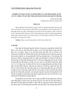
Globular-staged (G) embryos (75-150 µm in diameter, Fig. 1a), heart
shaped (H) embryos (75-200µm x 75-250 µm, Fig. 1b) and torpedo-shaped
(T) embryos (200-350 µm, Fig. 1c) were monitored. Mature torpedo shaped
embryos successfully germinated into cotyledonary embryo (Fig. 1d) which
further developed into cotyledonary leaves (Fig. 1e) after the fourth
generation. A heterogeneous population of somatic embryos appears,
showing non synchronous culture (Fig. 1f).
A fraction of somatic embryos differentiated directly into plantlets,
while the others produced secondary embryos after each subculture in a
repetitive way. The embryogenic callus retained its ability to grow and
produce somatic embryos for about a year.
40
European Scientific Journal September 2013 edition vol.9, No.27 ISSN: 1857 – 7881 (Print) e - ISSN 1857- 7431
Conversion of somatic embryos into plantlets
When the developed embryos were transferred to MS-XS, shoot
formation appeared, the length of the shoot was 0.5 cm after the third
subculture (Fig. 2a), and no further elongation appeared.
When regenerated shoots were transferred to several media
supplemented with various concentrations of IBA and IAA alone or in
combination with Kn (group A-D) for 3 generations, normal shoot length
increases to about 3cm (Fig. 2b). However, group A of regenerated plantlets
was the most successful. Shoot reached about 4.5 cm in length with green,
alternate acicular leaves and root was about 2 cm in length after 6
subcultures (Fig. 2c, d). When Kn was added (group B), roots begin to
appear at the first generation but was depleted at the second. Roots were
observed for group B, C and D regenerated plantlets at the first generation
but no further development occurred afterwards.
Abnormalities of I. mucronata embryogenic callus
Some of the embryos that had developed beyond the globular stage
were fused in pairs (Fig. 3a), early and late torpedo stage (Fig. 3b, c). Other
forms of anomalies may be present (Fig. 3d, e). Certain abnormality
appeared in some plantlets which seems dwarfed (Fig. 3f).
a
b
c
d
e
f
Figure 1: Stages of embryogenic callus of I. mucronata. a globular embryo (bar 30 µm). b
heart shaped embryo (bar 50 µm). c torpedo shaped embryo (bar 50 µm). d cotyledonary
embryo (bar 200 µm). e cotyledonary leaves (bar 1 cm). f embryos at different stages (bar 50
µm). (ET early torpedo)
41
European Scientific Journal September 2013 edition vol.9, No.27 ISSN: 1857 – 7881 (Print) e - ISSN 1857- 7431
a
b
c
d
Figure 2: Plantlet regeneration of I. mucronata from embryogenic callus. a &b shoot
formation. c root formation. d regenerated plantlet.
a
d
c
b
e
f
Figure 3: Anomalies in embryogenic callus of I. mucronata (bar 50 µm; a-e) a fused
globular (FG). b early torpedo (ET). c fused torpedo (FT). d abnormal heart embryo. e
anomalies in torpedo. f fused plantlet.
Embryogenic suspension culture
Biomass growing on suspension hormone free media (Fig. 4a) was
separated and examined microscopically revealing different embryo stages,
from globular to early cotyledonary stages (Fig. 4b-e). Globular embryos
appeared in the first generation (Fig. 4b) while heart shaped embryos (Fig.
42
European Scientific Journal September 2013 edition vol.9, No.27 ISSN: 1857 – 7881 (Print) e - ISSN 1857- 7431
4c) appeared in the second generation, which then differentiated into torpedo
stage (Fig. 4d). Moreover, heterogeneous embryos at different stages were
also monitored (Fig. 4-e)
Plantlet regeneration in ESC
When the embroids were transferred to liquid media supplemented
with 0.5 mg l-1 Kn, 5 mg l-1 ascorbic acid and 50 g l-1 sucrose in a trial for
embryo germination, shoots (1 cm length) appeared after the 4th subculture.
Unfortunately no further development appeared upon transfer of the
developed shoot to semi solid media supplemented with 2 mg l-1 IAA (group
A) (Fig. 4f). Abnormal embroids e.g. fused globular and torpedo shaped
were noticed at the 4th generation of embryogenic suspension (Fig. 5).
a
d
b
c
e
f
Figure 4: Somaticembryos of I. mucronata suspension culture. a Embryogenic suspension
biomass (bar: 1 cm) b globular embryos. c heart shaped embryos . d torpedo shaped
embryos. e different stages embryos (bar 50 µm, b-e). f undifferentiated plantlet.
a
b
Figure 5: Fused embryos in suspension culture of I. mucronata(bar 50 µm). a fused
globular. b fused torpedo.
43
European Scientific Journal September 2013 edition vol.9, No.27 ISSN: 1857 – 7881 (Print) e - ISSN 1857- 7431
Dry weight (g)
Growth dynamics of ESC
Growth curve of ESC based upon dry weight measurement is
represented in Fig. 6. It is noticed that the maximum dry weight was
achieved after 9 days and continued a stationary phase after that. Growth
parameters on dry weight basis were as following:
GI=1.381, RGR=0.04, µ=0.05 and dt= 13.06d
Investigation of total phenolic and flavonoids contents
When phenolic contents were estimated (Fig. 7), EC showed the
highest phenolic content as it represents 1.4 times the embryogenic
suspension culture and 1.6 the regenerated plantlets. Moreover, It represents
2.9 times the parent plant itself. Follow up of the phenolic content through
the whole generation of the 5th subculture of ESC on hormone free media,
revealed the gradual decrease till reaching minimum value at day 6 followed
by an increase at the 9th day reaching the highest level by day 13 then
decreasing again (Fig. 8). On the other hand, the lack of flavonoid content
was noticed in the regenerated plantlet and ESC while detected only in
embryogenic callus (293µg ml-1) which represents about 16 % of the parent
plant estimated previously (Al-Gendy et al. 2008).
0.7
0.6
0.5
0.4
0.3
0.2
0.1
0
0
3
6
9
13
15
Time (Day)
Figure 6: Growth curve of embryogenic
suspension culture of I. mucronata. Mean ± SD,
n=3
Figure
7:
Phenolic
contents
of
embryogenic callus and suspension
cultures of I. mucronata compared with
regenerated plantlet and parent plant.
Mean ± SD, n=3
44
Phenolic content (µg/g. DW)
European Scientific Journal September 2013 edition vol.9, No.27 ISSN: 1857 – 7881 (Print) e - ISSN 1857- 7431
6000
5000
4000
3000
2000
1000
0
0
3
6
9
12
15
Time (Day)
Figure 8: Follow up of phenolic content in I. mucronata embryogenic suspension culture
(ESC). Mean ± SD, n=3
Discussion
Indole acetic acid was the most successful for plantlet regeneration.
A previous study reported that IAA (2 mg l-1) and Kn (2 mg l-1) appeared to
be a good combination for shoot regeneration in Arachis hypogaea
(Narasimhulua and Reddy, 1983). Addition of 0.5 mg l-1Kn showed a
positive effect on regeneration when combined with0.1 mg l-1IAA and 0.5
mg l-1 6-benzylaminopurine.When the concentration of Kn was decreased
from 0.5 to 0.1 mg l-1, the percentage of regeneration was also decreased
from 80.60% and 62.2% to 6.0% and 14.6% in Pakistani wheat cultivars
Kohsar and Khyber-87, respectively. These results may justify the increased
embryogenesis when Kn was used at concentration 0.5 mg l-1(Noor et al.,
2009).Another report for in vitro regeneration of Citrus aurantifolia
(Rutaceae) revealed that IAA significantly influenced root proliferation and
shoot growth (Al-Khayri and Al-Bahrany, 2001).
Appearance of abnormal embryos has been observed in many
species. Reasons for their development are not well known. It has been
suggested that this phenomenon is attributed to the developmental plasticity
of somatic embryogenesis that is influenced by culture conditions. Possibly
somatic embryos that failed to convert into plantlets were inclined to produce
secondary embryos (Luo et al., 1999; Carman, 1990). In this study, these
abnormalities occurred in hormone free media, possibly due to failure to
convert into plantlets.
The lack of flavonoid content in the regenerated plantlet may be due
to the nature of flavonoids which are UV-B inducible (Cockell and
Knowland, 1999) while the lamps used in the in vitro growth chamber did
not provide wave lengths in the range of the UV radiation. An analogous
45
European Scientific Journal September 2013 edition vol.9, No.27 ISSN: 1857 – 7881 (Print) e - ISSN 1857- 7431
behavior was shown in callus cultures of Passiflora spp. where the UV-B
irradiation was able to increase the production of flavonoids (Antognoni et
al., 2007; Lucchesini et al., 2009).
Results seemed similar to that of thalamus-derived calluses of
Ranunculus asiaticus L. where non differentiating callus was characterized
by a high content of phenolic polymers and an elevated peroxidase and
polyphenol oxidase activity in comparison with differentiating callus (Beruto
et al., 1996).
Upon studying phenolic content of Echinacea. angustifolia, the yields
were the highest among the in vitro cultures and they were similar or higher
than leaves of adult plants (Lucchesini et al. 2009). These results are
matching with data obtained through this study (Fig. 7), where embryogenic
callus culture has the highest phenolic content compared with ESC,
regenerated plantlets and the parent plant.
Embryogenic suspension cultures have been established in only few
crops, including sweet potato, cowpea and horsegram (Naik and Murthy,
2010). However, the quality of somatic embryos with regard to their
germinability or conversion into plants has been generally very poor. This is
due to the apparently normal looking somatic embryos are actually
incomplete in development. (Bhojwani and Razdan, 1996; Amoo and
Ayisire, 2005; Naik and Murthy, 2010; Pescador et al., 2008; Yantcheva et
al., 1998). According to Canhoto et al. (1999), the most common
abnormalities encountered are embryo fusion. Fused globular and torpedo
shaped embryos were noticed at the 4th generation of I. mucronata
embryogenic suspension culture (Fig. 5).
In a study to compare ESC and NESC for Medicago sativa, NESC
gave a typical growth curve while in ESC the distinct phases were absent
(Hrubcová et al., 1994), this was the case in ESC of I. mucronata.
When estimating the growth parameters, GI of embryogenic
suspension is relatively low (1.38) when compared with non embryogenic
callus previously reported (Al-Gendy et al., 2008). It needs longer time to
reach double its initial weight (13.06 d) which is considered 1.6 the time
needed for non embryogenic callus. So, embryogenic suspension culture is
not a reliable method for obtaining biomass production.
Conclusion:
A protocol is established for the first time for somatic embryogenesis
in callus and suspension culture of I. mucronata that can be used for micro
propagation of this plant to save it from eradication, in addition to
comparison of phenolic and flavonoid contents in embryogenic callus and
suspension cultures with regenerated plantlets and parent plant.
46
European Scientific Journal September 2013 edition vol.9, No.27 ISSN: 1857 – 7881 (Print) e - ISSN 1857- 7431
Acknowledgement
The authors are grateful to October University for Modern Sciences
and Arts (MSA) for sponsorship and supplying the research facilities of this
research work.
References:
Ahmed, A. A. & Mabry, T. J. (1987). Flavonoids of Iphiona scabra.
Phytochemistry, 26, 1517-1518.
Ahmed, A. A.; Melek, F. R.; Seif El-Din, A. A. & Mabry, T. J. (1988).
Polysulphated flavonoids from Iphiona mucronata. Rev. Latinoamer Quim.,
19 (3), 107-109.
Al-Gendy, A. A.; El-Gindi, O. D. & Bakr, R. O. (2008). Production of
flavonoids in callus cultures of Iphiona mucronata, Astraceae. Egypt. J.
Biomed. Sci., 28, 142-150.
Al-Khayri, J. M. & Al-Bahrany, A. M. (2001). In vitro micropropagation of
Citrus aurantifolia (lime). Curr. Sci, 81(9, 10), 1242-1246.
Amoo, S. O. & Ayisire, B. E. (2005). Induction of callus and somatic
embryogenesis from cotyledon explants of Parkia biglobosa (Jacq.) Benth.
Afr. J. Biotechnol., 4 (1), 68-71.
Anderberg, A. (1985). The genus Iphiona (Compositae-Inuleae). Nord J Bot,
5, 169-194.
Antognoni, F.; Zheng, S.; Pagnucco, C.; Baraldi, R.; Poli, F. & Biondi, S.
(2007). Induction of flavonoid production by UV-B radiation in Passiflora
quadrangularis callus cultures. Fitoterapia, 78, 345–352.
Beruto, M.; Curir, P. & Debergh, P. (1996). Callus growth and somatic
embryogenesis in thalamus tissue of Ranunculus asiaticus L. cultivated in
vitro: Cytokinin effect and phenol metabolism. In Vitro Cell Dev Biol –
Plant, 32 (3), 154-160.
Bhojwani, S. S. & Razdan, M. K. (1996). Plant tissue culture: theory and
practical, a revised edition. Elsevier science, Netherlands, 148–150.
Carman, J. G. (1990). Embryogenic cells in plant tissue cultures: occurrence
and behavior. InVitroCellDevBiol, 26, 746-753.
Cockell, C. S. & Knowland, J. (1999). Ultraviolet radiation screening
compounds. Biol Rev, 74, 311–345.
Godoy-Hernández, G. & Vázquez-Flota, F. (2006). Growth measurements
estimation of cell division and cell expansion. In: Loyola-Vargas VM,
Vázquez-Flota F (ed) “ Plant Cell Culture Protocols”. 2nd edn. Humana Press
Inc.
Hrubcová, M; Cvikrová, M. & Eder, J. (1994).Peroxidase activities and
contents of phenolic acids in embryogenic and non embryogenic alfalfa cell
suspension. Biol Plantarum,36 (2), 175-182.
47
European Scientific Journal September 2013 edition vol.9, No.27 ISSN: 1857 – 7881 (Print) e - ISSN 1857- 7431
Kouakou, T. H.; Waffo-Téguo, P.; Kouadio, Y. J.; Valls, J.; Richard, T.;
Decendit, A. & Mérillon, J. M. (2007). Phenolic compounds and somatic
embryogenesis in cotton (Gossypium hirsutum L.). PlantCellTissOrganCult,
90, 25-29.
Kosalec, I.; Bakmaz, M.; Pepeljnjak, S. & Vladimir-Knezevic, S. (2004).
Quantitative analysis of the flavonoids in raw propolis from northern
Croatia. Acatpharma, 54,65-72.
Lucchesini, M.; Bertoli, A.; Mensuali-Sodi, A. & Pistelli, L. (2009).
Establishment of in vitro tissue cultures from Echinacea angustifolia D.C.
adult plants for the production of phytochemical compounds. SciHort122 (3),
484-490.
Luo, J.; Jia, J.; Gu, Y. & Liu, J. (1999). High frequency somatic
embryogenesis and plant regeneration in callus cultures of Astragalus
adsurgens Pall. PlantSci, 143, 93-99.
May, R. A. & Trigiano, R. N. (1991). Somatic embryogenesis and plant
regeneration from leaves of Dendranthema grandiflora. J Am Soc Hort Sci,
116 (2), 366-371.
Merxmuller, H.; Leins, P. & Roesseler, H. (1977). Inuleae: systematic
review. In: Heywood JB, Harborne JB, Turner BL (ed) The Biology and
Chemistry of Compositae. Academic press, New York, pp 590-593.
Monnier, M. (1990). Induction of embryogenesis in suspension culture. In:
Pollard JW, Walker JM (ed). Methods in molecular biology. Plant cell and
tissue culture. The Humana Press. Vol. 6, 149-157.
Murashige, T. & Skoog, F. (1962). A revised medium for rapid growth and
bioassay with tobacco tissue cultures. PhysiolPlant, 15(3), 473-497.
Naik, P. M. & Murthy, H. N. (2010). Somatic embryogenesis and plant
regeneration from cell suspension culture of niger (Guizotia abyssinica
Cass.). Acta Physiol Plant, 32, 75–79.
Narasimhulu, S. B. & Reddy, G. M. (1983). Plantlet regeneration from
different callus cultures of Arachis hypogaea L. Plant Sci Lett, 31(2-3), 157163.
Noor, S.; Ali, G. M.; Rashid, U.; Arshad, M.; Ali, S. & Zafar, Y. (2009).
Optimization of callus induction and regeneration system for Pakistani wheat
cultivars Kohsar and Khyber-87. Afr. J. Biotechnol., 8 (20), 5565-5569.
Parsaeimehr, A.;Sargsyan, E. & Javidnia, K. (2010). A comparative study of
the antibacterial, antifungal and antioxidant activity and total content of
phenolic compounds of cell cultures and wild plants of three endemic species
of Ephedra. Molecules, 15(3), 1668-1678.
Pescador, R.; Kerbauy, G. B.; Viviani, D. & Kraus, J. E. (2008).Anomalous
somatic embryos in Acca sellowiana (O. Berg) Burret (Myrtaceae).Revista
Brasil Bot, 31 (1), 155-164.
48
European Scientific Journal September 2013 edition vol.9, No.27 ISSN: 1857 – 7881 (Print) e - ISSN 1857- 7431
Sellappan, S. & Akoh, C. C. (2002).Flavonoids and antioxidant capacity of
Georgia-grown Vidalia onions. J Agric Food Chem 50,5338-5342.
Shohael, A. M.; Hahn, E. J. & Paek, K. Y. (2007). Somatic embryogenesis
and secondary metabolite production through bioreactor culture of siberian
ginseng (Eleutherococcus senticosus). Acta Hort. (ISHS) 764, 181-186.
Verpoorte, R.; Van der Heijden, R.; ten Hoopen, H. J. G. & Memmelink, G.
(1998). Metabolic engineering for the improvement of plant secondary
metabolite production. Plant Tiss. Cult. Biotechnol, 4, 3 -19.
Yantcheva, A.; Vlahova, M. & Antanassov, A. (1998).Direct somatic
embryogenesis and plant regeneration of carnation (Dianthus caryophyllus
L.). Plant Cell Rep, 18, 148–153.
Zahran, M. A. & Willis, A. J. (2009). The vegetation of Egypt, 2nd edition,
Springer.
49