Báo cáo khoa học: Injectable nanoparticles for efficient drug delivery
Bạn đang xem bản rút gọn của tài liệu. Xem và tải ngay bản đầy đủ của tài liệu tại đây (925.43 KB, 20 trang )
Injectable nanoparticles for drug delivery
TABLE OF CONTENTS
INTRODUCTION
Organic Nanoparticle Technology has been concerned than ever before recent
years due to its application to drug delivery, common to a number of therapeutic areas
and targets. Earlier researches on liposomes and emulsions were the examples of
enhancements that drug delivery could confer on established agents such as
doxorubicin and amphotericin. The disposition of nanoparticles was changed in vivo,
but the drug molecular structure was not transformed. For broader applicability,
nanoparticles have been sticked with additional features in order to enhance their
ability to targeting organs. Unlike microparticulates, nanoparticulates are sufficiently
small to avoid embolism related to intravenous (i.v) delivery, and can also be used for
the less invasive parenteral routes.[1]
A large proportion of i.v. drugs in development are antineoplastic agents or
antiinflammatory compounds. While they are fewer in number, there is a need for
improved antimicrobial agents as well, although many companies are exiting this area.
Opportunities for enhancement in these specific therapeutic areas will be considered
from a biological barrier perspective. Additionally, medical benefits arising from the
ability to target to specific organs will also be shown. The limitations of predicate
dosage form platforms need noted, which define the opportunities of nanoparticulates
to address unmet needs.[1]
Performer: Nguyễn Văn Tú - PhD Student
Bach Khoa University
Injectable nanoparticles for drug delivery
CONTENTS
I. GENERALITY OF INJECTABLE ROUTES
The goal of drug therapy is to prevent, cure, or control various disease states. To
achieve this goal, adequate drug doses must be delivered to the target tissues so that
therapeutic yet nontoxic levels are obtained. Pharmacokinetics examines the
movement of a drug over time through the body. Pharmacological as well as
toxicological actions of drugs are primarily related to the plasma concentrations of
drugs. Thus, the clinician must recognize that the speed of onset of drug action, the
intensity of the drug's effect, and the duration of drug action are controlled by four
fundamental pathways of drug movement and modification in the body (Figure 1).
First, drug absorption from the site of administration (Absorption) permits entry
of the therapeutic agent (either directly or indirectly) into plasma. Second, the drug
may then reversibly leave the bloodstream and distribute into the interstitial and
intracellular fluids (Distribution). Third, the drug may be metabolized by the liver,
kidney, or other tissues (Metabolism). Finally, the drug and its metabolites are
removed from the body in urine, bile, or feces (Elimination). This chapter describes
how knowledge of these four processes (Absorption, Distribution, Metabolism, and
Elimination) influences the clinician's decision of the route of administration for a
specific drug, the amount and frequency of each dose, and the dosing intervals.[2]
Performer: Nguyễn Văn Tú - PhD Student
2
Bach Khoa University
Injectable nanoparticles for drug delivery
Figure 1. Four processes of drug inside the body
The route of administration is determined primarily by the properties of the
drug (for example, water or lipid solubility, ionization, etc.) and by the therapeutic
objectives (for example, the desirability of a rapid onset of action or the need for longterm administration or restriction to a local site). There are two major routes of drug
administration, enteral and parenteral. (Figure 2 illustrates the subcategories of these
routes as well as other methods of drug administration.) [2]
Figure 2. Commonly used routes of drug administration. IV = intravenous;
Performer: Nguyễn Văn Tú - PhD Student
3
Bach Khoa University
Injectable nanoparticles for drug delivery
IM = intramuscular; SC = subcutaneous
Enteral administration, or administering a drug by mouth, is the simplest and
most common means of administering drugs. When the drug is given in the mouth, it
may be swallowed, allowing oral delivery, or it may be placed under the tongue,
facilitating direct absorption into the bloodstream.
The parenteral route introduces drugs directly across the body's barrier defenses
into the systemic circulation or other vascular tissue. Parenteral administration is used
for drugs that are poorly absorbed from the GI tract (for example heparin) and for
agents that are unstable in the GI tract (for example, insulin). Parenteral administration
is also used for treatment of unconscious patients and under circumstances that require
a rapid onset of action. In addition, these routes have the highest bioavailability and
are not subject to first-pass metabolism or harsh GI environments. Parenteral
administration provides the most control over the actual dose of drug delivered to the
body. However, these routes are irreversible and may cause pain, fear, and infections.
The three major parenteral routes are intravascular (intravenous or intra-arterial),
intramuscular, and subcutaneous (see Figure 1.2). Each route has advantages and
drawbacks. [2]
1. Intravenous (IV)
Injection is the most common parenteral route. For drugs that are not absorbed
orally, such as the neuromuscular blocker atracurium, there is often no other choice.
With IV administration, the drug avoids the GI tract and therefore, first-pass
metabolism by the liver. Intravenous delivery permits a rapid effect and a maximal
degree of control over the circulating levels of the drug. However, unlike drugs in the
GI tract, those that are injected cannot be recalled by strategies such as emesis or by
binding to activated charcoal. Intravenous injection may inadvertently introduce
bacteria through contamination at the site of injection. IV injection may also induce
hemolysis or cause other adverse reactions by the too-rapid delivery of high
concentrations of drug to the plasma and tissues. Therefore, the rate of infusion must
be carefully controlled. Similar concerns apply to intra-arterially injected drugs. [2]
2. Intramuscular (IM)
Performer: Nguyễn Văn Tú - PhD Student
4
Bach Khoa University
Injectable nanoparticles for drug delivery
Drugs administered IM can be aqueous solutions or specialized depot
preparations often a suspension of drug in a nonaqueous vehicle such as polyethylene
glycol. Absorption of drugs in an aqueous solution is fast, whereas that from depot
preparations is slow. As the vehicle diffuses out of the muscle, the drug precipitates at
the site of injection. The drug then dissolves slowly, providing a sustained dose over
an extended period of time. An example is sustained-release haloperidol decanoate,
which slowly diffuses from the muscle and produces an extended neuroleptic effect.
3. Subcutaneous (SC)
This route of administration, like that of IM injection, requires absorption and is
somewhat slower than the IV route. Subcutaneous injection minimizes the risks
associated with intravascular injection. [Note: Minute amounts of epinephrine are
sometimes combined with a drug to restrict its area of action. Epinephrine acts as a
local vasoconstrictor and decreases removal of a drug, such as lidocaine, from the site
of administration.] Other examples of drugs utilizing SC administration include solids,
such as a single rod containing the contraceptive etonogestrel that is implanted for
long-term activity, and also programmable mechanical pumps that can be implanted to
deliver insulin in diabetic patients. [2]
II.
BIOLOGICAL
BARRIERS
IMPOSED
BY
THE
MONOCYTE
PHAGOCYTIC SYSTEM (MPS)
Based upon an understanding of compromised vasculature, the requirements of
a drug delivery system intended for targeting to sites of tumor, infection, or
inflammation can be specified. There is an upper limit placed upon the size of the
particle, permitting diffusion through the vascular pores. The range of pore sizes is
300–700 nm, depending upon the tumor type, and therefore targeting particles should
be substantially smaller, preferably <250 nm. The particles should be designed to
target the pores rather than suffer less productive competitive encounters, the major
one being that of entrapment by the monocyte phagocytic system (MPS). [1]
1. Entrapment: Phagocytosis
The MPS system consists of fixed macrophage cells in key tissues, such as
liver, kidney, lung, bone marrow, and spleen, as well as circulating monocytes,
Performer: Nguyễn Văn Tú - PhD Student
5
Bach Khoa University
Injectable nanoparticles for drug delivery
macrophages, and PMN cells. These are designed to rid the body of bacterial, viral,
and particulate waste. The first step in the MPS removal process involves deposition of
specific circulating blood proteins onto the particle, termed
opsonization, which
subsequently signal receptors on the macrophages and PMN for particle uptake
(Figure 3).
Figure 3. The entrapment of Phagocytosis towards
particulates
Following opsonin docking on the receptors, an intracytoplasmic process is
activated, reorganizing actin filaments, causing the extension of pseudopodia to project
from the phagocyte, surrounding the particle. The pseudopodia follow the contours of
the particle as guided by further receptor docking onto the opsonized particle.
Provided the particle is smaller than approximately 8 mm, the spreading pseudopodia
will eventually meet, totally engulfing the particle. The particle is then encased in an
intracytoplasmic vacuole, termed a phagosome, formed from a remnant of the
spreading pseudopodia.
While this process of phagocytosis is applicable to particles as small as 500 nm,
a similar receptor-mediated endocytosis is more generally available to many different
kinds of cells. This extends to particles as small as 100 nm and robably smaller. Non–
receptor-mediated pincocytosis also becomes more prominent as particle size
decreases from 1100 down to 100 nm. [1]
2. Escape
Over the course of 15–30 minutes, the pH of the phagosome decreases from 7.4
to 4–5, as digestive enzymes are also added by docking vacuoles. Eventually, the
phagosome unites with a lysosome, emptying its contents into the low pH
Performer: Nguyễn Văn Tú - PhD Student
6
Bach Khoa University
Injectable nanoparticles for drug delivery
environment. If the particle is not metabolizable or soluble, it will remain in the
phagocyte. There are several ways in which phagocytized particles may escape the
lysosome to enter the cytoplasm, and from there, the extracellular milieu. If the pH–
solubility characteristic of the particle is such that it simply dissolves in the low pH
environment of the lysosome, then the particle will dissolve.
If additionally, the solubilized constituents are soluble in phospholipid
membranes,they may then dissolveinto the lysosomal membrane and enter the
cytoplasm, diffusing down a concentration gradient. By the same process, the
dissolved constituents may dissolve into the cytoplasmic membrane and diffuse
intotheextracellularspace. Itraconazole nanosuspensionexhibits this behavior, and is
able to vacate the phagolysosomal compartment, as from a depot, to provide sustained
release to thesystemiccirculation.
Alternatively, theparticlecoating may feature endosomolytic agents, which
cause the lysosomal membrane to rupture, thus emptying the contents of the lysosome,
including the particle, into the cytosol. [1]
3. Targeting or Evasion
Depending on the pharmacokinetic and targeting needs, nanoparticulate dosage
forms may be engineered to either target or evade the MPS. Targeting may be
accomplished passively, simply by ensuring that the nanoparticulate remains intact to
be phagocytized minutes after i.v. infusion. Alternatively, targeting motifs may be
intentionally added to the coating of the particle, for the purpose of actively docking
with particular macrophage receptors, thus triggering phagocytosis. Evasion of the
MPS is most commonly performed by inhibiting the initial opsonization process. This
is accomplished by coating the nanoparticles with a molecular layer that prevents
deposition of the opsonizing proteins. The result is a significantly prolonged
circulation time, than would otherwise occur. This affords sufficient time for the
particle to encounter and diffuse through vascular pores, resulting in higher ratios of
drug concentration in sites of tumor, infection, or inflammation, relative to normal
tissue. This increases the therapeutic index by increasing local site efficacy and
decreasing systemic toxicity. [1]
4. Avoidance
Performer: Nguyễn Văn Tú - PhD Student
7
Bach Khoa University
Injectable nanoparticles for drug delivery
Optimization of coating for minimizing MPS uptake has been exceedingly well
studied, and utilizes predominantly hydrophilic polymers that are attached by various
means to the particle surface. There is precedent for this from nature, where a strain of
Pseudomonas aeruginosa is known to elaborate a viscous polyuronic acid
polysaccharide, which interferes with phagocytosis by virtue of its hydrophilicity. The
coating most often used in drug delivery applications features polymers of ethylene
oxide. These may be adsorbed onto preexisting nanoparticulates, by using triblock
copolymers, containing a central hydrophobic polyoxypropylene segment, flanked by
hydrophilic polyoxyethylene chains on either side. The hydrophobic portion permits
physical adsorption onto hydrophobic surfaces of nanoparticles enabling the
hydrophilic chains to project into the aqueous medium. The steric barrier inhibits
opsonic protein deposition. Consistent with this concept, it has been found that the
hydrophilic chains should be sufficiently long (98 or more units of ethylene oxide) to
create a corona of sufficient thickness to prevent protein deposition. And the
hydrophobic section should be sufficiently long (greater than 67 propylene oxide
units) to resist shear detachment following administration in the blood. Certain
inconsistencies with the brush-like theory have been raised, namely that the
experimentally effective grafting density, polymer chain length, and poly(ethylene
oxide) (PEO) molecular weight are too low compared with required theoretical values.
It is argued that surface bonding is at least as important as steric barrier effects, as
shown by studies with phenoxy-substituted dextran polymers. Despite success in this
area, much remains to be done. [1]
The polymers that have proved most effective for prolonging circulation time,
poloxamine-908, poloxamer-407, etc., are not approved for use in injectable drugs.
Furthermore, although they work well with polystyrene model nanoparticles,
poloxamers and poloxamines do not prolong circulation time for a wide variety of
nanoparticles with more hydrophilic surfaces such as albumin and PLGA. For this
reason graft copolymers, primarily involving poly(ethylene glycol) (PEG), have been
studied. PEG coating employs the same ethylene oxide repeat unit found to be
effective in poloxamer, but is covalently bonded to the polymer comprising the bulk of
the nanoparticle. Because it is tethered to the surface of the nanoparticle it is therefore
Performer: Nguyễn Văn Tú - PhD Student
8
Bach Khoa University
Injectable nanoparticles for drug delivery
expected to avoid the desorption issues found with the free surfactants. PEG–PLGA
copolymer was found to extend the half-life of incorporated albumin from 14 minutes,
found with non-PEGylated PLGA nanoparticles, to 4.5 hours. The systematic variation
of both components of the polymer was studied. The PEG moiety was shown to repel
the deposition of the opsonizing protein complement, as shown with Western blot
using antiopsonin antibodies, but was less effective in repelling Immunoglobulin G
(IgG). [1]
III. FUNCTIONALITY FOR TARGETING DELIVERY
1. Coating Functionality
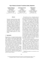
One of the more elaborate examples of coated nanoparticles elegantly
incorporates features designed to accomplish all of the drug loading, MPS avoidance,
active targeting, endocytotic uptake, and endosomal escape processes. A cyclodextrincontaining polycation of imidazole was designed to electrostatically complex with a
catalytic oligonucleotide, a DNAzyme, forming sub-100-nm particles termed
‘‘polyplexes.’’ The positively charged particles can interact with the negatively
charged cell surface proteoglycans for endocytotic uptake. Further, imidazole had been
demonstrated to enhance endosomal escape. However, neutralization of the excess
charge was required for minimizing nonspecific uptake, to enhance efficiency of active
targeting. This was accomplished with addition of the anionic glutamate functionality
to adamantane–PEG conjugates, which forms inclusion complexes with the exposed
cyclodextrins (Figure 4). [1, 3]
Performer: Nguyễn Văn Tú - PhD Student
9
Bach Khoa University
Injectable nanoparticles for drug delivery
Figure 4. Assembly of Polyplex fomulations.
(A)-Polyplex (B)-PEG-Polyplex (C)-Tf-PEG-Polyplex
The exposed PEG chains confer long circulation in biological fluids. Because
transferrin is often upregulated in rapidly growing cells, active targeting was
considered by adding transferrin–PEG–adamantane conjugates. Biodistribution in an
HT-29, high transferrin uptake, tumor xenograft mouse model was followed
subsequent to different routes of administration. Intraperitoneal injection indicated
high levels remaining in the peritoneum; presumably mobility was limited by their
size, even at 30–50 nm. Subcutaneous injection did not result in fluorescence outside
of the injection site. But i.v. delivery showed high levels in tumor, liver and kidney, all
organs rich in transferrin receptor activity. Polyplexes delivered by i.v. were
internalized by the tumor cells. [3]
2. External assistance in targeting
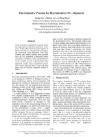
An alternate approach that has been explored to provide targeting functionality
to nanoparticles is via the use of an external energy source. For example, Rudge et al.
described a nanoparticulate system that was responsive to an external magnetic field.
The particles were comprised of activated carbon to allow adsorption and loading of
Performer: Nguyễn Văn Tú - PhD Student
10
Bach Khoa University
Injectable nanoparticles for drug delivery
drug and metallic iron to provide magnetically triggered targeting of the particles.
Good loading efficiency could be obtained for a number of drugs including
doxorubicin, mitomycin C, methotrexate, and camptothecin (Figure 5). [4]
Figure 5. Administration of MTCs to patient
In vivo studies using magnetic doxorubicin particles showed that efficient
targeting was achieved by injecting the particles using an arterial catheter, and then
homing the particles to a specific tissue, by using a strong magnetic field. In another
study, a much higher concentration of mitoxantrone was obtained in the tumor area, by
using only 50% and 20% of the normal dose by the use of magnetic drug targeting.
Ultrasound triggered drug delivery has also been adopted to provide targeted release of
drug to tumors. Nanoparticles and micelles accumulate into the tumor as a result of
passive targeting and the EPR effect. Ultrasound is then applied to trigger the release
of the drug so that the entire drug load is delivered within the tumor (Figure 6). [5]
Performer: Nguyễn Văn Tú - PhD Student
11
Bach Khoa University
Injectable nanoparticles for drug delivery
Figure 6. The four components of the ultrasound/micelle modality of drug targeting
IV. TYPES OF INJECTABLE NANOMATERIAL CARRIERS
There are three main components to an effective drug delivery nanoparticle: the
core material of the nanoparticle, the therapeutic payload, and surface modifiers.
Although a generalized structure does not accurately portray all nanomedicines, one
may be used to aid in understanding the objective of each portion of a nanomedicine
carrier. Nanomedicine carriers generally have the ability to load either hydrophobic or
hydrophilic therapeutics. Thus, suitable carrier materials have to be thoughtfully
selected for every therapeutic. However, some carrier materials have both hydrophobic
and hydrophilic regions. These materials could be effectively used to design
nanocarriers for delivery of multiple drugs. Additionally, the nanoparticle core material
must be non-toxic, non-immunogenic, and should be easily eliminated from the body
to avoid toxic accumulation and side effects. The core material must also possess a
release mechanism for the therapeutic payload after the carrier has reached its
destination. [6]
1. Liposomes
Liposomes are composed of lipid or phospholipid molecules containing a
hydrophilic head region and hydrophobic tail region that have aggregated together to
form an enclosed bilayer particle with an aqueous center and lipid membrane (Figure 7).
Performer: Nguyễn Văn Tú - PhD Student
12
Bach Khoa University
Injectable nanoparticles for drug delivery
Figure 7. A type of liposomes
Liposomes have been receiving attention as therapeutic carriers for over 40
years and have been studied as carriers for anticancer drugs, antifungal drugs,
analgesics, and gene therapies as well as for vaccines. They offer the ability to deliver
both hydrophilic drugs (in the aqueous center) and lipid-soluble drugs (within the
bilayer structure). This sort of therapeutic loading does not occupy surface
functionality groups that may further be used to attach targeting ligands and/or
biocompatibility agents such as PEG, chitosan, silk-fibroin, and polyvinyl alcohol
(PVA). These agents create stealth liposomes—liposomes that avoid MPS uptake, thus
having increased circulation times. Furthermore, the phospholipids being used to
create the liposome may be changed or modified to customize the properties of the
liposomal surface and membrane layer. [6]
2. Polymeric based carriers
Polymer carriers offer a large versatility in both structure and physiochemical
properties. A major reason for this versatility is the wide variety of monomers that may
be used to form the polymer architectures. Some of the commonly used structures as
injectable nanocarriers include polymersomes, dendrimers, and cyclodextrincontaining polymers (CDPs). Polymersomes are structurally similar to liposomes, but
they are formed from amphiphilic block copolymers. Dendrimers are multi-branched
Performer: Nguyễn Văn Tú - PhD Student
13
Bach Khoa University
Injectable nanoparticles for drug delivery
polymer structures extending out from a core. CDPs are polymers that contain
cyclodextrin molecules within the core structure or attached as side chains. [6]
Furthermore, certain polymers contain chemical groups that have the ability to
adapt accordingly to the current environment resulting in a change of properties in the
overall polymer itself. These polymers are referred to as responsive or “smart
polymers”. Some common environmental stimuli include pH, ionic strength, chemical
agents, mechanical stress, temperature, electromagnetic radiation, and electric field.
Common corresponding changes include optical clarity, conductivity, surface
chemistry, shape, permeability, mechanical properties, and phase separation. These
corresponding changes in properties result in the release of therapeutics. Additionally,
hydrophobic, hydrophilic, and electrostatic interactions are used between the polymers
themselves, polymers and therapeutics, and polymers and attachment molecules to aid
in targeting, biocompatibility, and to form more stable structures to increase the
efficacy of the nanocarriers. [6]
Performer: Nguyễn Văn Tú - PhD Student
14
Bach Khoa University
Injectable nanoparticles for drug delivery
Figure 8. Assembly of Polymersome, Dendrimer and Cyclodextrin
3. Inorganic based carriers
Another material that is getting much attention in the field of nanomedicine is
carbon nanotubes (CNTs). Although CNTs are not naturally water-soluble, they may
be treated with acids to create terminal carboxylic groups and/or have covalent
attachments of hydrophilic groups to their surface via functionalization chemistry to
increase solubility and circulation time. In one particular study, the covalent
attachment of PEG with an average 17 molecular weight of 1500 Dalton (PEGSWNTs) led to a circulation time of 22.5 hours in mice post-intravenous exposure
(Figure 9). [7]
Figure 9. TEM image of PEG-SWNTs.
It should also be noted that higher molecular weight PEG chain attachment
resulted in lower RES uptake, longer circulation times, and lower amounts of SWNTs
present in both the liver and spleen. Although preliminary studies show some
promising results for CNTs, much more biocompatibility information is needed to see
the full extent of effects for CNTs throughout the body.
In addition, Gold nanoparticles (AuNps) have recently emerged as an attractive
candidate for delivery of small drug molecules or biomolecules, such as proteins, or
RNA or DNA into target cells. Advantages of AuNps over polymeric nanoparticle gene
delivery agents include (i) ease of preparation of mono-dispersed particles of size
ranging from 1 to 150nm; (ii) non–toxicity of the gold core; (iii) easy functionalization
Performer: Nguyễn Văn Tú - PhD Student
15
Bach Khoa University
Injectable nanoparticles for drug delivery
of small molecules or nucleic acids via covalent or noncovalent interactions; and (iv)
ability to release the attached drug at remote places using their photo-physical
properties (Figure 10). [8]
Figure 10. Synthesis scheme for the preparation of DOX conjugated Au NPs.
V. CLINICAL TRIALS
Of all the nanocarriers above, liposomes are the most developed and currently
possess the greatest amount of clinical trials with some formulations currently in the
marketplace. This is probably due to the fact that the other materials have not been
investigated for the same duration and are relatively new in comparison. For this
reason, polymer based materials, CNTs and AuNps should not be overlooked as
nanomedicine delivery materials just because of the relatively low number of recent
clinical trials; however, CNTs do still have much to prove with respect to safety and
long-term biodistribution in order to be considered viable nanocarriers. Recent clinical
trials are shown in Figure 11.
Some of the more promising trials include Genexol-PM, which is an
amphiphilic diblock copolymer (PEG-(D,L-lactic acid)) forming a micelle that delivers
Paclitaxel for various types of cancers. Clinical trials are currently in phase 4 using
Genexol–PM for recurrent breast cancer and phase 3 for breast cancer. The liposomal
formulation AmBiosome® consisting of the antifungal Amphotericin B is currently in
phase 4 trials for fungal infections associated with acute leukemia and for central line
fungal infections. ThermoDox, a thermally sensitive doxorubicin-loaded liposome is
Performer: Nguyễn Văn Tú - PhD Student
16
Bach Khoa University
Injectable nanoparticles for drug delivery
currently in phase 3 trials for hepatocellular carcinoma. Lastly, Caelyx, a doxorubicin
HCL loaded liposome that is PEGylated, is currently in phase 4 trials for ovarian
neoplasms. [6]
Performer: Nguyễn Văn Tú - PhD Student
17
Bach Khoa University
Injectable nanoparticles for drug delivery
Performer: Nguyễn Văn Tú - PhD Student
18
Bach Khoa University
Injectable nanoparticles for drug delivery
Figure 11. Some of prominent applications using nanoparticles in drug delivery
Performer: Nguyễn Văn Tú - PhD Student
19
Bach Khoa University
Injectable nanoparticles for drug delivery
CONCLUSION
Injectable routes for drug delivery have a number of advantages as well as
drawbacks. However, the usage of nanoparticulate in injectable delivery have been
attended, especially in cancer therapy. For efficiency, nanoparticles can be
functionalized depending upon the targets and the characteristics of human cell, it can
be either coated by receptors which target the specific tumuors or used the assistance
of external energy sources for helping targeting therapy. A number of systems of
nanomaterials utilized in drug delivery including liposomes, polymeric based carriers
and inorganic based carriers. Of all systems, liposomes have been developed and
possess the greatest amount of the clinical trials currently in the market.
REFERENCES
1.
2.
CHAUBAL, B.R.a.M.V., Injectable Nanoparticles for Efficient Drug Delivery.
Vol. 159. 2006: Taylor & Francis Group, LLC. 222-255.
Finkel, R.C., Michelle A.; Cubeddu, Luigi X, Lippincott's Illustrated Reviews:
Pharmacology, 4th Edition, R.C. Finkel, Michelle A.; Cubeddu, Luigi X, Editor
2009, Lippincott Williams & Wilkins.
Performer: Nguyễn Văn Tú - PhD Student
20
Bach Khoa University
Injectable nanoparticles for drug delivery
3.
4.
5.
6.
7.
8.
al, S.H.P.e., Targeted Delivery of RNA-Cleaving DNA Enzyme (DNAzyme) to
Tumor Tissue by Transferrin-Modified, Cyclodextrin-Based Particles. Cancer
Biology & Therapy, 2004. 3(7): p. 11.
S. Rudge , C.P., C. Vessely, J. Koda, S. Stevens, L. Catterall, Adsorption and
desorption of chemotherapeutic drugs from a magnetically targeted carrier
(MTC). Journal of Controlled Release, 2001. 74: p. 6.
N.Y. Rapoport a, D.A. Christensen a, H.D. Fain b, L. Barrows b, Z. Gao a,
Ultrasound-triggered drug targeting of tumors in vitro and in vivo. Ultrasonics,
2004. 42: p. 8.
David M. Webster, P.S., Mark E. Byrne, Injectable Nanomaterials for Drug
Delivery: Carriers, Targeting Moieties, and Therapeutics. European Journal of
Pharmaceutics and Biopharmaceutics, 2012.
S.-T. Yang, J.-H.L., J. Wang, Dr. H.-F. Sun, Prof. Y. Liu, Dr. H. Wang,
Covalently PEGylated Carbon Nanotubes with Stealth Character In Vivo.
Nanotubes, 2008. 4(7): p. 5.
Agha Zeeshan Mirza a, Hina Shamshadb, Preparation and characterization of
doxorubicin functionalized gold nanoparticles. European Journal of Medicinal
Chemistry, 2011. 46: p. 4.
Performer: Nguyễn Văn Tú - PhD Student
21
Bach Khoa University