Ebook Handbook of vitamins (4th edition) Part 2
Bạn đang xem bản rút gọn của tài liệu. Xem và tải ngay bản đầy đủ của tài liệu tại đây (5.5 MB, 306 trang )
Robert B. Rucker/Handbook of Vitamins, Fourth Edition 4022_C009 Final Proof page 290 5.5.2007 4:54pm Compositor Name: BMani
290
Handbook of Vitamins, Fourth Edition
that pantothenic acid was required for the growth of certain bacteria and yeast
[1,17,20,22,23]. Next, Elvehjem and associates [21] and Jukes and associates demonstrated
that pantothenic acid was a growth factor for rats and chicks [2,16,35,36]. Early nutritional
studies in animals also demonstrated that there was loss of fur color in black and brown rats
and an usual dermatitis that occurred in chickens fed pantothenate-deficient diets; thus, at
one point pantothenate was known as the antigray or antidermatitis factor [37].
Williams coined the name pantothenic acid from the Greek meaning ‘‘from everywhere’’
to indicate its widespread occurrence in foodstuffs. The eventual characterization and synthesis of pantothenic acid by Williams in 1940 took advantage of observations that the
antidermatitis factor present in acid extracts of various food sources, i.e., pantothenic acid,
did not bind to fuller’s earth (a highly adsorbent claylike substance consisting of hydrated
aluminum silicates) under acidic conditions [22,23]. Using chromatographic and fractionation
procedures, which were typical of the 1930s and 1940s (solvent-dependent chemical partitioning), Williams isolated several grams of pantothenic acid for structural determination from
250 kg of liver as starting material [22,23]. With this information, a number of research
groups contributed to the chemical synthesis and commercial preparation of pantothenic
acid. Pantothenate and its derivatives are now produced mainly through chemical synthesis
and the global market in the past decade was >7 Â 106 kg=year [38].
As emphasized throughout this chapter, pantothenic acid, which is sometimes designated
as vitamin B5, is the core of the structure of coenzyme A (CoA), an essential cofactor in
pathways important to oxidative respiration, lipid metabolism, and the synthesis of many
secondary metabolites such as steroids, acetylated compounds (e.g., acetylated amino acids,
carbohydrates), and prostaglandins and prostaglandin-like compounds. In addition, the
phosphopantetheine moiety (a pantothenic acid derivative derived from CoA metabolism)
is incorporated into the prosthetic group of the acyl carrier proteins (ACP) used in fatty acid
synthases, polyketide synthases, lysine synthesis in yeast and bacteria, and nonribosomal
peptide synthetases. Coenzyme A was discovered as the cofactor essential for the acetylation
of sulfonamides and choline in the early 1950s [39–42]. In the mid-1970s, pantothenic acid was
identified as a component of ACP in the fatty acid synthesis (FAS) complex [43–46]. These
developments, in addition to a steady series of observations throughout this period on the
effects of pantothenic acid deficiency in humans and other animals, provide the foundation
for our current understanding of this vitamin.
CHEMICAL PERSPECTIVES AND NOMENCLATURE
Pantothenic acid [b-alanine-N-4-dihydroxy-3,3-dimethyl-1-oxobutyl)-(R); vitamin B5; CAS
Registry Number 79-83-4] is synthesized by microorganisms via an amide linkage of pantoic
acid and b-alanine subunits (Figure 9.1). Pantothenic acid is an essential metabolite for all
biological systems; however, the biosynthesis of pantothenic acid is limited to plants, bacteria,
eubacteria, and archaea (Figure 9.2). It is worth noting that the biosynthesis pathway for
pantothenic acid in microorganisms and plants is also viewed as a strong candidate for the
discovery of novel antibiotic and herbicidal compounds [38].
Pure pantothenic acid is water soluble, viscous, and yellow. It is stable at neutral pH, but
is readily destroyed by acid, alkali, and heat. Calcium pantothenate, a white, odorless,
crystalline substance, is the form of pantothenic acid usually found in commercial vitamin
supplements due to greater stability than the pure acid. The structure elucidation of pantothenate was based on the identification of a lactone formed by degradation of pantothenate. Initial analytical work revealed an a-hydroxy acid that was readily lactonized. Stiller
et al. [17] identified the lactone as a-hydroxy-b,b-dimethyl-x-butyrolactone (pantoyl lactone
or pantolactone), which aided in the structural elucidation of pantothenate.
Robert B. Rucker/Handbook of Vitamins, Fourth Edition 4022_C009 Final Proof page 291 5.5.2007 4:54pm Compositor Name: BMani
291
Pantothenic Acid
Pantothenic acid
NH2
OH
O
N
N
CH2
O
P
—
—
—
—
O
H
O
—
N
N
O
O−
P
O−
O
N
CH3 CH3
O
O
NH—CH2
Pantoic acid
CH2—SH
H
H
OH −O—P—O−
—
—
O
Pantetheine
Coenzyme A
FIGURE 9.1 Structural components of coenzyme A.
O
O
OH
α-Ketovaleric acid
Ketopantoate
hydroxymethyltransferase
O
O
OH
HO
Ketopantoic acid
Aspartic acid
Ketopantoate
reductase
OH
O
OH
HO
Pantoic acid
OH
β-Alanine
Pantothenic
synthetase
O
COOH
HO
N
H
Pantothenic acid
FIGURE 9.2 Pathway for the biosynthesis of pantothenic acid found in plants, bacteria (including
archaea), and eubacteria.
Robert B. Rucker/Handbook of Vitamins, Fourth Edition 4022_C009 Final Proof page 292 5.5.2007 4:54pm Compositor Name: BMani
292
Handbook of Vitamins, Fourth Edition
FOOD SOURCES AND REQUIREMENTS
PANTOTHENIC ACID REQUIREMENTS
Although limited, available data suggest that at intakes of 4–6 mg of pantothenic acid per
day, serum levels of pantothenic acid are maintained in young adults and no known signs of
deficiency are observed. The U.S. recommended dietary allowance (RDA) for pantothenic
acid, which is used for determining daily percent values on nutritional supplement and food
labels, is 10 mg=day [47].
Pantothenic acid is found in edible animal and plant tissues ranging from 10 to 50 mg=g of
tissue. Thus, it is possible to meet the current daily recommended intake for adults with a
mixed diet containing as little as 100 to 200 g of solid food; i.e., the equivalent of a mixed diet
corresponding to 600 to 1200 kcal or 2.4 to 4.8 MJ. In this regard, the typical Western diet
usually contains 6 mg or more of available pantothenic acid [37,48]. Table 9.1 gives the
current recommended amounts of pantothenic acid for humans, expressed as dietary reference intakes (DRI) [47]. Moreover, when expressed on a per energy intake equivalent basis,
the need for pantothenic acid is remarkably constant across species [49]. Although in mice
small amounts of pantothenic acid are synthesized by intestinal bacteria, the contribution of
bacterial synthesis to human pantothenic acid status is not known and probably small [28,50].
Regrettably, relatively little quantitative information on the enteric synthesis of pantothenic
acid exists.
FOOD SOURCES
Chicken, beef, potatoes, oat cereals, tomatoes, eggs, broccoli, and whole grains are major
sources of pantothenic acid. Refined grains have a lower content. Table 9.2 contains some
typical values for pantothenic acid in selected food. The processing and refining of grains
TABLE 9.1
Pantothenic Acid Dietary Reference Intakes (RDI)a
Category
0 through 6 months
7 through 12 months
Children
1 through 3 years
4 through 8 years
Girls and boys
9 through 13 years
14 through 18 years
Women and men
19 years and older
Pregnancy
14 through 50 years
Lactation
14 through 50 years
Recommendation
1.7 mg=day ~0.2 mg=kg
1.8 mg=day ~0.2 mg=kg
2 mg=day
3 mg=day
4 mg=day
5 mg=day
5 mg=day
6 mg=day
7 mg=day
Note: There is no evidence of toxicity associated; thus, the
lowest observed adverse-effect level (LOAEL) and an associated
no observed adverse-effect level (NOAEL) have not been
determined.
a
Recommendation of the Food and Nutrition Board of the
Institute of Medicine of the U.S. National Academy of Sciences.
Robert B. Rucker/Handbook of Vitamins, Fourth Edition 4022_C009 Final Proof page 293 5.5.2007 4:54pm Compositor Name: BMani
293
Pantothenic Acid
TABLE 9.2
Pantothenic Content in Selected Foods
Ingredient
Beer
Soft drinks
Wine
Wheat bran
Boiled rice
Soy flour
Raw eggs
Cooked fish
Lobster
Oysters
Salmon
Tuna
Apples
Apricots, bananas
Dates
Grapes
Lemon and orange juice
Plums
Prunes
Strawberries
Beef
Chicken boiled
Liver
Kidney
Pork
Cheese
Milk (bovine)
Milk (human)
Almonds
Peanuts
Walnuts
Peanut butter
~Amount (mg=100 g or mL of Edible Portion)
<0.1
<0.03
0.02–0.04
2–3
0.2–0.3
1.5–2.5
1.5–2.0
0.2–0.5
1.5
0.5
0.5
0.4
0.05
0.1–0.2
0.8
0.04
0.1
0.2
0.5
0.3
0.5–1.2
0.3–1.0
5–7
4–6
0.5–1.0
1.5
1
0.3–0.4
2–3
2–3
1
5–8
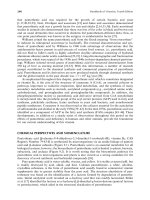
can produce as much as a 50% loss of pantothenic acid [51–53]. In keeping with the proposed
requirements for humans, human milk contains ~5–6 mg of pantothenic acid per 1000 kcal
[54–56]. It has been estimated that for every milligram of pantothenic acid consumed in the
diet, ~0.4 mg can be transported into milk when lactation is active. Because the pantothenic
content of milk correlates well with maternal intakes of pantothenic acid, the possibility
does exist that pantothenic acid deficiency may occur in infants consuming milk produced
by mothers deficient in the vitamin (e.g., those who consume predominantly refined cereals).
Because of the widespread distribution of pantothenic acid in foods and apparently the diets
of adults have to be markedly devoid of pantothenic acid to induce deficiency, the need for
aggressive fortification of pantothenic acid may never become a high priority.
INTESTINAL ABSORPTION AND MAINTENANCE
The vast majority of pantothenic acid in food is present as CoA or 40 -phosphopantetheine. In
order to be absorbed, these substances must first be hydrolyzed [50]. This occurs in the
Robert B. Rucker/Handbook of Vitamins, Fourth Edition 4022_C009 Final Proof page 294 5.5.2007 4:54pm Compositor Name: BMani
294
Handbook of Vitamins, Fourth Edition
intestinal lumen by the sequential activity of two hydrolases, pyrophosphatase and phosphatase, with pantotheine as the product. Intestinal phosphatases and nucleosidases are capable
of very efficient hydrolysis of CoA so that near-quantitative release of pantothenic acid occurs
as a normal part of digestion. Pantotheine is either absorbed as is, or further metabolized
to pantothenic acid by a third intestinal hydrolase, pantothenase [57–60]. In rats, pantothenic
acid absorption was initially found to be absorbed in all sections of the small intestine by
simple diffusion [28,50,61]. However, subsequent work in rats and chicks indicated that at
low concentrations, the vitamin is absorbed by a saturable, sodium-dependent transport
mechanism [62]. Further, the overall Km for pantothenic acid intestinal uptake is 10–20 mM.
At an intake of ~10–15 mg of coenzyme A, the amount of coenzyme A in a typical meal, the
pantothenic acid concentration in luminal fluid would be ~1–2 mM. At this concentration,
pantothenic acid would not saturate the transport system and should be efficiently and
actively absorbed [61].
Researchers have demonstrated that pantothenic acid shares a common membrane
transport system in the small intestine with another vitamin, biotin [61,63–67]. Experiments
using Caco-2 cell monolayers as a model of intestinal absorption have established that
pantothenic acid uptake is inhibited competitively by biotin and vice versa [61,63–67].
Similar relationships were observed in transport experiments involving the blood–brain
barrier [61,68,69], heart [70–73], and placenta [74–77]. For example, membrane transport
pathways for transplacental transfer of pantothenate were investigated by Grassl [77] assessing the possible presence of a Naþ–pantothenate cotransport mechanism in the maternal
facing membrane of human placental epithelial cells. The presence of Naþ–pantothenate
cotransport was determined from radiolabeled tracer flux measurements of pantothenate
uptake using preparations of purified brush-border membrane vesicles. Compared with
other cations, the imposition of an inward Naþ gradient stimulated vesicle uptake of pantothenate to levels ~40-fold greater than those observed at equilibrium. The effect of biotin on
the kinetics of Naþ-dependent pantothenate uptake and the effect of pantothenate on the
kinetics of Naþ-dependent biotin uptake suggest that placental absorption of biotin and
pantothenate from the maternal circulation also occurs by a common Naþ cotransport
mechanism.
After absorption, pantothenic acid enters the circulation from which it is taken up by
cells in a manner similar to that of intestinal absorption (see the following section). The
vitamin is excreted in the urine primarily as pantothenic acid [27,78–85]. This occurs after its
release from CoA by a series of hydrolysis reactions that cleave off the phosphate and
b-mercaptoethylamine moieties.
CELLULAR REGULATION OF PANTOTHENIC ACID, COA,
AND THE IMPORTANCE OF PANTOTHENIC KINASE
CELLULAR TRANSPORT
AND
MAINTENANCE
Said and others [61,63–67] have observed that similar to enterocytes, other epithelial cells take
up pantothenic acid in a manner that is inhibited by Na-K-ATPase inhibitors, such as
ouabain, and is in competition with biotin. In most instances, activity of this transporter is
sensitive to phosphokinase C (PKC)- and A (PKA)-mediated activation and inhibition. For
example, pretreatment of epithelial cells with phorbol 12-myristate 13-acetate (PMA), but not
with its negative control (4a-PMA) or with 1,2-dioctanoyl-sn-glycerol, both activators of
PKC, causes significant inhibition in uptake, whereas pretreatment of cells with staurosporine
and chelerythrine, inhibitors of PKC, promotes stimulation in uptake [67]. These findings
point toward the involvement of a PKC-mediated pathway in the regulation of biotin and
pantothenic acid uptake by epithelial cells.
Robert B. Rucker/Handbook of Vitamins, Fourth Edition 4022_C009 Final Proof page 295 5.5.2007 4:54pm Compositor Name: BMani
295
Pantothenic Acid
PANTOTHENIC ACID KINASE
Following uptake, the maintenance of pantothenic acid cellular concentration depends on its
incorporation into CoASH and pantotheine. The most important control step in this process is
the phosphorylation of pantothenic acid to 40 -phosphopantothenic acid by pantothenic acid
kinase [86–101] (Figure 9.3). There are four members in the human PanK family: PanK1,
PanK2, PanK3, and PanK4, which are located on chromosomes 10q23.31, 20p13, 5q35, and
1p36.32, respectively [98]. Pantothenic acid kinases possess a broad pH optimum (between pH 6
and 9). The Km for pantothenic acid in the liver enzyme of most animals is ~20 mM. Mg-ATP is
the nucleotide substrate for this phosphorylation reaction with a Km of ~0.6 mM [88,102–112].
The relationships involving the various isoforms are complex. Two murine PanK1s exist,
mPanK1a and mPanK1b [86,88,95,97,100]. These two transcripts are the result of an alternate splicing of the same gene. PanK1 localizes predominantly in heart, liver, and kidney
[86,88,95,97,100]. PanK2 is ubiquitously expressed with the highest levels in retinal and infant
basal ganglia [95,113–115]. PanK3 is limited to the liver, but expressed at a high level [88,95].
The expression of PanK4 occurs in most tissues with a high concentration in muscle
[88,95]. Metabolic labeling experiments in rat heart support the role of PanK in controlling
the flux of the CoA biosynthesis. For example, enhanced mPanK1b expression reduced the
intracellular pantothenate pool and triggered a 13-fold increase in intracellular CoA content.
PanK1b activity in vitro was stimulated by CoA and strongly inhibited by acetyl CoA,
illustrating the differential modulation of mPanK1b activity by pathway end products and
supporting the concept that the expression or activity of PanK is a determining factor in the
physiological regulation of the intracellular CoA concentration [100].
Pantothenic acid kinase is activated and inhibited nonspecifically by various anions. More
significantly, feedback inhibition of the kinase by CoA or CoA derivatives governs flux
through the subsequent steps in the CoA synthesis pathway and defines the upper threshold
for intracellular CoA cofactor levels. Inhibition by acetyl CoA is slightly greater than that of
free CoA. The inhibition by free CoA is uncompetitive with respect to pantothenate concentration; Ki for inhibition of 0.2 mM. Interestingly, L-carnitine, important for the transport of
fatty acids into mitochondria, is a nonessential activator of pantothenic acid kinase. Carnitine
Coenzyme A synthesis
Carnitine
Reverses the
inhibition
by CoA
Pantothenate
ATP
Pantothenate kinase
ADP
CoA
4Ј-Phosphopantothenate
ATP
Inhibitor
4Ј-Phosphopantothenoylcysteine synthetase
ADP + Pi
4Ј-Phosphopantothenoylcysteine
4Ј-Phosphopantothenoylcysteine decarboxylase
CO
2
3Ј,5Ј-ADP
Coenzyme A hydrolase
4Ј-Phosphopantetheine
ATP
Adenylyltransferase (dephospho-CoA-pyrophosphorylase)
PPi
Dephosphocoenzyme A
ATP
Dephosphocoenzyme A kinase
ADP
Coenzyme A (CoA or CoASH)
FIGURE 9.3 Coenzyme A metabolism and importance of pantothenic acid kinase.
Robert B. Rucker/Handbook of Vitamins, Fourth Edition 4022_C009 Final Proof page 296 5.5.2007 4:54pm Compositor Name: BMani
296
Handbook of Vitamins, Fourth Edition
has no effect by itself, but specifically reverses the inhibition by CoA. In heart, the free
carnitine content varies directly with the phosphorylation of pantothenic acid. Thus,
these properties of the kinase provide a potential mechanism for the control of CoA synthesis
and regulation of cellular pantothenic acid content, i.e., feedback inhibition by CoA and
its acyl esters that is reversed by changes in the concentration of free carnitine
[71,107,108,111,116].
In this regard, it is important to underscore that the free concentration of acyl CoA in
cells is low and variable because the bulk of acyl derivatives are protein bound. Moreover,
similar to CoA, carnitine exists in both free and acylated forms and reversal of kinase
inhibition by CoA does not occur when carnitine is acylated [107]. The ratio of free to
acylated carnitine varies considerably depending on feeding and hormonal influences, with
insulin of particular importance. Fasting and diabetes (states of low insulin) increase pantothenic acid kinase activity and the total content of CoA [102,105,110]. Perfusion of heart
preparations or incubation of liver cells with glucose, pyruvate, or palmitate markedly
inhibits pantothenic acid phosphorylation because of reduction in free carnitine and increases
in the free and acylated forms of CoA [116].
COA FORMATION
For CoA synthesis, the additional steps include the addition of adenine and ribose
30 -phosphate to produce CoA composed of 40 -phosphopantetheine linked by an anhydride
bond to adenosine 50 -monophosphate, modified by a 30 -hydroxyl phosphate (Figure 9.3). In
yeast and perhaps higher organisms (for which details of the pathway require further resolution), these steps are carried out on a protein complex with multifunctional catalytic sites
[117–120]. Important enzymatic features of this complex in yeast include dephospho-CoApyrophosphorylase activity, which catalyzes the reaction between 40 -phosphopantetheine and
ATP to form 40 -dephospho-CoA; dephospho-CoA-kinase activity, which catalyzes the ATPdependent final step in CoA synthesis; and CoA hydrolase activity, which catalyzes the
hydrolysis of CoA to 30 ,50 -ADP and 40 -phosphopantetheine. This sequence of reactions is
referred to as the CoA=40 -phosphopantetheine cycle and provides a mechanism by which the
40 -phosphopantetheine can be recycled to form CoA [107–110]. Each turn of the cycle utilizes
two molecules of ATP and produces one molecule of ADP, one molecule of pyrophosphate,
and one molecule of 30 ,50 -ADP. Although some enzymes of the pathway were identified
relatively rapidly, it was not possible to identify all enzymes using traditional methods.
Hence, the use of bacterial mutants and the application of molecular biology have been
essential in resolving key features of the pathway shown in Figure 9.3.
As CoA holds a central position in cellular metabolism, it may therefore be assumed to be
an ancient molecule [119]. Starting from the known Escherichia coli pathway and known
human enzymes required for the biosynthesis of CoA, phylogenetic profiles and chromosomal proximity methods have led to the conclusion that the topology of CoA synthesis from
common precursors is essentially conserved across the three domains of life [119].
COA REGULATION
In animal tissue, the levels of CoA cover a wide range and change in response to signals
arising from hormones, nutrients, and cellular metabolites. Hepatic CoA levels are among the
most responsive to such changes, ranging from 100 to 500 nmol=g liver. In decreasing order:
heart > kidney > diaphragm > skeletal muscle contain CoA in concentrations ranging from
100 to 50 nmol=g [43,103,106,108,121,122]. Fasting results in high levels of long-chain
fatty acyl CoA thioesters, whereas glucose feeding results in nonacylated CoA derivatives.
The total CoA levels decrease in response to insulin, but increase in response to glucagon. The
Robert B. Rucker/Handbook of Vitamins, Fourth Edition 4022_C009 Final Proof page 297 5.5.2007 4:54pm Compositor Name: BMani
Pantothenic Acid
297
transfer of activated acyl moieties across organelle membranes, to and from the CoA pools in
mitochondria, cytosol, and peroxisomes occurs through the carnitine transferase system and
ABC-like transporters [123–125].
The concentration of nonacylated CoA determines the rate of oxidation-dependent
energy production in both mitochondria and peroxisomes, and the interorganelle transport
of CoA-linked metabolites helps to maintain CoA availability. Although much remains to be
investigated regarding the relative roles, various compartments play a role in CoA regulation;
available evidence suggests that mitochondria are the principle sites of CoA synthesis. For
example, PanK2s localization in mitochondria is proposed to initiate intramitochondrial CoA
biosynthesis.
CoA synthase is also of importance in this process. 40 -phosphopantetheine adenylyltransferase and dephospho CoA kinase activities are both catalyzed by CoA synthase [126]. The
full-length CoA synthase is associated with the mitochondrial outer membrane, whereas the
removal of the N-terminal region relocates the enzyme to the cytosol. Phosphatidylcholine
and phosphatidylethanolamine, which are principle components of the mitochondrial outer
membrane, are potent activators of both enzymatic activities of CoA synthase. Taken
together, it may be inferred that CoA synthesis is regulated by phospholipids and intimately
linked to mitochondrial function [118]. At steady state, cytosolic CoA concentrations range
from 0.02 to 0.15 mM, mitochondrial concentrations range from 2 to 5 mM, and peroxisomal
concentration are ~0.5 mM CoA [106,108].
ACYL CARRIER PROTEIN
ACP is also referred to as a ‘‘macro-cofactor’’ because in bacteria, yeast, and plants, it is
composed of a dissociable polypeptide chain (MW ~8500–8700 Da) to which 40 -phosphopantetheine is attached [43,44,127]. However, in higher animals, ACP is most often associated
with a fatty acid synthase complex that is composed of two very large protein subunits
(MW ~250,000 Da each). The carrier segment or domain of the fatty acid synthetic complex
is also called ACP, i.e., one of seven functional or catalytic domains on each of the two subunits
that comprise fatty acid synthase (Table 9.3).
In addition to fatty acid production and catabolism, in yeast, bacteria, and plants, capable
of essential amino acid synthesis, proteins with 40 -phosphopantetheine attachment sites are
utilized. An example is aminoadipic acid reductase (e.g., LYS2 in yeast). The pantetheine
transferase (LYS5), which aids in the activation of aminoadipic acid reductase, has also
been isolated and cloned from a human source, i.e., a putative human homolog to the
LYS5 gene [128].
Regarding ACP assembly to form holo-ACP, apo-ACP is posttranslationally modified
via transfer of 40 -phosphopantetheine from CoA to a serine residue on apo-ACP
[126,127,129]. The resulting holo-ACP is then active as the central coenzyme of fatty acid
biosynthesis, either as individual subunit in bacterial systems or as a specific domain in
the fatty acid synthetase complex in higher animals (Figure 9.4). Moreover, the transfer
of the 40 -phosphopantetheine moiety of CoA to acyl carrier proteins may also serve as an
alternate to CoA degradation or catabolism, i.e., ACP formation has the potential of
providing an additional strategy for coordination of CoA levels [117,118,129].
In summary, the regulation of pantothenic acid kinase is complex and occurs via allosteric
and transcriptional mechanisms. Multiple approaches to regulating this important enzyme
are of obvious importance given the central roles and importance of both ACP and CoA to
intermediary metabolism, protein processing, and gene regulation. In addition to the allosteric controls, transcriptional regulation by peroxisome proliferator activated receptor transcription factors, sterol regulatory element binding proteins (SREBP), and interaction with
the glucose response element [95] are also essential.
Robert B. Rucker/Handbook of Vitamins, Fourth Edition 4022_C009 Final Proof page 298 5.5.2007 4:54pm Compositor Name: BMani
298
Handbook of Vitamins, Fourth Edition
TABLE 9.3
Catalytic Sites Associated with the Fatty Acid Synthase Complex
Catalytic Site
Function
Acetyl transferase
Malonyl transferase
3-Oxoacyl synthetase
Oxoacyl reductase
3-Hydroxyacyl dehydratase
Enoyl reductase
Thioester hydrolase
Catalyzes the transfer of an activated acetyl group on CoA to the sulfidryl group
of 40 -phosphopantetheine (ACP domain). In the next step, the acetyl group is
transferred to a second cysteine-derived sulfidryl group near active site of
3-oxoacyl synthase (see step 3) leaving the 40 -phosphopantetheine sulfhydryl
group free for step 2
Catalyzes the transfer of successive incoming malonyl groups to
40 -phosphopantetheine
Catalyzes the first condensation reaction in the process. The acetyl moiety
(transferred in step 1) occurs with decarboxylation and condensation to yield a
3-oxobutryl (acetoacetyl) derivative. In the subsequent series of cycles, the
newly formed acyl moieties react with the malonyl group added at each cycle
(see step 6)
Catalyzes reductions of acetoacetyl or 3-oxoacyl intermediates. The first cycle of
this reaction generates D-hydroxybutyrate, and in subsequent cycles,
hydroxyfatty acids
Catalyzes the removal of a molecule of water from the 3-hydroxyacyl derivatives
produced in step 4 to form enoyl derivatives
Catalyzes the reduction of the enoyl derivatives (step 5). This acyl group is
transferred to the sulfidryl group adjacent to 3-oxoacyl synthase, as described
in step 1, until a 16-carbon palmitoyl group is formed. This group, still attached
to the 40 -phosphopantetheine arm, is high-affinity substrate for the remaining
enzyme of the complex, thioester hydrolase
This enzyme liberates palmitic acid (step 6) from the 40 -phosphopantetheine arm
SELECTED PHYSIOLOGIC FUNCTIONS OF ACP AND COA
To reiterate, the functions of pantothenic acid as a vitamin are inexorably linked to processes
that utilize CoA as a substrate and cosubstrate, particularly given that the bulk of
40 -phosphopantotheine incorporated into ACP also derives from transfer reactions that
require CoA as substrate. Descriptions of the hundreds of reactions involving CoA in acetyl
and acyl transfers are beyond the scope of a chapter specifically focused on pantothenic acid.
However, the following descriptions (Table 9.4) were chosen to underscore how pantothenic
acid as a component of CoA and ACP is central to virtually all aspects of metabolism.
COA AND ACP AS HIGH-ENERGY INTERMEDIATES
Intermediates arising from the transfer reactions catalyzed by CoA and 40 -phosphopantetheine
in ACP are ‘‘high-energy’’ compounds [130]. Thioesters (–S–CO–R) are thermodynamically
Phosphopantetheinyl transferase
Coenzyme A
Adenosine 3Ј,5Ј-bisphosphate
Apo-ACP
FIGURE 9.4 Pantethenylation of acyl carrier protein.
Holo-ACP
Robert B. Rucker/Handbook of Vitamins, Fourth Edition 4022_C009 Final Proof page 299 5.5.2007 4:54pm Compositor Name: BMani
299
Pantothenic Acid
TABLE 9.4
Functions of CoA and ACP
Function
Importance
Carbohydrate-related citric acid cycle transfer reactions
Acetylation of sugars (e.g., N-acetylglucosamine)
Lipid-related
Phospholipid biosynthesis
Isoprenoid biosynthesis
Steroid biosynthesis
Fatty acid elongation
Acyl (fatty acid) and triacyl glyceride synthesis
Protein-related
Protein acetylation
Protein acylation (e.g., myristic and palmitic acid,
and prenyl moiety additions)
Oxidative metabolism
Production of carbohydrates important to cell structure
Cell membrane formation and structure
Cholesterol and bile salt production
Steroid hormone production
Ability to modify cell membrane fluidity
Energy storage
Altered protein conformation; activation of certain
hormones and enzymes, e.g., adrenocorticotropin
transcriptional regulation, e.g., acetylation of histone
Compartmentalization and activation of hormones and
transcription factors
less stable than typical esters (–O–CO–R) or amides (–N–CO–R). The double-bond character
of the C¼¼O bond in –S–C¼¼O–R does not extend significantly into the C–S bond, i.e., in thiol
esters the d-orbitals of sulfur do not overlap with the p-orbitals of carbon. This causes
thioesters to have relatively high-energy potential, and for most reactions involving CoA or
ACP, no additional energy, for example, from ATP hydrolysis, is required for transfer of the
acetyl or acyl group. At pH 7.0, the ÀDG of hydrolysis is ~7.5 kcal for acetyl coenzyme A and
10.5 kcal for acetoacetyl CoA, compared with 7–8 kcal for the hydrolysis of adenosine
triphosphate to AMP plus PPi or ADP plus Pi. CoA or ACP also reacts with acetyl or acyl
groups to form thioesters. The pKa of the thiol in CoA–SH is ~10 (ROH ~ 16); at physiological
pH, reasonable amounts of CoA–S– can be formed. CoA–SH is a potent nucleophile and more
nucleophilic than RO–; moreover, RS– is a much better leaving group than RO–. Therefore,
there is no mesomeric effect that makes the carbonyl group more polar than in regular ester
[R–O–CO–R0 ] or amide bonds [R–N–CO–R0 ].
O−
O
O−
C+
SCoA
S+
SCoA
CoA
Their reactivity toward nucleophiles lies between esters and anhydrides. Thiol esters are easier
to enolize than esters, i.e., the a-hydrogens are more acidic.
O
O
H
B
SCoA
SCoA
H
H
O
O
O−
SCoA
SCoA
As such, acetyl CoA is involved in Claisen condensations, which is the basis of fatty acid,
polyketide, phenol, terpene, and steroid biosynthesis. Coenzyme A is also central to the
balance between carbohydrate metabolism and fat metabolism. Carbohydrate metabolism
Robert B. Rucker/Handbook of Vitamins, Fourth Edition 4022_C009 Final Proof page 300 5.5.2007 4:54pm Compositor Name: BMani
300
Handbook of Vitamins, Fourth Edition
needs some CoA for the citric acid cycle to continue, and fat metabolism needs a larger
amount of CoA for breaking down fatty acid chains during b-oxidation [120].
SYNTHETIC VERSUS CATABOLIC PROCESSES INVOLVING PANTETHEINE
As a fundamental distinction, CoA is involved in a broad array of acetyl and acyl transfer
reactions and processes related to primarily oxidative metabolism and catabolism, whereas
ACP is involved in synthetic reactions (Table 9.4). The adenosyl moiety of CoA provides a
site for tight binding to CoA-requiring enzymes, while allowing the 40 -phosphopantetheine
portion to serve as a flexible arm to move substrates from one catalytic center to another
[43,120]. Similarly, when pantothenic acid (as 40 -phosphopantetheine) in ACP is used in
transfer reactions, it also functions as a flexible arm that allows for an orderly and systematic
presentation of thiol ester derivatives to each of the active centers of the FAS complex
described in the previous section. A FAS system also exists in mitochondria [131]. The
mitochondrial FAS pathway is novel in that it is similar to the FAS pathway in bacteria
(designated the ‘‘type ii’’ pathway), for example, discrete soluble protein catalyzes each step of
the reaction cycle rather than a multidomain complex.
ACETYLATIONS AS REGULATORY SIGNALS
The addition of an acetyl group into an amino acid –[NH2] or –[C¼¼O–OH] function can
markedly alter chemical properties. The same is true for biogenic amines, carbohydrates,
complex lipids and hormones, xenobiotics, and drugs [132–137]. Specific compounds range
from acetylcholine to melatonin to structural carbohydrates which are subject to O-linked
acetylations. Examples include acetylated sialic acids (under the control of two groups of
enzymes, O-acetyltransferases and 9-O-acetylesterases), cell surface antigens, and a wide
variety of lipopolysaccharides, and N-acetylgangliosides. Acetylation is critical to cell–cell
surface and cell surface protein–protein interactions (e.g., antigenic sites and determinants).
Of the hundreds of examples of covalently modified proteins, acetylation may be the most
common [138,139]. Acetylations are catalyzed by a wide range of acetyltransferases that
transfer acetyl groups from acetyl CoA to amino groups. Acetylation can alter enzymatic
activity, stability, DNA binding, protein–protein=peptide interactions [140–145].
Amino-terminal acetylations occur cotranslationally and posttranslationally on processed
eukaryotic regulatory peptides [140–150]. Proteins with serine and alanine termini are the
most frequently acetylated, although methionine, glycine, and threonine may also be targets.
This type of acetylation is usually irreversible and occurs shortly after the initiation of
translation. The biological significance of amino-terminal modification varies in that
some proteins require acetylation for function whereas others do not have an absolute
requirement. In some cases, the process may be promiscuous, given the large number of
proteins that may be acetylated. For example, it is estimated that over 50% of all proteins are
acetylated [149].
Lysine residues are also target for acetylations [143]. Lysine acetylations also occur
posttranslationally. Histones, transcription factors, cotranscriptional activators, nuclear
receptors, and a-tubulin are proteins in which acetylation of specific lysyl residues
modulates or alters function [147,148,150]. Acetylation occurs on internal lysine residues
within these proteins, and is balanced by the action of a large number of deacetylases [141].
The deacetylases are NAD-dependent. Instead of water, the NAD-dependent deacetylases
use a highly reactive ADP-ribose intermediate as a recipient for the acetyl group. The
products of the reaction are nicotinamide, acetyl ADP-ribose, and a deacetylated
substrate [145]. As an example of an important function, regions of chromatin that are
inactive exist as hypo-acetylated heterochromatin-like (tightly packaged) domains. Therefore,
Robert B. Rucker/Handbook of Vitamins, Fourth Edition 4022_C009 Final Proof page 301 5.5.2007 4:54pm Compositor Name: BMani
Pantothenic Acid
301
acetylation–deacetylation results in different states of chromatin configuration and is an
important regulator of gene expression [145].
Other nonhistone proteins and transcription fractions that are reversibly acetylated have
been implicated in protein–protein interactions and have been shown to facilitate specific
binding of regulatory proteins, such as steroid hormone receptors or that modulate transcription by altering protein–protein interactions (e.g., high-mobility group proteins: HMG1 and
HMG2). From a regulatory perspective, although there is no clear evidence that acetyltransferases act in classical cascade sequences (e.g., similar to phosphorylation or dephosphorylation signals), acetylations do alter the charge of the targeted lysyl group in a given protein.
Such modifications can markedly influence or cause changes in protein structure.
ACYLATION REACTIONS
Another type of CoA facilitated posttranslational modification is acylation. Acylations occur
by covalent attachment of lipid groups to change the polarity and strengthen the association
of an acylated protein with membranes, both intra- and extracellularly. To date, the best
characterized acylation pathways are those involving S-acyl linkages to proteins. Work
with Ras proteins has shown that the S-acylation–deacylation cycle along with prenylation
and carboxylmethylation may regulate the cycling of Ras between intracellular membrane
compartments [151,152]. Indeed, many signaling proteins (e.g., receptors, G-proteins,
protein tyrosine kinases, and other cell membrane ‘‘scaffolding’’ molecules) are acylated.
Examples of acylations include S-acylation [153] (predominately the addition of a
palmitoyl group), N-terminal myristoylations [109], and C-terminal prenylations and internal
prenylations [154].
PANTOTHENIC ACID DEFICIENCY, CLINICAL RELATIONSHIPS,
AND POTENTIAL INTERACTIONS INVOLVING POLYMORPHISMS
Pantothenic acid deficiency would be expected to result in generalized malaise, perturbations
in CoA and lipid metabolism, and mitochondrial dysfunction. In turn, altered homeostasis of
CoA would be expected to be associated with a number of disease states; indeed CoA has
been described as a component of diabetes, alcoholism, and Reye syndrome [37,43]. Changes
in or responses to hormones important to lipid metabolism (e.g., glucocorticoids, insulin,
glucagon, and PPAR agonists, such as clofibrate) also occur with either pantothenic acid
deficiency or in response to pantothenic acid kinase inhibitors. To reiterate, severe deficiencies of pantothenate are difficult to achieve (e.g., even commercial ‘‘vitamin-free’’ casein can
contain up to 3 mg pantothenate=kg [155]). Nevertheless, under conditions of mild pantothenate deficiency in which weight differences between groups are not observed, serum triglyceride and free fatty acid levels are elevated, a reflection of reduced CoA levels.
In deficient states, pantothenate is reasonably conserved, particularly when there is prior
exposure to the vitamin. For example, in studies using rodent embryos explanted at 9.0, 9.5,
and 10.5 days and cultured for periods of 2 days or more in vitamin-free serum, some type of
vitamin augmentation was necessary for normal growth [156]. However, lack of vitamins has
a more marked effect on the younger embryos than on those explanted at 10.5 days.
Experiments with media deficient in individual vitamins show that for normal development,
9.0 day embryos required a number of vitamins and biofactors (e.g., pantothenic acid,
riboflavin, inositol, folic acid, and niacinamide); however, 10.5 day embryos need only
riboflavin added to serum using growth and closure of the hindbrain as indices. In animals,
the classical signs of deficiency include growth retardation and dermatitis as a secondary
consequence of altered lipid metabolism [6,7,9,12,13,29,157–167]. Neurological, immunological [6,167], hematological, reproductive [29,162,168], and gastrointestinal pathologies
Robert B. Rucker/Handbook of Vitamins, Fourth Edition 4022_C009 Final Proof page 302 5.5.2007 4:54pm Compositor Name: BMani
302
Handbook of Vitamins, Fourth Edition
TABLE 9.5
Effects of Pantothenic Acid Deficiency in Selected Species
Species
Symptoms
Chicken
Dermatitis around beak, feet, and eyes; poor feathering; spinal cord myelin degeneration; involution of
the thymus; fatty degeneration of the liver
Anorectic behavior; listlessness; fused gill lamellae; reproductive failure
Dermatitis; loss of hair color with alopecia; hemorrhagic necrosis of the adrenals; duodenal ulcer;
spastic gait; anemia; leukopenia; impaired antibody production; gonadal atrophy with infertility
Anorexia; diarrhea; acute encephalopathy; coma; hypoglycemia; leukocytosis; hyperammonemia;
hyperlactemia; hepatic steatosis; mitochondrial enlargement
Dermatitis; hair loss; diarrhea with impaired sodium, potassium, and glucose absorption;
lachrymation; ulcerative colitis; spinal cord and peripheral nerve lesions with spastic gait
Fish
Rat
Dog
Pig
[169] have been reported. The effects of pantothenic acid deficiency in different species are
summarized in Table 9.5.
What is known about pantothenic acid deficiency in humans comes primarily from
two sources. First, during World War II, malnourished prisoners of war in Japan, Burma,
and the Philippines experienced numbness and burning sensations in their feet. While
these individuals suffered multiple deficiencies, numbness and burning sensations were only
reversed on pantothenic acid supplementation [170]. Second, experimental pantothenic
acid deficiency has been induced in both animals and humans by administration of the pantothenic acid kinase inhibitor, v-methylpantothenate, in combination with a diet low in
pantothenic acid [24,159,171–175]. Observed symptoms in humans also included numbness and burning of the hands and feet, as well as some of the other symptoms listed in
Table 9.5. Another pantothenic acid antagonist, calcium hopantenate, has been shown to
induce encephalopathy with hepatic steatosis and a Reye-like syndrome in both dogs and
humans [176].
With respect to temporal expression of pantothenic acid deficiency, if 5 mg or more is
needed per day by humans, it may be predicted that with a severe deficiency of pantothenic
acid, ~6 weeks would be required in an adult before clear signs of deficiency are observed.
A daily loss of 4–6 mg of pantothenic acid represents a 1%–2% loss of the body pool of
pantothenic acid in humans. For example, for many water-soluble vitamins (at a loss of 1%–2%
of the body pool) 1–2 months of depletion results in deficiency signs [37,49]. In this regard,
from the limited studies on pantothenic acid depletion ~6 weeks of severe depletion are
required before urinary pantothenic acid decreases to a basal level of excretion [79,177,178].
With regard to clinical applications, claims for pantothenic acid range from prevention
and treatment of graying hair (based on the observation that pantothenic acid deficiency in
rodents causes fur to gray) to improved athletic performance. Several studies have indicated
that pantetheine, in doses ranging from 500 to 1200 mg=day, may lower total serum cholesterol, low-density lipoprotein cholesterol, and triacylglycerols [25,179–189]. Oral administration of pantothenic acid and application of pantothenol ointment to the skin seems to
accelerate the closure of skin wounds and increase the strength of scar tissue in animal models
[190–192].
H
—
OH
N
H—O
O
CH3
CH3
Structure of pantothenol
OH
Robert B. Rucker/Handbook of Vitamins, Fourth Edition 4022_C009 Final Proof page 303 5.5.2007 4:54pm Compositor Name: BMani
303
Pantothenic Acid
However, the results are equivocal in humans. In a randomized, double-blind study
examining the effect of supplementing patients undergoing surgery for tattoo removal with
pantothenic acid did not demonstrate any significant improvement in the wound-healing
process [191]. Papers may also be found on lupus erythematosus and pantothenic acid
deficiency. Procainamide, hydralazine, and isoniazid are known to cause drug-induced
lupus erythematosus. Because these drugs are metabolized via CoA-dependent acetylation,
it is argued that there is an increased demand for CoA, which causes a pantothenic acid
deficit. However, clinical trials involving pantothenic acid supplementation and given diseases, lupus in particular, have yet to show promise [193–199].
Polymorphisms or gene defects in enzymes involved in CoA synthesis pathway exist, and
result in disease states, such as Hallervorden–Spatz syndrome or pantothenate kinase–
associated neurodegeneration [89,91,96,113–115]. This disease results from mutations in
PanK2, which is the most abundantly expressed form in the brain and localized in mitochondria. This autosomal recessive neurodegenerative disorder is characterized clinically by
dystonia and optic atrophy or pigmentary retinopathy with iron deposits in the basal ganglia
and globus pallidus [114,115].
PHARMACOLOGY
Several pantothenate-related compounds have been recommended as inhibitors of Staphylococcus aureus infections or proliferation of malarial parasites. Most of these analogs retain the
2,4-dihydroxy-3,3-dimethylbutyramide core of pantothenic acid. Many analogs are relatively
specific, inhibiting the proliferation of human cells only at concentrations several fold higher
than those required for inhibition of parasite or bacterial growth. The structures and chemical
characteristics of selected analogs are provided in Figure 9.5.
Some classic observations utilizing pantothenic acid antagonists such as v-methyl-pantothenic acid and calcium hopantenate were mentioned in the previous section. Tragic lessons
were learned utilizing these compounds. In moderate doses, v-methyl-pantothenic acid can be
potentially lethal [24]. Similarly, calcium hopantenate administration may cause fatal and
acute encephalopathy ([176]).
As was noted in the previous section, pantothenic acid supplementation has also been
associated with lipid-lowering effects, but pantothenic acid administration does not compete
with the excellent drugs that are currently available, although it is conceivable that the
OH
R
OH
CH3 CH3
OH
O
O P
O
ATP
ADP + Pi
O
R
O
CH3 CH3 O
R = —OH
—NH —NH
HCH
n
CH3
FIGURE 9.5 Pantothenic acid analogs that have potential as CoA synthesis inhibitors. Modifying the
carboxyl moiety of pantothenic acid by the addition of an aromatic or acyl group in amide linkage
results in a derivative that is effective as an inhibitor of 40 -phosphopantothenoylcysteine synthetase and
subsequent transferases (see Figure 9.2).
Robert B. Rucker/Handbook of Vitamins, Fourth Edition 4022_C009 Final Proof page 304 5.5.2007 4:54pm Compositor Name: BMani
304
Handbook of Vitamins, Fourth Edition
combination of pantetheine and an appropriate peroxisomal activated regulator receptor
agonists or coactivator may be of utility in normalizing lipid metabolism [95,179].
Regarding other applications, amelioration of the adverse effects of valproic acid on
ketogenesis and liver CoA metabolism by cotreatment with pantothenate and carnitine has
proven successful in developing mice. Valproic acid (CH3–CH2–CH2]2¼¼CH–COOH) is a
Food and Drug Administration (FDA)-approved drug used in the treatment of epilepsy
and has been used in the treatment of manic episodes associated with bipolar disorder.
Considering the side effects of valproic acid (nausea, tremors, and liver failure), pantothenic
acid supplementation has been suggested to have some promise in modulating such symptoms
when valproic acid is the drug chosen [200–205].
TOXICITY
Pantothenic acid is generally safe, even at extremely high doses. Excesses are mostly excreted
in the urine. Very high oral doses (>1 g=day) of pantothenic acid may be associated with
diarrhea and gastrointestinal disturbances. However, there are no reports of acute toxic
effects in humans, or commonly available pharmaceutical forms of pantothenic acid, other
than gastrointestinal disturbance. Indeed, no data are available that suggest neurotoxicity,
carcinogenicity, genotoxicity, or reproductive toxicity. Calcium pantothenate, sodium pantothenate, and panthenol are not mutagenic in bacterial tests.
In animals, young rats fed 50 mg=day (~0.5 g=kg bw=day) as calcium pantothenate for
190 days had no adverse effects. When bred, their offspring were maintained using the same
diets with no signs of abnormal growth or gross pathology. Similar studies in mice (both oral
and i.p.) have led to the same conclusions. In the early 1940s, Unna and Greslin [15,18]
reported acute and chronic toxicity tests with D-calcium pantothenate in mice, rats, dogs, and
monkeys. Acute oral LD50 values were 10,000 mg=kg bw, mice, and rats, with lethal doses
producing death by respiratory failure. An oral dose of 1000 mg=kg bw produced no toxic
signs in dogs or in one monkey. Oral dosing (500 or 2000 mg=kg bw=day to rats, 50 mg=kg
bw=day to dogs, 200–250 mg=kg bw=day to monkeys) for 6 months produced no toxic signs,
weight loss, or evidence of histopathological changes at autopsy [206].
In humans, Welsh [193,195] reported that giving patients high doses of pantothenic acid
derivatives ( 10–15 g) with the goal of treating symptoms of lupus erythematosus (see
previous section) had no side effects other than transient nausea and gastric distress. Likewise, Goldman [207] described the use of panthenol for the treatment of lupus erythematosus
at various dosage levels up to 8–10 g=day, for periods ranging from 5 days to 6 months with
few side effects. Webster [70] carried out a randomized, double-blind, placebo-controlled,
crossover study to assess the effects of pantothenic acid on exercise performance in six highly
trained cyclists. For each subject, two testing (cycling performance) sessions were carried
out, separated by a 21 day washout period. One testing session was carried out immediately
after 7 days supplementation with pantotheine derivatives at ~2 g=day or placebo. No significant
differences were identified between assessed parameters of cycling performance and no side
effects of the therapy were reported. In summary, high doses of pantothenic acid, 100–500
times the normal requirements, appear well tolerated.
STATUS DETERMINATION
Whole blood concentration and urinary excretion reflects pantothenic acid status. In humans,
whole blood concentrations typically range from 1.6 to 2.7 mmol=L [37,47,85,208] and a value
<1 mmol=L is considered low. Urinary excretion is considered a more reliable indicator of
status because it is more closely related to dietary intake [79]. Excretion of <1 mg pantothenic
Robert B. Rucker/Handbook of Vitamins, Fourth Edition 4022_C009 Final Proof page 305 5.5.2007 4:54pm Compositor Name: BMani
Pantothenic Acid
305
acid per day in urine is considered low. Plasma level of the vitamin is a poor indicator of
status because it is not highly correlated with changes in intake or status.
Pantothenic acid concentrations in whole blood, plasma, and urine are measured by
microbiological assay employing Lactobacillus plantarum. For whole blood, enzyme pretreatment is required to convert CoA to free pantothenic acid since L. plantarum does not respond
to CoA. Other methods that have been employed to assess pantothenic acid status include
radioimmunoassay, ELISA, and gas chromatography [52,53,84,85,209,210].
ACKNOWLEDGMENT
Preparation was supported in part by NIH training grant DK07355-24.
REFERENCES
1. Williams, R.J., Lyman, C.M., Goodyear, G.H., Truesdail, J.H., Holaday, D.: Pantothenic acid, a
growth determinant of universal biological occurrence. J. Am. Chem. Soc., 1933, 55:2912–2927.
2. Jukes, T.H.: The pantothenic acid requirements of the chick. J. Biol. Chem., 1939, 129:225–231.
3. Jukes, T.H.: The pantothenic acid requirement of the chick. J. Biol. Chem., 1939, 120:225–231.
4. SubbaRow, Y., Hitchings, G.H.: Pantothenic acid as a factor in rat nutrition. J. Am. Chem. Soc.,
1939, 61:1615–1618.
5. Milligan, J.L., Briggs, G.M.: Replacement of pantothenic acid by panthenol in chick diets. Poult.
Sci., 1949, 28:202–205.
6. Ludovici, P.P., Axelrod, A.E., Carter, B.B.: Circulating antibodies in vitamin efficiency states;
pantothenic acid deficiency. Proc. Soc. Exp. Biol. Med., 1949, 72:81–83.
7. Becker, E.R., Brodine, C.E., Marousek, A.A.: Eyelid lesion of chicks in acute dietary deficiency
resulting from blood-induced Plasmodium lophurae infection; role of pantothenic acid and biotin.
J. Infect. Dis., 1949, 85:230–238.
8. Kratzer, F.H., Williams, D.E.: The pantothenic acid requirement for poultry and early growth.
Poult. Sci., 1948, 27:518–523.
9. Wintrobe, M.M., Follis, R.H., Alcayaga, R., Paulson, M., Humphreys, S.: Pantothenic acid
deficiency in swine with particular reference to the effects on growth and on the alimentary
tract. Bull. Johns Hopkins Hospital, 1943, 73:313–319.
10. Schock, N.W., Sebrall, W.H.: The effects of changes in concentration of pantothenate on the work
output of perfused frog muscles. Am. J. Physiol., 1944, 142:274–278.
11. Glusman, M.: The syndrome ‘‘burning feet’’ (nutritional melagia) as a manifestation of nutritional
deficiency. Am. J. Med., 1947, 3:211–223.
12. Sullivan, M., Nicholls, J.: Nutritional dermatoses in the rat. VI. The effects of pantothenic acid
deficiency. Arch. Dermatol. Syphilol., 1942, 45:917–932.
13. Schaefer, A.E., McKibbin, J.M., Elvehjem, C.A.: Pantothenic acid deficiency in dogs. J. Biol.
Chem., 1942, 143:321–330.
14. Francis, J.P., Axelrod, A.E., Elvehjem, C.E.: The metabolism of pyruvate by liver from
pantothenic acid and biotin deficient rats. J. Biol. Chem., 1942, 145:237–340.
15. Unna, K., Greslin, J.G.: Studies on the toxicity and pharmacology of pantothenic acid.
J. Pharmacol. Exp. Ther., 1941, 73:85–90.
16. Spies, T.D., Stanberry, S.R., Williams, R.J., Jukes, T.H., Babcock, S.H.: Pantothenic acid in
human nutrition. J. Am. Med. Assoc., 1940, 115:523–524.
17. Stiller, E.T., Harris, S.A., Finkelstein, J., Keresztesy, J.C.: Pantothenic acid. VIII. The total
synthesis of pure pantothenic acid. J. Am. Chem. Soc., 1940, 62:1785–1790.
18. Unna, K., Greslin, J.G.: Toxicity of pantothenic acid. Proc. Soc. Exp. Biol. Med., 1940, 45:311–312.
19. Williams, R.J., Major, R.T.: The structure of pantothenic acid. J. Am. Chem. Soc., 1940, 61:1615.
20. Williams, R.J., Major, R.T.: The structure of pantothenic acid. Science, 1940, 91:246–248.
21. Wooley, D.W., Waisman, H.A., Elvehjem, C.A.: Nature and partial synthesis of the chick
antidermatitic factor. J. Am. Chem. Soc., 1939, 61:977–978.
Robert B. Rucker/Handbook of Vitamins, Fourth Edition 4022_C009 Final Proof page 306 5.5.2007 4:54pm Compositor Name: BMani
306
Handbook of Vitamins, Fourth Edition
22. Williams, R.J., Truesdail, J.H., Weinstock, H.H., Jr., Rohrmann, E., Lyman, C.M., McBurney,
C.H.: Pantothenic acid. II. Its concentration and purification from liver. J. Am. Chem. Soc., 1939,
60:2719–2723.
23. Williams, R.J.: Pantothenic acid—vitamin. Science, 1939, 89:486.
24. Hodges, R.E., Bean, W.B., Ohlson, M.A., Bleiler, R.: Human pantothenic acid deficiency produced by omega-methyl pantothenic acid. J. Clin. Invest., 1959, 38:1421–1425.
25. Kimura, S., Furukawa, Y., Wakasugi, J., Ishihara, Y., Nakayama, A.: Antagonism of L(À)pantothenic acid on lipid metabolism in animals. J. Nutr. Sci. Vitaminol., (Tokyo) 1980, 26:113–117.
26. Pietrzik, K., Hesse, C., Hotzel, D.: [Influencing of acetylation and corticosterone biosynthesis
through long-term pantothenic acid deficiency in rats]. Int. J. Vitam. Nutr. Res., 1975, 45:251–261.
27. Pietrzik, K., Hesse, C.H., Zur Wiesch, E.S., Hotzel, D.: [Urinary excretion of pantothenic acid as a
measurement of nutritional requirements]. Int. J. Vitam. Nutr. Res., 1975, 45:153–162.
28. Stein, E.D., Diamond, J.M.: Do dietary levels of pantothenic acid regulate its intestinal uptake in
mice? J. Nutr., 1989, 119:1973–1983.
29. Drell, W., Dunn, M.S.: Production of pantothenic acid deficiency syndrome in mice with methylpantothenic acid. Arch. Biochem., 1951, 33:110–119.
30. McDowell, M.E., Leveille, GA: Feeding experiments with algae. Fed. Proc., 1963, 22:1431–1438.
31. Sarett, H.P., Barboriak, J.J.: Inhibition of D-pantothenate by L-pantothenate in the rat. Am. J.
Clin. Nutr., 1963, 13:378–384.
32. Wells, I.C., Hogan, J.M.: Effects of dietary deficiencies of lipotropic factors on plasma cholesterol
esterification and tissue cholesterol in rats. J. Nutr., 1968, 95:55–62.
33. Williams, M.A., Chu, L.C., McIntosh, D.J., Hincenbergs, I.: Effects of dietary fat level on
pantothenate depletion and liver fatty acid composition in the rat. J. Nutr., 1968, 94:377–382.
34. Pietrzik, K., Hesse, C., Schulze zur Wiesch, E., Hotzel, D.: [Experimental pantothenic acid
deficiency in the rat]. Nahrung, 1974, 18(5):491–502.
35. Emerson, G.A.: Agnes Fay Morgan and early nutrition discoveries in California. Fed. Proc., 1977,
36:1911–1914.
36. Jukes, T.H.: Dilworth Wayne Woolley (1914–1966)—a biographical sketch. J. Nutr., 1974,
104:507–511.
37. Bender, D.A.: Optimum nutrition: thiamin, biotin and pantothenate. Proc. Nutr. Soc., 1999,
58:427–433.
38. Webb, M., Abeil C: Biosynthesis of pantothenate. Nat. Prod. Rep., 2004, 21:695–721.
39. Chantrenne, H., Lipmann, F.: Coenzyme A dependence and acetyl donor function of the
pyruvate–formate exchange system. J. Biol. Chem., 1950, 187:757–767.
40. Lipmann, F., Kaplan, N.O., Novelli, G.D., Tuttle, L.C., Guirard, B.M.: Isolation of coenzyme A.
J. Biol. Chem., 1950, 186:235–243.
41. Lipmann, F.: On chemistry and function of coenzyme A. Bacteriol. Rev., 1953, 17:1–16.
42. Lipmann, F., Jones, M.E., Black, S., Flynn, R.M.: The mechanism of the ATP-CoA-acetate
reaction. J. Cell. Physiol., 1953, 41(Suppl 1):109–112.
43. Tahiliani, A.G., Beinlich, C.J.: Pantothenic acid in health and disease. Vitam. Horm., 1991,
46:165–228.
44. Roncari, D.A.: Mammalian fatty acid synthetase. II. Modification of purified human liver
complex activity. Can. J. Biochem., 1975, 53:135–142.
45. Prescott, D.J., Vagelos, P.R.: Acyl carrier protein. Adv. Enzymol. Relat. Areas Mol. Biol., 1972,
36:269–311.
46. Knudsen, J.: Acyl-CoA-binding protein (ACBP) and its relation to fatty acid-binding protein
(FABP): an overview. Mol. Cell. Biochem., 1990, 98:217–223.
47. Panel on Folate OBV, and Choline, Subcommittee on Upper Reference Levels of Nutrients
FNB, IOM: Dietary Reference Intakes for Thiamin, Riboflavin, Niacin, Vitamin B6, Folate,
Vitamin B12 , Pantothenic Acid, Biotin, and Choline. Washington, DC: National Academy Press,
2000, 1998:1–592.
48. Tarr, J.B., Tamura, T., Stokstad, E.L.: Availability of vitamin B6 and pantothenate in an average
American diet in man. Am. J. Clin. Nutr., 1981, 34:1328–1337.
49. Rucker, R.B., Steinberg, F.M.: Vitamin requirements: relationship to basal metabolic need and
functions. Biochem. Mol. Biol. Educ., 2002, 30:86–89.
Robert B. Rucker/Handbook of Vitamins, Fourth Edition 4022_C009 Final Proof page 307 5.5.2007 4:54pm Compositor Name: BMani
Pantothenic Acid
307
50. Shibata, K., Gross, C.J., Henderson, L.M.: Hydrolysis and absorption of pantothenate and its
coenzymes in the rat small intestine. J. Nutr., 1983, 113:2107–2115.
51. Schroeder, H.A.: Losses of vitamins and trace minerals resulting from processing and preservation
of foods. Am. J. Clin. Nutr., 1971, 24:562–573.
52. Walsh, J.H., Wyse, B.W., Hansen, R.G.: Pantothenic acid content of 75 processed and cooked
foods. J. Am. Diet. Assoc., 1981, 78:140–144.
53. Walsh, J.H., Wyse, B.W., Hansen, R.G.: Pantothenic acid content of a nursing home diet. Ann.
Nutr. Metab., 1981, 25:178–181.
54. Nichols, E.L.,Nichols,V.N.: Human milk: nutritional resource. Prog. Clin. Biol. Res., 1981, 61:109–146.
55. Johnston, L., Vaughan, L., Fox, H.M.: Pantothenic acid content of human milk. Am. J. Clin.
Nutr., 1981, 34(10):2205–2209.
56. Deodhar, A.D., Ramakrishnan, C.V.: Studies on human lactation (Relation between the dietary
intake of lactating women and the chemical composition of milk with regard to vitamin content).
J. Trop. Pediatr., 1960, 6:44–47.
57. Airas, R.K.: Kinetic study on the reaction mechanism of pantothenase: existence of an acylenzyme intermediate and role of general acid catalysis. Biochemistry, 1978, 17:4932–4938.
58. Airas, R.K.: Pantothenase. Methods Enzymol., 1979, 62:267–275.
59. Airas, R.K.: Pantothenase-based assay of pantothenic acid. Anal. Biochem., 1983, 134(1):122–125.
60. Airas, R.K.: Pantothenase-based assay of pantothenic acid. Methods Enzymol., 1986, 122:33–35.
61. Said, H.M.: Cellular uptake of biotin: mechanisms and regulation. J. Nutr., 1999,
129(Suppl):490S–493S.
62. Fenstermacher, D.K., Rose, R.C.: Absorption of pantothenic acid in rat and chick intestine. Am.
J. Physiol., 1986, 250:G155–G160.
63. Balamurugan, K., Ortiz, A., Said, H.M.: Biotin uptake by human intestinal and liver epithelial
cells: role of the SMVT system. Am. J. Physiol. Gastrointest. Liver Physiol., 2003, 285:G73–G77.
64. Balamurugan, K., Vaziri, N.D., Said, H.M.: Biotin uptake by human proximal tubular epithelial
cells: cellular and molecular aspects. Am. J. Physiol. Renal Physiol., 2005, 288:F823–F831.
65. Chatterjee, N.S., Kumar, C.K., Ortiz, A., Rubin, S.A., Said, H.M.: Molecular mechanism of the
intestinal biotin transport process. Am. J. Physiol., 1999, 277:C605–C613.
66. Dey, S., Subramanian, V.S., Chatterjee, N.S., Rubin, S.A., Said, H.M.: Characterization of the 50
regulatory region of the human sodium-dependent multivitamin transporter, hSMVT. Biochim.
Biophys. Acta, 2002, 1574:187–192.
67. Said, H.M., Ortiz, A., McCloud, E., Dyer, D., Moyer, M.P., Rubin, S.: Biotin uptake by human
colonic epithelial NCM460 cells: a carrier-mediated process shared with pantothenic acid. Am. J.
Physiol., 1998, 275:C1365–C1371.
68. Park, S., Sinko, P.J.: The blood–brain barrier sodium-dependent multivitamin transporter: a
molecular functional in vitro–in situ correlation. Drug Metab. Dispos., 2005, 33:1547–1554.
69. Spector, R., Sivesind, C., Kinzenbaw, D.: Pantothenic acid transport through the blood–brain
barrier. J. Neurochem., 1986, 47:966–971.
70. Webster, M.J.: Physiological and performance responses to supplementation with thiamin and
pantothenic acid derivatives. Eur. J. Appl. Physiol. Occup. Physiol., 1998, 77(6):486–491.
71. Beinlich, C.J., Naumovitz, R.D., Song, W.O., Neely, J.R.: Myocardial metabolism of pantothenic
acid in chronically diabetic rats. J. Mol. Cell. Cardiol., 1990, 22(3):323–332.
72. Beinlich, C.J., Robishaw, J.D., Neely, J.R.: Metabolism of pantothenic acid in hearts of diabetic
rats. J. Mol. Cell Cardiol., 1989, 21(7):641–649.
73. Lopaschuk, G.D., Michalak, M., Tsang, H.: Regulation of pantothenic acid transport in the heart.
Involvement of a Naþ-cotransport system. J. Biol. Chem., 1987, 262(8):3615–3619.
74. Wang, H., Huang, W., Fei, Y.J., Xia, H., Yang-Feng, T.L., Leibach, F.H., Devoe, L.D.,
Ganapathy, V., Prasad, P.D.: Human placental Naþ-dependent multivitamin transporter. Cloning, functional expression, gene structure, and chromosomal localization. J. Biol. Chem., 1999,
274:14875–14883.
75. Prasad, P.D., Wang, H., Kekuda, R., Fujita, T., Fei, Y.J., Devoe, L.D., Leibach, F.H.,
Ganapathy, V.: Cloning and functional expression of a cDNA encoding a mammalian sodiumdependent vitamin transporter mediating the uptake of pantothenate, biotin, and lipoate. J. Biol.
Chem., 1998, 273:7501–7506.
Robert B. Rucker/Handbook of Vitamins, Fourth Edition 4022_C009 Final Proof page 308 5.5.2007 4:54pm Compositor Name: BMani
308
Handbook of Vitamins, Fourth Edition
76. Prasad, P.D., Ramamoorthy, S., Leibach, F.H., Ganapathy, V.: Characterization of a sodiumdependent vitamin transporter mediating the uptake of pantothenate, biotin and lipoate in human
placental choriocarcinoma cells. Placenta, 1997, 18:527–533.
77. Grassl, S.M.: Human placental brush-border membrane Na(þ)–pantothenate cotransport. J. Biol.
Chem., 1992, 267:22902–22906.
78. Cohenour, S.H., Calloway, D.H.: Blood, urine, and dietary pantothenic acid levels of pregnant
teenagers. Am. J. Clin. Nutr., 1972, 25:512–517.
79. Fry, P.C., Fox, H.M., Tao, H.G.: Metabolic response to a pantothenic acid deficient diet in
humans. J. Nutr. Sci. Vitaminol., (Tokyo) 1976, 22:339–346.
80. Srinivasan, V., Belavady, B.: Nutritional status of pantothenic acid in Indian pregnant and nursing
women. Int. J. Vitam. Nutr. Res., 1976, 46:433–438.
81. Tao, H.G., Fox, H.M.: Measurements of urinary pantothenic acid excretions of alcoholic patients.
J. Nutr. Sci. Vitaminol., (Tokyo) 1976, 22:333–337.
82. Duke, M.L., Kies, C, Fox, H.M.: Niacin and pantothenic acid excretions of humans fed a lowmethionine, plant-based diet. J. Nutr. Sci. Vitaminol., (Tokyo) 1977, 23:481–489.
83. Pietrzik, K., Hotzel, D.: Studies for the evaluation of pantothenic acid requirement. Nutr. Metab.,
1977, 21(Suppl 1):23–24.
84. Eissenstat, B.R., Wyse, B.W., Hansen R.G.: Pantothenic acid status of adolescents. Am. J. Clin.
Nutr., 1986, 44:931–937.
85. Song, W.O., Wyse, B.W., Hansen, R.G.: Pantothenic acid status of pregnant and lactating women.
J. Am. Diet Assoc., 1985, 85:192–198.
86. Virga, K.G., Zhang, Y.M., Leonardi, R., Ivey, R.A., Hevener, K., Park, H.W., Jackowski, S.,
Rock, C.O., Lee, R.E.: Structure–activity relationships and enzyme inhibition of pantothenamidetype pantothenate kinase inhibitors. Bioorg. Med. Chem., 2006, 14:1007–1020.
87. Li, Y., Chang, Y., Zhang, L., Feng, Q., Liu, Z., Zhang, Y., Zuo, J., Meng, Y., Fang, F.: High
glucose upregulates pantothenate kinase 4 (PanK4) and thus affects M2-type pyruvate kinase
(Pkm2). Mol. Cell. Biochem., 2005, 277:117–125.
88. Zhang, Y.M., Rock, C.O., Jackowski, S.: Feedback regulation of murine pantothenate kinase 3 by
coenzyme A and coenzyme A thioesters. J. Biol. Chem., 2005, 280:32594–32601.
89. Kapoor, S., Hortnagel, K., Gogia, S., Paul, R., Malhotra, V., Prakash, A.: Pantothenate kinase
associated neurodegeneration (Hallervorden–Spatz syndrome). Indian J. Pediatr., 2005, 72:261–263.
90. Brand, L.A., Strauss, E.: Characterization of a new pantothenate kinase isoform from Helicobacter
pylori. J. Biol. Chem., 2005, 280:20185–20188.
91. Le Gall, J.Y., Jouanolle, A.M., Fergelot, P., Mosser, J., David, V.: Genetics of hereditary iron
overload. Bull. Acad. Natl. Med., 2004, 188:247–262; discussion 262–243.
92. Vadali, R.V., Bennett, G.N., San, K.Y.: Applicability of CoA=acetyl-CoA manipulation system to
enhance isoamyl acetate production in Escherichia coli. Metab. Eng., 2004, 6:294–299.
93. Healy, D.G., Abou-Sleiman, P.M., Wood, N.W.: PINK, PANK, or PARK? A clinician’s guide to
familial parkinsonism. Lancet Neurol., 2004, 3:652–662.
94. Lin, H., Vadali, R.V., Bennett, G.N., San, K.Y.: Increasing the acetyl-CoA pool in the presence of
overexpressed phosphoenolpyruvate carboxylase or pyruvate carboxylase enhances succinate
production in Escherichia coli. Biotechnol. Prog., 2004, 20:1599–1604.
95. Ramaswamy, G., Karim, M.A., Murti, K.G., Jackowski, S.: PPARalpha controls the intracellular
coenzyme A concentration via regulation of PANK1 alpha gene expression. J. Lipid Res., 2004,
45:17–31.
96. Cossu, G., Melis, M., Floris, G., Hayflick, S.J., Spissu, A.: Hallervorden Spatz syndrome
(pantothenate kinase associated neurodegeneration) in two Sardinian brother with homozygous
mutation in PANK2 gene. J. Neurol., 2002, 249:1599–1600.
97. Rock, C.O., Karim, M.A., Zhang, Y.M., Jackowski, S.: The murine pantothenate kinase (Pank1)
gene encodes two differentially regulated pantothenate kinase isozymes. Gene, 2002, 291:35–43.
98. Ni, X., Ma, Y., Cheng, H., Jiang, M., Ying, K., Xie, Y., Mao, Y.: Cloning and characterization of
a novel human pantothenate kinase gene. Int. J. Biochem. Cell. Biol., 2002, 34:109–115.
99. Yun, M., Park, C.G., Kim, J.Y., Rock, C.O., Jackowski, S., Park, H.W.: Structural basis for the
feedback regulation of Escherichia coli pantothenate kinase by coenzyme A. J. Biol. Chem., 2000,
275(36):28093–28099.
Robert B. Rucker/Handbook of Vitamins, Fourth Edition 4022_C009 Final Proof page 309 5.5.2007 4:54pm Compositor Name: BMani
Pantothenic Acid
309
100. Rock, C.O., Calder, R.B., Karim, M.A., Jackowski, S.: Pantothenate kinase regulation of the
intracellular concentration of coenzyme A. J. Biol. Chem., 2000, 275:1377–1383.
101. Calder, R.B., Williams, R.S., Ramaswamy, G., Rock, C.O., Campbell, E., Unkles, S.E., Kinghorn,
J.R., Jackowski, S: Cloning and characterization of a eukaryotic pantothenate kinase gene (panK)
from Aspergillus nidulans. J. Biol. Chem., 1999, 274:2014–2020.
102. Reibel, D.K., Wyse, B.W., Berkich, D.A., Neely, J.R.: Regulation of coenzyme A synthesis in
heart muscle: effects of diabetes and fasting. Am. J. Physiol., 1981, 240:H606–H611.
103. Reibel, D.K., Wyse, B.W., Berkich, D.A., Palko, W.M., Neely, J.R.: Effects of diabetes and fasting
on pantothenic acid metabolism in rats. Am. J. Physiol., 1981, 240:E597–E601.
104. Halvorsen, O., Skrede, S.: Regulation of the biosynthesis of CoA at the level of pantothenate
kinase. Eur. J. Biochem., 1982, 124:211–215.
105. Robishaw, J.D., Berkich, D., Neely, J.R.: Rate-limiting step and control of coenzyme A synthesis
in cardiac muscle. J. Biol. Chem., 1982, 257:10967–10972.
106. Robishaw, J.D., Neely, J.R.: Pantothenate kinase and control of CoA synthesis in heart. Am. J.
Physiol., 1984, 246:H532–H541.
107. Fisher, M.N., Robishaw, J.D., Neely, J.R.: The properties and regulation of pantothenate kinase
from rat heart. J. Biol. Chem., 1985, 260:15745–15751.
108. Robishaw, J.D., Neely, J.R.: Coenzyme A metabolism. Am. J. Physiol., 1985, 248:E1–E9.
109. Shoji, S., Kubota, Y.: [Function of protein myristoylation in cellular regulation and viral proliferation]. Yakugaku Zasshi, 1989, 109:71–85.
110. Kirschbaum, N., Clemons, R., Marino, K.A., Sheedy, G., Nguyen, M.L., Smith, C.M.: Pantothenate kinase activity in livers of genetically diabetic mice (db=db) and hormonally treated cultured
rat hepatocytes. J. Nutr., 1990, 120:1376–1386.
111. Renstrom, B., Liedtke, A.J., Nellis, S.H.: The effects of pantothenic acid, cysteine and dithiothreitol in intact, reperfused pig hearts. Mol. Cell. Biochem., 1991, 105:27–35.
112. Saleheen, D., Nazir, A., Khanum, S., Haider, S.R., Frossard, P.: A novel mutation in a patient
with pantothenate kinase–associated neurodegeneration. Can. Med. Assoc. J., 2006, 13:173–174.
113. Kuo, Y.M., Duncan, J.L., Westaway, S.K., Yang, H., Nune, G., Xu, E.Y., Hayflick, S.J.,
Gitschier, J.: Deficiency of pantothenate kinase 2 (Pank2) in mice leads to retinal degeneration
and azoospermia. Hum. Mol. Genet., 2005, 14:49–57.
114. Gordon, N.: Pantothenate kinase-associated neurodegeneration (Hallervorden–Spatz syndrome).
Eur. J. Paediatr. Neurol., 2002, 6:243–247.
115. Hayflick, S.J.: Unraveling the Hallervorden–Spatz syndrome: pantothenate kinase-associated
neurodegeneration is the name. Curr. Opin. Pediatr., 2003, 15:572–577.
116. Lopaschuk, G.D., Hansen, C.A., Neely, J.R.: Fatty acid metabolism in hearts containing elevated
levels of CoA. Am. J. Physiol., 1986, 250:H351–H359.
117. Stuible, H.P., Meier, S., Wagner, C., Hannappel, E., Schweizer, E.: A novel phosphopantetheine:protein transferase activating yeast mitochondrial acyl carrier protein. J. Biol. Chem., 1998,
273:22334–22339.
118. Zhyvoloup, A., Nemazanyy, I., Panasyuk, G., Valovka, T., Fenton, T., Rebholz, H., Wang, M.L.,
Foxon, R., Lyzogubov, V., Usenko, V., et al.: Subcellular localization and regulation of coenzyme
A synthase. J. Biol. Chem., 2003, 278:50316–50321.
119. Genschel, U.: Coenzyme A biosynthesis: reconstruction of the pathway in archaea and an evolutionary scenario based on comparative genomics. Mol. Biol. Evol., 2004, 21:1242–1251.
120. Leonardi, R., Zhang, Y.M., Rock, C.O., Jackowski, S.: Coenzyme A: back in action. Prog. Lipid
Res., 2005, 44:125–153.
121. Shiau, S.Y., Hsu, C.W.: Dietary pantothenic acid requirement of juvenile grass shrimp, Penaeus
monodon. J. Nutr., 1999, 129:718–721.
122. Wittwer, C.T., Schweitzer, C., Pearson, J., Song, W.O., Windham, C.T., Wyse, B.W., Hansen,
R.G.: Enzymes for liberation of pantothenic acid in blood: use of plasma pantetheinase. Am. J.
Clin. Nutr., 1989, 50:1072–1078.
123. Hettema, E.H., van Roermund, C.W., Distel, B., van den Berg, M., Vilela, C., Rodrigues-Pousada,
C., Wanders, R.J., Tabak, H.F.: The ABC transporter proteins Pat1 and Pat2 are required for
import of long-chain fatty acids into peroxisomes of Saccharomyces cerevisiae. EMBO J., 1996,
15:3813–3822.
Robert B. Rucker/Handbook of Vitamins, Fourth Edition 4022_C009 Final Proof page 310 5.5.2007 4:54pm Compositor Name: BMani
310
Handbook of Vitamins, Fourth Edition
124. Webb, E., Claas, K., Downs, D.: thiBPQ encodes an ABC transporter required for transport
of thiamine and thiamine pyrophosphate in Salmonella typhimurium. J. Biol. Chem., 1998,
273:8946–8950.
125. Neubauer, H., Pantel, I., Lindgren, P.E., Gotz, F.: Characterization of the molybdate transport
system ModABC of Staphylococcus carnosus. Arch. Microbiol., 1999, 172:109–115.
126. Aghajanian, S., Worrall, D.M.: Identification and characterization of the gene encoding the
human phosphopantetheine adenylyltransferase and dephospho-CoA kinase bifunctional enzyme
(CoA synthase). Biochem. J., 2002, 365:13–18.
127. Lornitzo, F.A., Qureshi, A.A., Porter, J.W.: Subunits of fatty acid synthetase complexes. Enzymatic activities and properties of the half-molecular weight nonidentical subunits of pigeon liver
fatty acid synthetase. J. Biol. Chem., 1975, 250:4520–4529.
128. Praphanphoj, V., Sacksteder, K.A., Gould, S.J., Thomas, G.H., Geraghty, M.T.: Identification of
the alpha-aminoadipic semialdehyde dehydrogenase-phosphopantetheinyl transferase gene, the
human ortholog of the yeast LYS5 gene. Mol. Genet. Metab., 2001, 72:336–342.
129. Bucovaz, E.T., Macleod, R.M., Morrison, J.C., Whybrew, W.D.: The coenzyme A-synthesizing
protein complex and its proposed role in CoA biosynthesis in bakers’ yeast. Biochimie, 1997,
79:787–798.
130. Srivastava, D.K., Bernhard, S.A.: Biophysical chemistry of metabolic reaction sequences in concentrated enzyme solution and in the cell. Annu. Rev. Biophys. Biophys. Chem., 1987, 16:175–204.
131. Schneider, R., Brors, B., Massow, M., Weiss, H.: Mitochondrial fatty acid synthesis: a relic of
endosymbiotic origin and a specialized means for respiration. FEBS. Lett., 1997, 407:249–252.
132. Randy, L., Rose, E.H.: Metabolism of toxicants. In: A Textbook of Modern Toxicology, Ernest
Hodgson, ed., New York: John Wiley & Sons, 2004, 111–148.
133. Gilroy, D.W., Perretti, M.: Aspirin and steroids: new mechanistic findings and avenues for drug
discovery. Curr. Opin. Pharmacol., 2005, 5:405–411.
134. Seiler, N.: Catabolism of polyamines. Amino Acids, 2004, 26:217–233.
135. Caldovic, L., Tuchman, M.: N-Acetylglutamate and its changing role through evolution. Biochem.
J., 2003, 372(Pt 2):279–290.
136. Baker, R.R.: Lipid acetylation reactions and the metabolism of platelet-activating factor. Neurochem. Res., 2000, 25:677–683.
137. King, C.M., Land S.J., Jones, R.F., Debiec-Rychter, M., Lee, M.S., Wang, C.Y.: Role of
acetyltransferases in the metabolism and carcinogenicity of aromatic amines. Mutat. Res., 1997,
376:123–128.
138. Yan, S.C., Grinnell, B.W., Wold, F.: Post-translational modifications of proteins: some problems
left to solve. Trends Biochem. Sci., 1989, 14:264–268.
139. Rucker, R.B., Wold, F.: Cofactors in and as posttranslational protein modifications. FASEB J.,
1988, 2:2252–2261.
140. Grant, P.A., Berger, S.L.: Histone acetyltransferase complexes. Semin. Cell Dev. Biol., 1999,
10:169–177.
141. Fu, M., Wang, C., Wang, J., Zafonte, B.T., Lisanti, M.P., Pestell, R.G.: Acetylation in hormone
signaling and the cell cycle. Cytokine Growth Factor Rev., 2002, 13:259–276.
142. Lo, W.S., Henry, K.W., Schwartz, M.F., Berger, S.L.: Histone modification patterns during gene
activation. Methods Enzymol., 2004, 377:130–153.
143. Yang, X.J.: The diverse superfamily of lysine acetyltransferases and their roles in leukemia and
other diseases. Nucleic Acids Res., 2004, 32:959–976.
144. Nemajerova, A., Erster, S., Moll, U.M.: The post-translational phosphorylation and acetylation
modification profile is not the determining factor in targeting endogenous stress-induced p53 to
mitochondria. Cell Death Differ., 2005, 12:197–200.
145. Yasui, K., Matsuyama, T., Ito, T.: [Transcriptional regulation by post-translational modification
of histone]. Seikagaku, 2005, 77:498–504.
146. Wallis, N.G., Perham, R.N.: Structural dependence of post-translational modification and reductive acetylation of the lipoyl domain of the pyruvate dehydrogenase multienzyme complex. J. Mol.
Biol., 1994, 236:209–216.
147. Kozminski, K.G., Diener, D.R., Rosenbaum, J.L.: High level expression of nonacetylatable alphatubulin in Chlamydomonas reinhardtii. Cell Motil. Cytoskeleton, 1993, 25:158–170.
Robert B. Rucker/Handbook of Vitamins, Fourth Edition 4022_C009 Final Proof page 311 5.5.2007 4:54pm Compositor Name: BMani
Pantothenic Acid
311
148. Edde, B., Rossier, J., Le Caer, J.P., Berwald-Netter, Y., Koulakoff, A., Gros, F., Denoulet, P.: A
combination of posttranslational modifications is responsible for the production of neuronal
alpha-tubulin heterogeneity. J. Cell. Biochem., 1991, 46:134–142.
149. Stadtman, E.R.: Covalent modification reactions are marking steps in protein turnover. Biochemistry, 1990, 29:6323–6331.
150. Troitskii, G.V.: [Post-synthetic modification of proteins]. Ukr. Biokhim. Zh., 1985, 57:81–98.
151. Desrosiers, R.R., Gauthier, F., Lanthier, J., Beliveau, R.: Modulation of Rho and cytoskeletal
protein attachment to membranes by a prenylcysteine analog. J. Biol. Chem., 2000, 275:14949–14957.
152. Rowinsky, E.K., Windle, J.J., Von Hoff, D.D.: Ras protein farnesyltransferase: a strategic target
for anticancer therapeutic development. J. Clin. Oncol., 1999, 17:3631–3652.
153. Smotrys, J.E., Linder, M.E.: Palmitoylation of intracellular signaling proteins: regulation and
function. Annu. Rev. Biochem., 2004, 73:559–587.
154. Maurer-Stroh, S., Washietl, S., Eisenhaber, F.: Protein prenyltransferases: anchor size, pseudogenes and parasites. Biol. Chem., 2003, 384:977–989.
155. Wittwer, C.T., Beck, S., Peterson, M., Davidson, R., Wilson, D.E., Hansen, R.G.: Mild pantothenate deficiency in rats elevates serum triglyceride and free fatty acid levels. J. Nutr., 1990,
120:719–725.
156. Cockroft, D.L.: Changes with gestational age in the nutritional requirements of postimplantation
rat embryos in culture. Teratology, 1988, 38:281–290.
157. Nelson, M.M., Sulon, E., Becks, H., Wainwright, W.W., Evans, H.M.: Changes in endochondral
ossification of the tibia accompanying acute pantothenic acid deficiency in young rats. Proc. Soc.
Exp. Biol. Med., 1950, 73:31–36.
158. Krehl, W.A.: Pantothenic acid in nutrition. Nutr. Rev., 1953, 11(8):225–228.
159. Bean, W.B., Hodges, R.E.: Pantothenic acid deficiency induced in human subjects. Proc. Soc. Exp.
Biol. Med., 1954, 86:693–698.
160. Macdonald, R.A., Jones, R.S., Pechet, G.S.: Folic acid deficiency and hemochromatosis. Arch.
Pathol., 1965, 80:153–160.
161. Brown, R.V.: Vitamin deficiency and voluntary alcohol consumption in mice. Q. J. Stud. Alcohol.,
1969, 30:592–597.
162. Gries, C.L., Scott, M.L.: The pathology of thiamin, riboflavin, pantothenic acid and niacin
deficiencies in the chick. J. Nutr., 1972, 102:1269–1285.
163. Moran, J.R., Greene, H.L.: The B vitamins and vitamin C in human nutrition. II. ‘Conditional’
B vitamins and vitamin C. Am. J. Dis. Child., 1979, 133:308–314.
164. Wilson, R.P., Bowser, P.R., Poe, W.E.: Dietary pantothenic acid requirement of fingerling channel
catfish. J. Nutr., 1983, 113:2124–2128.
165. Blair, R., Newsome, F.: Involvement of water-soluble vitamins in diseases of swine. J. Anim. Sci.,
1985, 60:1508–1517.
166. Masumoto, T., Hardy, R.W., Stickney, R.R.: Pantothenic acid deficiency detection in rainbow
trout (Oncorhynchus mykiss). J. Nutr., 1994, 124:430–435.
167. Axelrod, A.E.: Immune processes in vitamin deficiency states. Am. J. Clin. Nutr., 1971, 24:265–271.
168. Gontzea, I.: [Congenital malformations due to defective diet. II. Etiopathogenetic considerations].
Nutr. Dieta Eur. Rev. Nutr. Diet., 1969, 11:145–156.
169. Nelson, R.A.: Intestinal transport, coenzyme A, and colitis in pantothenic acid deficiency. Am. J.
Clin. Nutr., 1968, 21:495–501.
170. Davenport, R.E., Spaide, J., Hodges, R.E.: An evaluation of various survival rations. Am. J. Clin.
Nutr., 1971, 24:513–523.
171. Bean, W.B., Hodges, R.E., Daum, K.: Pantothenic acid deficiency induced in human subjects.
J. Clin. Invest., 1955, 34:1073–1084.
172. Thornton, G.H., Bean, W.B., Hodges, R.E.: The effect of pantothenic acid deficiency of gastric
secretion and motility. J. Clin. Invest., 1955, 34:1085–1091.
173. Hodges, R.E., Ohlson, M.A., Bean, W.B.: Pantothenic acid deficiency in man. J. Clin. Invest.,
1958, 37:1642–1657.
174. Hodges, R.E., Bean, W.B., Ohlson, M.A., Bleiler, RE.: Factors affecting human antibody
response. III. Immunologic responses of men deficient in pantothenic acid. Am. J. Clin. Nutr.,
1962, 11:85–93.
Robert B. Rucker/Handbook of Vitamins, Fourth Edition 4022_C009 Final Proof page 312 5.5.2007 4:54pm Compositor Name: BMani
312
Handbook of Vitamins, Fourth Edition
175. Hodges, R.E., Bean, W.B., Ohlson, M.A., Bleiler, R.E: Factors affecting human antibody
response. V. Combined deficiencies of pantothenic acid and pyridoxine. Am. J. Clin. Nutr., 1962,
11:187–199.
176. Shimizu, S., Miura, M., Kawaguchi, T., Nakamura, Y., Takei, M., Miyahara, H., Furuta, K.,
Sodeyama, T., Kiyosawa, K., Furuta, S.: [A case of Reye’s syndrome occurred during administration of calcium hopantenate]. Nippon Shokakibyo Gakkai Zasshi, 1990, 87:2540–2544.
177. Annous, K.F., Song, W.O.: Pantothenic acid uptake and metabolism by red blood cells of rats.
J. Nutr., 1995, 125:2586–2593.
178. Fox, H.M., Linkswiler, H.: Pantothenic acid excretion on three levels of intake. J. Nutr., 1961,
75:451–454.
179. Binaghi, P., Cellina, G., Lo Cicero, G., Bruschi, F., Porcaro, E., Penotti, M.: [Evaluation of the
cholesterol-lowering effectiveness of pantethine in women in perimenopausal age]. Minerva Med.,
1990, 81:475–479.
180. Nieman, D.C., Underwood, B.C., Sherman, K.M., Arabatzis, K., Barbosa, JC., Johnson, M.,
Shultz, T.D.: Dietary status of Seventh-Day Adventist vegetarian and non-vegetarian elderly
women. J. Am. Diet. Assoc., 1989, 89:1763–1769.
181. Cighetti, G., Del Puppo, M., Paroni, R., Galli Kienle, M.: Modulation of HMG-CoA reductase
activity by pantetheine=pantethine. Biochim. Biophys. Acta, 1988, 963:389–393.
182. Eto, M., Watanabe, K., Chonan, N., Ishii, K.: Lowering effect of pantethine on plasma betathromboglobulin and lipids in diabetes mellitus. Artery, 1987, 15:1–12.
183. Cighetti, G., Del Puppo, M., Paroni, R., Fiorica, E., Galli Kienle, M.: Pantethine inhibits
cholesterol and fatty acid syntheses and stimulates carbon dioxide formation in isolated rat
hepatocytes. J. Lipid Res., 1987, 28:152–161.
184. Arsenio, L., Bodria, P., Bossi, S., Lateana, M., Strata, A.: [Clinical use of pantethine by parenteral
route in the treatment of hyperlipidemia]. Acta Biomed. Ateneo Parmense, 1987, 58:143–152.
185. Cighetti, G., Del Puppo, M., Paroni, R., Galli, G., Kienle, M.G.: Effects of pantethine on
cholesterol synthesis from mevalonate in isolated rat hepatocytes. Atherosclerosis, 1986,
60:67–77.
186. Bertolini, S., Donati, C., Elicio, N., Daga, A., Cuzzolaro, S., Marcenaro, A., Saturnino, M.,
Balestreri, R.: Lipoprotein changes induced by pantethine in hyperlipoproteinemic patients: adults
and children. Int. J. Clin. Pharmacol. Ther. Toxicol., 1986, 24:630–637.
187. Arsenio, L., Bodria, P., Magnati, G., Strata, A., Trovato, R: Effectiveness of long-term treatment
with pantethine in patients with dyslipidemia. Clin. Ther., 1986, 8:537–545.
188. Miccoli, R., Marchetti, P., Sampietro, T., Benzi, T., Tognarelli, M., Navalesi, R.: Effects of
pantethine on lipids and apolipoproteins in hypercholesterolemic diabetic and nondiabetic
patients. Curr. Ther. Res., 1984, 36:545–549.
189. Gaddi, A., Descovich, G.C., Noseda, G., Fragiacomo, C., Colombo, L., Craveri, A., Montanari,
G., Sirtori, C.R.: Controlled evaluation of pantethine, a natural hypolipidemic compound, in
patients with different forms of hyperlipoproteinemia. Atherosclerosis, 1984, 50:73–83.
190. Vaxman, F., Olender, S., Lambert, A., Nisand, G., Aprahamian, M., Bruch, J.F., Didier, E.,
Volkmar, P., Grenier, J.F.: Effect of pantothenic acid and ascorbic acid supplementation on
human skin wound healing process. A double-blind, prospective and randomized trial. Eur.
Surg. Res., 1995, 27:158–166.
191. Vaxman, F., Olender, S., Lambert, A., Nisand, G., Grenier, J.F.: Can the wound healing process
be improved by vitamin supplementation? Experimental study on humans. Eur. Surg. Res., 1996,
28:306–314.
192. Weimann, B.I., Hermann, D.: Studies on wound healing: effects of calcium D-pantothenate on the
migration, proliferation and protein synthesis of human dermal fibroblasts in culture. Int. J.
Vitam. Nutr. Res., 1999, 69:113–119.
193. Welsh, A.L.: Lupus erythematosus treatment by combined use of massive amounts of calcium
pantothenate or panthenol with synthetic vitamin E. AMA Arch. Derm. Syphilol., 1952, 65:137–148.
194. Cochrane, T., Leslie, G.: The treatment of lupus erythematosus with calcium pantothenate and
panthenol. J. Invest. Dermatol., 1952, 18:365–367.
195. Welsh, A.L.: Lupus erythematosus: treatment by combined use of massive amounts of pantothenic
acid and vitamin E. AMA Arch. Derm. Syphilol., 1954, 70:181–198.
Robert B. Rucker/Handbook of Vitamins, Fourth Edition 4022_C009 Final Proof page 313 5.5.2007 4:54pm Compositor Name: BMani
Pantothenic Acid
313
196. Slepyan, A.H., Frost, D.V., Overby, L.R., Fredrickson, R.L., Osterberg, A.E.: The diagnosis of
lupus erythematosus; probable significance of pantothenate blood levels. AMA Arch. Derm., 1957,
75:845–850.
197. Tishchenko, L.D.: Combined treatment of lupus erythematosus patients with resochin and calcium
pantothenate. Vestn. Dermatol. Venerol., 1963, 37:16–20.
198. Rodesch, P.: The smooth tongue of the aged. Acta Stomatol. Belg., 1968, 65:155–187.
199. Leung, L.H.: Systemic lupus erythematosus: a combined deficiency disease. Med. Hypotheses,
2004, 62:922–924.
200. Dawson, J.E., Raymond, A.M., Winn, L.M.: Folic acid and pantothenic acid protection against
valproic acid-induced neural tube defects in CD-1 mice. Toxicol. Appl. Pharmacol., 2006,
211:124–132.
201. Baggot, P.J., Kalamarides, J.A., Shoemaker, J.D.: Valproate-induced biochemical abnormalities in
pregnancy corrected by vitamins: a case report. Epilepsia, 1999, 40:512–515.
202. Sato, M., Shirota, M., Nagao, T.: Pantothenic acid decreases valproic acid-induced neural tube
defects in mice (I). Teratology, 1995, 52:143–148.
203. Thurston, J.H., Hauhart, R.E.: Vitamins to prevent neural-tube defects. N. Engl. J. Med., 1993,
328:1641–1642.
204. Thurston, J.H., Hauhart, R.E.: Reversal of the adverse chronic effects of the unsaturated
derivative of valproic acid—2-n-propyl-4-pentenoic acid—on ketogenesis and liver coenzyme A
metabolism by a single injection of pantothenate, carnitine, and acetylcysteine in developing mice.
Pediatr. Res., 1993, 33:72–76.
205. Thurston, J.H., Hauhart, R.E.: Amelioration of adverse effects of valproic acid on ketogenesis and
liver coenzyme A metabolism by co-treatment with pantothenate and carnitine in developing mice:
possible clinical significance. Pediatr. Res., 1992, 31:419–423.
206. Scientific Committee on Food: Opinion of the Scientific Committee on Food on the tolerable
upper intake level of pantothenic acid. Report of the European Commission: Health and consumer
protection directorate-general, SCF=CS=NUT=UPPLEV=61 Final: 1–6.
207. Goldman, L.: Intensive panthenol therapy of lupus erythematosus. J. Invest. Dermatol., 1950,
15:291–293.
208. Sauberlich, H.: Pantothenic acid. In: Laboratory Tests for the Assessment of Nutritional Status, 2nd
edn. Boca Raton: CRC Press, 1999, 175–183.
209. Gonthier, A., Boullanger, P., Fayol, V., Hartmann D.J.: Development of an ELISA for pantothenic acid (vitamin B5) for application in the nutrition and biological fields. J. Immunoassay, 1998,
19:167–194.
210. Rychlik, M.: Pantothenic acid quantification by a stable isotope dilution assay based on liquid
chromatography–tandem mass spectrometry. Analyst, 2003, 128:832–837.
Robert B. Rucker/Handbook of Vitamins, Fourth Edition 4022_C009 Final Proof page 314 5.5.2007 4:54pm Compositor Name: BMani