Ebook Atlas of anatomic pathology Part 1
Bạn đang xem bản rút gọn của tài liệu. Xem và tải ngay bản đầy đủ của tài liệu tại đây (12.79 MB, 97 trang )
Saul Suster
Editor
Atlas of
Mediastinal
Pathology
123
Atlas of Anatomic Pathology
Series Editor
Liang Cheng
For further volumes:
/>
Saul Suster
Atlas of Mediastinal
Pathology
Saul Suster, MD
Department of Pathology
Medical College of Wisconsin
Milwaukee, Wisconsin
USA
Atlas of Anatomic Pathology
ISBN 978-1-4939-2673-2
ISBN 978-1-4939-2674-9
DOI 10.1007/978-1-4939-2674-9
(eBook)
Library of Congress Control Number: 2015941333
Springer New York Heidelberg Dordrecht London
© Springer Science+Business Media, LLC 2015
This work is subject to copyright. All rights are reserved by the Publisher, whether the whole or part of the material is
concerned, specifically the rights of translation, reprinting, reuse of illustrations, recitation, broadcasting, reproduction
on microfilms or in any other physical way, and transmission or information storage and retrieval, electronic adaptation,
computer software, or by similar or dissimilar methodology now known or hereafter developed.
The use of general descriptive names, registered names, trademarks, service marks, etc. in this publication does not
imply, even in the absence of a specific statement, that such names are exempt from the relevant protective laws and
regulations and therefore free for general use.
The publisher, the authors and the editors are safe to assume that the advice and information in this book are believed
to be true and accurate at the date of publication. Neither the publisher nor the authors or the editors give a warranty,
express or implied, with respect to the material contained herein or for any errors or omissions that may have been
made.
Printed on acid-free paper
Springer Science+Business Media LLC New York is part of Springer Science+Business Media (www.springer.com)
This book is dedicated to my wife, Jenny Suster, my best friend and partner,
for always being there for me, and to our children, David Ilan and Dana
Deborah, for bringing light and meaning to our lives (B’H’).
Series Preface
One Picture Is Worth Ten Thousand Words
– Frederick Barnard, 1927
Remarkable progress has been made in anatomic and surgical pathology during the last 10
years. The ability of surgical pathologists to reach a definite diagnosis is now enhanced by
immunohistochemical and molecular techniques. Many new clinically important histopathologic entities and variants have been described using these techniques. Established diagnostic
entities are more fully defined for virtually every organ system. The emergence of personalized
medicine has also created a paradigm shift in surgical pathology. Both promptness and precision are required of modern pathologists. Newer diagnostic tests in anatomic pathology, however, cannot benefit the patient unless the pathologist recognizes the lesion and requests the
necessary special studies. An up-to-date Atlas encompassing the full spectrum of benign and
malignant lesions, their variants, and evidence-based diagnostic criteria for each organ system
is needed. This Atlas is not intended as a comprehensive source of detailed clinical information
concerning the entities shown. Clinical and therapeutic guidelines are served admirably by a
large number of excellent textbooks. This Atlas, however, is intended as a “first knowledge
base” in the quest for definitive and efficient diagnosis of both usual and unusual diseases.
The Atlas of Anatomic Pathology is presented to the reader as a quick reference guide for
diagnosis and classification of benign, congenital, inflammatory, nonneoplastic, and neoplastic
lesions organized by organ systems. Normal and variations of “normal” histology are illustrated for each organ. The Atlas focuses on visual diagnostic criteria and differential diagnosis.
The organization is intended to provide quick access to images and confirmatory tests for each
specific organ or site. The Atlas adopts the well-known and widely accepted terminology,
nomenclature, classification schemes, and staging algorithms.
This book Series is intended chiefly for use by pathologists in training and practicing surgical pathologists in their daily practice. It is also a useful resource for medical students, cytotechnologists, pathologist assistants, and other medical professionals with special interest in
anatomic pathology. We hope that our trainees, students, and readers at all levels of expertise
will learn, understand, and gain insight into the pathophysiology of disease processes through
this comprehensive resource. Macroscopic and histological images are aesthetically pleasing
in many ways. We hope that the new Series will serve as a virtual pathology museum for the
edification of our readers.
Indianapolis, IN, USA
Liang Cheng, MD
vii
Preface
The subject of this volume on mediastinal pathology has not enjoyed the status of a separate
subspecialty in pathology owing to the fact that, other than for thymomas and other rare forms
of thymic pathology, no other specific type of pathological processes are restricted to the mediastinum. As such, mediastinal pathology essentially has fallen in the realm of general pathology. The pathology of the mediastinum has, thus, been historically somewhat neglected. The
contents of this volume are the result of personal experience and observations the author
accrued over a period of more than 20 years, during which circumstances and opportunities
allowed me to be exposed to a wealth of material from many sources on this topic.
My interest in mediastinal pathology was initially sparked during my fellowship at Yale
University with Dr. Juan Rosai, who introduced me to the study of thymic epithelial neoplasms
and generously shared with me his vast collection of consultation cases accumulated over the
years. My interest in this topic was rekindled when I became an attending pathologist at the
Mount Sinai Hospital of Greater Miami due to the opportunity to collaborate with Dr. Cesar
Moran, at the time the Director of Mediastinal Pathology at the Armed Forces Institute of
Pathology in Washington, D.C., who generously made available to me the inexhaustible files
of mediastinal pathology stored at that institution.
It is inevitable that in a work like this the personal bias of its author should be introduced.
It is also inevitable that the terminology and the specific approach to many of the entities discussed will change with time. This volume expresses my current understanding of the field
with the acknowledgement that newer discoveries or observations may require that we adjust
our terminology or our point of view. The book is intended as a text and pictorial atlas but does
not presume to be comprehensive or to explore every topic in every detail; it is simply meant
to provide guidance and assistance for practicing pathologists for the diagnosis of mediastinal
conditions.
I am indebted to my teachers, colleagues, and students who have guided, taught, challenged, and stimulated me over the years. I am also in debt to the many pathologists who have
shared their difficult and challenging cases of mediastinal pathology with me in consultation.
I would also like to thank my Developmental Editor, Lee Klein from Springer for the patience
exhibited during the production of this volume on account of many delays. Finally, I must
make public my appreciation to my wife, Jenny Suster, who patiently put up with my absenteeism and self-absorption during the writing of this book and selflessly stimulated me to complete it.
Milwaukee, WI, USA
Saul Suster, MD
ix
Contents
1 Reactive, Developmental, Inflammatory, and Tumorlike Conditions . . . . . . . .
2 Thymic Epithelial Neoplasms . . . . . . . . . . . . . . . . . . . . . . . . . . . . . . . . . . . . . . . .
3 Neuroendocrine Neoplasms of the Thymus . . . . . . . . . . . . . . . . . . . . . . . . . . . . .
4 Neurogenic Tumors . . . . . . . . . . . . . . . . . . . . . . . . . . . . . . . . . . . . . . . . . . . . . . . .
5 Soft Tissue Tumors of the Mediastinum . . . . . . . . . . . . . . . . . . . . . . . . . . . . . . .
6 Germ Cell Tumors . . . . . . . . . . . . . . . . . . . . . . . . . . . . . . . . . . . . . . . . . . . . . . . . .
7 Lymphoproliferative Disorders . . . . . . . . . . . . . . . . . . . . . . . . . . . . . . . . . . . . . .
Index . . . . . . . . . . . . . . . . . . . . . . . . . . . . . . . . . . . . . . . . . . . . . . . . . . . . . . . . . . . . . . . .
1
19
57
89
111
157
185
219
xi
1
Reactive, Developmental,
Inflammatory, and Tumorlike
Conditions
A variety of reactive, developmental, inflammatory, and
tumorlike conditions can occur in the mediastinum
(Table 1.1). Congenital and developmental cysts can present
as a mass lesion in the mediastinum and may occur in a variety of settings. Congenital cysts usually occur in younger
patients and are unilocular and small; they can arise anywhere along the anatomic course of embryonic descent of
the thymus, including the neck. Developmental cysts can
arise from displaced or ectopic remnants, such as foregut
cysts and enteric duplication cysts. Acquired cysts can arise
as a result of underlying inflammatory processes and can
grow to be quite large and multiloculated. Thymic hyperplasia is another reactive process that presents as enlargement of
the thymus and clinically can simulate a malignancy.
Inflammatory conditions affecting the mediastinum include
acute and chronic inflammation (mediastinitis), granulomatous processes related to sarcoidosis (affecting primarily
mediastinal lymph nodes) or fungal infection, and end-stage
fibrosing inflammation resulting in idiopathic sclerosing
mediastinitis. Other developmental abnormalities that occur
with some frequency in the mediastinum are the presence of
ectopic thyroid or parathyroid tissue, which may give rise to
tumor growths secondary to hyperplasia, benign nodules, or
development of malignancy. Finally, a variety of benign
tumorlike conditions in this anatomic compartment can lead
to tumor masses that may be confused with malignancy,
including thymolipoma, thymofibrolipoma, and Castleman’s
disease.
S. Suster (ed.), Atlas of Mediastinal Pathology, Atlas of Anatomic Pathology,
DOI 10.1007/978-1-4939-2674-9_1, © Springer Science+Business Media, LLC 2015
1
2
1 Reactive, Developmental, Inflammatory, and Tumorlike Conditions
Table 1.1 Reactive, developmental, inflammatory, and tumorlike conditions of the mediastinum
Process
Reactive and developmental conditions
Inflammatory conditions
Ectopic growths
Tumorlike conditions
Condition
Congenital thymic cysts
Foregut cysts (bronchogenic, enteric)
Pericardial (mesothelial) cysts
Acquired multilocular thymic cysts
Thymic lymphoid hyperplasia in myasthenia gravis and other autoimmune disorders
“Pure” thymic hyperplasia
Physiologic thymic involution of the adult
Acute and chronic mediastinitis
Granulomatous mediastinitis
Idiopathic sclerosing mediastinitis
Ectopic thyroid tissue (multinodular goiter, adenomas, carcinomas)
Ectopic parathyroid tissue (hyperplasia, adenoma, carcinoma)
Thymolipoma
Thymofibrolipoma
Castleman’s disease
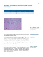
Fig. 1.1 Congenital thymic cyst shows a distended, unilocular cavity
lined by thin translucent walls and filled with fluid. Notice the portion
of normal thymus attached at the end of the cyst. This tumor developed
in a 15-year-old boy who presented with stridor and shortness of breath
owing to the large size of this particular cyst (approximately 10 cm in
greatest diameter)
Fig. 1.2 Histologic appearance of a congenital thymic cyst, showing a
single layer of flattened cuboidal epithelium. The lining in congenital
thymic cysts can also contain stratified squamous epithelium and sometimes columnar ciliated epithelium. The contents of the cyst usually
consist of clear, serous fluid. Note the absence of inflammation in the
wall of the cyst
1 Reactive, Developmental, Inflammatory, and Tumorlike Conditions
Fig. 1.3 Another area in the wall of a congenital thymic cyst. Notice a
small island of involuting thymic remnants beneath the lining in the
wall of the cyst (arrows). This thymic remnant is composed of blandappearing spindle cells with dispersed nuclear chromatin and scant
cytoplasm reminiscent of the involuting thymus. Thymic remnants are
rarely identified in the walls of congenital thymic cysts
Fig. 1.4 Foregut cyst of the mediastinum. This 4-cm cyst was incidentally found in a 23-year-old woman during a routine chest X-ray and
showed a thickened, fibrous wall with a smooth and shiny outer surface.
It was located at the bifurcation of the trachea and main bronchi and
was easily detached and removed by blunt dissection. Foregut cysts
most commonly arise in the middle or posterior mediastinum. They can
occasionally communicate with the trachea or main bronchi and are
more common in young adults and children, although older individuals
also may be affected
3
Fig. 1.5 Histologic examination of a foregut cyst shows a lining composed of columnar ciliated epithelium, with hyaline cartilage, smooth
muscle, and mucus glands in the walls of the cyst. Infection is a common complication and may lead to lung abscess formation in cases
associated with a tracheobronchial fistula
Fig. 1.6 At higher magnification, a portion of a foregut cyst shows
immature (spindled), stratified squamous epithelium with welldeveloped intercellular bridges showing partial maturation of the luminal surface toward ciliated columnar epithelium (arrow). The
identification of cartilage in the wall of the cyst is the most reliable way
to identify cysts that originate from the bronchi. Otherwise, the term
“foregut cyst” would be an appropriate designation
4
Fig. 1.7 Foregut cyst showing a lining composed of columnar ciliated
epithelium in close proximity with clusters of residual involuting thymic epithelium in the wall of the cyst (arrowheads). The thymic epithelium in such instances can undergo hyperplastic changes, not to be
confused with the development of thymoma
Fig. 1.8 Example of a foregut cyst of an enteric type presenting as a mass
in the posterior mediastinum in a 15-year-old boy with dysphagia. The
lining of the cyst is composed of gastric-type epithelium with a readily
identifiable muscularis propria. Occasionally, ciliated columnar epithelium can be identified in these cysts when they arise from the esophagus.
Another term used for these cysts is “enteric duplication cyst.” The lining
can be of either a gastric or enteric type. Rarely, some foregut cysts show
admixtures of gastric, enteric, and bronchial epithelium
1 Reactive, Developmental, Inflammatory, and Tumorlike Conditions
Fig. 1.9 Mesothelial (pericardial) cyst incidentally found at autopsy
(arrows). The cyst shows a smooth, translucent wall bulging from the
pericardium. The cyst was filled with clear, serous fluid. Similar cysts
can occur higher in the anterior mediastinum and arise from the pleural
reflection; these are designated as “pleural mesothelial cysts”
Fig. 1.10 Histologic appearance of a mesothelial cyst of the anterior
pericardium shows a single layer of round to polygonal mesothelial
cells. Rarely, the mesothelium can show foci of papillary mesothelial
hyperplasia, not to be confused with malignant mesothelioma
1 Reactive, Developmental, Inflammatory, and Tumorlike Conditions
5
Fig. 1.11 Cut surface of an acquired multilocular thymic cyst in a
56-year-old woman, showing multiple distended cystic cavities filled
with hemorrhagic fluid. The walls of the cyst are thickened and edematous and show pinpoint foci of hemorrhage and cholesterol granulomas.
These cysts can grow to very large proportions and become symptomatic. Extensive sampling is required to rule out the possibility of cystic
degeneration of an underlying malignant neoplasm
Fig. 1.13 Acquired multilocular thymic cyst at higher magnification,
showing the lining of the cyst in continuity with residual thymic epithelium inside the walls of the cyst (arrows). The residual thymic epithelium is accompanied by a small lymphocytic component and can be
seen to be in continuity with dilated Hassall’s corpuscles
Fig. 1.12 Histologic appearance of multilocular thymic cyst, showing
dilated cystic cavity surrounded by dense lymphoid aggregates. The lining of the cyst is made up of simple cuboidal epithelium to stratified
squamous epithelium. Focal areas of hemorrhage and inflammation are
commonly present in the wall of the cysts
Fig. 1.14 Detail of the lining in an acquired multilocular thymic cyst,
showing the cyst cavity lined by simple cuboidal epithelium originating
from a remnant of thymic tissue in the wall of the cyst. Notice the
admixture of the epithelium with small T lymphocytes
6
Fig. 1.15 More advanced stage in a multilocular thymic cyst, showing
dense fibrosis of the walls of the cyst secondary to chronic inflammation and netlike branching of hyperplastic thymic epithelium surrounded by the fibrous tissue
Fig. 1.16 Higher detail from an area of fibrosis in a multilocular thymic cyst, showing complex branching of thymic epithelium displaying
a sieve-like architecture, with thin, elongated strands of thymic epithelial cells circumscribing dense areas of collagen in a fibroepitheliomatous fashion. This appearance is very distinctive in long-standing
multilocular thymic cysts with prominent fibrotic changes in the walls
1 Reactive, Developmental, Inflammatory, and Tumorlike Conditions
Fig. 1.17 Acquired multilocular thymic cyst showing severe inflammation of the walls with pseudoepitheliomatous hyperplasia. Notice the
tongues and strands of squamous epithelium arising from the luminal
surface of the cyst and infiltrating into the wall of the cyst (arrows).
These reactive changes can sometimes be quite prominent and display
mild cytologic atypia and even mitotic figures, simulating an invasive
squamous cell carcinoma arising from the wall of the cyst
Fig. 1.18 Cholesterol cleft granuloma in the wall of an acquired multilocular thymic cyst. In addition to hemorrhage, fibrosis, and acute and
chronic inflammation, cholesterol cleft granulomas are a prominent feature often encountered in thymic cysts. Notice the admixture of foamy
macrophages and multinucleated giant cells with the cholesterol clefts
1 Reactive, Developmental, Inflammatory, and Tumorlike Conditions
Fig. 1.19 Acquired multilocular thymic cyst of lymphoepithelial type
shows thickened, fleshy, and edematous walls with a fish-flesh appearance due to dense lymphoid infiltrates. Such cysts are very similar to
lymphoepithelial cysts of the pancreas or salivary glands and are a common feature in children with AIDS, but they can also be seen in adult
patients who are not immunosuppressed
7
Fig. 1.21 At higher magnification, the thymic lymphoepithelial cyst
shows a cystic cavity lined by low cuboidal to stratified squamous epithelium and surrounded by a dense cuff of lymphoid tissue. In one area,
the lymphoid tissue is surrounding residual atrophic Hassall’s corpuscles (arrows)
Fig. 1.22 Thymic lymphoid follicular hyperplasia in a 45-year-old
patient with myasthenia gravis shows a slightly enlarged and thickened
thymus gland that weighed 65 g (normal for age, 25 ± 12 g). Notice the
normal-appearing, smooth outer surface and conserved normal shape of
the gland
Fig. 1.20 Multilocular thymic cyst of a lymphoepithelial type shows
cystically dilated cavities surrounded by dense lymphoid tissue with
well-developed lymphoid follicles containing germinal centers. The
main differential diagnosis for these lesions involves MALT lymphoma
of the thymus, which can also show prominent cystic changes and lymphoid follicles but which will have a monotonous population of mildly
atypical small lymphocytes infiltrating residual Hassall’s corpuscles to
form lymphoepithelial lesions
8
Fig. 1.23 Histologic appearance of lymphoid follicular hyperplasia in
a patient with myasthenia gravis, showing an enlarged lymphoid follicle with a prominent germinal center. Notice residual involuting
Hassall’s corpuscles at the periphery of the lymphoid follicle
Fig. 1.24 Gross appearance of “pure” (or “true”) thymic hyperplasia.
The thymus is enlarged to at least four times its normal size and weight.
The outer surface is smooth and shiny, and the enlargement is uniform.
This patient had no history of myasthenia gravis or other autoimmune
disorder; the lesion was found incidentally on a routine chest X-ray.
This process can also be seen as a complication of chemotherapy in
patients with Hodgkin lymphoma and germ cell tumors in adults. It can
also follow chemotherapy in cancer patients or antiretroviral therapy for
HIV infection
1 Reactive, Developmental, Inflammatory, and Tumorlike Conditions
Fig. 1.25 Histologic appearance of the thymus in “pure” thymic
hyperplasia shows an essentially normal thymus, with preservation of
the cortical-medullary architecture and no evidence of lymphoid follicular hyperplasia. The only abnormality is the increased size and
weight of the gland. The absence of sheeting and confluence of the
lymphocytes, the preservation of the normal architecture with abundant
Hassall’s corpuscles, and the absence of a thick fibrous capsule surrounding the organ distinguish this condition from a well-differentiated
lymphocyte-rich thymoma
Fig. 1.26 Involuting thymus of the adult in a 54-year-old woman who
died of other causes. The reverse process of thymic hyperplasia is thymic involution, which is a normal physiologic process that starts after
puberty and progresses into adulthood. The thymus loses volume and
becomes almost entirely replaced by fat, with only scattered microscopic islands of residual, atrophic thymic epithelium seen on histologic examination. It must be emphasized that the thymus does not
disappear under normal physiologic conditions; it remains as a welldefined structure in the mediastinum
1 Reactive, Developmental, Inflammatory, and Tumorlike Conditions
9
Fig. 1.27 Histologic appearance of the thymus in normal involution
shows small, residual islands of thymic epithelium surrounded by scant
lymphocytes. Notice that the thymic islands are separated by abundant
fatty tissue, which is replacing the original thymic parenchyma. Loss of
thymic parenchyma is experienced mainly at the expense of the immature T-lymphoid cell population, which is no longer being actively
recruited to the thymus for the programming of T-memory cells
Fig. 1.29 An elongated strand of involuting thymic epithelium
(arrows) is seen arising from a small island of residual thymus (lower
right). Sometimes these elongated strands of atrophic thymic epithelium can show complex branching and adopt a netlike configuration
like that observed in acquired multilocular thymic cysts
Fig. 1.28 At higher magnification, an involuting island of thymic epithelium from an adult shows residual strands of epithelial cells admixed
with scant small lymphocytes. Notice a small gland-like structure,
which is a normal part of the process. Occasionally, these microscopic
glandular structures can display a rosette-like or trabecular architecture
resembling neuroendocrine rests
Fig. 1.30 At higher magnification, an island of atrophic epithelium in
thymic involution shows clusters of small, oval to spindle cells with
scant cytoplasm and nuclei displaying evenly dispersed chromatin
without nucleoli. The cells in these islands of involuting thymus are
indistinguishable from those seen in spindle-cell thymomas
10
1 Reactive, Developmental, Inflammatory, and Tumorlike Conditions
Fig. 1.31 Another microscopic residual island of involuting thymic
epithelium shows cystic dilatation of a gland-like structure. Small cystic structures like these are commonly present in the involuting thymus
of adults but seldom grow to a significant size
Fig. 1.33 At higher magnification, idiopathic sclerosing mediastinitis
shows extensive deposition of fibrous connective tissue, with sparse
inflammatory infiltrate chiefly composed of plasma cells and small
lymphocytes
Fig. 1.32 Sclerosing mediastinitis encroaching on the trachea shows
dense, fibrous tissue replacing the mediastinal fat and containing scattered islands of inflammatory cells
Fig. 1.34 Entrapment of large nerve trunks by the fibrosing process is
a common feature observed in idiopathic sclerosing mediastinitis
1 Reactive, Developmental, Inflammatory, and Tumorlike Conditions
Fig. 1.35 Ropelike, linear strands of keloidal collagen flanked by scant
fibroblastic and inflammatory cells can sometimes be seen in idiopathic
sclerosing mediastinitis, creating a superficial resemblance to a solitary
fibrous tumor—a pitfall to be avoided in small biopsy samples
Fig. 1.36 The gross appearance of mediastinal thyroid goiter shows a
multinodular thyroid with a smooth outer surface. Ectopic goiters can
undergo the same spectrum of changes seen in thyroid goiters, including the development of thyroiditis and malignant transformation
11
Fig. 1.37 Scanning magnification of a mediastinal thyroid goiter
shows large, unencapsulated lobules containing follicles of varying
sizes filled with colloid
Fig. 1.38 At higher magnification, a hyperplastic nodule in a mediastinal thyroid goiter shows thyroid follicles filled with colloid and lined
by normal-appearing thyroid follicular cells with small, round nuclei
containing dispersed chromatin and small nucleoli
12
Fig. 1.39 Gross appearance of mediastinal ectopic parathyroid adenoma. Notice the smooth outer surface and pale tan-brown color of the
gland. Ectopic mediastinal parathyroid tissue can be an incidental
microscopic finding in tissues containing involuting thyroid removed
during thyroid surgery or mediastinal exploration for lymph nodes.
Rarely, these adenomas can be the site for the development of parathyroid carcinoma
1 Reactive, Developmental, Inflammatory, and Tumorlike Conditions
Fig. 1.41 The histologic appearance of an ectopic parathyroid adenoma in the mediastinum shows typical clear cell acinar architecture.
Notice remnants of involuting thymus in the vicinity (bottom left)
Fig. 1.42 At higher magnification, an ectopic mediastinal parathyroid
adenoma shows sheets of clear cells (top right) and cord-like, elongated
strands of involuted thymic tissue (bottom left) composed of small spindle cells admixed with scant lymphocytes
Fig. 1.40 This mediastinal ectopic parathyroid adenoma shows a
homogeneous, red-brown cut surface
1 Reactive, Developmental, Inflammatory, and Tumorlike Conditions
13
Fig. 1.43 The gross appearance of the cut surface of a mediastinal thymolipoma (lipomatous hamartoma) shows a well-circumscribed tumor
mass surrounded by a thin capsule and primarily composed of yellow,
lobulated fatty tissue
Fig. 1.45 A histologic variation of thymolipoma is characterized by
atrophic strands of involuting thymic epithelium embedded in abundant
hyalinized stroma and admixed with lobules of mature adipose tissue.
Such cases have been designated as “thymofibrolipoma”
Fig. 1.44 The histologic appearance of a thymolipoma shows small
residual islands of involuted thymic epithelium, with normal preservation of corticomedullary architecture surrounded by abundant mature
adipose tissue
Fig. 1.46 At higher magnification, thymofibrolipoma shows slender,
elongated strands of thymic epithelial cells composed of small oval to
spindle cells with scant cytoplasm and dispersed nuclear chromatin,
surrounded by abundant paucicellular, fibrous connective tissue