Atlas of Neuromuscular Diseases - part 5 ppt
Bạn đang xem bản rút gọn của tài liệu. Xem và tải ngay bản đầy đủ của tài liệu tại đây (946.92 KB, 46 trang )
189
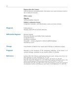
Thoracodorsal nerve
Genetic testing NCV/EMG Laboratory Imaging Biopsy
+
Fig. 29. Thoracodorsal nerve
anatomy.
1
Thoracodorsal nerve.
2
Latissimus dorsi muscle
190
Fibers stem from C5–C7 roots. (Only 50% of cases have fibers from C7.) The
fibers pass through the upper and middle trunks and the posterior cord, and
continues with the lower subscapular nerve.
Occasionally this nerve is a branch of the axillary and radial nerves.
A motor branch goes to the latissimus dorsi muscle, and may also innervate
the teres major muscle.
Both muscles are adductors and inward rotators of the scapulohumeral joint
and help to bring down the elevated arm (see Fig. 29).
Few clinical symptoms, weakness compensated in part by pectoralis major and
teres major muscles.
Signs:
Atrophy, and slight winging of the inferior margin of the scapula
Motor: Latissimus dorsi: weakness in adduction and medial rotation of shoulder
and arm.
Isolated lesion is very uncommon.
Neuralgic amyotrophy (rarely)
Plexus lesions: injury in association with posterior cord or more proximal
brachial plexus lesions.
EMG
Plexus: posterior cord lesions, upper/middle trunk lesions
Radicular: C5–C7 lesion
Conservative. Surgical interventions are not necessary because of the minor
dysfunction.
Due to this fact, this muscle can be used for grafting to the biceps brachii and
outward rotators of humeroscapular joint.
Good
Symptoms
Causes
Diagnosis
Differential diagnosis
Therapy
Prognosis
Anatomy
191
Patients note painless atrophy.
Weakness and atrophy of the pectoral muscle. Compensatory hypertrophy of
other chest muscles.
Lateral pectoral nerve:
Receives fibers from C5–7 (lateral cord of plexus) and supplies upper part of
pectoral muscle.
Medial pectoral nerve:
Receives fibers from C8/T1 and supplies lower part of pectoral muscle.
Aplasia
Entrapment in hypertrophies of minor pectoral muscle
Neck dissection
Weight lifting
Bird SJ (1996) Acute focal neuropathy in male weight lifters. Muscle Nerve 19: 897–899
Pectoral nerve
Causes
Reference
Anatomy
Symptoms
Signs
192
Thoracic spinal nerves
Symptoms
Signs
Pathogenesis
Anatomy
Genetic testing NCV/EMG Laboratory Imaging Biopsy
(+) + +
The twelve pairs of thoracic spinal nerves innervate all the muscles of the trunk
and surrounding skin, except the lumbar paraspinal muscles and overlying
skin. Dorsal and ventral rami can be affected.
Three groups: T1, T2–T6, T7–T12.
a) T1 and C8: first intercostal nerve
b) T2–T6: innervation of the chest wall
T2 is the intercostobrachial nerve (see also brachial plexus)
c) T7–11: Thoracoabdominal nerves
T12 is the subcostal nerve
Pain, sensory symptoms, depending on whether dorsal or ventral rami are
affected.
Muscle weakness may be difficult to assess, except in the case of abdominal
muscles, where bulging occurs during coughing or pressure elevation.
Metabolic:
Diabetic truncal neuropathy
Infectious:
Herpes: Pre-herpetic neuralgia (1–20 days before onset)
Herpetic neuralgia
Post-herpetic neuralgia
Lyme disease
Compressive:
Abdominal cutaneous nerve entrapment
Notalgia paresthetica: involvement of dorsal radicular branches
Thoracic disc disease (rare)
Neoplastic:
Invasion at the apex of the lung
Schwannoma
Vertebral metastases
Traumatic:
Trauma
193
Iatrogenic:
Postoperative (abdominal surgery, post mastectomy, and thoracotomy)
Laboratory: Fasting glucose, serology (herpes, borreliosis)
CSF examination (e.g., pleocytosis and antibodies in Lyme disease)
Imaging: vertebral column: plain X-ray, CT, MRI
Electrophysiology: NCV of intercostal nerves is difficult and not routinely done.
EMG: paraspinal muscles, intercostals, abdominal wall muscles
Local painful conditions of the vertebral column (disc herniation, spondylodis-
citis, metastasis)
“Intercostal neuralgia”
Muscle disease with abdominal weakness
Slipping rib/Cyriax syndrome
Depends on the etiology
Daffner KR, Saver JL, Biber MP (2001) Lyme polyradiculoneuropathy presenting as increas-
ing abdominal girth. Neurology 40: 373–375
Gilbert RW, Kim JH, Posner JB (1978) Epidural spinal cord compression from metastatic
tumor; diagnosis and treatment. Ann Neurol 3: 40–51
Love JJ, Schorn VG (1965) Thoracic disc protrusions. JAMA 191: 627–631
Stewart JD (1999) Thoracic spinal nerves. In: Stewart JD (ed) Focal peripheral neuropathies.
Lippincott, Philadelphia, pp 499–508
Vial C, Petiot P, Latombe D, et al (1993) Paralysie des muscles larges de l àbdomen due a
une maladie de Lyme.
Rev Neurol (Paris) 149: 810–812
Differential diagnosis
Therapy
References
Diagnosis
194
Differential diagnosis
The intercostal nerves are the ventral rami of the thoracic spinal nerves. They
innervate the intercostal (first 6) and abdominal muscles (lower 6), as well as
skin (via anterior and lateral branches). The first ventral ramus is part of the
brachial plexus.
Intercostobrachial nerve:
Originates from the lateral cutaneous nerve of the second and third intercostal
nerves to innervate the posterior part of the axilla.
Often anastomizes with the medial cutaneous nerve of the upper arm (stem-
ming from medial cord of brachial plexus).
The 7–11th ventral rami are called the thoracoabdominal nerves.
The 12th thoracic nerve is the subcostal nerve.
Radicular pain (beltlike)
Over the thorax cavity, no muscle weakness can be detected. However, bulging
of abdominal muscles may be apparent.
Abdominal cutaneous nerve entrapment
Diabetic truncal neuropathy
Herpes zoster
Notalgia paresthetica
Post-operatively: abdominal, retroperitoneal, and renal surgery.
Traumatic lesions
Thoracic disc trauma (rarely)
Vertebral metastasis
Laboratory: fasting glucose
Serology (herpes, Lyme disease)
Imaging: vertebral column, MRI
Electrophysiology is difficult in trunk nerves and muscles
Pain may be of intra-thoracic, intra-abdominal, or spinal origin.
Compartment syndrome of the rectus abdominis muscle
Intercostal nerves
Symptoms
Signs
Pathogenesis
Diagnosis
Anatomy
Genetic testing NCV/EMG Laboratory Imaging Biopsy
(+) + – Osseous structures of
vertebral column and ribs
195
Costochondritis
Head zones (referred pain)
Hernia
“Intercostal neuralgia”
Pseudoradicular pain
Rupture of the rectus abdominis muscle
Slipping rib
Thoraconeuralgia gravidarum
Depending on etiology
Krishnamurthy KB, Liu GT, Logigian EL (1993) Acute Lyme neuropathy presenting with
polyradicular pain, abdominal protrusion, and cranial neuropathy. Muscle Nerve 16:
1261–1264
Mumenthaler M, Schliack H, Stöhr M (1998) Läsionen der Rumpfnerven. In: Mumenthaler
M, Schliack H, Stöhr M (eds) Läsionen peripherer Nerven und radikuläre Syndrome.
Thieme, Stuttgart, pp 368–374
Staal A, van Gijn J, Spaans F (1999) The intercostal nerves. In: Staal A, van Gijn J, Spaans
F (eds) Mononeuropathies. Saunders, Londons, pp 84–86
Stewart J (2000) Thoracic spinal nerves. In: Stewart J (ed) Focal peripheral neuropathies.
Lippincott, Williams & Wilkins, Philadelphia, pp 499–508
Thomas JE (1972) Segmental zoster paresis: a disease profile.
Neurology 22: 459–466
Therapy
References
196
Symptoms
Signs
Differential diagnosis
Anatomy
Originates from lateral cutaneous nerve of second and third intercostal nerves
to innervate the posterior part of the axilla. This nerve often anastomizes with
the medial cutaneous nerve of the upper arm (from the medial cord of the
brachial plexus).
Pain in the axilla, chest wall, or thorax. Often occurs one or two months after
mastectomy. Reduced movement of the shoulder enhances pain.
Sensation is impaired in the axilla, chest wall, and proximal upper arm.
Operations in the axilla (removal of lymph nodes)
Following surgery for thoracic outlet syndrome
Lung tumors
Assa J (1974) The intercostobrachial nerve in radical mastectomy. J Surg Oncol 6: 123–126
Intercostobrachial nerve
Reference
197
Iliohypogastric nerve
Fig. 30. lliohypogastric nerve
anatomy.
1
lliohypogastric
nerve.
2
llioinguinal nerve.
3
Obturator nerve.
4
Genitofemo-
ral nerve
Fibers originate at L1, then emerge from the lateral border of the psoas, crossing
the lower border of the kidney, then the lateral abdominal wall. Then the nerve
crosses the transverse abdominal muscle above iliac crest and passes between
the transverse and oblique internal abdominal muscles. Finally two branches
are given off: the lateral anterior and anterior cutaneous nerves.
Burning and stabbing pain in the ilioinguinal region, which may radiate to-
wards the genital area or hip. Symptoms increase when walking.
Difficult to examine. Spontaneous bulging of abdominal wall. Sensory deficit
may be present. Tinel’s sign over a surgical scar may be observed. Slight flexion
of hip while standing.
Symptoms
Signs
Anatomy
198
Electrophysiology is not routinely available. Clinical distribution.
Spontaneous entrapment in abdominal wall, surgery, hernioraphy, appendecto-
my, abdominoplasty, nephrectomy, endometriosis.
Steroids locally, scar removal, neurolysis.
Diagnosis
Therapy
Differential diagnosis
199
Ilioinguinal nerve
Fig. 31. llioinguinal nerve anat-
omy. a A-female.
1
llioinguinal
nerve. b B-male.
1
lliohypogas-
tric nerve.
2
llioinguinal nerve
Fig. 32. Ilioinguinal nerve le-
sion after gynecologic surgery.
The sensory loss (marked with a
ball pen) reached almost the la-
bia
200
The ilioinguinal nerve originates with fibers from T12 and L1. The motor
component innervates the internal and external oblique muscles, and the
transverse abdominal muscle.
The sensory component covers the skin overlying the pubic symphysis, the
superomedial aspect of the femoral triangle, the anterior scrotal surface, and the
root of the penis/labia majora and mons pubis (Fig. 31).
Hyperesthesia, sometimes with significant pain over the lower abdominal
quadrant and the inguinal region and genitalia (Fig. 32).
Weakness of lower abdominal muscles, hernia.
Abdominal operations with a laterally placed incision
Biopsy
Endometriosis, leiomyoma, lipoma
Herniotomy
Iliac bone harvesting
Pregnancy, child birth
Spontaneous entrapment – “inguinal neuralgia“
Studies: no standard electrophysiologic techniques are available
Local anesthetic infiltration
Surgical exploration and resection of the nerve
Genitofemoral neuropathy
Inguinal pain syndrome
Iliohypogastric neuropathy
L1 radiculopathy (very rare)
Dawson DM (1990) Miscellaneous uncommon syndromes. In: Dawson DM (ed) Entrap-
ment neuropathies. Little Brown, Boston, pp 307–323
Komar J (1971) Das Ilioinguinalis Syndrom. Nervenarzt 42: 637–640
Mumenthaler M (1998) Läsionen einzelner Nerven im Beckenbereich und an den unteren
Extremitäten, 7. Aufl. G. Thieme Verlag, Stuttgart, pp 393–464
Purves JK, Miller JD (1986) Inguinal neuralgia; a review of 50 patients.
Can J Surg 29:
585–587
Stulz P, Pfeiffer KM (1982) Peripheral nerve injuries resulting from common surgical
procedures in the lower portion of the abdomen.
Arch Surg 117: 324–327
Clinical syndrome
Signs
Causes
Diagnosis
Therapy
Differential diagnosis
References
Anatomy
201
Genitofemoral nerve
Symptoms
Signs
Causes
Diagnosis
Differential diagnosis
Therapy
Prognosis
References
Anatomy
The nerve originates from the ventral primary rami of L1 and L2, then runs
along the psoas muscle to the inguinal ligament. In the inguinal canal the
genital branch runs with the ilioinguinal nerve, to supply the skin of the mons
pubis and labium majus. The genital branch also innervates the cremaster
muscle, while the femoral branch innervates the proximal anterior thigh.
May give rise to continuous pain, sometimes called “spermatic neuralgia”.
Can present as a post-operative inguinal neuralgia.
Paresthesias (may be painful) of the medial inguinal region, upper thigh, side of
scrotum, and labia majora.
Tenderness in the inguinal canal. Cremaster reflex unreliable.
Appendectomy
Bone graft removal
Hernioraphy
Nephrectomy
Trauma
Tumors (uncommon)
Tuberculosis
Varicocele testis
No electrophysiologic studies are available
Diagnostic anesthetic blockade
L1, 2 radiculopathy
Iliohypogastric neuropathy
Ilioinguinal neuropathy
Anesthetic blockade
Operative neurolysis
Good
Magee RK (1942) Genitofemoral causalgia (a new syndrome). Can Med Assoc J 46: 326–
329
Staal A, van Gijn J, Spaans F (1999) The genitofemoral nerve. In: Staal A, van Gijn J, Spaans
F (eds) Mononeuropathies. Saunders, London, pp 95–96
202
Superior and inferior gluteal nerves
Genetic testing NCV/EMG Laboratory Imaging Biopsy
++
Fig. 33. Superior gluteal nerve
anatomy.
1
Superior gluteal
nerve
Fig. 34. Trendelenburg’s sign,
indicating weakness of the hip
abductors (gluteus medius mus-
cle). A Standing on both feet the
pelvis remains in horizontal po-
sition. B When the patient
stands on his left leg, his pelvis
tilts to the right side. This patient
had a left gluteus medius nerve
lesion, caused by an iliac aneu-
rysm. Note that the greater glu-
teal muscles are not affected
203
Superior gluteal nerve:
Originates with the posterior branches from ventral rami of L4–S1, to innervate
the gluteus medius and minimus muscles.
Inferior gluteal nerve:
Originates with the posterior portions of L5 and S1, and ventral primary rami of
S2. It innervates the piriformis and gluteus maximus muscles.
Superior:
Causes Trendelenburg’s gait. Excessive drop of the non-weight-bearing limb
and a steppage gait on the unaffected side. Hip abduction is weak, sensation is
normal.
Inferior:
Causes buttock pain and weak hip extension (weakness getting up).
Superior:
Misplaced injection, trauma, hemorrhage, arthroplasty, aneurysm.
Inferior:
Rarely isolated, often associated with the sciatic nerve, occasionally with
pudendal nerve. Colorectal carcinoma, injections, trauma.
EMG, imaging
Sacral plexus lesion
Hip and pelvic pathology
Grisold W, Karnel F, Kumpan W, et al (1999) Iliac artery aneurysm causing isolated
superior gluteal nerve lesion. Muscle Nerve 22: 1717–1720
Rask MR (1980) Superior gluteal nerve entrapment syndrome. Muscle Nerve 3: 304–307
Wilbourn AJ, Lesser M (1983) Gluteal compartment syndrome producing sciatic and
gluteal mononeuropathies: a report of two cases. Electrencephal Clin Neurophysiol 55:
45–46
Symptoms and Signs
Pathogenesis
Diagnosis
Differential diagnosis
References
Anatomy
204
Pudendal nerve
Genetic testing NCV/EMG Laboratory Imaging Biopsy
+– +
Fig. 35. Pudendal nerve anato-
my. a
1
Pudendal nerve.
2
Perineal nerves.
3
Dorsal nerve
of clitoris.
4
Inferior rectal
nerves. b
1
Perineal nerves.
2
Pudendal nerves
205
Fig. 36. Pudendal nerve anato-
my. a
1
Dorsal nerve of penis.
2
Pudendal nerve.
3
Perineal
nerves. b
1
Perineal branch of
cutaneous femoral posterior
nerve.
2
Pudendal nerve.
3
Rec-
tal inferior nerves.
4
Bulbo
spongiosus muscle.
5
External
anal sphincter muscle
206
Clinical picture
Signs
Causes
Anatomy
The nerve originates from S2–S4, and passes through the sciatic foramen and
pudendal canal. Its terminal branches are the inferior rectal nerve (innervating
the levator ani, external anal sphincter muscles, and skin around the anus), the
perineal nerve (innervating the external urethral sphincter muscles, bulbocaver-
nosus, perineum, and dorsal aspect of scrotum/labia), and the terminal branch
of the pudendal nerve (providing sensation to the clitoris, glans penis, dorsal
region of the penis) (see Fig. 35 through 37).
Perineal sensory symptoms, sexual dysfunction.
Bilateral lesions may cause urinary or fecal incontinence, impotence/anor-
gasmy, and sensory disturbances.
Sphincter reflexes (anal, bulbocavernosus reflex absent)
Selective injury is rare
External compression:
Perineal, post-operative of hip fractures
Long bicycle rides
Suturing through sacrospinal ligament during colonoscopy
Stretch:
Straining during defecation
Childbirth
Pelvic fracture
Pelvic surgery
Fig. 37. Pudendal nerve anato-
my.
1
Cutaneous femoris poste-
rior nerve.
2
Labial/scrotal
nerves.
3
Anococcygeal nerve
207
Hip dislocation
Intraarticular foreign body
Polyneuropathy
Radicular lesion (S2–S4)
Sacral plexus
Structural abnormalities of the pelvic floor or viscera
EMG of external anal sphincter
Bulbocavernosus reflex
Pudendal SEP
Anorectal manometry, urodynamic examinations
Imaging
Amarenco G, Ismael SS, Bayle B, et al (2001) Electrophysiological analysis of pudendal
neuropathy following traction. Muscle Nerve 24: 116–119
Podnar S, Vodusek DB (2001) Standardization of anal sphincter electromyography: utilty of
motor unit potential parameters. Muscle Nerve 24: 946–951
References
Differential diagnosis
Diagnosis
209
Mononeuropathies: lower extremities
211
Obturator nerve
Fig. 38. Obturator nerve anato-
my.
1
Obturator nerve.
2
Cuta-
neous femoris posterior nerve.
3
Sapheneous nerve.
4
Calca-
neal nerve.
5
Sural nerve.
6
Lat-
eral plantar nerve.
7
Medial
plantar nerve
The obturator nerve fibers stem from L2–4, and course within the belly of the
psoas muscle, emerging on the medial side of the psoas, then passing over the
sacroiliac joint, and continuing along the wall of pelvis to the obturator canal.
Sensory loss, paresthesias, or radiating pain in the medial thigh. Disability in
walking due to impaired stabilization of the hip joint. The leg is held in an
Symptoms
Anatomy
212
abducted position, leading to a wide-based gait. The adductor tendon reflex
may be absent.
Neuralgic pain may be confused with osteitis.
Adductor weakness, with or without sensory deficits.
Compression: Obturator hernia, scar in thigh, labor, endometriosis, retroperito-
neal Schwannoma
Iatrogenic: Hip surgery, fixation of acetabular fracture, intrapelvic surgery
Laparoscopic dissection of pelvic nodes, gracilis flap, prostatectomy
Hypogastric artery aneursym
Metastatic cancer
Trauma: pelvic fracture, gunshot, retroperitoneal hematoma
Obturator nerve injury occurs commonly with a femoral nerve lesion. Causes
include retroperitoneal hematoma, cancer, hip arthroplasty, lymphoma.
EMG
Imaging
L2–L4 radiculopathy
Depends on etiology and type of nerve injury
Depends on etiology and type of nerve injury
Roger LR, Borkowski GP, Albers JW, et al (1993) Obturator mononeuropathy caused by
pelvic cancer: six cases. Neurology 43: 1489–1492
Sorenson EJ, Chen JJ, Daube JR (2002) Obturator neuropathy: causes and outcome. Muscle
Nerve 25: 605–607
Staal A, van Gijn J, Spaans F (1999) The obturator nerve. In: Staal A, van Gijn J, Spaans F
(eds) Mononeuropathies; examination, diagnosis and treatment. Saunders, London,
pp 109–111
Signs
Causes
Diagnosis
Differential diagnosis
Therapy
Prognosis
References
213
Femoral nerve
Genetic testing NCV/EMG Laboratory Imaging Biopsy
++ –+
Fig. 39. Femoral nerve anato-
my.
1
Femoral nerve.
2
Saphen-
ous nerve.
3
Patellar branch
214
The femoral nerve is derived from the lumbar plexus (originating from the
ventral roots of L2–L4). Proximal (intrapelvic) branches go to the psoas major
and iliacus muscles, passing through the inguinal ligament. Motor branches go
to the pectineus, sartorius and quadriceps muscles. Sensory branches to the
medial aspect of the thigh, anterior medial knee, and lower leg (saphenous
nerve) (see Fig. 39).
Sensory loss on the ventral thigh, perhaps with saphenal involvement (over the
tibial bone).
Buckling of the knee (on uneven surfaces) and falls (leg “collapses”). Sensory
symptoms may be mild or absent.
Pain is variable, depending on the cause of the neuropathy. Often felt in the
inguinal region or iliac fossa. Nerve trunk pain with or without sensory symp-
toms (e.g., in diabetes).
Atrophy and weakness of quadriceps muscles. Weakness of the psoas and
quadriceps muscles only occurs with proximal lesions. Decreased or absent
knee jerk. Sensory loss over anterior aspect of thigh and medial side of lower
leg.
Compressive:
Compression or stretch during surgery or obstetrical procedures: hip arthroplas-
ty, pseudoaneurysm in the groin, retraction in abdominal surgery, vaginal
hysterectomy in lithotomy position, laproscopic hernia repair, kidney trans-
plantation, abdominal hysterectomy, vaginal delivery (see Fig. 40).
Synovial cyst of hip.
Fig. 40. Femoral nerve lesion
after vascular surgery
Symptoms
Clinical syndrome
Causes
Anatomy
215
Iatrogenic:
Femoral arterial puncture, femoral catheterization, inadvertent suturing, local
infusions of chemotherapeutic agents, local anesthetic injections
Prolonged pressure: marked extension or flexion of hip in unconscious patients,
pregnancy (bilateral)
Idiopathic:
Inflammatory: heterotopic ossification, bursitis of iliopsoas muscle, lymph
nodes in ilioinguinal region, hip abscess
Metabolic: Diabetic femoral neuropathy is a misnomer; it should be called
diabetic lumbosacral plexopathy or diabetic polyradiculopathy.
Neoplastic: local tumors, perineuroma, malignant invasion
Traumatic: Penetrating injury
Vascular:
Anticoagulant therapy
Hematoma in psoas or iliacus muscle from rupture of an abdominal aortic
aneurysm
Trauma
Saphenous nerve lesions:
Bursitis of pes anserinus
Entrapment, medial side of knee
Entrapment by a branch of the femoral artery
Meniscectomy, arthroscopy
Neurolemmoma
EMG: quadriceps and iliac muscles, include paraspinal, iliopsoas, hip adductor
NCV: femoral nerve latencies and CMAPs
Sensory nerve conduction of the main trunk difficult
Sensory nerve conduction of saphenal nerve
Saphenous SEP (stimulation inferomedial to patella) is more reliable.
Neuroimaging: CT scan for psoas hematoma (has to be done acutely if hemato-
ma is suspected) or tumor infiltration of psoas muscle
MRI-femoral nerve tumors
Laboratory tests: fasting glucose, vasculitis serologies
Aneurysm of iliac artery
Irradiation of the inguinal area
L2–L4 radiculopathy
Mononeuropathy multiplex
Depends on the etiology.
Complete, postoperative lesions require surgical approach. Surgery is also
indicated for hematoma, depending upon the location and size. Otherwise,
conservative management.
Diagnosis
Differential diagnosis
Therapy