Ebook Master techniques in general surgery Gastric surgery Part 2
Bạn đang xem bản rút gọn của tài liệu. Xem và tải ngay bản đầy đủ của tài liệu tại đây (43.97 MB, 168 trang )
21
Robot-assisted Gastrectomy
with Lymph Node Dissection
for Gastric Cancer
Woo Jin Hyung, Yanghee Woo, and Kazutaka Obama
Introduction
Robotic surgery for gastric cancer is increasing. Many surgeons have adopted robotic
surgery to facilitate the technically challenging procedure of gastrectomy with D2 lymphadenectomy. With robotic gastric cancer surgery training, experienced laparoscopic
surgeons can safely provide the advantages of minimally invasive surgery to their
patients. Adherence to the oncologic principles of gastric cancer treatment ensures that
the long-term survival benefits of surgery will not be compromised.
INDICATIONS/CONTRAINDICATIONS
The indications for robotic surgery are similar to those of the conventionallaparoscopic
approach to gastric cancer. Early gastric cancer patients without parlgastrlc lymph node
(LN) involvement are ideal candidates for robotic gastrectomy with limited lymphadenectomy. Locally advanced gastric cancer without evidence of distant metastases
is a generally accepted indication for robotic gastrectomy and D2 lymphadenectomy.
Indication.s for robotic gastrectomy with limited lymphadenectomy:
cT1NoMo
Mucosal and submucosal tumors not eligible for endoscopic l"BSection
Failed endoscopic mucosal resection or endoscopic submucosal dissection
Indications for robotic gastrectomy requiring D2 lymphadenectomy:
cT1N1Mo
• cT:aNoMo; cTaNtMo
Cur1"8Dtly, there is no evidence to support robotic surgery for gastric cancer with
serosal involvement (T4a) or invasion of adjacent organs (T4b), or for palliative intent.
Intolerance to pneumoperitoneum is a contraindication.
219
220
Part II Procedures tor Neoplastic Disease
~ PREOPERATIVE WORK-UP
The preoperative work-up of patients undergoing robotic surgery for gastric cancer
requires complete evaluation of the patient's clinical status, confirmation pathologic
diagnosis, and estimation of the location and extent of disease. The preoperative workup will guide each step of the surgical decision-maldng process.
• Upper andoscopy with biopsy and with or without clipping proximal to the lesion
• Endoscopic ultrasound
• CT scan of the abdomen
~ SURGERY
Pertinent Anatomy
Robotic gastrectomy and lymphadenectomy requires the knowledge of gastric vessels
and the accompanying nodal stations as defined by the Japanese Gastric Cancer Association. The operative procedure is described relative to the dissection of the LN stations in D2 lymphadenectomy.
Operating Room Configuration
The operating room configuration is centered on the patient and the da Vinci Surgical
System (Sunnyvale, CA, USA). Relative position of the operating table, the surgeon
console, the anesthesia cart, the surgical cart, the assistant, the monitors, and the robot
during robotic gastrectomy are described.
• The robot system is positioned cephalad to the patianl
• The patient-side assistant is positioned to the lower left side of the patient on the
opposite side of the scrub nurse, scrub table, and the main assistant monitor.
• The vision systems rack is placed at the foot of the operating table.
• The surgeon's master console is positioned to grant the surgeon a view of the patient
Patient Positioning, Port Placement, Robot Docking, and
Preparation of the Operative Field
The patient is placed under general anesthesia, positioned supine with both arms tucked
to the patient side, and urinazy catheter is placed. The abdoman is prepared from the
nipple line to the suprapubic region and draped in the standard sterile fashion. Five ports,
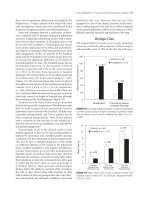
two 12 mm and three 8 mm, are used for robotic gastrectomy (Fig. 21.1). Port placements
may require minor adjustments for the patient's body habitus. Once the ports are placed,
the robot surgical cart is brought in from the head of the patiant, and the robot arms are
docked.
• The camera arm is docked to the infraumbilical port (C)
• The first arm holds the curved bipolar Maryland forceps
• The second and the third arms hold the ultrasonic shears or a monopolar device and
the Cadiere forceps, interchangeably.
Liver Retraction
The self-sustaining retraction of the left lobe of the liver is required during robotic
gastrectomy as in other upper abdominal surgeries. Adequate liver retraction is a prerequisite for complete dissection of the suprapancreatic lymphadenectomy and along
the lesser curve of the stomach. Several methods have bean described.
tahir99 - UnitedVRG
vip.persianss.ir
Cllaptar 21 Robot-assisted Gastrectomy with lymph Node Dissection for Gastric Cancer
221
figur• 21.1 Patient preparation.
A: Port placement After the
12-mm infraumbilical port is placed
using the Hasson tBchnique, the
patient is placed in 15° reverse
Trendelenburg position for the
insertion of the three 8-mm ports
and the 12-mm assist port under
direct visualization. B: Docking of
the robot anns. The robot arms
should be docked as indicated by
the numbers.
...
CD
1\'1
I
Ci
....
...
•,g
1\'1
a.
Cl
CD
...
z
lntnoperatiwe Tumor Localization to Determine die Resection Extent
Intraoperative tumor localization is required to determine the appropriate margin of
resection during robotic subtotal distal gastrectomy. Since robotic surgery is performed
for lesions without serosal involvement, the lesion cannot be readily detected during
the operation. Intraoperative tumor localization has bean achieved by several d:ifferent
methods including dye injection, intraoperative endoscopy, or laparoscopic ultrasound.
A successful technique using preoperatively placed endoclips and an intraoperative
abdominal x-ray is a simple and effective method.
~
...:.!!
;::,
"0
G)
....
Cl
a:
Procedure of 02 LN Dissection During Distal Subtotal Gastrectomy
Five Steps and Associated Anatomic Landmarks
1. Partial omentectomy and left side dissection of the greater curvature: left gastroepi·
ploic vessels
2. Right side dissection of the greater curvature and duodenal transection: head of
pancreas and right gastroepiploic vessels
3. Hepatoduodenal ligament dissection and approach to suprapancreatic area: right
gastric artery, proper hepatic artery (PHA), portal vein (PV), and celiac axis
4. Exposure of the root of the left gastric artery (LGA) and skeletonization of the splenic
vessels
5. Lesser curvature dissection: esophageal crus and cardia: proximal gastric resection
Partial Omentectomy and Left Side Dissection of die Greater Curvltllre
The exposure of the omentum can be achieved by creating a draping of the greater
omentum for safe division and retrieval of LN stations 4sb and 4d (Fig. 21.2A).
• Divide the greater omentum from the :midtransverse colon toward the lower pole of
the spleen.
• Carefully identify, ligate, and divide the left gastroepiploic vessels at their roots.
(Fig. 21.2B).
• Clear the greater curvature of the stomach from the proximal resection margin to the
short gastric vessels.
Right Side Dissection of die Greater Curvltllre and Duodenal Transection
Attention is directed to the right side of the patient for mobilization of the distal stomach from the head of the pancreas and dissection of the soft tissues containing LN
tahir99 - UnitedVRG
vip.persianss.ir
222
Part II Procedures tor Neoplastic Disease
Figwa 21.2 Left aide dissection of the greater curvature. A: Partial omentectomy begins from the distal greater curvature 4 to 5 em
from the gasttoepiploic vessels. B: The dissection is continued toward the lower pole of the spleen where the left gastroepiploic
vessels are divided and the short gastric vessels are encountered.
station 6 which is bordered by right gastroepiploic vein (RGEV), anterior superior pancreaticoduodenal vein (ASPDV), and the middle colic vein (Fig. 21.3).
• Release the connective tissues between the pancreas and the posterior stomach and
the duodenal attachments to the colon.
• Dissect the soft tissues on the head of the pancreas to identify, ligate, and divide the
RGEV as it joins the anterior superior pancreaticoduodenal vein. (Soft tissues anterior to and superior to the ASPDV and superior to the middle colic vein should be
retrieved on either side of the RGEV.)
• Identify, ligate, and divide the right gastroepiploic artery as it branches from the
gastroduodenal artery (GDA).
• Release the attachments between the duodenum and the pancreas along the GDA
until the common hepatic artery (CHA) is reached.
• Insert 4" x 4" gauze anterior to the head of pancreas to prevent injury to the GDA
and proceed to the suprapancreatic region.
• Clear the supraduodenal area and divide the duodenum approximately 2 em distal
to the pylorus using an endo-linear stapler.
This completes the intrapylorlc dissection.
Dissection of the Hepatoduodenal Ligament and Suprapancreatic Dissection
The en bloc retrieval of the suprapancreatic LNs is achieved by meticulous dissection
along the PHA, the PV, and the CHA after the ligation of the right gastric artery.
Figure Z1.3 Right aide dissection at
the head of the pancreas. The soft
tissues containing lymph nodes from
station 6 have been removed to reveal
the bordering vessels, the right gasttoepiploic vein IRGEV), anterior
superior pancreaticoduodenal vein
IASPDV), and the middle IMCVl colic
vein. The area of the 14v lymph node
station has also been dissected with
the superior mesenteric vein (SMVl
exposed.
tahir99 - UnitedVRG
vip.persianss.ir
Cllaptar 21 Robot-assisted Gastrectomy with lymph Node Dissection for Gastric Cancer
223
Figura 21.4 Dissection of the right
gastric artBry. The root of the right
gastric artery IRGA) has been iso·
lated after soft tissues containing
lymph nodas from station 5 have bean
dissected. CHA.. common hepatic
artery; PHA, proper hepatic artery
IPHA).
...
CD
1\'1
I
• Dissect the anterior surface of the PHA to identify, ligate, and divide the right gastric
artery at its origin for retrieval of LN station #5 (Fig. 21.4).
• Clear the soft tissues anterior and medial to the PHA until the PV is exposed medially for LN station 12a (Fig. 21.5A).
• The soft tissues around CHA contain LN station #8a.
• Proceed to identify, ligate, and divide the left gastric vein as it drains into the PV.
(In some patients the left gastric vein drains into the splenic vein and must be identified anterior to the splenic artery.)
• Skeletonize the CHA toward the celiac axis to retrieve the soft tissues around the
celiac artery, which contain LN station #9 (Fig. 21.5B).
Ci
....
...
•,g
1\'1
a.
Cl
CD
...
z
~
...:.!!
;::,
-a
G)
....
Cl
a:
Exposure of dae Left Gastric Artery and Skeletonization of dae Splenic Vessels
The dissection of the soft tissues along the LGA and splenic vessels ensures the retrieval
of LN station 'I and 11p, respectively. (Fig. 21.6)
• Divide the retroperitoneal attachments to the lesser curvature of the stomach to
improve access to the root of the LGA.
• Expose the root of the LGA by clearing the sWTounding soft tissues and securely
ligate and divide it.
Figure 21.5 Approach to the suprapancreatic lymph node dissection. A: En bloc LN dissection along PHA and CHA. Soft tissues
anterior to and medial to the PHA and medial to the portal vein IPVl are dissected en bloc with the soft tissues around the CHA to
retrieve the lymph nodes in stetions12a and 8a, respectively. 1: Skeletonization of the CHA toward celiac artery. The dissection
continues along the proximal CHA and splenic artery to clear the soft tissues surrounding the celiac artery for soft tissues containing lymph node station 9.
tahir99 - UnitedVRG
vip.persianss.ir
224
Part II Procedures tor Neoplastic Disease
Figur• 21.6 Root of the left gastric
arteJY ILGA) and skeletonized splenic
vessels. The soft tissues along the
celiac axis are cleared to identify the
root of the LGA and retrieve lymph
nodes from station 7. Dissection along
the splenic vassals continues half way
toward the spleen to retrieve the soft
tissues containing lymph nodes from
station 11p. SPA, splenic artery; SPV,
splenic vain.
• Skeletonize the anterior surface of the splenic artery and expose the anterior surface
of the splenic vein. (Dissection of LN station 11p is complete once the half-way point
on the splenic vessels or until the posterior gastric artery is reached.)
Lesser Curvature Dissection and Proximal Resection
The lesser curvature of the stomach is freed from the ratroparitonaum until the esophageal crus is reached. The soft tissues along the intraabdominal esophagus, the right
cardia, and the lesser curvature of the stomach, which contain LN stations 1 and 3, are
cleared to prepare for the proximal resection.
• Perform the truncal vagotomy at this time by dividing the anterior and posterior
branches of the vagus nerve.
• After the stomach is fully mobilized, transect the stomach using a 60-mm blue load
endo-linear stapler ensuring sufficient proximal margin (additional load for the stapler may be required.)
This completes the procedure of robotic D2 lymphadenectomy for distal subtotal
gastrectomy.
Procedure of 02 Lymphadenectomy During Total Gastrectomy
For advanced gastric cancer located in the upper body of the stomach, total gastrectomy
with D2 lymphadenectomy is recommended. D2 lymphadenectomy for proximal tumors
require the retrieval of the soft tissues encasing the splenic hilum, which contain LN
station 10. '1\vo options exist for retrieval of lymph station 10: a total gastrectomy with
splenectomy and a spleen-preserving total gastrectomy. While splenectomy-related postoperative complications, such as subphrenic abscesses and postsplenectomy syndrome,
are well known, complete dissection of the splenic hilum during spleen-preserving total
gastrectomy is a very complex procedure. Spleen preservation is recommended for experienced surgeons.
Spleen-Presatving Total Gastrectomy
Robotic spleen-preserving total gastrectomy requires three additional steps: the dissection of the distal splenic vessels (LN station 11d), the splenic hilum (LN station 10),
and the division of the short gastric vessels (LN station 2) (Fig. 21.7).
• After the division of the left gastroepiploic vessels, the short gastric vessels are
divided until the esophagophrenic ligament is reached and released.
• Approach the splenic hilum by identifying the distal splenic vessels behind the
distal pancreas and skeletonizing the vessels toward the spleen.
• Completely remove the soft tissues encasing the splenic hilum.
tahir99 - UnitedVRG
vip.persianss.ir
Cllapter 21 Robot-assisted Gastrectomy with Lymph Node Dissection for Gastric Cancer
225
Figur• 21.7 Complatad dissection of
tha splanic vassals and splanic hilum.
D21ymphadanactomy during splaanprasarving total g11tJactomy for
proximal !asians raquiras tha complata dissection of tha soft tinuas
along tha antira langth of tha splanic
vanals far ratriaval of lymph nodas
11d and tha splanic hilum for lymph
noda station 10.
Cll
• The remaining soft tissues along the distal splenic artery and vein can be approached
by completing the dissection from the proximal splenic vessels.
::z
a
u
'iii
...
..
'ii.
Tot•I G11trectomy witll Splenectomy
Total gastrectomy with splenectomy requires the full mobilization of the distal pancreas
and the spleen.
Free the splenic vessels from the distal pancreas.
Release the remaining splenic attachments by dividing the splenophrenic and splenorenalligaments.
Divide the splenic vessels behind the pancreas, approximately 5 to 6 em from the
celiac artery.
Cll
z
...
.:;!
00
!
....
::::0
..
ct
ID
u
Reconstruction
After robotic gastric resection and complete LN dissection, several methods for the creation of an intracorporeal or extracorporeal gastrointestinal anastomosis have been
described. The advantages and disadvantages to each approach exist. The appropriate
selection of the gastrointestinal reconstruction after robotic gastric cancer surgery depends
on the resection extent and remains a surgeon's preference. In. general, stapled anastomoses are preferred but sutured anastomosis using robot assistance is also an option. Regardless of the method and approach used, patient-side assistance is required for the
application of the stapler. Therefore, many methods used during laparoscopic gastroduodenostomy, gastrojejunostomy, and esophagojejunostomy can be applied aftar robotic gastric resections.
Gastroduodenostomy, gastrojejunostomy, or Rou:x-en-Y gastrojejunostomy
Intracorporeal or extracorporeal
Linear or circular staplers including transoral anvil placement
POSTOPERATIVE MANAGE:MENT
Postoperative management of patients who have undergone robotic gastrectomy involves
determination of when to resume oral intake, appropriate fluid maintenance, pain control, DVT prophylaxis, perioperative antibiotics, and blood work.
Return of gastrointestinal function is expected in 3 to 5 days in patients without
complications.
• Oral intake is resumed on postoperative day (POD) 2 and advanced as tolerated usually to liquid diet (POD 3), soft diet (POD 4), and regular diet (POD 5).
• Median length of hospital stay is usually 5 days without complications.
tahir99 - UnitedVRG
vip.persianss.ir
226
Part II Procedures tor Neoplastic Disease
)
CO:MPLICATIONS
The reported complication rates for robotic gastrectomy VSIY· The largest sarles evaluating the short-term outcomes of robotic and laparoscopic gastric cancer surgery report
wound-related issues, intraluminal bleeding and anastomotic leakage to be the most
common complications encountered after robotic gastrectomies. These complications
are not directly related to robot assistance since the port placements and anastomoses
are not performed using the robot
In general the morbidity and mortality associated with radical gastrectomies depend
on the extent of resection, LN dissection, experience of the surgeon, and the experience
of the institution where the surgery is baing performed. Many of the complications are
related to the extent of LN dissection and expectedly are higher with D2 lymphadenectomy than for Dl. Improved surgical outcomes have been reported with spleen-preserving
total gastrectomies when compared to total gastrectomy with splenectomy. No differences
in complication rates have been found between laparoscopic and robotic gastric cancer
surgeries.
Other possible complications are as follows:
•
•
•
•
•
•
Intra-abdominal fluid collections/abscesses
Intraluminal and intra-abdominal bleeding
Pancreatitis/pancreatic leak/pancreatic fistula
Anastomotic leak/stricture
Gastroparesis or ileus
Obstruction
3
RESULTS
Robotic surgery for gastric cancar treatment is a relatively novel field. Many studies
have studied laparoscopic versus open gastric cancer surgery and demonstrated many
benefits of minimally invasive surgery without the loss of oncologic standards. Comparison of robotic approach to laparoscopic approach is scarce, but preliminary evidence suggest that robotic gastric cancer surgery has mora benefits than laparoscopic
and open surgery for the patient and the surgeon. The short-term results of the robotic
gastrectomy from four major publications are shown in Table 21.1.
BflliBjits for the patient:
• Less pain
• Shorter length of hospital stay
Study A C•• ZH)
Study B (8 • H)
Study c ,. - 11)
Study D C• •7)
Open conversion
Nona
Nona
Nona
Nona
Resection extent
Oistalsubtatal gastrectomy
Total gastrectamy
Completion tatal
112
62
2
13
11
0
18
0
0
1
021ymphadanactomy
105
14
7
Operative time (min)
220±47
24
288 (255-3115)
259±39
420 (390-480)
300 (loo-900)
Estimated blood loss (cc)
92±153
30(1HOO)
30±15
Number of LN retrieved
42A± 15.5
28 (23-34)
41.1 ±10.9
Median LOS (days)
5
Swdy A11), Study B121. Study C(3), Study D141.
0
0
8
5
24(11~)
4
tahir99 - UnitedVRG
vip.persianss.ir
Chapter 21 Robot-assisted Gastrectomy with Lymph Node Dissection for Gastric Cancer
•
•
•
•
•
*lZ7
Decreased blood loss
Faster gastrointestinal recovery
Faster physical recovery
Better quality of life after surgery
Better cosmesis
Benefits for tb.e SW1feo.u:
•
•
•
•
•
Ergonomics
3D view
Control of four arms
Accuracy of dissection
Shorter learning curve
Disadvantages:
•
•
•
•
..•
Longer operative time
Initial cost of robot for hospital
Financial burden to patient
Limited training opportunities
.~
Cl
·~
tO
-a..
2
z
~ CONCLUSIONS
.e•
Robotic surgery for gastric cancer is a safe and feasible operation. The short-term benefits of robotic gastrectomy parallel that of laparoscopy. Surgical oncologists who treat
gastric cancer patients can readily adhere to the oncologic principles of gastric cancer
treatment including no touch technique, negative margins, adequate LN dissection, and
so on. The adoption of robotic surgery for the treatment of gastric cancer patients may
improve the quality of surgery for the patient and offer a shorter learning curve for the
surgeon.
-a
I!!
.....
:::1
0
s:.
Acknowledgments
This work was supported by a grant of the Korea Healthcare technology RI:D project,
Ministry of Health, Welfare, 1: Family AH'airs, Republic of Korea (1020410).
Recommended References and Readings
Anderson C, Ellenhom J, Hellan M, et al. Pilot series of robotassisted lapa:roscopic subtotal gastlectomy with extended lym·
phadenectomy fo:r gastric cancer. Surg Endosc. 2007;Z1(9):
1662-1666.
D'Annibale A, Pende V, P8l'D.Uza G, et al. Full robotic gastrectomy
with extended (D2) lymphadenectomy for gastric cancer: Surgical technique and preliminary results. J SUl1I Res. 2011;166(2):
e113--e120.
Hartgrink HH, Jansen EP, van Grieken NC, et al. Gastric cancar.
Lancet. 2009;374(9688):477-490.
Hur H, Kim JY, Cho YK, et al. Technical feasibility of robot-sewn
anastomosis in robotic surgery for gastric cancer. I Lo.paroendosc
Adv SUl1I Tech A. 2010;20(8):693-697.
Hyung WJ, Lim JS, Song J, et al. Laparoscopic spleen-preserving
splenic hilar lymph node dissection during total gastrectomy for
gastric cancer. JAm Coll Surg. 2008;207(2):e8-e11.
Hyung WJ, Song C, Cheong JH, et al. Pacto:rs influencing operation
time of laparoscopy-assiBted distal subtotal gasllectomy: Analysis of consecutive 100 initial cases. Eur1 Surg Oncol. 2007;33(3):
314-319.
Kim MC, Heo GU, Juug GJ. Robotic gastrectomy for gastric cancer:
sUigical techniques and clinical merits. Surg Bndosc. 2010;24(3):
610-5.
Kim HH, Hyung WJ, Cho GS, et al. Morbidity and mo.rtal.ity of lap81'oscopic ga.strectam.y versus open gastlectom.y for gastric cancar:
An interim report-a phase m multioentar, prospective, randomized 1nal (KLASS 'Iiial). Ann Surg. 2010;251(3):417-420.
Kim HI, Hyuug WJ, Lee CR, et al. Intraoperative portable abdominal
radiograph for tumor localization: a aimple and a.ccurate method
for laparoscopic gastrectomy. Surg Endosc. 2011;25(3):95&-983.
Patriti A, Ceccarelli G, Bellochi R. et al. Robot-assisted laparoscopic
total and partial gastric resection. with D2 lymph node dissection for adenocarcinoma. Surg Endosc. 2008;22(12):2753-2780.
Pugliese R, Maggioni D, Sansonna P, et al. Outcomes and survival
after lapa:roscopic gastrectomy for adenocarcinoma. Analysis on
65 patients operated on by conventional o:r robot-assisted minimal access procedures. Bur J Surg Oncol. 2009;35(3):281-288.
Song J, Kang WH, Oh SJ, et al. Role of robotic gasllectomy using da
VInci system compared with lapa:roscopic gasllectomy: Initial
experience of zo consecutive cases. Sur.f Endosc. 2009;Z3(6):
1204-1211.
Song J, Oh SJ, Kang WH, et al. Robot-asli.sted gastrectomy with lymph
node dissection. for gastric cancer: lessons learned from an initial
100 consecutive procedures. Ann Surg. 2009;249(8):927-932.
Woo Y, Hyung WJ, Pak ICH, et al. Robotic gastrectomies offer a
sound oncologic sUigical alternative for the treatment of early
gastric cancers comparing favorably with laparoscopic resections. Arc.h Surg. 2011;148(9):1066-1092.
tahir99 - UnitedVRG
vip.persianss.ir
tahir99 - UnitedVRG
vip.persianss.ir
22
Laparoscopic Resection
of Gastrointestinal
Stromal Tumors
R. Matthew Walsh
Gastrointestinal stromal tumors (GISTs) represent 1 o/o of all primary gastrointestinal
tumors and are the most common gastrointestinal tumor of mesenchymal origin. This
group of neoplasms represents an interesting aspect of cell biology as an early example
of a single gene mutation-induced neoplasm. The specific mutation occurs in the intracellular domain of the c-KIT proto-oncogene which is present in 80o/o to 95o/o of these
neoplasms. This allows the neoplasms to be distinguished from leiomyomas of the
stomach which are positive for desmin and negative for KIT.
GISTs occur anywhere in the gastrointestinal tract but are most common in the
stomach (50%) and small bowel (25o/o). They account for half of the submucosal lesions
seen on upper endoscopy because they arise from the muscular layer of the intestine
(Figs. 22.1 and 22.2). The median size at presentation is 5 em and symptomatic patients
in general present a decade earlier than asymptomatic patients with an overall median
age of 66 to 69 years. The most common presenting symptom is gastrointestinal bleeding which occurs in one-third of patients and could be occult or overt bleeding. The
next most common symptom is abdominal pain in 20% of patients. Additional presentations include an abdominal mass or incidental gastric mass on radiologic imaging
or endoscopy. The presence of multiple GISTs can suggest familial GIST. The endoscopic view can include a well circumscribed submucosal mass that may include a
deep ulceration for those presenting with gastrointestinal bleeding. And while this
endoscopic finding is sufficient in symptomatic patients to proceed with resection, it
is not specific.
Surgical resection is indicated for symptomatic GISTs, and biopsy is not required
when tumor dissemination may be a risk. One tenet of treatment centers around the
knowledge that all GISTs have malignant potential. Risk stratification is important to
consider both for the indication for resection and for adjuvant therapy. Risk stratification for resection centers on size. Autopsy series demonstrate a high prevalence (22o/o)
of small GISTs (<10 mm) in individuals over 50 years. Most of these small GISTs do
not progress rapidly into large macroscopic tumors despite the presence of a KIT mutation. It is currently recommended that in acceptable risk patients, any GIST >2 em
should be resected. Contraindications to resection from a tumor biology perspective
tahir99 - UnitedVRG
229
vip.persianss.ir
230
Part II Procedures tor Neoplastic Disease
figur• 22.1 Endoscopic Yiaw of a
submucosal mass. Tha endoscopic
Yiaw is typical but nat specific for
a GIST. Tha growth pattern can ba
intraluminal, exophytic to tha
stomach or both.
include patients with known metastatic disease or unresectable tumor due to size or
extended organ involvement that would lead to unacceptable morbidity or functional
deficit. This latter group is amendable to neoadjuvant imatinib mesylate to downsize
the tumor. This therapy typically lasts for 6 to 12 months with maximal response
defined as no further improvement between f:w'o successive CT scans.
~
PREOPERATIVE PLANNING
A component of preoperative planning involves a consideration of the accuracy of the
preoperative diagnosis for GIST. The differential diagnosis includes other submucosal
masses such as lipoma, carcinoids, and leiomyomas or sarcomas, and nongastric masses
which originate from the liver, pancreas, or spleen, as well as lymphoma or germ cell
tumors. The diagnostic yield of endoscopy with biopsy is 35%, endoscopic ultrasound
with fine needle aspiration (FNA) 84o/o, abdominal computed tomography 74o/o, and magnetic resonance imaging 91%. Endoscopic ultrasound is valuable in assessing the gastric
layer from which the lesion arises as well as providing access for biopsy if that is required.
Once an accurate diagnosis of GIST has been determined, preoperative planning
will be guided by size, location, and relative intra/extra gastric configuration. The interplay of all of these factors will determine the ultimate operative approach. A large
lesion that is very exophytic or pedunculated on the anterior wall of the gastric body
Figur• 22.2 Image obtained from
endoscopic ultrasound IEUS).
These tumors arise from tha
muscularis propria as damon·
stratud. They can have a dumbbell
configuration which is nat always
evident on EUS.
tahir99 - UnitedVRG
vip.persianss.ir
Cllaptar 22 Laparoscopic Resection of Gastrointastinal Stromal Tumors
231
is a straight-forward laparoscopic resection and would be an entirely different operative
approach from the same size lesion of the posterior antrum with an appreciable intragastric component which may require a standard distal gastrectomy. A posterior location may extend into the retroperitoneum requiring a pancreatic resection for complete
removal. Ti'ansgastric or intragastric procedures should be considered for posterior wall
or gastroesophageal junction tumors with an intragastric component A wide variety of
minimally invasive techniques are appropriate for GIST tumors which defies the concept of a single best approach for all patients. The integration and assessment of intraoperative endoscopy by the surgeon and diagnostic laparoscopy should guide operative
decisions regardless of the preoparative plan.
Preoperative planning does require consideration of special equipment for many
laparoscopic resections. A video endoscope, angled laparoscope, specimen retrieval
bags, and endoscopic linear staplers are standard fare. Intragastric procedures where
the operation occurs in an insuftlatad stomach with intragastric ports is a special operation which should be planned. It behooves the surgeon to be prepared with the following equipment if an intragastric approach is being contemplated.
Endoscopy Equipment
•
•
•
•
•
•
Diluted epinephrine
Sclerotherapy needle
Over-tube
Biopsy forceps
Endo-snare
Roth-net
Laparoscopic Equipment
•
•
•
•
•
Balloon stabilized 5-mm trocars
Needle drivers
tntrasonicshears
Hook cautery
Dual channel inputs for picture-in-picture
Robotic-assisted laparoscopy can also be performed for all manner of laparoscopic
resections of GISTs including intragastric procedures. Use of robotic techniques will be
determined by equipment availability and expertise.
&
SURGERY
Regardless of the specific operative approach, laparoscopic versus open, intragastric
versus transgastric, formal resection versus wedge resection, the same surgical objective
should be obtained: complete resection without tumor disruption. The principle goal
of resection is obtaining macroscopically negative margins. The need to achieve microscopically negative margins is uncertain, since outcomes are likely determined by biologic tumor behavior and not the microscopic margin. The presence of a positive
margin may be falsely interpreted based on specimen retraction, and re-excision is not
advised for a microscopically positive only (Rl) resection. Radical resection that would
include lymphadenectomy is not required to ensure good outcomes, but a formal resection may be required based on size and location to achieve the best functional outcome.
Extended resection should be done for contiguous organ involvement only to the degree
that an RO or Rl resection is accomplished. A laparoscopic approach to resection is
feasible, providing the same principles of traditional surgery are upheld: complete
resection without tumor disruption. It was due to concem for tumor disruption by
manipulation of the tumor that laparoscopy was initially discouraged, but its utility has
been borne out in many series.
tahir99 - UnitedVRG
vip.persianss.ir
232
Part II Procedures tor Neoplastic Disease
Positioning
Routine supine positioning is employed with video monitors at the head of the bed.
Rarely will the monitors be at the feet if an intragastric resection is entertained for an
antral lesion. Access to the mouth should be available for intraoperative endoscopy.
Use of a split-lag bed is purely basad on surgeon preference.
Technique
There are multiple techniques used for laparoscopic resection of gastric GISTs due to
the varied locations of the lesions. It also points to the ingenuity of techniques fostered
by minimal access surgery. General approaches will be discussed that are adaptable to
specific situations.
Llparoscopic Wedge Resections
In this general scenario, wide access to the abdominal cavity is required with trocars
positioned in a lazy "U" as used for most upper abdominal surgery (Fig. 22.3). This
involves typically five trocars, all 5-mm except for a 12-mm at the umbilicus for endoGIA stapler and extraction site. This approach is acceptable for all anterior wall masses
of any location and many posterior wall lesions accessible via transgastric approach or
via the lesser sac. There is a common misconception that a wedge resection requires
elevating the lesion and its gastric wall attachment using a stapler to transect both walls
of the stomach in a single firing (Fig. 22.4), and this technique is used most often by
the laparoscopic novice and is really useful for only the most exophytic of GISTs. The
resection of a spherical mass with a linear stapler will typically require a long staple
line (use and expense of multiple cartridges) resulting in an unnecessarily large gastric
deformity. It is typically a batter option to resect the mass with a rim of normal stomach
with any anergy source (cautery, endoscopic shears; Figure 22.5A) and reconstruct the
defect. The closure of the defect can be accomplished with a stapler (Fig. 22.5B) or
suturing which will result in a better functional result. The specimen should be placed
in a retrieval bag.
Figure 22.3 Typical ttocar positioning for most
laparoscopic gastric procedures !excluding
inttagastric mchniques).
Smm
5 mm
~J~
10m1m ~
5 mm
10mm
tahir99 - UnitedVRG
vip.persianss.ir
233
Cllaptar 22 Laparoscopic Resection of Gastrointastinal Stromal Tumors
.....
~
l
figure ZZ.4 A: Leperoscopic approach to a posterior wall GIST. The gastrocolic omentum is divided with endoshears to expose the
posterior well. A trensgesttic approach can reach the same lesion but does not allow assessment for exttegastric, retroperitllneel
extension and thus is reserved for posterior well lesions \."ith dominant intragastric component 1: A stBpled excision of the gastric
well containing a GIST often requires a long staple line and multiple firings til excise e spherical mass. This is ideal for exophytic
end smell lesions without causing excessive deformity.
lntngastric Raection
Resection of gastric GISTs can be performed while operating within the gastric lumen.
This requires trocar placement directly into the gastric lumen and insuffiation with C02
to distend the lumen. Endoscopic skills are important to allow b:ocar placement, suture
passage, and specimen retrieval. The best candidates for this approach are those with
...
CD
1\'1
I
Ci
....
...
•,g
1\'1
a.
Cl
CD
...
z
~
...:.!!
;::,
-a
G)
....
Cl
a:
-
~
1\'1
a..
lesions that are near the gasb:oesophageal junction or on the posterior wall of the proximal stomach. The lesion should be predominantly intragastric with realization that
lesions can have a "dumbbell" configuration that may be seen on endoscopic ulb:asound
or preoperative CT. A full-thickness resection of the gastric wall is feasible with the
inb:agastric approach as well as an enucleation that involves partial depth removal.
The operative sequence is as follows:
• Patient is in supine position and under general anesthesia.
• Endoscopy is performed to confirm position, particularly whether anterior or posterior wall, and selection of trocar location by digital indentation.
A
figure ZZ.5 A: Complete excision of anterior gasttic mass with hook cautery til achieve e negative margin. Avoid direct rettection of
lesion tD prevent disruption. B: Closure of e gastric well defect after excision of en anterior well GIST. The closure can be done in e
transverse fashion as well end compromises the lumen minimally. Sutures can also be used to elevate the corners of the defect
tahir99 - UnitedVRG
vip.persianss.ir
234
Part II Procedures tor Neoplastic Disease
Agur• 22.6 Endoscopic guidance
of balloon-tipped trocars into the
stomach with maximal triangula·
tion. Typical trocar positions (insst).
• General abdominallaparoscopy to look for exophytic component and metastases.
• 5-mm balloon tipped b:ocars into the stomach with the stomach maximally distended
with air under endoscopic visualization (Fig. 22.6). Three trocars are placed with
maximal triangulation secure with balloon insufllation so trocars do not migrate from
the stomach inadvertently.
• C02 insufflation into stomach.
• Endoscopic injection with a sclerotherapy needle of dilute epinephrine for hydrodissection and improved hemostasis. The hydrodissection allows for identification of
the precise border of the GIST if enucleation is anticipated. The delineation of the
lesion is clearly visible in all casas (Fig. 22.7).
Agure ZZ..'I Endoscopic injection of
the submucosa with epinephrine for
hemostasis and hydrodissection.
tahir99 - UnitedVRG
vip.persianss.ir
Cllaptar 22 Laparoscopic Resection of Gastrointastinal Stromal Tumors
235
figur• 22.8 Laparoscopic closure
of gastric wall defect with sutures
introduced by endoscopically
passed sutures.
...
CD
1\'1
I
Ci
....
...
•,g
1\'1
a.
Cl
CD
...
z
~
...:.!!
;::,
"0
G)
....
Cl
a:
• 5-mm instruments and camera for laparoscopic intragastric dissection. Typically the
hook cautery works well for flne dissection.
• Following excision the lesion is placed in the stomach and the gastric wall defect
repaired in all casas.
• The endoscope is re-inserted with an over-tuba. Vicrylsuturas of 4 inches length are
passed into the stomach with an endoscopic biopsy forceps.
• The gastric wall defect is closed with laparoscopic needle drivers and endoscopically
passed sutures (Fig. 22.8).
• The needles are removed orally. Any needed number of sutures can be passed as
necessary to complete the closure.
• The GIST is removed orally after endoscopic capture with a Roth-net or snare.
• The balloon trocars are deflated and withdrawn from the stomach into the peritoneal
cavity. The anterior gastric wall puncture sites are closed with standard laparoscopic
suturing (Fig. 22.9).
Laparoscopic Fonnal Gastric Resection
A standard type of gastric resection is rarely required for resection of a GIST. It is not
necessary from an oncologic perspective. This may be necessary for the size and compromising position of a GIST, typically an antral lesion whose resection and reconstruction would result in luminal compromise and outlet obstruction. An antractomy with
either Billroth I or II reconstruction is easier to accomplish as a planned excision rather
than following excision where the subsequent large defect needs to be reconstructed.
The laparoscopic approach to antrectomy or distal gastrectomy is similar to the
technique for a gastric cancer except that lymphadenectomy is not required. Should
this be undertaken due to tumor size it must be dona with the consideration of not
disrupting the tumor. Many lesions that require a standard type of resection or extended
resection due to tumor size are best done with open resection to avoid tumor disruption
since this is of greater consequence than a laparotomy incision.
Robotic Excision
All of the aforementioned laparoscopic techniques can be performed with robotic assistance. Robotic partial gastric resection can be utilized with robotic endoscopic shears or
23&
Part II Procedures tor Neoplastic Disease
Figwa 12.9 Laparoscopic closure
of trocar situs with balloon cath·
eturs removed from the stomach
into the abdominal cavity.
hook cautery. The defect can be easily sutured closed in two layers using robotic needle
drivers, thus avoiding any staplers (Fig. 22.10A and B). This robotic suturing is effective
regardless of the defect size or location and is a particularly good training procedure
for residents and fellows. The degrees-of-freedom of the robotic instruments is what
makes the defect size and location a straight-forward repair and is bast demonstrated
for intragastric suturing. The intragastric repair of the gastric wall defect is the most
tedious and time-consuming aspect of the intragastric operative approach which is
greatly improved with the robotic instruments. The standard 5-mm robotic trocars can
be placed intragastricly without need for balloon-stabilized ports due to the robotic
instrument recognition platform. A standard gastric resection is also well described
robotically for early gastric cancer and can be used in this similar situation. The use of
robotics in the resection of gastric GISTs is limited by the availability of the device,
surgical training, and one's imagination.
Figura 22.10 A: Endoscopic view of antral lesion that is posterior and intragastric. The light from the laparoscope shines through
the anterior gastric wall. B: Robotic closure of the excised defact that was approached transgastrically. The right robotic needle
driver is being passed through the pylorus to avoid outlet obstruction.
Cllaptar 22 Laparoscopic Resection of Gastrointastinal Stromal Tumors
POSTOPERATIVE MANAGE:MENT
The typical course of patients following laparoscopic GIST resection is notable for brief
hospitalizations and rapid recovery. The length of stay is typically less than 5 days. A
nasogastric tube is not routinely utilized nor is routine Gastrografln studies to interrogate for a leak. Patients are begun on a liquid diet the first postoparativa day and
advanced as tolerated. Antibiotic prophylaxis is used for 24 hours. A proton pump
inhibitor is used routinely for the 2 months to aid gastric mucosal healing and reduce
hemorrhage at the excision site. Any patient undergoing intragastric enucleation is
surveyed yearly, to include endoscopic ultrasound, for 5 years to identify local recurranee should that occur.
:l COMPLICATIONS
Procedure-specific complications are very infrequent, with many series reporting no
complications. Potential morbidity would include hemorrhage, leak, and inlet or outlet
obstruction.
• Hemorrhage can occur at the site of the resection if a partial thickness resection was
dane at an anastomosis or through a staple line. These bleeding complications can be
reduced with fibrin glue for a staple line, mucosal approximation of gastric wall defects,
and usa of postoperative acid-suppressive medications. Bleeding complications can be
diagnosed and managed with endoscopic techniques and rarely raoperation.
• Staple or suture line leaks are technical complications that are best avoided by maintaining meticulous technique. They should be suspected in a patient who is not
following the anticipated recovery path, is septic, unusually tender on examination,
or exhibiting delayed gastric emptying. It can be documented by oral contrastenhanced CT scan (perigastric abscess with or without contrast extravasation) or
Gastrografin swallow. Contained leaks can be managed with percutaneous drainage
and antibiotics; the others with reoperation.
• The lumen can be operatively compromised at both the gastroesophageal junction
and the antrum (pylorus). This can occur for lesions at both locations due to a large
excision or from the reconstruction. Usually this is a consequence of poor operative
selection or unsuspected narrowing with staplers. Symptoms are typically based on
precise location: dysphagia or gastric outlet obstruction. Reoperation with resection
and reconstruction is often required.
3
RESULTS
The patient outcome is typically a consequence of tumor biology, provided the basic
surgical tenets of GIST excision are maintained. There does not appear to be an inherent disadvantage to laparoscopic resection. Small retrospective comparative trials have
shown no adverse outcome from laparoscopic outcomes relative to resection, margin
status, morbidity, or tumor recurrence. The length of stay can be favorably impacted by
laparoscopic approaches.
Survival after surgery alone for GIST is favorable when compared to other intraabdominal sarcomas. The overall outcome for patients who undergo complete resection
with negative margins shows a 5-year disease-specific survival rata of 54% with a
median survival of 66 months. The two most important prognostic features of the pri·
mary tumor are its size and mitotic index, which provides for a consensus approach to
risk stratification.
An important consideration should be the use of i.matinib mesylata in the adjuvant setting. A seminal trial has been published that randomized patients with >3 em
237
238
Part II Procedures tor Neoplastic Disease
KIT-positive GISTs to 1 year of 400 mg imatinib following complete resection. At a
median follow-up of 20 months a clear improvement in recunence-free survival was
noted with imatinib, and the trial was stopped due to this interim analysis. There as
yet has been no improvement in overall survival. This study clearly demonstrates that
empiric adjuvant imatinib reduces rates of early recurrence, yet it is not clear whether
this strategy improves overall survival, whether longer therapy beyond 1 year is warranted, and what patient selection criteria for therapy would be used.
+..:., CONCLUSIONS
Laparoscopic approaches to resection of gastric GISTs are reasonable, providing complete
resection without violation of the tumor is achieved. There are a variety of operative
approaches available to achieve this goal. The specific operative approach should be
tailored to the patiant's specific size and GIST location to obtain the optimal functional
and oncologic outcomes.
Recommended References and Readings
Ageimy A, WUDsch PH, Hofsta.adter F. et sl. MiDuta gsstrlc sclerosing stromsl tumors (GIST tumorlet3) sre comm011 ill adults md
&9quently show c-KIT mutati011s. Am J Surg Pathol. 2007;31(1):
113-120.
Bonvslot S, Eldweny H, Pechoux C, et sl. Impsct of surgery on
advanced gastrointestinal tumors (GIST) in the imatinib era.
Ann Surg Oncol. 2006;13(12):1596-1603.
Catena F, DiBattista M, Fusaroll P. Laparoscopic treatment of Gastric
GIST: Report of 21 cases and literature's review. J Castmintest
Surg. 2008;12:561-568.
DeMatteo RP, Ballman KV, Ant011escu CR., et al. Adjuvant imatinib
mesylate after resection of localized, primary gastrointestinal
stromal tumour: a randomized, double-blind, placebo-controlled
trisl. lAncet. 2009;373(9869):1097-1104.
DeMsttao RP, Lewis JJ, LeUDg D, et sl. 'I\vo hundred gastrointestinsl
stromsl tumors: R8CU.lT9Ilce pe.ttams and prognostic fsctoJ:s for
survivsl. Amt Surg. 2000;231(1):51-58.
Demet:ri GD, BenjsmiD. RS, Blanke CD, et sl. NCCN Task Force
Report: Optimsl MansgemBilt of Pstients with Gsstrointestinsl
Stromsl Thmor (GIST) - Updste of the NCCN Clinics! Practice
Guidelines. JNCCN. 2007;5(Suppl 2):51-79.
Demetri GD, von Mehren M, Ant011escu CR, et sl. NCCN Task Force
report: Update on the mansgement of patients with gastrointestinal stromal tumors. J Notl Compr Cane Netw. 2010;8(Suppl 2):
51-41.
Dholakl.a C, Gould J. Minimally invasive resection of gastrointestinal stromal tumors. Surg Clin N Am. 2008;88:1009-1018.
Everett M, Gutman H. Surgical mansgement of gastrointestinal
stromal tumors: Analysis of outcome with respect to surgical
margins and technique. 1 Surg Oncol. 2008;98:588-593.
Glasco G, Velo D, Angriman I, et al. Gastrointestinal stromal tumors:
Report of an audit and review of the literature. Eur 1 Cancer
Prev. 2009;18:106-118.
Kim MC, Heo GU, Jung GJ. Robotic gastrectomy fur gastric amcer: SuJ'o
gl.cal techniques and clinic merits. SurgEndosc. 201G;Z4(3):610-615.
Learn PA, Sicklick JK, DeMstteo RP. Randomized clillicsl trisls in
gsstrointestinslstromsl tumms. Surg Oncol Clin N Am. 2010;19:
101-113.
Matthews BD, Wslsh RM, Karchar KW. Laparoscopic vs. open resection of gsst:ric stromsl tumors. Surg Endosc. 2002;18(5):803-807.
Miettinen M, Lasota J. Gastrointestinsl stromsl tumors: pathology
and p:ognosiB at diffarent sites. Ssmin Diagn Pathol. 2008;23(2):
70-83.
NishimuraJ, NakaJima K, Omori T, etal. Surgical strategy for gastric
gastrointestinal stromal tumors: Laparoscopic vs. open resection. Surg Endosc. 2007;21:875-878.
Novitsky YW, Kercher KW, Sing RF. LOilg·term outcomes of laparoscopic resection of gastric gastrointestional stromal tumors. Ann
Surg. 2006;243(6):738-745.
Raut CP, Ashley SW. How I do it: Surgical management of gastrointestinal stromal tumors. 1 Castrointest Surg. 2008;12:1592-1599.
Rosen MJ, Henifard BT. Endoluminal gastric surgery: The modern
ara of :minimslly invasive surgery. Surg Clin N Am. 2005;85:9891007.
Salem TB, Ahmed I. Gsstrointestinsl stromsl tumour-evolviDg COil·
cepts. Surgson. 2009;7(1):36-41.
Scarpa M, Bertin M, Ruffolo C, et sl. A systematic review 011 the
clinics! disgnosis of gsstrointest:insl stromsl tumors. I Surg
Oncol. 2008;98:384-392.
Wslsh RM, Ponsky J, Brody F, et sl. Combined endoscopiclle.paroscopic
intragastric resection of gastric stromal tumors. 1 Costrointest Surg.
2003;7(3):386-392.
23 Surgery for Gastrinoma
E. Christopher Ellison
INDICATIONS
Gastrinoma, abo known as the Zollinger-Ellison syndrome (ZES). is a rare cause of
ulcer disease. The incidence is about one case per million per year. The disease usually
occurs between the ages of 30 and :70, although cases in children and the elderly have
been reported. Sporadic cases dominate, accounting for :75% of all cases. Familial cases
occur in 25% of patients and are usually part of the multiple endocrine neoplasia type
1 (MEN1) syndrome. It is very important to establish whether the patient has sporadic
gastrinoma or MEN1, as the surgical treatment is different.
The diagnosis of gastrinoma is suggested by fasting hypergastrinemia off proton
pump inhibitors (PPis) in a patient with refractory ulcer disease, gastroesophageal
reflex, or diarrhea. The most common causes of hypergastrinemia are achlorhydria associated with atrophic gastritis and chronic use of PPis. PPis induce achlorhydria, and
hence, in the absence of negative feedback on the G cells in the gastric antrum, more
gastrin is released. To establish that the fasting hypergastrinemia is caused by gastrinoma, it is necessary to check for the presence of gastric acid. If the patient has a gastric pH of :7 off PPI.s, then ZES is excluded. If the patient has acid in the gastric aspirate,
then a secretin provocative test is indicated. In a patient with gastrinoma, secretin will
cause an increase in the gastrin. A positive test is defined as an increase of gastrin
greater than 110 pglmL over the baseline value (Fig. 23.1).
The contemporary surgical approach to gastrinoma is directed at tumor excision,
and no gastric procedure is performed. Surgery is recommended only after the diagnosis is clearly established and imaging has localized the tumor.
~ PREOPERATIVE PLANNING
'1\vo-thirds of gastrinomas are located in the gastrinoma triangle shown (Fig. 23.2). This
applies to both sporadic and MEN1 patients. In sporadic gastrinoma, the tumors are
located either in the duodenum (about half the cases) or the pancreas (nearly half the
cases), or both. Gastrinomas may also occur primarily in lymph nodes. About one half
of sporadic patients have tumors in both the duodenum and the pancreas. In those with
MEN1, there is a propensity for multiple tumors, and duodenal tumors are found in
239
240
Part II Procedures tor Neoplastic Disease
Figw• 2!.1 Sacnrtin stimulation test for
gastrinoma.
500
_.
~300
g_
c
!
200
100
0
2
10
Minutes
5
15
30
nearly all of these patients. The gastrin-producing pancreatic tumors are usually in the
head of the pancreas. Although many patiants have tumors in the body and tail of the
pancreas, most of these are nonfunctional.
Preoperative localization tests should be performed. Somatostatin scintigraphy and
CT scan are the initial tests. This may be supplemented with endoscopic ultrasound,
MRI, and selective arterial secretin stimulation. In one-third of casas these tests will be
negative. Exploration is clearly wSITanted in patients with positive localization tests.
An individualized approach is recommended for those with negative localization tests
and for patients with :MEN1.
Outline of the Surgical Procedure
The operative procedure is divided into three unique major steps and is based on the
principle of the gastrinoma triangle: step 1 is pancreatic exposure and managemant of
pancreatic tumors; step 2 is assessment for and resection of duodanal tumors; and step
3 is sampling of lymph nodes in the gastrinoma triangle.
Figur• 23.2 Tha gastrinoma triangle.
2111
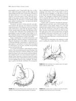
Sbrp 1: h::cctwik: ''i*"W'ii _ . ...... . , afJlu.arutlc'rau:wao
The o~ ~ t.kt ..,.,.. th.& .:bdom.et\ th!ol.t8b a rofdJh)e fnd«fan m.d 1*"'
n.o
fcnm, .. mrnual upJoz:dCIIII.
6l.l.l'pCID. DOXl pod'o:a. a w1clo ~ a:&&D.OU.Rl' to
lUIIy""'""'" tho duod..,um and allaw pelpllloo ol 0.. h.eo.d of tho"""""""'' NS>d Ia
lho _..,. of tho
tile 1 -..... 'l'hl.t 116ill--.:l by b!m.o:o..J pal.
~ o1 tho o:lld. lllo» by •·~nllllroooiiii.CL Noort lolocolodlll"" ol
my po.t>.OIMliC lmWM.I'........US.:.u.w ~II! po.o.OIMII.·
--11rmuah
!1!-
c>o<~aoct.,...,..,,lltom-..n...tod~an1holoMd01\e:odelsoattllo111m0f
m.olla pallcubr If the pm.cnllle dw:t Ia bxvolnd.
Stlp 1: .I
M'1M tiN- J)aaf!IM) "n:r:ttYft aM I• I t'dltt
lntri.Op«!U!n ~a.od.,..IIOP111.f1J! truldllumlntd01\ at lho duodonwn
rDta.f be dOCI.S to try to l.ooall&a a d.uod.eo.el bmurt. llawwflilf tb..& pnls:tnd tec'hntque k
- . . . of • IO!lJfmd""'' dll.odon.almo.y to , - pol,pdl!ll> ol tho du.odm.ol muoooo.
Tldo II tho 111.011 zolllblo ...tllod to ld
!llllllcauod !D tht lNn1
lllllllaef . . . .IJfofGMDI
sn
Tloo ~ Ia UI'D.O!Iy doa.o u .,. opoa pzocodDn>;
- · looolllotloa a lop«100<0plc II! ft>botle
~Law.....,
wltlllmpnmod P""'P'
toch.t>l4•• "'"1 bo ttllblda 6xt ..,..
~
l'lzo. .i ..
Tloo patloal. Ia placod !D 0.. wp!Do pcci!IOG. '!b.o loll am Ia lucbd m.ol the liFt ezm
II plooocl<81 "" """ boo«!d.
1'111. .
A !'Odphon!IVII ~ Comzal...,... -~~ UMt! !tutl..-..l
o1_..,). ~!olio -a...ta, a~lullo ODI!.Foloy cw1b- 110 pliocod.
t:.t-1
Ia a.old!tllm to.~ lapll'Otany..t, tho~ oqulpm.eal. olooulll bo ....n.hlo:
An""""-
1. IDirLap
out mel., odalt "PI*'
1. A blpol1r ccqolalta,t
locioi ..
-00.
A Jllldltno
fo awlo, ll.fiJ! tho lll>
'l1l.o N1lll.cl Up.
PucrHtic Ezt ssurund Mllllllllnt aC PucrHiic TilIIIII
........... ca~ •• o.,.
Tloo pot_.., eiona tho lltOOII.d pcxntcm ot tht duod.,..,l& tnn!Md llhorp\y. Wodlll
..... tho ci....W..... by tho Allil.ttaal. fodll- tbo ...... l!pt!oo., I'l!.o J:.odi.o:
,.,._14 oomplot> w:t.ox. tho I.e.~ """Ia-. I'l!.o daodonum m.ol b.oad ot tl!.o
pmtnU " " pal,pt.tod (l'l,p. a8 .! mel a8 .~
1J11111 r~.tMt.c .....
n.. ~ ou:ll"'""""- br olillply
from.
a.. _._. coloD.
-CO''"'-
Tlda Ia <:8lO!sd wldsly (PI& 23.5).'1'b.e body and lldl of tho~""""""" ..,. ~· 'l'b.e
111M~,..,II! 'botwOOD 0.. pooUitar woll of lho iMmach ll.fiJ! tho
of lh•
ptiii.CIOU arolll.dood w!tll. ~. I'l!.o .....t Alld .,.\olio: IJ1II!acl> of tho bood. ol
242
Part II Procedures tor Neoplastic Disease
figure ZU Incision for Kocher maneuver.
I
I
I
''
Incision-----"
\
' ,,......
---·
figure Z:U Duodenum and head of
pancreas lifted by surgeons left hand.
figure 23.5 Division of omentum to enter the
lesser sac and expose the pancreas.
Pancreas
Chapter 23 Surgery for Gastrinoma
figur. ZU Pancreas exposed in the Iasser sac.
Superior mesenteric
vessels
the pancreas are exposed by continued dissection of the omentum in order to expose
the vascular groove and the anterior portion of the superior mesenteric vein (Fig. 23.6).
In some cases to provide enhanced exposure the gastroepiploic vein is ligated with 2-0
silk and divided. Bimanual palpation of the pancreas is performed.
Uhrasound of die Pancreas
The ultrasound transducer is placed in a sterile covering, and gel is applied to the tip
of the probe. Transducers are designed to produce ultrasound waves of diHerent frequencies. The higher the frequency of the waves, the greater the resolution of the image
on the screen. Thus a 10-MHz transducer will produce a clearer image than a 5-MHz
transducer. Saline is instilled into the lesser sac to cover the pancreas. The pancreas is
examined for hypoechoic lesions (Fig. 23.7).
figur• ZU Intraoperative ultrasound
af the pancreas.
243