Ebook Oral medicine and pathology at a glance Part 2
Bạn đang xem bản rút gọn của tài liệu. Xem và tải ngay bản đầy đủ của tài liệu tại đây (5.34 MB, 69 trang )
9781405199858_4_C30.qxd
30
3/1/10
2:10
Page 52
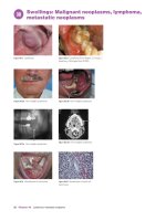
Swellings: Malignant neoplasms, lymphoma,
metastatic neoplasms
Figure 30.1 Lymphoma.
Figure 30.2 Lymphoma (from Bagan JV, Scully C.
Medicina y Patologia Oral, 2006).
Figure 30.3a Non-Hodgkin lymphoma.
Figure 30.3b Non-Hodgkin lymphoma.
Figure 30.3c Non-Hodgkin lymphoma.
Figure 30.4 Metastasis of carcinoma.
Figure 30.3d Non-Hodgkin lymphoma.
Figure 30.5 Metastasis of renal cell
carcinoma.
52
Chapter 30 Lymphoma, metastatic neoplasms
9781405199858_4_C30.qxd
3/1/10
2:10
Page 53
Lymphomas
Definition: Malignant neoplasm arising from lymphocytes; based on
the “Revised European-American Lymphoma classification” (REAL),
the WHO (2001, updated 2008) classified lymphomas in three broad
groups (B, T or NK (natural killer)) according to cell type, plus less
common groups e.g. Hodgkin lymphoma (HL).
Prevalence (approximate): Lymphomas are rare but, with the
increase in HIV disease, are becoming more common.
Age mainly affected: Young adults. However, African Burkitt lymphoma typically affects children < 12–13 years age.
Gender mainly affected: M > F.
Etiopathogenesis: Lymphomas affecting the oral cavity are mainly
B-cell lymphomas. Non-Hodgkin lymphoma (NHL) is more common
in immunosuppression/HIV and autoimmune disease and often associated with Epstein-Barr virus (EBV; human herpesvirus-4). Plasmablastic
lymphoma (polymorphic immunoblastic B lymphoproliferative disease)
is predisposed by HIV disease and may be EBV-related, as is African
Burkitt lymphoma (BL).
HL affects males predominantly and may have a family history, history
of EBV infection, or rarely HIV or the prolonged use of growth hormone.
T-cell/NK angiocentric lymphomas (lethal midline granuloma) are
related to EBV while T-cell lymphomas are occasionally associated
with HTLV-1.
CT scanning with PET, or gallium scan, are used to detect small
deposits (Figures 30.3a–d).
Biopsy/histopathology are mandatory. Lymphomas should be classified
by histopathology and immunochemistry, and staged for the most
appropriate therapy and prognostication, since some forms are indolent
and compatible with a long life even without treatment, whereas other
forms are aggressive.
Blood tests are performed to assess function of major organs, and
erythrocyte sedimentation rate (ESR) which helps prognosis.
Staging (Ann Arbor classification):
Stage I – involvement of a single lymph node region (I) or single
extralymphatic site (Ie)
Stage II – involvement of two or more lymph node regions on the
same side of the diaphragm (II) or of one lymph node region and a
contiguous extralymphatic site (IIe)
Stage III – involvement of lymph node regions on both sides of the
diaphragm, which may include the spleen (IIIs) and/or limited contiguous extralymphatic organ or site (IIIe, IIIes)
Stage IV – disseminated involvement of one or more extralymphatic
organs.
The absence of systemic symptoms is signified by adding “A” to the
stage; the presence of systemic symptoms is noted by adding “B” to the
stage. For localized extranodal extension from mass of nodes which
does not advance the stage, subscript “E” is added.
Diagnostic features
History
Oral: A lump or ulcer or loose teeth.
Extraoral: Night sweats, fatigue, weight loss, rashes, pruritus, painless enlargement of lymph nodes, pain following alcohol consumption,
back pain.
Clinical features
Between 2–10% of lymphomas present first in the oral cavity and, of
these, 80% are composed of follicular centre cells or post-follicular
cells. Lymphomas usually affect the pharynx or palate, but occasionally
the tongue, gingivae or lips; they may appear as swellings, which sometimes ulcerate and cause pain or sensory disturbance.
Oral: HL is rare and presents with enlarged rubbery lymph nodes,
often in the neck, fever, pruritus, weight loss and night sweats and in
advanced disease also with hepatosplenomegaly. NHL is more common,
presents similarly but may be extra-nodal and then presents with lumps
(Figure 30.1) or more usually non-healing painless ulcers (Figure 30.2),
especially in the fauces, palate or maxillary gingivae, or with bony
deposits, resulting in pain, anesthesia, swelling, tooth loosening, or
pathological fracture. Polymorphic immunoblastic B lymphoproliferative disease presents as diffuse lumps or nodules, especially in the fauces
or gingiva.
African BL commonly affects the jaws with massive swelling, which
ulcerates into the mouth, pain, paresthesia or increasing tooth mobility.
Discrete radiolucencies in the lower third molar region, destruction of
lamina dura and widening of the periodontal space or teeth, which may
appear to be “floating in air”, may be radiographic features.
Extraoral: Fever, pruritus, weight loss and night sweats and in advanced
disease also hepatosplenomegaly. Infections and other neoplasms are
commonly associated.
Differential diagnosis: lymph node involvement mimics reactive
immunoblastic processes (e.g. mononucleosis) and infections (e.g. Kikuchi
lymphadenitis).
Management
HL early stage disease (IA or IIA) is treated with radiotherapy or
chemotherapy. Patients with later disease (III, IVA, or IVB) are treated
with combination chemotherapy alone.
NHL is treated by combinations of radiotherapy or chemotherapy,
monoclonal antibodies, immunotherapy and hematological stem cell
transplantation.
Prognosis
HL has a 90% five-year survival; NHL has < 50% five-year survival.
Metastatic oral neoplasms
Metastases to the oral tissues are rare, accounting for only 1% of all oral
tumors and most appear in bone, especially the mandibular premolar or
molar area or condyle. Most metastases originate from carcinomas of
breast, lung, kidney, thyroid, stomach, liver, colon, bone or prostate.
Tumor deposits arise from lymphatic or hematogenous spread.
Metastases usually present as a lesion arising in the jaw, sometimes
only revealed coincidentally by imaging, at other times causing symptoms.
In up to one-third of patients the jaw lesions are the first manifestation of
the tumor. Many metastases are asymptomatic but others manifest with:
• pain
• paresthesia or hypoesthesia
• swelling (Figure 30.4)
• tooth mobility
• non-healing extraction sockets
• pathological fracture
• radiolucency or radiopacity.
Diagnosis is from history and clinical features supplemented by
imaging and histopathology (Figure 30.5).
Treatment is with radiotherapy, surgery or chemotherapy.
The prognosis is grave; the time from diagnosis of the metastasis to
death is often only months.
Lymphoma, metastatic neoplasms Chapter 30 53
9781405199858_4_C31.qxd
31
3/1/10
Page 54
Ulcers and erosions: Local causes,
drug-induced ulcers
Systemic
Drugs
2:11
Malignant
Ulcers
Aphthae
Local
Figure 31.2 Ulceration after biting the lip
in a convulsion.
Figure 31.3 Traumatic ulcer.
Figure 31.1 Causes of ulcers.
Figure 31.5 Nicorandil-induced ulcer.
Figure 31.4 Chronic traumatic ulcer.
Various infections or other systemic disorders, particularly those of
blood, gastrointestinal tract or skin can produce mouth ulcers.
Malignant neoplasms usually begin as swellings or lumps but may present as an ulcer. Mouth ulcers are often caused by trauma or burns, or
aphthae, sometimes by drugs.
A useful mnemonic is So Many Laws And Directives (Figure 31.1)
(Table 31.1).
Features that may aid diagnosis are ulcer numbers, shape, size, site,
base, associated erythema, margin, and pain. A single ulcer, especially
if persisting for three or more weeks is usually indicative of a chronic
problem such as malignant disease or serious infection (e.g. tuberculosis or a fungal infection).
Local causes
Oral ulceration due to local factors is common. The history is typically
of a single ulcer of short duration (5–10 days) with an obvious cause.
Trauma may cause ulceration – typically of the lateral tongue, or the lip
or buccal mucosa at the occlusal plane (Figure 31.2). Accidental cheek
biting of an anesthetized lower lip or tongue following a dental local
analgesic injection is fairly common in young children and those with
learning disability. Orthodontic appliances or, more commonly, dentures (especially if new) are responsible for many traumatic ulcers and
have been a problem in cleft-palate patients. Riga-Fede disease consists
of ulcers of the lingual frenum in neonates with natal lower incisors, but
similar ulcers may occur at other ages from coughing or cunnilingus.
Oral purpura or ulceration may be seen on the palate in fellatio. The possibility of some other etiology for ulcers should always be borne in mind;
child abuse may cause ulcers, especially over the upper labial frena.
Self-mutilation may be seen in patients who have psychological
problems (Figure 31.3), learning or sensory impairment, or Lesch54
Chapter 31 Ulcers and erosions
Figure 31.6 Methotrexate-induced
ulceration.
Nyhan syndrome. Chronic trauma may cause a well-defined ulcer with
a whitish keratotic halo (Figure 31.4); the differential diagnosis may
then include a neoplasm, lichen planus or lupus erythematosus.
Thermal burns, especially of the tongue and palate (e.g. “pizza burn”
– now more common with microwave oven use), chemical burns, and
irradiation mucositis may be seen.
Ulcers of local cause usually heal spontaneously within 7–14 days if
the cause is removed. Maintenance of good oral hygiene and the use of
hot saline mouthbaths and 0.2% aqueous chlorhexidine gluconate
mouthwash aid healing. A 0.1% benzydamine mouthwash may help
give relief. Occasionally, particularly in self-induced trauma, mechanical protection with a plastic guard may help.
Patients should be reviewed within three weeks to ensure healing has
occurred. Any patient with a single ulcer lasting more than 2–3 weeks
should be regarded with suspicion and investigated further; biopsy may
be indicated.
Eosinophilic ulcer (traumatic eosinophilic
granuloma; traumatic ulcerative
granulomatous disease)
Eosinophilic ulcer is a rare, self-limiting ulcer that often appears on the
tongue in children or older adults. The etiology remains obscure, but it
may be associated with trauma, though drug reaction or an allergic
response have also been suggested. Histopathological features include
an extensive inflammatory cell infiltration with many eosinophilic cells
throughout the submucosa and histological similarities to CD30+ Tlymphoproliferative disorders. The peripheral blood eosinophil count,
however, is normal. Diagnosis and treatment is with either conservative
excision or incisional biopsy.
9781405199858_4_C31.qxd
3/1/10
2:11
Page 55
Drug-induced ulcers (stomatitis
medicamentosa)
A wide range of drugs can occasionally induce mouth ulcers, by a variety
of effects. In some, there may also be lesions on skin or other mucosae.
Drugs particularly implicated include:
• antianginal drugs such as nicorandil (Figure 31.5)
• antibiotics (metronidazole, penicillin, erythromycin, tetracycline)
• anticonvulsants (clonazepam, hydantoins, lamotrigine)
• antidepressants (imipramine, fluoxetine)
• antihypertensives (captopril, enalapril, propranolol)
• anti-inflammatory agents such as NSAIDs (aspirin, ibuprofen, indometacin, naproxen)
• antimalarials (chloroquine)
• antimitotic drugs used in chemotherapy (Figure 31.6) (cisplatin,
ciclosporin, doxorubicin, methotrexate, vincristine)
• antiretrovirals (ritonavir, saquinavir, zidovudine).
Oral use of caustics or agents such as cocaine can cause erosions or
ulcers. Chemical burns due, for example, to holding mouthwashes in
the mouth or drugs against the buccal mucosa, can cause white sloughing lesions. Suggested associations of oral LP with systemic disease
such as diabetes mellitus and hypertension (Grinspan syndrome) are
most probably explained by drug-induced lichenoid lesions (Chapter 39).
Erythema multiforme and toxic epidermal necrolysis (Chapter 36)
may be drug-induced. Pemphigoid can be induced by penicillamine
and furosemide. Pemphigus can be induced by captopril and other
drugs (mercaptopropionyl glycine, penicillamine, penicillins, piroxicam,
pyritinol, rifampicin, 5 thiopyridoxine, tiopronine).
Diagnosis is made from the drug history and testing the effect of
withdrawal. Skin patch tests are rarely of practical value.
Ulcers caused by drugs usually resolve in 10–14 days if the offending
drug can be identified and withdrawn. Treat ulceration symptomatically
with topical benzydamine and possibly chlorhexidine.
Table 31.1 Causes of oral ulceration.
Systemic
Blood
Infections
Gastrointestinal
Skin and
connective tissue
Anemia
Sideropenia
Hypoplasminogenemia
Neutropenias
Leukemias
Myelofibrosis
Myelodysplasia
Multiple myeloma
Giant-cell arteritis
Periarteritis nodosa
Aspergillosis
Atypical mycobacterial infections
Blastomycosis
Coccidioidomycosis
Cryptococcosis
Cytomegalovirus infection
Gram-negative bacteria
Hand, foot and mouth disease
Herpangina
Herpes simplex
Histoplasmosis
HIV infection
Infectious mononucleosis
Leishmaniasis
Lepromatous leprosy
Mucormycosis
Necrotising ulcerative gingivitis
Paracoccidioidomycosis
Syphilis
Tuberculosis
Tularemia
Varicella-zoster
Celiac disease
Crohn disease
Orofacial granulomatosis
Ulcerative colitis
Dermatitis herpetiformis
Epidermolysis bullosa
Epidermolysis bullosa acquisita
Chronic ulcerative stomatitis
Graft-versus-host disease
Erythema multiforme
Lichen planus
Linear IgA disease
Pemphigoid
Pemphigus
Felty syndrome
Lupus erythematosus
Mixed connective tissue disease
Reiter disease
Malignant
Local
Aphthae
Drugs & others
Carcinoma and
other malignant
tumors
Langerhans cell
histiocytoses
Wegener
granulomatosis
Burns (chemical,
electrical, thermal,
radiation)
Trauma (may be
artifactual)
Recurrent aphthous
stomatitis
Drugs:
Cytotoxics, NSAIDs,
nicorandil, many others
Aphthous-like ulcers
(including Behçet
syndrome/MAGIC
syndrome, Sweet
syndrome and acute
febrile illness of
childhood (PFAPA:
Periodic fever, aphthae,
pharyngitis, adenitis))
Other conditions:
Angiolymphoid
hyperplasia with
eosinophilia,
hypereosinophilic
syndrome,
necrotizing
sialometaplasia
Ulcers and erosions
Chapter 31 55
9781405199858_4_C32.qxd
32
3/1/10
2:12
Page 56
Ulcers and erosions: Aphthae
1
Antigens
in epithelium
cross-react
with oral
streptococci
Stratified
squamous
epithelium
Aphthae
4
Cytotoxic
leukocytes
and cytokines
attack
epithelium
3
T and
NK cells
recruited
2
HLA
expression
on epithelium
Figure 32.2 Recurrent aphthous stomatis
(RAS) minor.
Figure 32.1 Aphthae pathogenesis.
Figure 32.3 RAS major.
Figure 32.4 RAS herpetiform ulcers.
Recurrent
oral
ulceration
Low risk
No
Topical
corticosteroids
Lesions on
mucosae
other than
oral, or skin
or systemic
disease?
Yes
High risk
Response?
Yes
No
Shared care
Specialist
form, or heat-shock protein. Cell-mediated immune mechanisms appear
to be involved in the pathogenesis: helper T-cells predominate early on,
with some natural killer cells. Cytotoxic cells then appear and there
is evidence for an antibody-dependent cellular cytotoxicity reaction
(Figure 32.1).
Etiological factors can include stress, trauma, various foods (nuts,
chocolate, potato crisps) and cessation of tobacco smoking. A minority
(about 10–20%) of patients attending outpatient clinics with RAS have
an underlying hematinic deficiency, usually a low serum iron or ferritin,
or deficiency of a B vitamin (e.g. folate or B12). Some women have
RAS clearly related to the progestogen level fall in the luteal phase of
the menstrual cycle, and regress in pregnancy.
Ulcers similar to aphthae (aphthous-like ulcers) are also seen in other
conditions (Chapter 33).
Figure 32.5 Recurrent oral ulcers: management.
Diagnostic features
Definition: Aphthae are recurrent mouth ulcers which typically start in
childhood, have a natural history to improve with age and are unassociated with systemic disease.
Prevalence (approximate): 25–60% of the population.
Age mainly affected: Children and young adults.
Gender mainly affected: F > M.
Etiopathogenesis: There may be a family history and weak HLA associations suggesting a genetic predisposition. This determines a minor
degree of immunological dysregulation with immunological reactivity
to unidentified antigens, possibly microbial, such as cross-reacting
antigens between the oral mucosa and Streptococcus sanguis or its L
56
Chapter 32 Aphthae
History
Oral: Aphthae often begin with a tingling or burning sensation at the site
of the future ulcer, progressing to form a red spot, followed by an ulcer.
Extraoral: None (by definition).
Clinical features
Oral: Aphthae typically:
• start in childhood or adolescence
• are multiple
• are ovoid or round
• recur
• have a yellowish depressed floor
9781405199858_4_C32.qxd
3/1/10
2:12
Page 57
• have a pronounced red inflammatory halo.
Aphthae may present different clinical appearances and behaviors.
Minor aphthae (Mikulicz’s aphthae) (Figure 32.2) are:
• small, 2–4 mm in diameter
• last 7–10 days
• tend not to be seen on gingiva, palate or dorsum of tongue
• heal with no obvious scarring
• most patients develop not more than six ulcers at any single episode.
Major aphthae (Sutton’s ulcers) are less common, much larger, and
more persistent than minor aphthae, and can affect the soft palate and
dorsum of tongue as well as other sites (Figure 32.3). Sometimes termed
periadenitis mucosa necrotica recurrens (PMNR), major aphthae:
• can exceed 1 cm in diameter
• are most common on the palate, fauces and lips,
• can take months to heal
• may leave scars on healing
• at any one episode there are usually fewer than six ulcers present.
Herpetiform ulcers clinically resemble herpetic stomatitis (Figure 32.4).
They:
• start as multiple pinpoint aphthae
• enlarge and fuse to produce irregular ulcers
• can be seen on any mucosa, but especially on the tongue ventrum.
Extraoral: The presence of extraoral manifestions means there is
another diagnosis.
Differential diagnosis: From aphthous-like ulcers.
Investigations: There is no specific diagnostic test of value. Blood
tests, to exclude identifiable causes, may include:
• full blood count
• hemoglobin assay
• white cell count and differential
• red cell indices
• iron studies
• red cell folate level
• serum vitamin B12 measurements
• serum anti-tissue transglutaminase antibodies.
Rarely, biopsy may be indicated to establish definitive diagnosis,
since single aphthae may mimic carcinoma and pemphigus may start
with aphthous-like ulceration. Histopathology shows an ulcer covered
by fibrinous exudate infiltrated by polymorphonuclears overlying granulation tissue with dilated capillaries and edema over a fibroblastic
repair reaction.
Management
Treatment aims are to:
• reduce pain
• reduce ulcer duration
• increase disease-free intervals.
Features that might suggest a systemic background, and indicate
specialist referral (Figure 32.5) include:
• Any suggestion of systemic disease from extraoral features such as:
— genital, skin or ocular lesions
— gastrointestinal complaints (e.g. pain, altered bowel habits, blood
in feces)
— weight loss
— weakness
— chronic cough
— fever
— lymphadenopathy
— hepatomegaly
— splenomegaly
• An atypical history such as:
— onset of ulcers in later adult life
— exacerbation of ulceration
— severe aphthae
— aphthae unresponsive to topical hydrocortisone or triamcinolone
• Presence of other oral lesions, especially:
— candidosis (including angular stomatitis)
— glossitis
— purpura or gingival bleeding
— gingival swelling
— necrotizing gingivitis
— herpetic lesions
— hairy leukoplakia
— Kaposi sarcoma.
Predisposing factors should be corrected. If there is an obvious relationship to certain foods, the causal food should be excluded from the
diet. Good oral hygiene should be maintained; chlorhexidine or triclosan
mouthwashes help achieve this and may help reduce ulcer duration.
Topical minocycline, doxycycline or other tetracycline mouth rinses
may be of benefit.
Ulcer pain can usually be reduced, and the time to healing reduced,
with hydrocortisone hemisuccinate pellets or triamcinolone acetonide
in carboxymethylcellulose paste; failing the success of these, a stronger
topical corticosteroid (e.g. beclometasone, betamethasone, clobetasol,
fluticasone, mometasone) or systemic corticosteroid (e.g. prednisolone)
may be required.
There are multiple other available therapies, including carbenoxolone, dapsone, cromoglicate, levamisole, colchicine, pentoxifylline,
thalidomide and many others, but generally their efficacy has not been
well proven or they have unacceptable adverse effects. Topical tacrolimus may be effective but randomized trials are awaited.
Prognosis
The natural history is of spontaneous resolution with age.
Aphthae Chapter 32 57
9781405199858_4_C33.qxd
33
3/1/10
2:12
Page 58
Ulcers and erosions: Aphthous-like ulcers
Recurrent oral
ulceration
Extraoral lesions?
No
Yes
Fever?
No
Yes
Auto-inflammatory syndromes
Ulcers in one oral site
No
RAS or aphthous-like
ulcers (blood,
gastrointestinal,
immune, skin diseases)
Behçet syndrome,
neutropenia,
bullous disease
Yes
RAS, trauma or herpesvirus
Figure 33.1 Recurrent ulcers diagnosis.
Figure 33.2 Behçet syndrome.
Table 33.1 Behçet syndrome manifestations.
Major
Minor
Mouth ulcers: (90–100% of cases)
Genital ulcers
Arthralgia
Thrombophlebitis – superficial
or deep migratory
Intestinal lesions ; discrete
bowel ulcerations
Lung involvement; pneumonitis
Hematuria and proteinuria
Ocular lesions
CNS lesions
Skin lesions and pathergy
58
Chapter 33 Aphthous-like ulcers
9781405199858_4_C33.qxd
3/1/10
2:12
Page 59
Aphthous-like ulcers (ALU) are lesions that clinically resemble recurrent aphthous stomatitis but present atypically (e.g. commencement
after adolescence, with fever, strong family history, or failing to resolve
with age, or associated with systemic disease) (Figure 33.1).
Such ulcers may be seen in Behçet syndrome; immunodeficiencies,
e.g. HIV/AIDS and neutropenias; autoinflammatory syndromes, e.g.
periodic fever, aphthous stomatitis, pharyngitis and cervical adenitis
(PFAPA); hematological diseases; gastrointestinal disorders; dermatological disorders; drugs; and infections such as HIV and infectious
mononucleosis.
Behçet syndrome is especially important since the mouth ulcers so
closely resemble aphthae, that it must be excluded in people who have
recurrent mouth ulcers.
Behçet syndrome (BS, Behçet disease)
Definition: Aphthous-like ulcers associated with systemic disease.
Prevalence (approximate): Rare, most common in people from the
Mediterranean and Middle East, Central Asia, China, Korea and Japan
(the “silk road” from Europe to the Far East).
Age mainly affected: Young adults.
Gender mainly affected: M > F.
Etiopathogenesis: BS is an immunological disorder with a genetic
background. There are familial cases and often associations with HLAB5101. The many immunological findings include:
• T-helper (CD4) to T-suppressor (CD8) cell ratio decreased.
• Circulating autoantibodies against intermediate filaments, cardiolipin and neutrophil cytoplasm.
• Mononuclear cells initiate antibody-dependent cellular cytotoxicity
to oral epithelial cells, and there is disturbed natural killer cell activity.
Also involved are hyperactive polymorphonuclear leukocytes and
cytokines (interleukins, tumor necrosis factor).
• Immune (antigen-antibody) complexes circulate and are deposited
in blood vessel walls, initiating leukocytoclastic vasculitis. Many of the
clinical features (erythema nodosum, arthralgia, uveitis) are common
to established immune complex disease. The antigen responsible may
include herpes simplex virus or streptococcal antigens. Heat-shock
proteins have also been implicated.
Nearly all of the features of BS are due to the blood vessel inflammation
which can produce widespread effects in many tissues, from mucosae, skin,
and eyes (uvea and retina), to brain, blood vessels, joints, skin, and bowels.
Clinical features
Most patients present with oral, genital and ocular disease but many
other tissues can be affected.
History
Oral: Recurrent ulcers.
Extraoral: Non-specific signs and symptoms may precede mucosal
ulceration by up to five years. Sore throats, myalgias and migratory
erythralgias are common. Malaise, anorexia, weight loss, weakness,
headache, sweating, lymphadenopathy, large joint arthralgia and pain
in substernal and temporal regions may occur.
Eyes: Impaired visual acuity; uveitis (anterior uveitis) with conjunctivitis (early) and hypopyon (late), retinal vasculitis (posterior uveitis), and
optic atrophy. Both eyes are eventually involved and blindness may result.
Skin: Acneiform rashes; pustules at venepuncture sites (pathergy);
pseudofolliculitis and erythema nodosum (tender red nodules over shins).
Neurological: Headache, psychiatric, motor or sensory manifestations; meningoencephalitis, cerebral infarction (stroke), psychosis,
cranial nerve palsies, cerebellar and spinal cord lesions.
Venous thrombosis: Raised von Willebrand factor can cause thrombosis of large veins (vena cavae or dural sinuses).
Arthritis: Joint swelling, stiffness, pain, and tenderness occur in
about half of patients at sometime during their lives. Most commonly
affected are knees, wrists, ankles, and elbows.
Differential diagnosis: Inflammatory bowel diseases, connective
tissue diseases, syphilis, Reiter syndrome (reactive arthritis).
Diagnosis
The diagnosis is difficult because:
• symptoms rarely appear simultaneously
• many other disorders have similar symptoms
• there is no single pathological diagnostic test to diagnose BS.
BS is therefore diagnosed clinically and there are three levels of
certainty for diagnosis:
(1) International Study Group diagnostic guidelines (for research)
(2) Practical clinical diagnosis (generally agreed pattern)
(3) “Suspected” or “Possible” diagnosis (incomplete pattern of symptoms).
Practical clinical diagnostic criteria include recurrent oral ulceration
(at least three episodes in 12 months) plus two or more other major
manifestations (criteria: Table 33.1).
Findings of HLA-B5101 and pathergy are supportive of the diagnosis, as are antibodies to cardiolipin and neutrophil cytoplasm. Brain
MRI may show focal lesions or enlargement of ventricles or subarachnoid spaces but can be normal even in the presence of neurological
involvement. Biopsy of the skin or oral and genital ulcers is rarely indicated but reveals lymphocytic and plasma cell invasion in the prickle
cell layer of the epithelium. Vessel walls show IgM and C3 immune
deposits and, occasionally, necrotizing vasculitis.
Disease activity may be assessed by raised erythrocyte sedimentation rate, serum levels of acute phase proteins (e.g. CRP) or antibodies
to intermediate filaments.
Management
BS rarely spontaneously remits. Patients with suspected BS should
be referred early for specialist advice and treatment. Multidisciplinary
care is often required, involving oral physicians, dermatologists,
rheumatologists, ophthalmologists, neurologists, gynecologists and
urologists.
Oral ulcers: Are treated as for aphthae.
Systemic manifestations: These are treated with aspirin, anticoagulants
and immunosuppression (using colchicine, corticosteroids, azathioprine,
ciclosporin, dapsone, rebamipide or pentoxifylline). Interferon alfa or
anti-TNF therapy (e.g. thalidomide, infliximab, etanercept) are increasingly used.
Clinical features
Oral: Aphthous-like ulcers often affect the palate (Figure 33.2).
Extraoral: Genital, ocular, cutaneous, neurological, and vascular lesions
are common.
Genitals: Ulcers resemble aphthae, affect the scrotum and penis of
males and vulva of women and can scar.
Prognosis
BS has considerable morbidity especially in terms of ocular and neurological disease, with a relapsing and remitting but variable course.
Mortality can occur from neurological, vascular, bowel, or cardiopulmonary involvement or as a complication of therapy.
Aphthous-like ulcers
Chapter 33 59
9781405199858_4_C34.qxd
34
3/1/10
2:13
Page 60
Ulcers and erosions: Blood diseases,
gastrointestinal disorders
Figure 34.2 Aphthous-like ulcers in
celiac disease.
Figure 34.1 Leukemia.
Figure 34.3 Unilateral angular stomatitis.
Table 34.1 Leukemias.
Myelogenous leukemia (“myeloid”
or “non-lymphocytic”)
Lymphocytic leukemia (“lymphoblastic”)
Type
Acronym
Age mainly affected
Treatment
% 5-year survival
Acute lymphoblastic
ALL
Most common childhood leukemia
Chemotherapy and radiation
85 in children
50 in adults
Chronic lymphocytic
CLL
Adults > 55
Chemotherapy and corticosteroids
75
Aphthous-like and other mouth ulcers may be seen in disorders affecting the blood or gastrointestinal system.
Blood diseases
Ulcers may be seen in anemia and leukocyte defects (neutropenia,
agranulocytopenia, leukemia, myelodysplastic syndromes or chronic
granulomatous disease). In leukocyte defects there may also be severe
gingivitis, rapid periodontal breakdown, as well as infections – mainly
viral and fungal – and lymphadenopathy. Chemotherapy treatment and
hematopoietic stem cell (bone marrow) transplantation can also produce oral ulceration and infections.
Leukemias
Definition: Malignant leukocyte proliferation (Greek leukos, “white”;
aima, “blood”); there are several types (Table 34.1).
Prevalence (approximate): Uncommon.
Age mainly affected: 50–60% of leukemias are acute, affect mainly
children or young adults. CML is seen mainly in middle-aged adults;
CLL is seen mainly in the elderly.
60
Chapter 34 Blood diseases disorders
Acute myelogenous
AML
Adult males
Chemotherapy
40
Chronic myelogenous
CML
Adults
Imatinib
90
Gender mainly affected: M = F.
Etiopathogenesis: Ionizing radiation, immunosuppression, chemicals
(e.g. hair dyes; benzene), chromosomal disorders (e.g. Down syndrome),
retroviruses (rarely). Fanconi anemia predisposes to AML.
Diagnostic features
History
Oral: Ulcers, infections.
Extraoral: Pallor, fatigue, bruising, infections.
Clinical features
Oral: Oral purpura (petechiae and ecchymoses) and spontaneous gingival
hemorrhage.
Mouth ulcers: Associated with cytotoxic therapy, with viral, bacterial or fungal infection, or non-specific (Figure 34.1). Herpes simplex
or zoster-varicella virus ulcers are common.
Microbial infections, mainly fungal and viral, are common in the
mouth and can be a significant problem. Candidosis is extremely
common. Herpes labialis is also common.
9781405199858_4_C34.qxd
3/1/10
2:13
Page 61
Simple odontogenic infections can spread widely and be difficult to
control.
Non-odontogenic oral infections can involve a range of bacteria,
including Staphylococcus aureus, Pseudomonas aeruginosa, Klebsiella
pneumoniae, Staphylococcus epidermidis, Escherichia coli, and
Enterococcus spp. especially in acute leukemias, and may act as a
portal for septicemia.
Other occasional findings include mucosal pallor, paresthesia
(particularly of the lower lip), facial palsy, extrusion of teeth or bone,
painful swellings over the mandible and parotid swelling (Mikulicz
syndrome). Leukemic deposits occasionally cause swelling, e.g. gingival swelling is a feature especially of myelomonocytic leukemia.
Extraoral: Anemia, purpura, infections, lymphadenopathy,
hepatosplenomegaly.
Differential diagnosis: Other causes of ulcers and purpura.
Blood picture and bone marrow biopsy are mandatory investigations.
Management
Therapy for leukemia includes chemotherapy (Table 34.1), cladribine,
pentostatin, rituximab, radiotherapy, bone marrow or stem cell transplant, monoclonal antibodies and corticosteroids. Supportive care includes
oral hygiene and topical analgesics; aciclovir for herpetic infections;
antifungals for candidosis.
Prognosis
Good for many, with a five-year survival rate about 50%. In children
with ALL this is 85% (Table 34.1).
Gastrointestinal disorders
Malabsorption states (pernicious anemia, Crohn disease and celiac
disease) may precipitate mouth ulcers in a small minority of patients.
Oral lesions, termed pyostomatitis vegetans, are deep fissures, pustules
and papillary projections seen rarely, mostly in patients with inflammatory bowel disease, i.e. ulcerative colitis or Crohn disease. The course
of these lesions tends to follow that of the associated bowel disease.
Although the oral lesions may respond partially to topical therapy (e.g.
corticosteroids), systemic treatment is often needed.
Age mainly affected: From childhood (not always recognized).
Gender mainly affected: M = F.
Etiopathogenesis: A genetically determined hypersensitivity to
gliadin, a gluten protein constituent of wheat, barley and rye that affects
the jejunum. Most patients have the variant HLA-DQ2 or DQ8 alleles
(DQ2.5 has high frequency in peoples of North and Western Europe,
where celiac disease is most common). Viral exposures, i.e. adenovirus
type 12, may trigger an immunologic response in persons genetically
susceptible to celiac disease.
Tissue transglutaminase modifies gliadin to a protein that causes an
immunological cross-reaction with jejunal tissue, causing inflammation and loss of villi (villous atrophy), thus leading to malabsorption.
Diagnostic features
History
Oral: Ulcers, angular cheilitis or sore mouth. Symmetrically distributed
enamel hypoplastic defects are common.
Extraoral: Patients may fail to thrive and/or have chronic diarrhea, or
malabsorption (e.g. fatigue, anemia, osteopenia and sometimes a bleeding tendency) but many appear otherwise well. Associated autoimmune
conditions such as diabetes mellitus type 1 and thyroid disease are
common and dermatitis herpetiformis and/or IgA deficiency may
occasionally be seen.
Clinical features
Oral: Perhaps 3% of patients with aphthous-like ulcers have celiac
disease (Figure 34.2) and other oral features may include angular stomatitis (Figure 34.3); glossitis or burning mouth syndrome; and dental
hypoplasia.
Extraoral: Symptomless or diarrhea and malabsorption, and weight
loss.
Differential diagnosis: From inflammatory bowel disease.
A blood picture and hematinic assay results may suggest malabsorption, but the first-line investigation is assay of serum antibodies against
tissue transaminase (anti-tTG), possibly followed by HLA-DQ2 and
DQ8, and small bowel biopsy.
Management
Celiac disease (gluten sensitive
enteropathy)
Definition: A hypersensitivity to gluten, affecting the small intestine
(Greek, koiliakos = abdominal).
Prevalence (approximate): < 1% of the population, but more commonly in ethnic groups such as Celtic descendants, rare in people of
African, Japanese and Chinese descent.
Nutritional deficiencies should be rectified and the patient must thereafter adhere strictly to a gluten-free diet, i.e. no wheat, barley or rye,
when oral lesions invariably resolve or ameliorate. Corn and rice are safe.
Prognosis
Good, but celiac disease predisposes to small intestine adenocarcinoma
and lymphoma.
Blood diseases disorders
Chapter 34 61
9781405199858_4_C35.qxd
35
3/1/10
2:14
Page 62
Ulcers and erosions: Infections
Figure 35.1 Acute necrotizing gingivitis.
Figure 35.3 Rash of secondary syphilis.
Figure 35.2 Secondary syphilis.
Figure 35.4 Syphilis 20 × .
Herpesviruses and many other viruses can cause mouth ulceration
(see Chapters 9 and 10) typically in children, and present with multiple
ulcers and an acute febrile illness. EBV can also cause ulceration (see
Chapter 60). Acute necrotizing gingivitis is a bacterial infection seen
mainly where hygiene and/or nutrition are poor or in HIV/AIDS, especially
in resource-poor areas. Chronic bacterial (e.g. syphilis, tuberculosis),
fungal (e.g. histoplasmosis) and parasitic (e.g. leishmaniasis) infections
may cause chronic ulceration, mainly in adults, again especially in
resource-poor areas and in HIV/AIDS.
Hand, foot and mouth disease (HFM;
vesicular stomatitis with exanthem)
Definition: Oropharyngeal vesicles and ulcers, with vesicles on hands
and/or feet.
Prevalence (approximate): Uncommonly reported.
Age mainly affected: Children; epidemics common in Asia and
Australia. Sometimes seen in immunocompromised adults.
Gender mainly affected: M = F.
Etiopathogenesis: Picornaviridae (Coxsackie virus A16 usually, but
A5, A7, A9 and A10 or B9, or other enteroviruses).
Clinical features
Oral: Shallow, painful, small ulcers mainly on tongue or buccal mucosa.
Extraoral: Non-itchy rash develops over 1–2 days on the palms of the
hands and soles of the feet, sometimes also on buttocks and/or genitalia.
The rash is of flat or raised red spots, sometimes with small, painful
vesicles.
Differential diagnosis: Herpetic stomatitis; herpangina.
Investigations
This is a clinical diagnosis. Serology is confirmatory but rarely required.
Management
No specific treatment is available. Mouth lesions can be treated symptomatically. Skin vesicles heal spontaneously in about one week. Aspirin
should be avoided in children.
Prognosis
Good. Small mortality from encephalitis, meningitis, paralysis, or
pulmonary edema/ hemorrhage.
Herpangina
Diagnostic features
History
Oral: Infection may be subclinical. The incubation period is up to a week.
One or two days after fever onset, painful mouth sores develop.
Extraoral: Fever, headache, malaise, anorexia, diarrhea.
62
Chapter 35 Ulcers and erosions: Infections
Definition: An acute febrile illness associated with vesicles and ulcers
in oropharynx (Latin, herp = an itch, angina = choking).
Prevalence (approximate): Uncommonly reported. Epidemics reported
worldwide (most recently in Japan, with some fatalities).
Age mainly affected: Children.
9781405199858_4_C35.qxd
3/1/10
2:14
Page 63
Gender mainly affected: M = F.
Etiopathogenesis: Enteroviruses, mainly Coxsackie A1-A6, A8, A10,
A12, A16 or A22, but similar syndromes can be caused by B1–5 and
ECHOviruses (9 or 17).
Diagnostic features
Clinical features
Oral: Painful ulceration starting on interdental papillae (Figure 35.1);
pronounced gingival bleeding; halitosis; sialorrhea.
Extraoral: Occasional fever and cervical lymphadenopathy.
Differential diagnosis: Herpetic stomatitis, leukemia.
Investigations: Smear is optional.
History
Oral: Sore mouth.
Extraoral: Fever, malaise, headache, sore throat lasting 3–6 days.
Management
Clinical features
Oral: Vesicular eruption mainly on fauces and soft palate, which rupture to leave round, painful, shallow ulcers.
Extraoral: None.
Differential diagnosis: Herpetic stomatitis; HFM.
Prognosis
Investigations
This is a clinical diagnosis. Coxsackievirus A may be recovered from
the nasopharynx, feces, blood, urine, and cerebrospinal fluid.
Management
As for HFM.
Prognosis
Good. Rarely complicated by CNS lesions and cardiopulmonary
failure.
Bacterial infections
Acute necrotizing ulcerative gingivitis
(Vincent disease; acute ulcerative
gingivitis, AUG, ANG, ANUG)
Definition: Painful gingival ulceration, affecting mainly the interdental
papillae.
Prevalence (approximate): Uncommon, except in children in developing countries, especially Sub-Saharan Africa and India; 4–16% in
HIV infected patients.
Age mainly affected: Young adults.
Gender mainly affected: M > F.
Etiopathogenesis: Proliferation of anaerobic fusiform bacteria and
spirochaetes (variously Borrelia vincentii, Fusobacterium necrophorum, Prevotella intermedia, Fusobacterum nucleatum, Porphyromonas
gingivalis as well as Treponema and Selemonas spp. and sometimes
others. e.g. Stenotrophomonas maltophilia, Pseudomonas aeruginosa,
Bacteroides fragilis, and Staphylococcus aureus). Predisposing factors
include:
• poor oral hygiene
• smoking
• malnutrition
• immune defects.
Diagnostic features
History
Oral: Sore gingivae; bleeding, mouth odor.
Extraoral: None.
Oral debridement and hygiene instruction; peroxide or perborate mouthwashes; metronidazole (penicillin in pregnant females); periodontal
assessment.
Good, but a rapid progression of the lesion to the cheek in malnourished
or immunosuppressed patients with infection may lead to cancrum oris
(noma, or “neglected third world disease”).
Syphilis
In primary syphilis, a primary chancre (hard or Hunterian chancre) may
involve the lip, tongue or palate. A small, firm, pink macule changes to
a papule which ulcerates to form a painless round ulcer with a raised
margin and indurated base. Chancres heal spontaneously in 3–8 weeks
but are highly infectious and are associated with enlarged, painless
regional lymph nodes.
In secondary syphilis, which follows after 6–8 weeks, about one-third
of patients have highly infectious painless ulcers (mucous patches and
snail-track ulcers) (Figure 35.2). Rash (Figure 35.3) and lymphadenopathy are common and lesions show a dense plasma cell infiltrate
(Figure 35.4).
In tertiary syphilis a localized granuloma (gumma) that varies in size
from a pinhead to several centimeters may arise, affecting particularly
palate or tongue. Gummas break down to form deep chronic punchedout ulcers that are not infectious.
More common is leukoplakia on the dorsum of the tongue which has
been considered as having a high potential for malignant change but this
is contraversial.
Congenital syphilis may present with dental anomalies such as
Hutchinson teeth.
Gonorrhea
Oropharyngeal asymptomatic carriage of gonococci is found in around
4% of those attending clinics for sexually transmitted infections. Mucosal
erythema, sometimes with edema and ulceration may occur.
Tuberculosis
The most common oral presentation in pulmonary tuberculosis is a lump
or chronic ulcer, usually of the dorsum of tongue, but jaw lesions or
cervical lymphadenitis may be seen. Atypical mycobacterial ulcers, are
caused particularly by Mycobacterium avium-intracellulare, often as
a complication of HIV/AIDS, occasionally in apparently healthy individuals. Cervicofacial infection is occasionally caused by M. chelonei,
usually as lymph node abscesses, or occasionally as intraoral swellings.
Tuberculosis is a notifiable disease in the UK (the Proper Officer of the
local authority must be notified).
Ulcers and erosions: Infections
Chapter 35 63
9781405199858_4_C36.qxd
36
1/1/04
9:49
Page 64
Ulcers and erosions: Erythema multiforme,
toxic epidermal necrolysis and
Stevens-Johnson syndrome
Genetics
Stratified
squamous
epithelium
Infections,
esp. herpes
simplex
1
Antigenic
change in
epithelium
Erythema multiforme
Immune
disorders
4
Vasculitis,
cytotoxic
cells and
complement
attack
epithelium
Drugs
Erythema multiforme
Food additives
Figure 36.1 Erythema multiforme etiology.
3
Complement
activated
leukocytes
recruited
2
Antigenantibody
complexes
form
Figure 36.2 Erythema multiforme pathogenesis.
Conjunctiva
Nasal
Oral
Genital
Erythema multiforme
affects epithelia
Figure 36.4 Erythema multiforme.
Figure 36.5 Erythema multiforme
target lesions.
Skin
Figure 36.3 Erythema multiforme.
Table 36.1 Main causal factors in erythema multiforme.
Erythema
multiforme
Low risk
No
Topical
corticosteroids
Lesions on
mucosae
other than
oral, or skin
or systemic
disease?
Yes
High risk
Response?
Yes
No
Shared care
Figure 36.6 Erythema multiforme treatment.
64
Chapter 36 Mucocutaneous conditions
Specialist
Micro-organisms
Drugs*
Chemicals
Immune factors
Herpes simplex
virus
Mycoplasma
pneumoniae
Allopurinol
Aminopenicillins
Anticonvulsants
Barbiturates
Cephalosporins
Corticosteroids
Quinolones
Oxicam NSAIDS
Protease inhibitors
Sulfonamides
Benzoates
Nitrobenzene
Perfumes
Terpenes
BCG
Graft versus
host diseases
Hepatitis B
immunization
Inflammatory
bowel disease
Polyarteritis
nodosa
Sarcoidosis
Systemic lupus
erythematosus
* Incriminated in TEN (toxic epidermal necrolysis) and SJS (StevensJohnson syndrome).
9781405199858_4_C36.qxd
1/1/04
9:49
Page 65
Erythema multiforme
Definition: Erythema multiforme (EM) is a mucocutaneous condition
mediated by antigen-antibody (immune complex – mainly IgM) deposition in the superficial microvasculature of skin and mucous membranes.
Prevalence (approximate): Uncommon.
Age mainly affected: Younger adults in second and third decades.
Gender mainly affected: M > F.
Etiopathogenesis: There may be a genetic predisposition, with various HLA associations (e.g. patients with extensive mucosal involvement
may have HLA-DQB1*0402). A putative immunological hypersensitivity reaction usually to various micro-organisms or drugs (Figure 36.1)
(Table 36.1), results in immune complexes and the ingress of cytotoxic
CD8 T lymphocytes, inducing keratinocyte apoptosis and satellite cell
˙
necrosis (Figure 36.2).
Diagnostic features
EM minor (accounts for 80%) is a mild, self-limiting rash usually
affecting one mucosa. EM major is a severe, life-threatening variant
that overlaps with toxic epidermal necrolysis (see below) and involves
multiple mucous membranes and epithelia (Figure 36.3).
History
Oral: Often recurrent attacks, classically with serosanguinous exudates
on the lips for 10–14 days once or twice a year.
Extraoral: EM minor may cause a mild rash. EM major causes
widespread lesions also affecting eyes, pharynx, larynx, esophagus,
skin and genitals, with bullous, target-like lesions and other rashes,
pneumonia, arthritis, nephritis or myocarditis.
Clinical features
Oral: Most patients with EM (70%), of either minor or major forms,
have oral lesions which begin as erythematous macules that blister and
break down to irregular, extensive, painful erosions with extensive surrounding erythema, typically most pronounced in the anterior mouth
(Figure 36.4). The labial mucosa is often involved, and a serosanguinous exudate leads to crusting of the swollen lips.
Extraoral: Rash; typically target, or iris-like (Figure 36.5) but, in EM
major, may be bullous.
Ocular changes: Resemble those of pemphigoid; dry eyes and symblepharon may result.
Genital changes: Include balanitis, urethritis and vulval ulcers.
Differential diagnosis: Viral stomatitides, pemphigus, toxic epidermal
necrolysis and subepithelial immune blistering disorders (pemphigoid
and others).
Investigations: The diagnosis is mainly clinical; the Nikolsky sign
is negative. There are no specific diagnostic tests. HLA-DQ3 may be a
helpful marker for distinguishing herpes-associated EM from other
diseases with EM-like lesions. Blood tests may be helpful (serology for
Mycoplasma pneumoniae or HSV (or DNA or immunostain studies), or
other micro-organisms).
Biopsy/histopathology of perilesional tissue with immunostaining
and histological examination may help but not invariably, since the
histopathology is extremely variable. The most typical features are
intraepithelial blisters due to areas of intercellular edema, which coalesce to form vesicles. There is a variable inflammatory reaction in the
corium, sometimes with subepithelial vesiculation. Thus there may be
intra- or subepithelial vesiculation. Sometimes eosinophilic coagula
develop within the upper epithelium, forming large, round, eosinophilic
bodies which are fibrinous in nature. True vasculitis is rare in early
lesions but sometimes there is a perivascular infiltrate. In later lesions
there is perivascular cuffing and sometimes vasculitis, and the whole
epithelium becomes necrotic and sloughs. When there is an extensive
inflammatory overlay the interpretation is difficult. Immunostaining
shows fibrin and C3 at the epithelial basement membrane zone, and
perivascular IgM, C3 and fibrin, but is not specific.
Management
Spontaneous healing can be slow, taking up to 2–3 weeks in EM minor
and up to six weeks in EM major, and thus treatment is indicated and
specialist care may be required (Figure 36.6). No specific therapy is
available but supportive care is important; a liquid diet and even intravenous fluid therapy may be necessary. Oral hygiene should be improved
with 0.2% aqueous chlorhexidine mouthbaths. The use of corticosteroids is controversial:
• EM minor may respond to topical corticosteroids, although systemic
corticosteroids may still be required.
• EM major should be treated with systemic corticosteroids (prednisolone) and/or azathioprine, ciclosporin, levamisole, thalidomide or
other immunomodulatory drugs.
Antimicrobials may be indicated.
• Aciclovir or valaciclovir is used in herpes-associated EM.
• Tetracycline is used in EM related to Mycoplasma pneumoniae.
Prognosis
Most cases resolve without sequelae in 2–4 weeks. Some recur.
Toxic epidermal necrolysis (TEN, Lyell
syndrome) and Stevens-Johnson
syndrome (SJS)
Toxic epidermal necrolysis (TEN) is a rare, potentially lethal mucocutaneous condition in which the skin peels off in swaths, with 30% or
more epithelial detachment. Stevens-Johnson syndrome (SJS) is a milder
form, with epithelial detachment involving less than 10% of body
surface. Both TEN and SJS usually affect the mouth, and early on. They
involve two or more mucosal surfaces and present with blisters that
arise on erythematous or purpuric macules. Fever is common. Mucous
membrane involvement can result in gastrointestinal hemorrhage,
respiratory failure, and ocular and genitourinary complications.
Typically these conditions arise as adverse drug reactions (e.g. to
NSAIDs, allopurinol, antiretrovirals, anticonvulsants (including carbamazepine) or sulfonamides). Most cases occur within the first four
weeks of drug exposure. Family members may also react similarly if
exposed to the offending drug. There is a strong association between
HLA-B*1502 and carbamazepine-induced TEN among Han Chinese.
These conditions must be differentiated mainly from paraneoplastic
syndromes, and the staphylococcal scalded skin syndrome.
Treatment is withdrawal of culprit drugs, and urgent specialist referral to a burns or intensive care unit for treatment (with intravenous
immunoglobulins, ciclosporin, cyclophosphamide, plasmapheresis,
infliximab, ulinastatin (protease inhibitor of neutrophil elastase) or
pentoxyfylline), supportive management, and nutritional support.
Prognosis
TEN is fatal in 30% and SJS in 5% of cases.
Mucocutaneous conditions
Chapter 36 65
9781405199858_4_C37.qxd
37
3/1/10
2:15
Page 66
White lesions: Candidosis (candidiasis)
White patch
Developmental
Neoplastic
WHITE
LESION
Candidosis,
materia alba
or burn
Traumatic
YES
Rubs off with gauze?
YES
NO
Diffuse?
Lichen planus
White sponge nevus
Proliferative verrucous
leukoplakia
Inflammatory
Figure 37.1 Causes of white lesions.
NO
Leukoplakia
Frictional keratosis
Dyskeratosis congenita
Lupus erythematosus
Syphilis
Figure 37.2 White patch diagnosis.
LOCAL
Antimicrobials
Dental appliances
Corticosteroids
Dry mouth
Radiation
Smoking
SYSTEMIC
Anemia
Diabetes
Immune defects
Immunosuppressive or
cytotoxic drugs
Figure 37.3 Factors predisposing to candidosis.
Figure 37.4 Pseudomembranous
Figure 37.5 Candidosis.
candidosis.
Table 37.1 Causes of oral white lesions.
Acquired
Infective
Mucocutaneous
diseases
Neoplastic
and possibly
pre-neoplastic
Others
Developmental
Candidosis
and candidal
leukoplakia
Hairy leukoplakia
Koplik spots
(early measles)
Papillomas
Syphilitic
leukoplakia
Lichen planus
and lichenoid
lesions
Lupus
erythematosus
Carcinoma
Leukoplakias
Burns
Friction
Grafts
Materia
alba
Darier disease
Dyskeratosis
congenita
Pachyonychia
congenita
White sponge
nevus
Figure 37.6a Candidosis
in HIV/AIDS before
wiping with gauze.
Figure 37.6b Candidosis
after wiping with gauze.
White patches may be produced by epithelial debris (e.g. “material
alba” – white debris which accumulates where oral hygiene is lacking),
sloughing (e.g. burns), or epithelial thickening – rarely inherited but
more commonly acquired (Figure 37.1) (Table 37.1). Superficial conditions such as debris or candidosis can be wiped away with a dry gauze
(Figure 37.2).
Acute pseudomembranous candidosis
(Also called “thrush” in UK and some other countries.)
Definition: Lesions consist of white flecks, plaques or nodules,
which will wipe off with gauze.
Prevalence (approximate): Uncommon.
66
Chapter 37 White lesions: Candidosis
Age mainly affected: Neonates and adults.
Gender mainly affected: M = F.
Etiopathogenesis: Candida albicans is a harmless commensal yeast
in the mouths of nearly 50% of the population (carriers). Oropharyngeal
candidosis may be seen in healthy neonates as they have yet to acquire
immunity. Local ecological changes such as a disturbance in the oral
flora (e.g. by antibiotics, xerostomia), or a decrease in immune defences
(e.g. by immunosuppressive treatment or immune defects (HIV/AIDS,
leukemias, lymphomas, cancer, diabetes)), can allow Candida to
become an opportunistic pathogen (Figure 37.3). There is also an
increase in non-albicans species (e.g. Candida glabrata, C. tropicalis,
C. krusei ).
9781405199858_4_C37.qxd
3/1/10
2:15
Page 67
Diagnostic features
History
Oral: Sometimes soreness.
Extraoral: Soreness.
Clinical features
Oral: Candidosis presents anywhere but especially in the upper buccal
vestibule (Figure 37.4) and the palate (Figure 37.5). White or creamy
plaques that can be wiped off to leave a red base are typical (Figures 37.6a
and b). Red lesions may occur. Lesions may thus be white, mixed white
and red, or red.
Extraoral: Other mucosae, nails and skin may be affected if the cause
is generalized, such as an immune defect.
Differential diagnosis: Lichen planus, hairy leukoplakia, leukoplakia,
Koplik or Fordyce spots.
Investigations
The diagnosis is clinical usually, but a Periodic acid Schiff (PAS) or
Gram-stained smear (hyphae) or oral rinse may help. Visible hyphae
or blastospheres on potassium hydroxide mount indicate Candida
infection. Culture is diagnostic.
Blood tests for an immune defect may be warranted.
Management
Treat predisposing cause and, for mild to moderate cases in otherwise
healthy people, give two weeks of topical antifungals such as nystatin
oral suspension or ointment (for perioral), or amphotericin lozenges,
or miconazole oral gel or mucoadhesive buccal tablets. In moderate to
severe cases, or the immunocompromised, fluconazole, itraconazole
or voriconazole are indicated. In refractory cases, check to ensure
that the patient is not immunocompromised or the organism is not
azole-resistant.
Prognosis
Depends on cause.
Chronic hyperplastic candidosis (Candidal
leukoplakia)
Definition: Leukoplakia and/or erythroplakia associated with candidosis.
Prevalence (approximate): Uncommon.
Age mainly affected; Middle-age and older.
Gender mainly affected: M = F.
Etiopathogenesis: Candida albicans can produce nitrosamines and can
induce epithelial proliferation and dysplasia. Co-factors, such as smoking, vitamin deficiency and immune suppression, may contribute.
Diagnostic features
History
Oral: Often symptomless.
Clinical features
Oral: A tough adherent white leukoplakia-like plaque. The plaque is
variable in thickness and often rough or irregular in texture, or nodular
with an erythematous background (speckled leukoplakia). The usual sites
are the dorsum of the tongue or the post-commissural buccal mucosa.
Differential diagnosis: Thrush, leukoplakia, keratosis, lichen planus.
scraping. PAS or Gram-staining then show candidal hyphae embedded
in clumps of detached epithelial cells. Biopsy/histopathology are indicated, and show a parakeratotic plaque infiltrated by polymorphs,
spongiform pustules, and acanthosis. The candidal hyphae may not be
easily seen in the hematoxylin and eosin stained slide but as in acute
candidosis are readily visualized with PAS. The epithelium shows
downgrowths of blunt or club-shaped rete ridges and there is thinning
of the suprapapillary epithelium with a resemblance to psoriasis (“psoriasiform hyperplasia”). The basement membrane zone may be thick
and prominent and there is variable inflammation in the corium.
Management and prognosis
Candidal leukoplakia may be potentially malignant. Persons with
leukoplakia should be advised to stop any tobacco/alcohol/betel habits,
and should be encouraged to have a diet rich in fruit and vegetables.
Antifungal treatment is indicated but, if the lesion fails to resolve, it
is best to remove it by excision or laser.
Chronic mucocutaneous candidosis
(CMC)
Definition: A heterogeneous group of syndromes characterized by
persistent cutaneous, oral and other mucosal candidosis, with little
propensity for systemic dissemination.
Prevalence (approximate): Rare.
Age mainly affected: From early pre-school childhood.
Gender mainly affected: M = F.
Etiopathogenesis: Various, usually congenital, cellular immune
defects underly CMC, sometimes generalized, sometimes restricted to
Candida (this is not one single entity). Decreased interleukin 2 (IL-2)
and interferon-gamma (TH 1 cytokines) and increased IL-10 may be
implicated.
Hypoparathyroidism (with dental defects), diabetes, hypoadrenocorticism, and hypothyroidism may be seen in one variant
– candidosis-endocrinopathy syndrome (CES). Autoimmune
polyendocrinopathy-candidosis-ectodermal dystrophy (APECED) has
significant morbidity from endocrinopathies or other autoimmune diseases. In thymoma (thymus tumor) and diseases such as myasthenia
gravis, myositis, aplastic anemia, neutropenia and hypogammaglobulinemia, CMC may develop in adult life.
Diagnostic features
History
Oral: Symptomless or sore.
Extraoral: Symptomless or sore.
Clinical features
Oral: White plaques which become widespread, thick and adherent.
Oral carcinoma may occasionally supervene.
Extraoral: Candidal infections of nails (paronychia and onychomycosis), scalp, trunk, hands and feet. HPV infections may also be prevalent.
Differential diagnosis: Candidosis, lichen planus, leukoplakia.
Investigations: Immunological testing, endocrinological testing.
Management
Sytemic antifungals.
Prognosis
Investigations
The plaque cannot be wiped off, but fragments can be detached by firm
Lesions tend to recur and Candida readily becomes drug-resistant but
disseminated invasive infections and mycotic aneurysms are rare.
White lesions: Candidosis Chapter 37 67
9781405199858_4_C38.qxd
38
3/1/10
2:15
Page 68
White lesions: Keratosis, leukoplakia
Figure 38.1 Biting causing keratosis.
Tobacco
Figure 38.2 Biting mucosa
causing keratosis.
Alcohol
Candida
Betel
Figure 38.5 Leukoplakia etiology.
Figure 38.8 Verrucous
Leukoplakia
HPV
EBV in hairy
leukoplakia
Radiation
leukoplakia.
Figure 38.4 Frictional keratosis.
Syphilis
Leukoplakia
Sanguinarine
Other
factors
Figure 38.3 Cheek chewing.
Figure 38.7 Homogeneous
leukoplakia.
Figure 38.6 Leukoplakia: infective causes.
Figure 38.9 Leukoplakia that
proved to be carcinoma.
Figure 38.10b Keratosis and atrophy.
Figure 38.10a Acanthosis and
hyperparakeratosis.
Leukoplakia
Tobacco a
possible cause?
Definition: White lesion caused by repeated trauma.
Prevalence (approximate): Common.
Age mainly affected: Middle-age and older.
Gender mainly affected: M > F.
Etiopathogenesis: Etiological factors include prolonged abrasion
(e.g. sharp tooth, dental appliance, toothbrushing, mastication, cheek
biting). Bilateral alveolar ridge keratosis (BARK) may be seen in edentulous areas. An occlusal line (linea alba) is often seen on the lateral
tongue (Figure 38.1) and in the buccal mucosae (Figure 38.2), as is
cheek-biting (morsicatio buccarum or morsicatio mucosa oris, MMO),
most prevalent in anxious females (Figure 38.3). Rarely self-mutilation
is seen in psychiatric disorders (Figure 38.4), learning impairment or
some rare syndromes.
No
Yes
Cease habit. Review
at 4 weeks
Resolving?
Yes
No
Biopsy
Yes
Definable lesion,
e.g. lichen planus?
Manage
No
Moderate or
severe dysplasia?
Watchful waiting
No
Yes
Excise
Figure 38.11 Leukoplakia management.
68
Chapter 38 Keratosis, leukoplakia
Diagnostic features
Clinical features
Linea alba is typically thin, white with occasional petechiae and may be
seen in isolation or sometimes with crenation of the margins of the
tongue, from pressure. Cheek-biting causes white and red lesions with
a shredded surface, in the labial and/or buccal mucosa near the occlusal
line. Keratoses on edentulous ridges (BARK), especially in the partially dentate, are presumably caused by friction from mastication.
Differential diagnosis: Leukoplakia, lichen planus, leukoedema,
white-sponge nevus, smokeless tobacco keratosis, chemical keratosis,
and hairy leukoplakia.
9781405199858_4_C38.qxd
3/1/10
2:15
Page 69
The diagnosis is clinical. Histopathology can confirm lack of dysplasia and shows acanthosis and hyperkeratosis (usually orthokeratosis);
spinous layer cells often demonstrate intraepithelial edema and occasional vacuolated cells resembling koilocytes.
infections such as candidosis, syphilis and HPV (Figure 38.6). Dietary
fibre, fruit and vegetables appear to be protective.
Where a specific etiological factor cannot be identified, the term idiopathic leukoplakia is used.
Management
Diagnostic features
Apart from removing irritants and ceasing habits, no treatment is
required.
History
Oral: Most are symptomless.
Prognosis
Clinical features
Oral: May occur as white single localized, multiple, or diffuse widespread lesions. Most are smooth plaques (homogeneous leukoplakias)
(Figure 38.7) seen on the lip, buccal mucosae, or gingivae; others are
non-homogeneous. Of these some are warty (verrucous leukoplakia)
(Figure 38.8); some are mixed white and red lesions (speckled
leukoplakias or erythroleukoplakia). Whether homogeneous and
non-homogeneous leukoplakias are independent disease entities or a
continuum of progressive clinical phases is unclear.
A poorer prognosis is suggested by:
• surface nodularity
• erythema
• ulceration
• increased firmness and induration
• unexplained hemorrhage.
Differential diagnosis: Carcinoma, lichen planus, chronic cheekbiting, keratosis, stomatitis nicotina, leukoedema, white sponge nevus.
There is no evidence that continued minor trauma alone has any carcinogenic potential.
Tobacco-related keratosis
Definition: White hyperkeratotic lesions caused by tobacco-chewing or
snuff-dipping.
Prevalence (approximate): Uncommon.
Age mainly affected: Adults.
Gender mainly affected: M = F.
Etiopathogenesis: Tobacco-chewing or snuff-dipping (holding flavored
tobacco powder in the vestibule) causes white edematous and hyperkeratotic wrinkled white plaque lesions (verrucous keratoses) in up
to 20% of users. Oral snuff appears to cause more severe clinical
changes than does tobacco-chewing, but dysplasia is more likely in
chewers. Lesions can, after several decades of use, progress to verrucous carcinoma.
Diagnostic features
Clinical features
Oral: Typically a white lesion in the buccal sulcus adjacent to where
snuff is placed, often with some gingival recession.
Differential diagnosis: Leukoplakia, lichen planus, leukoedema.
The diagnosis is usually obvious from the habit, but biopsy/
histopathology may be reassuring in excluding dysplasia. Biopsy
shows pronounced hyperparakeratosis and intraepithelial edema in the
superficial epithelium.
Investigations
Biopsy is mandatory; histological findings range from hyperkeratosis
and hyperplasia to atrophy and dysplasia to carcinoma (Figure 38.9).
Histopathological evidence of dysplasia is not a requirement for the
diagnosis (Figures 38.10a and b), but appears to be the feature most
predictive of malignant potential. The most appropriate area to biopsy
is not easy; guidance can be obtained by selecting any associated red
area or using toluidine blue staining (Chapter 3).
Management and prognosis
Management
The patient should stop the habit.
Prognosis
Snuff dippers’ lesions usually resolve on stopping the habit, even after
25 years of use, but any residual keratosis after two months should be
considered a leukoplakia and viewed with suspicion.
Leukoplakia
Definition: “A predominantly white lesion of the oral mucosa that
cannot be characterized as any other definable lesion” – a clinical term,
without any histological connotation, to characterize white lesions
that cannot be rubbed off with gauze or diagnosed as another specific
disease entity. Leukoplakia is a potentially malignant disorder; it does
not include frictional lesions or those associated with restorations or
cheek-biting.
Prevalence (approximate): Up to 3% of adults.
Age mainly affected: Adults.
Gender mainly affected: M > F.
Etiopathogenesis: Most affected patients use tobacco or betel or
drink alcohol (Figure 38.5). Less common identified causes include
Persons with leukoplakia should be advised to stop any tobacco/
alcohol/betel habits, and should be encouraged to have a diet rich in fruit
and vegetables (70% of lesions then disappear or regress within 12 months).
The malignant transformation rates range from 3 to 33% over 15
years; up to 10% of those with moderate and 25% of those with severe
dysplasia develop carcinoma in a ten-year period (estimated annual
cancer rate 1%). Dysplasia appears to be the best predictor. Dysplastic
lesions do not have any specific clinical appearance but, where erythroplasia is present, or the lesions are verrucous or nodular or speckled,
then severe dysplasia or carcinomas may be seen. Site is also relevant;
leukoplakias in the floor of mouth/ventrum of tongue and lip appear to
be the most sinister. The most extensive follow-up studies suggest that
idiopathic leukoplakia has the highest risk of developing cancer; malignant change appears to be more frequent among non-smokers.
Any dysplasia must be taken seriously but, even in studies of leukoplakias which on incisional biopsy showed no dysplasia but were excised,
up to 10% had carcinoma in the excision specimens. Most experts
therefore remove these lesions (with scalpel or laser) (Figure 38.11).
Occasionally patients are treated by cryosurgery, photodynamic therapy or topical cytotoxic agents (e.g. bleomycin).
The patient should be followed regularly (at 6 –12 months intervals).
Keratosis, leukoplakia Chapter 38 69
9781405199858_4_C39.qxd
39
3/1/10
2:16
Page 70
White lesions: Hairy leukoplakia, lichen planus
IDIOPATHIC
DENTAL
MATERIALS
Amalgam
Gold
LICHEN PLANUS
DRUGS
Non-steroidals
Antihypertensives
Antidiabetics
Antimalarials
Figure 39.1 Hairy leukoplakia.
Figure 39.2 Lichen planus and lichenoid lesions etiology.
1
Antigenic
change
in epithelium
Stratified
squamous
epithelium
DISEASES
Graft versus
host
Liver disease
HIV
Hepatitis C
Lichen planus
4
Cytotoxic
T cells
attack
epithelium
3
T cells
recruited
2
HLA
expression
on
epithelium
Figure 39.3 Lichen planus pathogenesis.
Hair
Oral
Skin
Figure 39.5a Lichen planus.
Figure 39.5b Lichen planus.
Figure 39.6 Lichen planus.
Figure 39.7 Lichen planus.
Genital
Nails
Lichen planus can
affect stratified
squamous epithelia
and appendages
Figure 39.4 Lichen planus.
Hairy leukoplakia
Figure 39.8 Histological
features of LP.
Lichen
planus
Definition: Bilateral white tongue lesions.
Prevalence (approximate): Uncommon.
Age mainly affected: Adult.
Gender mainly affected: M > F.
Etiopathogenesis: Epstein-Barr virus, usually in an immunocompromised patient, especially in HIV/AIDS. Cases have been reported in
patients with hematological malignancies or organ transplants.
Diagnostic features
Low risk
No
Topical
corticosteroids
Lesions on
mucosae
other than
oral, or skin?
Yes
High risk
Response?
Yes
Specialist
No
Shared care
Figure 39.9 Lichen planus management.
70
Chapter 39 Hairy leukoplakia, lichen planus
Clinical features
Oral: Vertically corrugated symptomless white lesions on the margins,
dorsal or ventral surfaces of the tongue (Figure 39.1).
Extraoral: Maybe lesions of HIV/AIDS or immunodeficiency.
Differential diagnosis: Frictional keratosis, lichen planus, tobaccoassociated leukoplakia, geographic tongue.
Investigations
• HIV serotest.
• Biopsy/histopathology shows irregular parakeratosis and vacuolated
cells with dark pyknotic nuclei (koilocytes-like) in the stratum spinosum.
9781405199858_4_C39.qxd
3/1/10
2:16
Page 71
Epithelial nuclei stain positively immunocytochemically and in situ
hybridization for EBV capsid antigen.
Management
Anti-retroviral (ART) and anti-herpes agents (mainly valaciclovir and
famciclovir) may clear the lesion. Topical therapy with podophyllin 25%
and retinoids may also help. Cryotherapy has been reported as successful.
Prognosis
Appears to be benign, and self-limiting, but recurrences are common.
Lichen planus (LP) and lichenoid
reactions
Definition: A mucocutaneous disorder characterized variably by oral,
genital and/or skin lesions.
Prevalence (approximate): Possibly 1% of the population.
Age mainly affected: Middle-age and older.
Gender mainly affected: F > M.
Etiopathogenesis: A minority of cases have an identifiable offending
agent such as drugs (e.g. antihypertensives, antidiabetics, gold salts,
non-steroidal anti-inflammatory agents, antimalarials) or dental materials (amalgam, gold or others), or may arise in graft-versus-host disease
(GVHD), HIV infection or hepatitis C (Figure 39.2). These are often
termed lichenoid lesions. The etiology in most patients, however, is
unclear (idiopathic LP).
Upregulation of epithelial basement membrane extracellular matrix
proteins and the secretion of cytokines and intercellular adhesion
molecules by keratinocytes facilitates ingress of T-lymphocytes which
attack stratified squamous epithelia (Figure 39.3). Auto-cytotoxic CD8+
T-cells bind to keratinocytes and trigger the programmed cell death
(apoptosis) of basal cells via tumor-necrosis factor alpha (TNF-alpha)
and interferon gamma (IFN-gamma). TNF-alpha stimulates activation of
nuclear factor kappa B (NF-kB) and production of inflammatory cytokines.
Inhibition of transforming growth factor beta which normally causes
keratinocyte proliferation can lead to atrophic forms of LP. Genetic
polymorphism of IFN-gamma is a risk factor for development of oral
lesions, whereas TNF-alpha allele may be a risk factor for LP affecting
mouth and skin.
Diagnostic features
History
Oral: Lesions may be asymptomatic or may cause soreness, especially
if atrophic or erosive.
Extraoral: Typically an itchy rash, or genital soreness (Figure 39.4).
Clinical features
Oral: Typically, lesions are:
• bilateral
• posterior in the buccal (cheek) mucosa
• sometimes on the tongue, floor of mouth or gingivae
• rare on the palate
Presentations typically include white:
• network of raised white lines or striae (reticular pattern)
(Figures 39.5a–b and Figure 39.6)
• papules
• plaques, simulating leukoplakia
Erosions are less common, persistent, irregular, and painful, with
a yellowish slough (plus white lesions). Red atrophic areas and/or
desquamative gingivitis may be seen.
Some lesions may be associated with hyperpigmentation.
Lichenoid oral lesions clinically and histologically resemble LP but
may:
• be unilateral
• be associated with erosions
• affect particularly the palate and tongue.
Extraoral: LP may also affect:
• Skin; itchy (pruritic), purple, polygonal, papules especially on the
flexor surface of the wrists (Figure 39.7). These may have white
Wickham striae. Trauma may induce lesions (Koebner phenomenon).
• Genitals; white or erosive lesions (if there is also oral involvement,
these are termed vulvovaginal-gingival or penile-gingival syndromes).
• Esophagus; white or erosive lesions.
• Nails; ridging.
• Hair; loss.
Differential diagnosis: Lupus erythematosus, leukoplakia, keratosis,
malignancy, chronic ulcerative stomatitis, pemphigus, pemphigoid.
Investigations
Optional Blood tests may help exclude liver disease (hepatitis C) and
diabetes.
Biopsy/histopathology; history and clinical appearance are usually
highly indicative of the diagnosis but lesional biopsy is often indicated,
particularly to differentiate from other conditions and exclude malignancy. Histological features of LP may include (Figure 39.8):
• a dense subepithelial cellular infiltrate including mostly T-lymphocytes
• hyperkeratosis and thickening of the granular cell layer
• basal cell liquefaction degeneration and colloid bodies
• “saw-tooth” appearance of rete pegs
• immunostaining for fibrin at the epithelial basement membrane
zone.
It can be a problem in histopathology to characterize lichen,
lichenoid and microscopically similar lesions, including sometimes
leukoplakia.
Management
Predisposing factors should be excluded. If amalgams might be
implicated, it may be worthwhile considering removing them. If
drugs are implicated, the physician should be consulted as to possible
alternatives.
Oral lesions may respond to the more potent topical corticosteroids
(e.g. clobetasol, beclomethasone, or budesonide). Antifungals may be
helpful.
Widespread, or severe, or recalcitrant lesions can be managed with
intralesional or stronger topical corticosteroids.
Specialist referral may be indicated if there is concern about
malignancy, extraoral lesions, diagnosis, or recalcitrant oral lesions
(Figure 39.9). Topical tacrolimus or ciclosporin, or systemic immunosuppressive agents (e.g. corticosteroids, azathioprine, ciclosporin or
dapsone) or vitamin A derivatives (e.g. isotretinoin) may be required.
Persons with lichen planus should be advised to stop any tobacco/
alcohol/betel habits, and should be encouraged to have a diet rich in
fruit and vegetables.
Prognosis
Oral LP is often persistent but benign. Although controversial it is
generally accepted that there is about a < 3% chance of malignant
transformation over five years, predominantly in those with longstanding LP.
Hairy leukoplakia, lichen planus
Chapter 39 71
9781405199858_4_C40.qxd
40
3/1/10
2:17
Page 72
Salivary conditions: Salivary swelling and
salivary excess
Salivary gland swelling
Yes
Swallowing
impaired
Yes
Single gland swollen?
No
Discrete
swelling within
gland?
No
Fever
Pus
expressed
from ducts?
Yes
Yes
Lump
increasing in
size or
causing
pain?
No
Acute
sialadenitis
Saliva
excess
Dry
eyes
and
dry
mouth?
No
No
Mumps or
sarcoidosis
Calculus,
sarcoidosis,
neoplasm or
other cause
Neuromuscular
control impaired
Yes
Anatomy
deranged
Sjögren’s syndrome
or HIV salivary
gland disease
Size or pain
increases
with meals?
Yes
Neoplasm,
intraglandular
lymph node
enlargement,
cyst, calculus
or other cause
No
Figure 40.2 Causes of drooling.
Yes
Calculus or
other
obstruction
No
Hyposalivation?
No
Sialosis, sarcoidosis,
Mikulicz disease,
recurrent parotitis,
salivary calculus
Yes
Drugs,
sarcoidosis,
Sjögren’s
syndrome,
HIV salivary
gland
disease
Figure 40.1 Diagnosis of salivary swelling.
Figure 40.3 Drooling in learning impairment.
Box 40.1 Causes of excess saliva.
Psychogenic (usually)
Painful lesions in the mouth
Drugs or poisoning
Foreign bodies in the mouth
Poor neuromuscular coordination
Others
Table 40.1 Causes of salivary gland swelling.
Duct
obstruction
Inflammatory
Neoplasms
Hypertrophy
Deposits
Drugs
Calculus
(sialolithiasis)
Mucus plug
Other
Actinomycosis
Ascending sialadenitis
Lymphadenitis
Mikulicz disease
(lymphoepithelial lesion
and syndrome)
Radiation sialadenitis
Recurrent parotitis
Sarcoidosis
Sjögren syndrome
Tuberculosis
Viral sialadenitis
Salivary
Other
Sialosis
(sialadenosis)
Amyloidosis
Hemochromatosis
Antihypertensives
Chlorhexidine
Cytotoxic drugs
Iodine
72
Chapter 40 Salivary excess
9781405199858_4_C40.qxd
3/1/10
2:17
Page 73
Perhaps 700–1000 ml of saliva are produced each day, most by the
parotid, submandibular and sublingual glands (major salivary glands).
Parotid saliva makes the bulk of the stimulated saliva and the submandibular gland produces 70% of resting saliva. Mucus glands (minor
salivary glands) in the lips, palate and elsewhere produce mainly mucin
and immunoglobulin A (IgA). Functions of saliva include facilitating
lubrication in the mouth, pharynx and esophagus, and assisting swallowing, speech, digestion (amylase), and defense against infections
(mainly IgA, lysozyme and histatins).
Saliva is produced in response to taste, masticatory or psychogenic
stimuli. Control is mainly via the parasympathetic innervation.
The main complaints related to salivary glands are dry mouth and pain,
but sialorrhea and salivary gland swelling (Table 40.1) can be concerns.
Salivary swelling
Swellings may be caused by salivary gland duct obstruction (e.g. stone/
calculus); inflammation (e.g. sialadenitis; HIV/AIDS; Sjögren syndrome; sarcoidosis); neoplasm; sialosis; deposits; drugs; because of
tumor infiltration from elsewhere, or because of salivary lymph node
enlargement.
An algorithm for diagnosis of swellings is shown in Figure 40.1. The
diagnosis of salivary complaints is from the history and examination
often supplemented by investigations, especially imaging (Table 40.2).
Fine needle aspiration (FNA) is useful to determine if a major gland
enlargement is caused by tumor, lymphoma or reactive process. Labial
gland biopsy may assist in diagnosis of Sjögren syndrome.
Saliva excess (sialorrhea, hypersialia,
hypersalivation, ptyalism) and drooling
Sialorrhea describes increased salivary flow: Drooling is the overflowing of saliva from the mouth not usually associated with increased
saliva production.
Causes: Painful lesions or foreign bodies in the mouth, drugs (e.g.
anticholinesterases (insecticides and nerve agents); antipsychotics, and
cholinergic agonists used to treat dementia and myasthenia gravis);
toxins (e.g. mercury and thallium); and rarely other causes (e.g. rabies)
may be implicated. Sialorrhea is an uncommon subjective complaint but
objective evidence is even less common, and the problem is sometimes
perceived rather than real. Causes of sialorrhea are shown in Box 40.1.
Drooling is normal in healthy infants, but usually stops by about
18 months and is considered abnormal if it persists beyond the age of
four years. It may be due to oral motor dysfunction; a deficit of the oral
sphincter; inadequate swallowing capacity (e.g. esophageal obstruction);
and, less frequently, sialorrhea (Figure 40.2). Drooling is common in
neurological conditions (e.g. Alzheimer disease, cerebral palsy, Down
syndrome, learning impairment, Parkinsonism, stroke, facial palsy,
pseudobulbar palsy, or bulbar palsy).
Drooling may not only be unesthetic (Figure 40.3) but can also affect
speech and eating, and lead to functional, social, psychological, and
clinical consequences for patients, families, and caregivers. Saliva soils
clothing and patients may have perioral skin breakdown and infections,
disturbed speech and eating, and can occasionally develop aspirationrelated and pulmonary complications.
Diagnosis
Absolute quantification of saliva spill or intraoral pooling by volumetric measurement can help guide treatment; a subjective estimate can be
made by counting the bibs or items of clothing soiled each day.
Management
Management options range from conservative therapy to medication,
radiation, or surgery, and often a combination is needed. Pharmacological treatment (anti-cholinergic drugs, e.g. atropinics such as hyoscine
or ipratropium or adrenergic stimulators, e.g. clonidine) decreases
salivation. Botulinum toxin serotype A injections may have a positive
outcome. Persistent drooling is managed by redirecting the submandibular duct flow to the back of the mouth; or duct ligation (mainly
parotid); or gland removal or neurectomy.
Table 40.2 Investigations used in salivary gland disease.
Procedure
Advantages
Disadvantages
Comments
Blood tests
Simple
Rarely reflect local disease
CT
MRI
Radiography (plain)
Expensive; radiation
Expensive
Radiation
Salivary gland biopsy
Can examine several glands
Can examine several glands
Lower occlusal and oblique lateral or
DPT may show submandibular calculi
Soft PA film may show parotid calculi
Gives histopathology
Can confirm systemic disease (e.g. Sjögren
syndrome or rheumatoid arthritis)
Useful for investigating space occupying lesions
Useful for investigating space occupying lesions
Calculi may not be radio-opaque
Sialography
—
Sialometry (salivary
flow rates)b
Scintigraphy and
radiosialometry
Simple
Time consuming, crude, insensitive,
radiation
May cause pain or sialadenitis
Imprecise
Measures radionuclidec uptake
Radiosialometry more quantitative
Radiation taken up by, and rarely
damages, thyroid gland
Ultrasonography
Non-invasive, inexpensive
User-dependent
Invasive
Minor gland biopsy may cause
hypoesthesia. Major gland biopsy may
result in facial palsy or salivary fistula
Labial gland biopsy is simple and reflects
changes in other salivary (and exocrine) glands
Major gland biopsy may be diagnostic in
localised gland disease
Fine needle biopsy may be useful
Helps eliminate gross structural damage,
calculi or stenoses a
Rapid clinical procedure to confirm or refute
xerostomia
High uptake (hotspots) may reveal tumor
Also shows duct patency, gland vascularity and
function
Increasingly used
a
Combined sialography with CT or MRI may be useful particularly in diagnosis and localisation of neoplasms.
Unstimulated whole salivary flow rate (UWSFR) usually used; flow rates < 1.5 ml/15 min are low. Alternatively, stimulate parotid salivary flow with 1 ml 10%
citric acid on the tongue or pilocarpine 2.5 mg oral or IV; flow rates < 1 ml/minute may signify reduced salivary function.
c
Usually technetium pertechnetate.
b
Salivary excess
Chapter 40 73
9781405199858_4_C41.qxd
41
3/1/10
2:17
Page 74
Salivary conditions: Dry mouth
Figure 41.1 Dry mouth: cheilitis.
Figure 41.2 Dry mouth: candidosis.
Dry mouth
Chewing gum
Lemon candies
Salivary substitutes
Figure 41.3 Dry mouth: angular stomatitis.
Improved adequately?
YES
NO
Persist in dietary
control and oral hygiene
Figure 41.4 Dry mouth: caries.
Figure 41.5 Dry mouth management.
Dry mouth (xerostomia; hyposalivation) is a common subjective complaint but objective evidence of hyposalivation is less common, and the
complaint is sometimes perceived rather than real.
The main causes are iatrogenic (anticholinergic or sympathomimetic
drugs, e.g. tricyclics, phenothiazines and antihistamines; irradiation of
salivary glands including incidentally by 131 iodine therapy; cytotoxic
agents; or graft-versus-host disease) and non-iatrogenic (dehydration,
e.g. uncontrolled diabetes; Sjögren’s syndrome; sarcoidosis; HIV disease,
rarely cystic fibrosis or salivary gland aplasia) (Table 41.1). Sialosis,
calculi or removal of a single major gland rarely cause xerostomia.
Sequelae of hyposalivation include caries, candidosis and ascending
sialadenitis.
Diagnosis
Dryness may present because of:
• difficulty eating dry foods (the cracker sign)
• difficulties in controlling dentures in speech and swallowing
74
Chapter 41 Dry mouth
Pilocarpine
5 mg/3 times a day
• disturbed taste
• soreness, often due to cheilitis (Figure 41.1), candidosis (Figure 41.2)
or angular stomatitis (Figure 41.3)
• caries (Figure 41.4)
• sialadenitis.
Mouth dryness may be recognized by:
• the clicking quality of speech as the tongue tends to stick to the palate
• the mucosa tending to stick to a dental mirror
• the mouth may appear dry and glazed; the tongue may develop a characteristic lobulated, usually red, surface with depapillation
• there may be lack of the usual pooling of saliva in the floor of mouth
• thin lines of frothy saliva.
Salivary flow rates will confirm the presence, and degree of xerostomia. Whole saliva collected without stimulation by allowing the patient
to dribble into a sterile container over a measured period is now
regarded as the best form of sialometry. A value below 1.5 ml/15 mins
is regarded as abnormal. Parotid output after stimulation with 10%
9781405199858_4_C41.qxd
3/1/10
2:17
Page 75
citric acid can also be objectively determined using a suction (Lashley
or Carlsson-Crittenden) cup over the parotid duct orifice or by cannulation of the duct, but has no advantage. A value below 1.0 ml/min is
regarded as abnormal.
since they may cause other cholinergic effects such as bradycardia,
sweating and urge to urinate
• transglossal electrical stimulation
The lips should be protected with petroleum jelly. Complications
which should be avoided/treated include:
Management
It is wise for the patient with dry mouth to use a soft/moist diet and
avoid:
• dry foods such as biscuits
• drugs that may produce xerostomia, such as:
— tricyclics
— alcohol
— smoking
Salivary substitutes (mouth wetting agents) may help symptomatically (Figure 41.5). Various are available including:
• water
• methylcellulose
• mucin; artificial saliva
Salivation may be stimulated by using:
• chewing gums (containing sorbitol, not sucrose)
• diabetic sweets
• cholinergic drugs that stimulate salivation (sialogogues), such as
pilocarpine or cevimeline; these should be supervised by the specialist
Dental caries
• Control of dietary sucrose intake.
• Daily use of fluorides (1% sodium fluoride gels or 0.4% stannous
fluoride gels) and remineralising casein phosphopeptide-calcium phosphate preparations.
Candidosis
• Dentures should be left out of the mouth at night and stored for a
limited period in sodium hypochlorite solution or chlorhexidine.
• An antifungal such as miconazole gel or amphotericin or nystatin
ointment should be spread on the denture before re-insertion and a topical antifungal preparation such as nystatin or amphotericin suspension
or lozenges used. Fluconazole is also effective.
Bacterial sialadenitis
• Acute sialadenitis needs treating with antibiotics such as amoxicillin/clavulanate or flucloxacillin.
Table 41.1 Causes of dry mouth.
Diseases
Iatrogenic
Drugs
Procedures
Drugs with anticholinergic or
sympathomimetic effects
Irradiation
Chemotherapy
Bone marrow transplantation
Graft-versus-host disease
Inflammatory
Infective
Other disorders affecting
salivary glands
Dehydration
Psychogenic
Sjögren syndrome
Sarcoidosis
HIV infection
HCV infection
HTLV-1 infection
Other infections
Amyloidosis or other deposits
Cystic fibrosis
Dysautonomia
Ectodermal dysplasia
Salivary gland aplasia
Chronic kidney disease
Diabetes insipidus
Diabetes mellitus
Diarrhea and vomiting
Hyperparathyroidism
Severe hemorrhage
Anxiety states
Bulimia nervosa
Depression
Hypochondriasis
Dry mouth Chapter 41 75
9781405199858_4_C42.qxd
42
3/1/10
2:18
Page 76
Salivary conditions: Sjögren syndrome
Viruses
DRY EYES
Keratoconjunctivitis
sicca
SS-2
Liver
disease
Sjögren syndrome
GVHD
DRY MOUTH
Xerostomia
CONNECTIVE
TISSUE
DISEASE
DRY EYES
Keratoconjunctivitis
sicca
SS-1
DRY MOUTH
Xerostomia
Figure 42.3 Primary Sjögren syndrome
Genetics
(sicca syndrome).
Figure 42.1 Causes of Sjögren syndrome (SS).
Figure 42.2 Secondary Sjögren syndrome
(SS-2).
Figure 42.5 Dry mouth.
Figure 42.4 Dry mouth.
Figure 42.7 Hand deformities
in rheumatoid arthritis in SS.
Figure 42.6 Salivary swelling
Dry mouth
in SS.
Reduced salivary flow (measured by sialometry)
with dry eyes (measured by Schirmer test)
Biopsy of labial salivary glands
(> 1 focus of lymphocytes in 4 mm2)
No
Laboratory test
ANA, ENA
SS-A and SS-B
and
Yes
Positive
Negative
Sicca syndrome
Sjögren’s syndrome
Others, autoimmune
diseases associates
Yes
Secondary
Sjögren
syndrome
No
Primary
Sjögren
syndrome
Figure 42.8 Algorithm for diagnosis of SS.
76
Chapter 42 Sjögren syndrome
If biopsy of labial
salivary glands – positive
(> 1 focus of lymphocytes
in 4 mm2 )
Consider an incomplete
form of Sjögren
syndrome and ask for
laboratory test in a
review some months later
Figure 42.9 Sjögren
syndrome focal
lymphocytic adenitis.
Definition: The association of dry mouth (xerostomia) and dry eyes
(keratoconjunctivitis sicca).
Prevalence (approximate): Uncommon.
Age mainly affected: Older people.
Gender mainly affected: F > M.
Etiopathogenesis: A benign autoimmune inflammatory exocrinopathy
(epithelitis) directed against alpha fodrin, a cytoskeletal protein involved
in actin binding, with lymphocyte-mediated destruction of salivary,
lacrimal and other exocrine glands. Tumor necrosis factor (TNF), interferon (IFN) and B cell activating factor (BAFF) are implicated. A viral
etiology, possibly human retrovirus 5 (HRV-5), and a genetic predisposition may be implicated. A SS type of disease may follow HIV, EBV,
HCV, or Helicobacter pylori infection, or graft-versus-host disease
(Figure 42.1).