Ebook Oral medicine and pathology at a glance Part 1
Bạn đang xem bản rút gọn của tài liệu. Xem và tải ngay bản đầy đủ của tài liệu tại đây (3.01 MB, 61 trang )
9781405199858_1_pre.qxd
3/1/10
2:28
Page iv
9781405199858_1_pre.qxd
3/1/10
2:28
Page i
Oral Medicine and Pathology at a Glance
9781405199858_1_pre.qxd
3/1/10
2:28
Page ii
9781405199858_1_pre.qxd
3/3/10
10:15
Page iii
Oral Medicine and
Pathology at a Glance
Professor Crispian Scully CBE, MD, PhD, MDS, MRCS, BSc, FDSRCS, FDSRCPS, FFDRCSI,
FDSRCSE, FRCPath, FMedSci, FHEA, FUCL, DSc, DChD, DMed(HC), Dr HC
Professor of Oral Medicine, Pathology and Microbiology, University of London; Director
(Special Projects) UCL-Eastman Dental Institute; Professor of Special Care Dentistry;
Chair of Division of Maxillofacial Diagnostic, Medical and Surgical Sciences
President-elect: International Academy of Oral Oncology (IAOO)
Visiting Professor, Universities of Bristol, Edinburgh and Helsinki
Professor Oslei Paes de Almeida DDS, MSc, PhD
Department of Oral Diagnosis and Pathology, Dental School of Piracicaba, University of
Campinas, São Paulo, Brasil
Professor Jose Bagan MD, PhD, MDS
Professor of Oral Medicine. Valencia University, Department of Stomatology,
University General Hospital, Valencia, Spain
Professor Pedro Diz Dios MD, DDS, PhD
Senior Lecturer in Special Needs Dentistry
Head of Special Needs Dentistry Section, School of Medicine and Dentistry,
Santiago de Compostela University, Spain
Honorary Visiting Professor at UCL-Eastman Dental Institute,
University College of London (UK)
Professor Adalberto Mosqueda Taylor DDS, MSc
Professor of Oral Pathology and Medicine,
Health Care Department,
Universidad Autónoma Metropolitana Xochimilco,
Honorary Professor at National Institute of Cancerology,
Mexico, DF
A John Wiley & Sons, Ltd., Publication
9781405199858_1_pre.qxd
3/1/10
2:28
Page iv
This edition first published 2010
© 2010 Blackwell Publishing Ltd
Blackwell Publishing was acquired by John Wiley & Sons in February 2007. Blackwell’s publishing
programme has been merged with Wiley’s global Scientific, Technical, and Medical business to form
Wiley-Blackwell.
Registered office
John Wiley & Sons Ltd, The Atrium, Southern Gate, Chichester, West Sussex, PO19 8SQ, United Kingdom
Editorial offices
9600 Garsington Road, Oxford, OX4 2DQ, United Kingdom
2121 State Avenue, Ames, Iowa 50014-8300, USA
For details of our global editorial offices, for customer services and for information about how to apply
for permission to reuse the copyright material in this book please see our website at www.wiley.com/
wiley-blackwell.
The right of the author to be identified as the author of this work has been asserted in accordance with the
Copyright, Designs and Patents Act 1988.
All rights reserved. No part of this publication may be reproduced, stored in a retrieval system, or
transmitted, in any form or by any means, electronic, mechanical, photocopying, recording or otherwise,
except as permitted by the UK Copyright, Designs and Patents Act 1988, without the prior permission of
the publisher.
Wiley also publishes its books in a variety of electronic formats. Some content that appears in print
may not be available in electronic books.
Designations used by companies to distinguish their products are often claimed as trademarks. All brand
names and product names used in this book are trade names, service marks, trademarks or registered
trademarks of their respective owners. The publisher is not associated with any product or vendor
mentioned in this book. This publication is designed to provide accurate and authoritative information
in regard to the subject matter covered. It is sold on the understanding that the publisher is not engaged in
rendering professional services. If professional advice or other expert assistance is required, the services
of a competent professional should be sought.
Library of Congress Cataloging-in-Publication Data
Oral medicine and pathology at a glance / Crispian Scully . . . [et al.].
p. ; cm. – (At a glance series)
Includes index.
ISBN 978-1-4051-9985-8 (pbk. : alk. paper)
1. Oral medicine–Handbooks, manuals, etc. 2. Mouth–Pathophysiology–Handbooks, manuals, etc.
I. Scully, Crispian. II. Series: At a glance series (Oxford, England).
[DNLM: 1. Jaw Diseases–pathology–Handbooks. 2. Mouth Diseases–pathology–Handbooks.
WU 49 O627 2010]
RC815.O677 2010
617.5′22–dc22
2009037338
A catalogue record for this book is available from the British Library.
Set in 9/11.5pt Times by Graphicraft Limited, Hong Kong
Printed in Singapore
1
2010
9781405199858_1_pre.qxd
3/1/10
2:29
Page v
Contents
Preface vii
1 Examination of extraoral tissues 2
Head and neck 3
Cranial nerves 3
Limbs 3
2 Examination of mouth, jaws, temporomandibular region
and salivary glands 4
Mouth 5
Jaws 5
Temporomandibular joint (TMJ) 5
Salivary glands 5
3 Investigations: Histopathology 6
Mucosal biopsy 7
Brush biopsy 7
Labial salivary gland biopsy 7
4 Investigations: Microbiology 8
5 Investigations: Imaging 10
6 Investigations: Blood tests 12
Referring a patient for specialist opinion 12
7 Anatomical variants and developmental anomalies 14
Fordyce spots (“Fordyce granules”) 15
Fissured tongue (scrotal or plicated tongue) 15
Stafne cyst or bone cavity 15
Torus palatinus 15
Torus mandibularis 15
Varicosities 15
8 Blisters 16
Angina bullosa hemorrhagica (localized oral purpura;
traumatic oral hemophlyctenosis) 17
9 Blisters, infections: Herpes simplex virus 18
Herpes simplex 18
Recurrent herpes labialis 19
Recurrent intraoral herpes 19
10 Blisters infections: Varicella zoster virus 20
Chickenpox (varicella) 21
Zoster (shingles) 21
11 Blisters, skin diseases: Pemphigus 22
Pemphigus 23
12 Blisters, skin diseases: Pemphigoid 24
13 Pigmented lesions 26
Superficial discoloration 26
Hairy tongue (black hairy tongue; lingua villosa nigra) 27
14 Pigmented lesions: Ethnic pigmentation and tattoos 28
Ethnic pigmentation 29
Foreign body tattoos 29
15 Pigmented lesions: Melanotic macule 30
16 Pigmented lesions: Nevus and others 31
Adenocorticotrophic hormone effects (ACTH) 31
17 Pigmented lesions: Malignant melanoma 32
18 Red and purple lesions 34
Purpura 34
19 Red and purple lesions: Desquamative gingivitis,
mucositis 35
Desquamative gingivitis 35
Mucositis 35
20 Red and purple lesions: Erythematous candidosis 36
Acute candidosis 36
Chronic candidosis 37
Denture-related stomatitis (denture sore mouth; chronic
atrophic candidosis) 37
Angular stomatitis (angular cheilitis; perleche) 37
Median rhomboid glossitis (central papillary atrophy
of the tongue) 37
21 Red and purple lesions: Angiomas 38
Hemangioma 38
Venous lake (venous varix; senile hemangioma of lip) 38
Lymphangioma 38
22 Red and purple lesions: Proliferative vascular lesions,
Kaposi sarcoma 39
Proliferative vascular lesions 39
Kaposi sarcoma 39
23 Red and purple lesions: Erythroplakia 40
Erythroplakia (erythroplasia) 40
24 Red and purple lesions: Erythema migrans (lingual erythema
migrans; benign migratory glossitis; geographical
tongue; continental tongue) 41
25 Swellings: Hereditary conditions, drug-induced
swellings 42
Hereditary gingival fibromatosis (HGF) 43
C1 esterase inhibitor deficiency (hereditary angioedema) 43
Drug-induced gingival swelling 43
26 Swellings: Infections, human papilloma virus 44
Papilloma 44
Warts (verrucae) 45
Multifocal epithelial hyperplasia (Heck disease) 45
Koilocytic dysplasia 45
HPV and oral cancer 45
27 Swellings: Granulomatous conditions 46
Sarcoidosis 46
Crohn disease and orofacial granulomatosis 46
28 Swellings: Reactive lesions 48
Denture-induced hyperplasia (epulis fissuratum) 49
Fibroepithelial polyp (fibrous lump) 49
Fibroma 49
Giant cell epulis (peripheral giant cell granuloma) 49
Pyogenic granuloma 49
29 Swellings: Malignant neoplasms, oral squamous
cell carcinoma (OSCC) 50
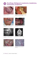
30 Swellings: Malignant neoplasms, lymphoma, metastatic
neoplasms 52
Lymphomas 53
Metastatic oral neoplasms 53
31 Ulcers and erosions: Local causes, drug-induced
ulcers 54
Local causes 54
Eosinophilic ulcer (traumatic eosinophilic granuloma;
traumatic ulcerative granulomatous disease) 54
Drug-induced ulcers (stomatitis medicamentosa) 55
32 Ulcers and erosions: Aphthae 56
33 Ulcers and erosions: Aphthous-like ulcers 58
Behçet syndrome (BS, Behçet disease) 59
v
9781405199858_1_pre.qxd
3/1/10
2:29
Page vi
34 Ulcers and erosions: Blood diseases, gastrointestinal
disorders 60
Blood diseases 60
Leukemias 60
Gastrointestinal disorders 61
Celiac disease (gluten sensitive enteropathy) 61
35 Ulcers and erosions: Infections 62
Hand, foot and mouth disease (HFM; vesicular stomatitis
with exanthem) 62
Herpangina 62
Bacterial infections 63
Acute necrotizing ulcerative gingivitis (Vincent disease;
acute ulcerative gingivitis, AUG, ANG, ANUG) 63
Syphilis 63
Gonorrhea 63
Tuberculosis 63
36 Ulcers and erosions: Erythema multiforme, toxic epidermal
necrolysis and Stevens-Johnson syndrome 64
Erythema multiforme 65
Toxic epidermal necrolysis (TEN, Lyell syndrome) and
Stevens-Johnson syndrome (SJS) 65
37 White lesions: Candidosis (candidiasis) 66
Acute pseudomembranous candidosis 66
Chronic hyperplastic candidosis (Candidal leukoplakia) 67
Chronic mucocutaneous candidosis (CMC) 67
38 White lesions: Keratosis, leukoplakia 68
Tobacco-related keratosis 69
Leukoplakia 69
39 White lesions: Hairy leukoplakia, lichen planus 70
Hairy leukoplakia 70
Lichen planus (LP) and lichenoid reactions 71
40 Salivary conditions: Salivary swelling and salivary excess 72
Salivary swelling 73
Saliva excess (sialorrhea, hypersialia, hypersalivation,
ptyalism) and drooling 73
41 Salivary conditions: Dry mouth 74
42 Salivary conditions: Sjögren syndrome 76
43 Salivary conditions: Sialolithiasis, sialadenitis 78
Sialolithiasis 78
Sialadenitis 78
Sialadenitis: Acute viral (mumps) 78
Sialadenitis: Acute bacterial ascending 79
Sialadenitis: Chronic bacterial 79
Sialadenitis: Recurrent parotitis of childhood 79
44 Salivary conditions: Neoplasms 80
Benign neoplasms (adenomas) 81
Malignant neoplasms 81
45 Salivary conditions: Mucoceles, sialosis 82
Mucoceles (mucous cyst; mucus extravasation phenomenon;
myxoid cyst) 83
Sialosis (sialadenosis) 83
46 Neck swelling 84
Discrete swellings in the neck 85
Cervical lymphadenopathy 85
Unexplained lymphadenopathy 85
Diffuse swelling of the neck 85
47 Neck swelling: Cervical lymphadenopathy in generalized
lymphadenopathy 86
Systemic infections 87
vi
Contents
48
49
50
51
52
53
54
55
56
57
58
59
60
Inflammatory disorders (not known to be infective) 87
Neoplastic causes 87
Drugs 87
Others 87
Neurological conditions: Bell palsy, and trigeminal sensory
loss 88
Bell palsy 89
Trigeminal sensory loss 89
Neurological conditions and pain: Local, referred and
vascular 90
Local causes of orofacial pain 90
Referred causes of orofacial pain 91
Vascular causes of orofacial pain 91
Neurological conditions and pain: Trigeminal
neuralgia 92
Trigeminal neuralgia 93
Neurological conditions and pain: Psychogenic (idiopathic
facial pain, idiopathic odontalgia and burning mouth
syndrome (oral dysesthesia)) 94
Persistent idiopathic, or unexplained (atypical) facial
pain (IFP) 95
Burning mouth “syndrome” (BMS, glossopyrosis,
glossodynia, oral dysesthesia, scalded mouth syndrome,
or stomatodynia) 95
Jaw conditions: Temporomandibular pain-dysfunction 96
Temporomandibular joint pain-dysfunction syndrome
(TMPD), myofascial pain dysfunction (MFD), facial
arthromyalgia (FAM), mandibular dysfunction,
or mandibular stress syndrome 97
Jaw bone conditions: Radiolucencies and radiopacities 98
Radiolucencies 98
Radiopacities 99
Mixed radiolucent and radiopaque lesions 99
Jaw bone conditions: Odontogenic diseases and cysts 100
Odontogenic infections 101
Odontogenic cysts 101
Jaw bone conditions: Odontogenic tumors 102
Benign odontogenic tumors 102
Malignant odontogenic tumors 103
Jaw conditions: Bone disorders 104
Non-neoplastic diseases 105
Neoplastic disorders 105
Jaw bone conditions: Fibro-osseous lesions 106
Osseous dysplasia, cemento-osseous dysplasia (COD),
periapical cemental or cemento-osseous dysplasia
(PCD) 107
Cherubism 107
Fibrous dysplasia 107
Hypercementosis 107
Ossifying fibroma (cemento-ossifying fibroma) 107
Paget disease of bone 107
Maxillary sinus conditions 108
Rhinosinusitis (sinusitis) 109
Neoplasms 109
Oral malodor 110
Human immunodeficiency virus (HIV) infection and
AIDS 112
Index 115
9781405199858_1_pre.qxd
3/1/10
2:29
Page vii
Preface
At a Glance books are used by students as introductory texts at the start
of a course, or for revision purposes in the run up to examinations.
The premise of the series is that the books should cover core information for undergraduates – and this information is broken down into
“bite-size chunks”. The books will therefore be the foundations for
use in practice.
Oral medicine and pathology are subjects which vary across the world
in their autonomy, strength, and official recognition, and whose remit
varies somewhat from the treatment of oral diseases in ambulatory
patients to the care of patients with a wide range of medical and surgical
disorders. Oral diseases are seen worldwide, and with increasing global
travel and migrations, conditions more common in the tropics are now
seen in most countries.
The aim of this book is to offer an overview of aspects of oral
medicine and pathology, with an emphasis on oral health care provision
in general practice. Intended outcomes are that, having read this book,
readers should be more aware of the immediate steps needed to make
the diagnosis and arrange patient management.
The authors are specialists and teachers in oral medicine and pathology from two continents, Europe and the Americas, whose focus
ranges from mainly in oral medicine to largely in oral pathology, whose
experience covers all these conditions and have between them taught in
North America, South America, Europe, the Middle East, and the
Antipodes. The authors have a common philosophy of recognizing that
the mouth is only part of the patient; that prevention and early diagnosis are crucial; that care of the patient is not simply attention to the oral
problem; that patients should be empowered in their health care; and
that the care is best delivered by a multidisciplinary team, of which oral
health care providers are an integral and important part.
The book includes the most important conditions in oral medicine
and pathology (those causing pain or affecting the mucosae, salivary
glands, or jaws) essential for students – those that are most common and
those that are dangerous or even potentially lethal, and is intended to
represent current practice at most major centers across the world. The
intimate connection with general medicine is highlighted by the various
eponymous conditions highlighted in this book. Being restricted by size
and cost, this book does not strive to be comprehensive or to include
material that is usually covered in courses in Applied Basic Sciences or
Human Disease, and does not include diseases of the teeth, or the basics
of history taking – only specific relevant points in the text.
Clinicians should bear in mind, however, that the history gives the
diagnosis in about 80% of cases. The history is followed by thorough
physical examination and often then by investigations, whereupon a
diagnosis or at least a differential diagnosis is formulated. Management
follows and is usually medical or surgical.
The diagnosis and management is discussed here and, in many cases,
practitioners who have the competence can undertake the care; in other
cases or if in doubt, it is better that the practitioner refers the patient to a
specialist in oral medicine, for an opinion, shared care, or for care by the
specialist. Reliable evidence for the effectiveness of many treatment
regimens is becoming available but data are sparse and there are thus
still many gaps in knowledge, especially in relation to many of the newer
biological response modifiers.
The material included in this book is all new, but we have drawn on
publications by the authors, especially from Scully C (2008) Oral and
Maxillofacial Medicine 2nd edition, Churchill Livingstone, Edinburgh,
Scully C, Flint SF, Porter SR, Moos K (2004) Atlas of Oral and
Maxillofacial Diseases 3rd edition, Taylor and Francis, London, and
Brown J and Scully C (2004) Advances in oral health care imaging.
Private Dentistry, 9, 1, 86–90; 2, 67–71 and 3, 78–79.
We thank our patients and also thank Dr Derren Ready (UCL) for
microbiology images, and Dr Jane Luker (Bristol) for checking our
advice on modern imaging.
Crispian Scully
Oslei Paes de Almeida
Jose Vicente Bagan
Pedro Diz Dios
Adalberto Mosqueda Taylor
vii
9781405199858_1_pre.qxd
3/1/10
2:29
Page viii
“What one knows, one sees”
Goethe (1749–1832)
viii
9781405199858_1_pre.qxd
3/1/10
2:29
Page ix
9781405199858_4_C01.qxd
1
1/1/04
6:15
Page 2
Examination of extraoral tissues
Figure 1.2 Hereditary hemorrhagic telangiectasia.
Figure 1.1 Down syndrome facies.
Figure 1.3 Cutaneous odontogenic fistula.
Figure 1.4a Lipoma.
Figure 1.4b Scan of lipoma.
Figure 1.5 Hereditary hemorrhagic telangiectasia
(same patient as in Figure 1.2).
2
Chapter 1 Examination of extraoral tissues
Figure 1.6 Purpura on arm.
9781405199858_4_C01.qxd
1/1/04
6:15
Page 3
This book does not include the basics of history taking, only specific
relevant points in the text. Bear in mind that the history gives the
diagnosis in about 80% of cases.
Following the history, during which the clinician will note the patient’s
conscious level, any anxiety, appearance, communication, posture,
breathing, movements, behavior, sweating, weight loss or wasting
(Figure 1.1), physical examination is indicated. This necessitates touching the patient; therefore, informed consent and confidentiality are
required, a chaperone available, and religious and cultural aspects should
be borne in mind (see Scully and Wilson).
Relevant medical problems may even be manifest in the fully clothed
patient – where changes affect the head and neck, cranial nerves, or
limbs. Therefore, while there is no rigid system for examination, the
clinician should ensure that these areas are checked.
Head and neck
Pupil size should be noted (e.g. dilated in anxiety or cocaine abuse,
constricted in heroin abuse).
Facial color should be noted:
• pallor (e.g. anemia)
• rashes (e.g. viral infections, lupus) (Figure 1.2)
• erythema (e.g. anxiety, alcoholism, polycythemia)
Swellings, sinuses or fistulas should be noted (Figure 1.3).
Facial symmetry is examined for evidence of enlarged masseter
muscles (masseteric hypertrophy) suggestive of clenching or bruxism.
Neck swellings should be elicited, followed by careful palpation of
lymph nodes (and salivary and thyroid glands), searching for swelling
and/or tenderness, by observing the patient from in front, noting any
obvious asymmetry or swelling (Figure 1.4a and b), then standing
behind the seated patient to palpate the nodes. Systematically, each
region needs to be examined lightly with the pulps of the fingers, trying
to roll the nodes against harder underlying structures.
Some information can be gained by the texture and nature of the
lymphadenopathy; nodes that are tender may be inflammatory (lymphadenitis), while those that are increasing in size and are hard, or fixed
to adjacent tissues, may be malignant.
Cranial nerves
The cranial nerves should be examined, in particular facial movement
and corneal reflex should be tested and facial sensation determined
(Table 1.1). Movement of the mouth as the patient speaks is important,
especially when they allow themselves the luxury of some emotional
expression.
Facial movement is tested out by asking the patient to:
• close their eyes; any palsy may become obvious, with the affected
eyelid failing to close and the globe turning up so that only the white of
the eye shows (Bell sign)
• close their eyes tightly against your attempts to open them, and note
the degree of force required to part the eyelids
• wrinkle their forehead, and check any difference between the two sides
• smile
• bare the teeth or purse the lips
• blow out the cheeks
• whistle
The muscles of the upper face (around the eyes and forehead) are
bilaterally innervated and thus loss of wrinkles on one-half of the
forehead or absence of blinking suggests a lesion in the lower motor
neurone.
Corneal reflex depends on the integrity both of the trigeminal and
facial nerves – a defect of either will give a negative response. This is
tested by gently touching the cornea with a wisp of cotton wool twisted
to a point. Normally, this procedure causes a blink but, provided that the
patient does not actually see the cotton wool, no blink follows if the
cornea is anesthetic from a lesion involving the ophthalmic division of
the trigeminal nerve, or if there is facial palsy.
Facial sensation is tested by determining the response to light touch
(cotton wool) and pin–prick (gently pricking the skin with a sterile pin,
probe or needle without drawing blood). It is important to test sensation
in all parts of the facial skin but the most common defect is numb chin,
due to a lesion affecting the mandibular division of the trigeminal.
Occasionally, a patient complains of hemifacial or complete facial
hypoesthesia (reduced sensation) or anesthesia (complete loss of sensation). If the corneal reflex is retained or there is apparent anesthesia over
the angle of the mandible (an area not innervated by the trigeminal
nerve), then the symptoms are probably functional (non-organic, i.e.
psychogenic).
Limbs
Hands may reveal rashes (Figure 1.5), purpura (Figure 1.6), pigmentation or conditions such as arthritis and Raynaud phenomenon. Finger
clubbing may reveal systemic disease. Nail changes may reveal anxiety
(nail biting), or disease such as koilonychia (spoon-shaped nails), in
iron deficiency.
The operator should then ensure that all relevant oral areas are
examined, in a systematic fashion.
Reference
Scully C and Wilson N (2006). Culturally Sensitive Oral Healthcare.
Quintessence, London.
Table 1.1 Cranial nerve examination.
Cranial nerve
Examination
I
II
Olfactory
Optic
III
Oculomotor
IV
V
Trochlear
Trigeminal
VI
VII
Abducens
Facial
Sense of smell for common odors
Visual acuity (Snellen types ±
ophthalmoscopy); nystagmus
Visual fields (by confrontation)
Pupil responses to light and
accommodation
Eye movements
Pupil responses
Eye movements
Sensation over face ± corneal reflex ±
taste sensation
Motor power of masticatory muscles;
jaw jerk
Eye movements
Motor power of facial muscles
Corneal reflex ± taste sensation
Hearing (tuning fork at 256 Hz)
Balance
Gag reflex
Taste sensation
Gag reflex
Motor power of trapezius and sternomastoid
Motor power of tongue
VIII Vestibulocochlear
IX
Glossopharyngeal
X
XI
XII
Vagus
Accessory
Hypoglossal
Examination of extraoral tissues
Chapter 1 3
9781405199858_4_C02.qxd
2
1/1/04
6:15
Page 4
Examination of mouth, jaws,
temporomandibular region and salivary glands
Figure 2.1a Portable miniature operative light.
Figure 2.1b ENT headlight.
Figure 2.2a Teeth and gingivae.
Figure 2.2b Buccal mucosa.
Figure 2.2c Buccal mucosa.
Figure 2.2d Palate.
Figure 2.2e Tongue dorsum.
Figure 2.2f Tongue ventrum and floor
of mouth.
LIPS
Herpes labialis
Cheilitis
Mucoceles
Granulomatous conditions
PALATE
Torus palatinus
Stomatitis nicotina
Pemphigoid
Pemphigus
Teeth
Soft palate
Uvula
T il
Tonsils
Tongue
Lips
BUCCAL MUCOSA
Leukoedema
Linea alba
Cheek biting
Aphthae
Lichen planus
Figure 2.4 Toluidine blue.
TONGUE
Geographic tongue
Glossitis
Burning tongue syndrome
Aphthae
Figure 2.5 Chemiluminescent illumination
Figure 2.3 Common diseases.
4
Chapter 2 Examination of mouth
system (ViziLite).
9781405199858_4_C02.qxd
1/1/04
6:15
Page 5
The lips are best first inspected. Complete visualization intraorally
requires a good light; this can be a conventional dental unit light, or
special loupes or ENT light (Figures 2.1a and b). If the patient wears
a dental appliance, this should be removed to examine beneath.
Mouth
The dentition and occlusion should be examined. Study models on a
semi- or fully-adjustable articulator may be needed. This is discussed in
basic dental textbooks.
All mucosae should be examined, beginning away from the focus of
complaint or location of known lesions. Labial, buccal, floor of the mouth,
ventrum of tongue, dorsal surface of tongue, hard and soft palate mucosae,
gingivae and teeth should be examined in sequence, recording lesions
on a diagram (Figures 2.2a–f). Lesions are described as in Table 2.1.
Some conditions are found only in, or typically in, certain sites
(Figure 2.3).
Mucosal lesions are not always readily visualized and, among
attempts to aid this, are:
• toluidine blue (vital) staining
• chemiluminescent illumination
• fluorescence spectroscopy and imaging
Toluidine blue staining (Figure 2.4) stains mainly pathological areas
blue. The patient rinses for 20 seconds with 1% acetic acid to clean the
area; then 20 seconds with plain water; then 60 seconds with 1% aqueous toluidine blue solution; then again 20 seconds with a 1% acetic acid;
and finally with water for 20 seconds.
Chemiluminescent illumination relies on fluorophores that naturally
occur in cells after rinsing the mouth with 1% acetic acid (Figure 2.5)
using excitation with a suitable wavelength.
Fluorescence spectroscopy is where tissues are illuminated with light,
and lesions change the fluorophore concentration and light scattering
and absorption, and their visibility may thus be enhanced.
Jaws
Jaw deformities or lumps may be best confirmed by inspection from
above (maxillae/zygomas) or behind (mandible), then palpated to
detect swelling or tenderness. The maxillary sinuses can be examined
by palpation for tenderness. X-ray (Waters projection), computed
tomography (CT), magnetic resonance imaging (MRI), transillumination or endoscopy can help.
Temporomandibular joint (TMJ)
Check:
• opening and closing paths
• opening extent (inter-incisal distance at maximum mouth opening)
• excursions
• joint noises
• condyles, by palpating them with a finger, via the external auditory
meatus
• masticatory muscles on both sides; masseters, by intraoral–extraoral
compression between finger and thumb, palpate the masseter bimanually
by placing a finger of one hand intraorally and the index and middle fingers
of the other hand on the cheek over the masseter; note any hypertrophy.
Temporalis: Check by direct palpation of the temporal region.
Palpate the temporal origin along the anterior border of the ascending
mandibular ramus, asking the patient to clench their teeth.
Lateral pterygoid (lower head): Check by placing a little finger up
behind the maxillary tuberosity (the “pterygoid sign”). Examine the muscle
indirectly by asking the patient to open the jaw against resistance and to
move the jaw to one side while applying gentle resistance.
Medial pterygoid: Check intraorally lingually to the mandibular
ramus.
Salivary glands
Oral dryness (scarce or frothy saliva; absence of saliva pool in floor of
mouth, reduced flow from Stensen duct, food residues; lipstick on teeth;
mirror sticks to mucosa) should be excluded. Salivary function assessment is discussed in Chapter 40.
Major salivary glands (parotids and submandibulars) should be
inspected and palpated for evidence of enlargement:
• Parotids are palpated using fingers placed over the glands in front of
the ears, to detect pain or swelling. Early enlargement of the parotid
gland is characterized by outward deflection of the lower part of the ear
lobe, which is best observed by looking at the patient from behind.
• Submandibulars are palpated bimanually between fingers inside the
mouth and extraorally.
Table 2.1 Main descriptive terms applied to orofacial and skin lesions.
Term
Meaning
Atrophy
Bulla
Reduction in tissue mass
Visible fluid accumulation within or beneath
epithelium (blister)
Scar: A permanent mark after healing
Closed cavity (epithelial lining)
Loss of superficial epithelial thickness (commonly
follows a blister)
Macular area of hemorrhage > 2 cm in diameter
(i.e. a bruise)
Loss of most of epithelial thickness (often follows
a blister)
Redness of mucosa (from atrophy, inflammation,
vascular congestion or increased perfusion)
Splitting off of epithelial keratin in scales or sheets
Formation of excessive fibrous tissue
Linear gap or slit
Abnormal connection, lined by epithelium between
two epithelium lined organs
Skin pustule or abscess
Death of tissue
Localized collection of blood
Heaped-up scar
Circumscribed alteration in color or texture, not raised
A colored lesion present from birth
Solid mass under/within mucosa or skin > 0.5 cm in
diameter
Circumscribed palpable elevation < 0.5 cm in diameter
Punctate hemorrhagic spot 1–2 mm in diameter
Elevated area of mucosa or skin > 0.5 cm in diameter
Visible accumulation of pus in epithelium
Fibrous tissue replacement of another tissue
Induration of submucosal and/or subcutaneous tissues
A pouch or cavity in any organ or tissue
Swelling caused by normal or pathological material
or cells
Loss of epithelium with loss of some underlying tissues
Area of edema, compressible and usually evanescent
Small (< 0.5 cm) visible fluid accumulation in
epithelium
Area of edema, compressible and usually evanescent
Cicatrix
Cyst
Desquamation
Ecchymosis
Erosion
Erythema
Exfoliation
Fibrosis
Fissure
Fistula
Furuncle
Gangrene
Hematoma
Keloid
Macule
Nevus
Nodule
Papule
Petechia
Plaque
Pustule
Scar
Sclerosis
Sinus
Tumor
Ulcer
Urticaria*
Vesicle
Weal*
*same
Examination of mouth Chapter 2 5
9781405199858_4_C03.qxd
3
1/1/04
6:16
Page 6
Investigations: Histopathology
Figure 3.1a Pemphigoid.
Figure 3.1b Erythroleukoplakia.
Figure 3.1d White sponge nevus. Typical
Figure 3.2 Biopsy kit.
perinuclear halo 40 ×.
Figure 3.1c White sponge nevus.
Figure 3.3 Scalpel and punch.
Figure 3.4 Excision
biopsy of a lump.
Table 3.1 Biopsy of oral lesions.
Type of lesion
Biopsy
Lesional area to biopsy
Preferred method
Blister
Incisional
Margin/perilesional or
whole blister
Scalpel
Carcinoma (suspected)
Margin
Erosion
Margin/perilesional
Erythroplakia
Lesion
Granulomatous
Deep
Leukoplakia
Any red area
Lichenoid
Punch or scalpel
Box 3.1 Indications for
biopsy
Indications for biopsy include
lesions that:
• have neoplastic or potentially
malignant features
• are enlarging
• persist > 3 weeks
• are of uncertain etiology
• fail to respond to treatment
• cause concern.
Lesion
Lump (mucosal)
Excisional
Mucocele
Excisional
Pigmented
Excisional
Salivary major gland swelling
FNAC or FNAB
Salivary minor gland swelling
Palate – incisional
Lip – excisional
Labial gland biopsy for
xerostomia diagnosis
– incisional
Scalpel
Ulcer
Incisional
Margin/perilesional
Scalpel
Scalpel
US guidance
Figure 3.5 Brush biopsy (oral CDx).
6
Chapter 3 Investigations: Histopathology
9781405199858_4_C03.qxd
1/1/04
6:16
Page 7
Having taken a careful history and completed the clinical examination,
the clinician is often in a position to formulate the diagnosis, or at least
a list of differential diagnoses. In the latter case, the diagnosis is provisional, and another opinion (e.g. specialist referral) or investigations
may be necessary to reach a firm diagnosis.
Informed consent and confidentiality is required for all investigations.
Biopsy is the removal of tissue usually for diagnosis by histopathological examination (Box 3.1). Practitioners who have the competence
and confidence can undertake mucosal biopsy but in other cases it may
be better to refer.
Methods for biopsy include (Table 3.1):
• Incisional biopsy – sampling using a disposable tissue punch (a
round-shaped knife) or scalpel. Punches are light, easy to use and less
likely than a scalpel to damage anyone. Most biopsies can be performed
with a 3 or 5 mm punch, without suturing.
• Excisional biopsy – scalpel or laser removal of the whole lesion.
• Needle biopsy (mainly for lymph nodes and lumps):
— fine-needle cutting biopsy (FNCB) using wide-bore needle
— fine-needle aspiration biopsy (FNA or FNAB) or cytology
(FNAC), using 22 gauge needle, sometimes as ultrasound-guided
fine-needle aspiration cytology (US-FNAC).
— curettage; scraping (e.g. from a bone cavity).
• Remove the required tissue.
• Snap-freeze specimen in liquid nitrogen or place in Michel solution
if for immunostaining; if for other staining, place it in 10% neutral
buffered formalin (Table 3.2).
• Label specimen and request form carefully and follow the postal
regulations if the specimen is to be mailed.
• Suture if necessary, using a fine needle and resorbable suture (e.g.
Polyglycolic acid suture (Vicryl* Rapide)), or black silk (Figure 3.4).
Direct immunofluorescence is a qualitative technique used to detect
immune deposits (antibodies and/or complement) in the tissues, using
fluorescein stain which fluoresces apple green under ultraviolet light,
and is useful in the diagnosis, particularly of bullous disorders.
Indirect immunofluorescence is a qualitative and quantitative technique used to detect immune components (circulating antibodies and/or
complement) in the serum. It is a two or more stage technique requiring
patient serum and animal tissue.
Other techniques
Immunohistochemistry, polymerase chain reaction (PCR) in situ
hybridization (ISH), and fluorescent ISH (FISH) are also used, especially in diagnosis of infections or neoplasms.
Brush biopsy
Mucosal biopsy
In most incisional biopsies it is preferrable to sample the lesional margin or perilesional area, as sampling an ulcer is rarely helpful since the
epithelium has been lost. In suspected malignant mucosal lesions it can
be difficult to decide which is the best part to biopsy but, generally, red
areas (erythroplakia) are where dysplasia is most likely and therefore
are best sampled (Figures 3.1a–d). It can be helpful to stain the mucosa
before biopsy with toluidine blue:
• Give a local analgesic (Figure 3.2).
• Use a scalpel when a bullous disorder is suspected as a punch might
tear the fragile tissue (Figure 3.3).
• Hold the tissue with suture or forceps to avoid squeezing and causing
crush artifacts.
This uses a cytobrush as a sampling device to reach deeper layers
of the oral epithelium (Figure 3.5), evaluating the cells obtained by
computer-assisted image analysis. Major limitations are cost and high
false-negative rates.
Labial salivary gland biopsy
• Give local analgesia.
• Make a linear mucosal incision to one side of the midline in the lower
labial mucosa or an X-shaped incision over the swelling which overlies
the salivary gland.
• Excise at least four lobules of salivary gland.
• Suture the wound if necessary.
Table 3.2 Frequently used tissue stains.
Stain
Constituents
Stains
Used for
Congo red
H&E
Sodium salt of benzidine diazo
Hematoxylin (basic stain)
Eosin (acidic stain)
Diagnosis of amyloidosis
Most histopathology
Mucicarmine
Carmine and aluminium hydroxide
Amyloid apple-green under polarized light
Cell nuclei (basophilic) stain blue/purple
Cytoplasm, connective tissue and other
extracellular substances (eosinophilic)
stain pink/red
Acid mucins stain pink
Papanicolaou
(Pap) staining
PAS
Combination of hematoxylin, eosin
Y, Orange G, Light Green SF and
Bismark brown
Periodic acid Schiff
Nuclei stain blue, cytoplasm of basal cells
light blue, intermediate cells orange-red
and superficial yellow
Carbohydrates stain purple
Prussian blue
Potassium ferrocyanide and acid
Iron stains blue or purple
Romanowsky stains (Wright,
Jenner, Leishman, Giemsa)
Silver staining
Eosin Y, methylene blue (methanol
and glycerol)
Silver nitrate
Leukocytes stain purple
Sudan stains
Van Gieson stain
Sudan III, IV and Black B, Oil Red O
Picric acid and acid fuchsin
Lipids stain black or red
Collagen stains red
Muscle stains yellow
Nuclei stain black
Proteins and DNA stain brown/black
Muco-epidermoid
carcinoma, Cryptococcus
Smears for cytopathology
Fungal hyphae, glycogen,
mucus
Iron in bone marrow and
other biopsy specimens
Inspection of blood cells
Fungi, some bacteria
(syphilis, rhinoscleroma),
collagen, reticulin
Lipid deposits
Collagen in vessels, liver
and bone marrow
Investigations: Histopathology
Chapter 3 7
9781405199858_4_C04.qxd
4
1/1/04
6:18
Page 8
Investigations: Microbiology
Figure 4.1b Candida hyphae PAS staining.
Figure 4.1a Unstained Candida albicans.
Figure 4.1c Candidosis (silver stain).
Figure 4.1d Candida colonies.
Table 4.1 Common microbiological stains.
Figure 4.1e Histoplasmosis silver impregnation.
Figure 4.1f CMV immunohistochemistry.
8
Chapter 4 Investigations: Microbiology
Stain
Main components
Main uses
Acid fast (Ziehl-Neelsen
and Kinyoun stains)
Carbol fuchsin and
methylene blue
Differentiates bacteria with
waxy cell walls, e.g.
Mycobacterium tuberculosis,
Mycobacterium leprae,
and Mycobacterium
avium-intracellulare complex
from those that do not
Gomori methenamine
silver (GMS)
Silver
Stains carbohydrates in fungi
Gram
Crystal violet, Gram’s
iodine and safranin
Stains Gram-positive bacteria
(e.g. Staphylococci), and
Gram-negative bacteria (e.g.
Escherichia coli) based on
differences in cell wall
structure
Periodic acid Schiff
(PAS)
Periodic acid selectively
oxidizes glucose, creating
aldehydes that react with
Schiff to produce a purplemagenta colour
Stains carbohydrates in fungi
9781405199858_4_C04.qxd
1/1/04
6:18
Page 9
Informed consent and confidentiality is required for all investigations.
Testing for infections can be a very sensitive issue, especially in the
case of Human Immunodeficiency Virus (HIV) infections, tuberculosis
and sexually transmitted infections (e.g. Syphilis, Herpes, Anogenital
warts, Gonorrhea). HIV testing in particular remains voluntary and
confidential, and patients must be counseled properly beforehand. It has
been recommended in the UK that patients should be offered and
encouraged to accept HIV testing in a wider range of settings than is
currently the case; that patients with specific indicator conditions
should be routinely recommended to have an HIV test; and that all doctors, nurses and midwives should be able to obtain informed consent
for an HIV test in the same way that they currently do for any other
medical investigation (The British HIV Association; British Association
of Sexual Health and HIV; and British Infection Society).
Microbiological diagnosis is based on either demonstration of the
micro-organism or its components (antigens or nucleic acids), or on the
demonstration in the serum of a specific antibody response.
Whenever an early diagnosis is important for the institution of
therapy or some other measure (e.g. infection control), methods that
demonstrate the organism or its components are best used as results are
more speedily obtained.
Micro-organisms can be demonstrated directly in samples or tissues
by microscopy using various stains (Table 4.1).
Direct cytological smears and histopathology are sometimes used, as
is growth after inoculation in cultures (Figures 4.1a–f), but rapid and
sensitive techniques for detecting antigens and nucleic acids have very
much come to the fore (Table 4.2). Antigen tests use, for example,
ELISA (Enzyme-Linked ImmunoSorbent Assay), latex agglutination,
or immunofluorescence. Nucleic acids are usually detected by polymerase chain reaction (PCR) or variants on that technology.
Microbial specimen handling is important to ensure reliable results.
Specimens should be collected before antimicrobials are started and
always handled and labelled as a biohazard. If pus is present, a sample
should be sent in a sterile container, in preference to a swab. If tuberculosis is suspected, this must be clearly indicated on the request form. If
the microbiological specimen cannot be dealt with within two hours,
the swab should be placed in transport medium and kept in the refrigerator at 4°C (not a freezer) until dealt with by the microbiology department. Swabs for viral infections must be sent in viral transport medium;
dry swabs are no use. Acute and convalescent serum samples should be
taken for serological diagnosis of infections. The convalescent serum is
collected 2–3 weeks after the acute illness.
Laboratory tests available to help the diagnosis of oral diseases are
shown in Table 4.2, but many infections are diagnosed provisionally
on clinical grounds. Laboratory confirmation may help diagnosis
and management and, in the case of HIV, syphilis and tuberculosis is
mandatory.
Reference
The British HIV Association; British Association of Sexual Health
and HIV; and British Infection Society. />file1031097.pdf. Accessed 24 March 2009.
Table 4.2 Laboratory diagnostic tests for oral microbial infections*.
Micro-organism
Candidosis
Coxsackie
Cytomegalovirus
(CMV; HHV-5)
Epstein-Barr
virus (EBV)
Diagnostic tests
Main
Other tests
Culture in Saboraud dextrose agar
for identification
Coxsackie IgM
CMV IgM
Speciation tests such as germ tube tests
and culture on CROM agar
EBNA IgG
Monospot (Paul-Bunnell heterophile
antibody test) is 98% sensitive
False negatives common in patients
< 5 years (when anti-VCA IgM should
be assayed)
Mouth washing for culture
Scrapings of lesions reveal HSV by EM
and multinucleate giant
Tzanc cells
Herpes simplex
viruses (HSV)
Immunofluorescence testing (IF) and enzyme
linked immunosorbent assays (ELISA),
Immunostaining will give same day results
Nucleic acid (PCR)
Herpes varicella-zoster
virus (VZV)
Immunostaining
Nucleic acid (PCR)
Mumps
Syphilis
Mumps IgM
Serology
Non-specific Reagin tests (VDRL and RPR tests)
Specific tests for treponemal antibodies
(TPI, FTA-Abs, hemagglutination tests
(HATTS and MHA-TP))
Fluorescence staining (auramine-rhodamine) or
Ziehl-Neelsen staining or nucleic acid probes
Tuberculosis
API kits give more definitive
identification
Immunostaining (Figure 4.1f)
Scrapings of lesions reveal VZV by EM
and multinucleate giant
Tzanc cells
Serum amylase raised
Fluorescent antibody staining of smear
Nucleic acid amplification tests
(NAAT) PCR to detect TB DNA
Interferon-γ (interferon-gamma) release
assays (IGRAs)
Serology: HSV IgG and IgM in
primary infection
HSV specific IgG alone in
reactivation
Western blot is confirmatory
Serology: VZV IgM in primary
and recurrent infections
Mumps IgG later
Dark ground microscopy
Culture
MB/BacT, BACTEC 9000,
and the Mycobacterial Growth
Indicator Tube (MGIT)
ELISA
Adenosine deaminase
* See Chapter 60 for HIV-testing.
Investigations: Microbiology
Chapter 4 9
9781405199858_4_C05.qxd
5
1/1/04
6:19
Page 10
Investigations: Imaging
Figure 5.3 Gorlin-Goltz syndrome: keratocystic odontogenic tumor.
Figure 5.1 Bone Scan:
Mandibular squamous
cell carcinoma.
Figure 5.2a CT: osteosarcoma.
Figure 5.4 Periapical
radiography: periapical
granuloma.
Figure 5.2b CT: ameloblastoma.
Figure 5.8 Sialogram in sialolithiasis.
Figure 5.5 MRI: head and neck.
Figure 5.6 MRI: pleomorphic adenoma T1.
Figure 5.9 Ultrasound scan. Submandibular salivary
Figure 5.7 Salivary scintiscan normal.
10
Chapter 5 Investigations: Imaging
gland. Courtesy of J. Brown, C. Scully and Private
Dentistry.
9781405199858_4_C05.qxd
1/1/04
6:19
Page 11
Informed consent and confidentiality is required for all investigations.
Because of the adverse effects of ionising radiation and the cumulative effect of radiation hazard, clinicians requesting examination or
investigation using X-rays must satisfy themselves that each investigation is necessary and that the benefit outweighs the risk.
Ultrasound and magnetic resonance imaging avoid radiation hazards.
Angiography is a relatively high radiation dose invasive technique and
MRI angiography is often used in its place. Angiography use should
first be discussed with a radiologist, but it can be useful in diagnosis of:
• vascular anomalies or tumors
• parotid gland deep lobe tumors
Arthrography has been used in the past for diagnosis of suspected TMJ
internal derangements but, in most centres, it has been superceded by MRI.
Bone scintiscanning is a high radiation dose technique and often
other imaging modalities can be more appropriately used. It is essential
to discuss with a radiologist prior to referring the patient, but it can be
useful in diagnosis of:
• bone invasion or metastases (Figure 5.1)
• condylar or coronoid hyperplasia
• fibro-osseous disease
• other bone disease
Computed axial tomography (CT or CAT) shows the bone and teeth
white, and can be useful in diagnosis of:
• hard tissue lesions (Figures 5.2a and b)
• paranasal sinuses diseases
• lesions in complex anatomical areas inaccessible to conventional
radiographs
• tumor spread, to exclude cranial base or intracranial pathology
• TMJ disorders (Cone Beam Computed Tomography (CBCT) is
especially helpful)
Disadvantages of CT are mainly that it:
• gives a fairly high radiation exposure (CT of the head can give the
equivalent exposure to about 100 chest radiographs)
• is expensive
• gives artfacts (star artfacts) when imaging the jaws if amalgam, other
metal restorations or implants are present
Cone beam CT is becoming widely utilised for imaging bone/dental
pathology of the jaws but is not recommended for imaging soft tissue
lesions. It has the advantage of a lower radiation dose to the patient than
conventional CT.
Dental panoramic tomography (DPT; or orthopantomography [OPTG])
is a specialized tomographic technique used to produce a flat representation of both jaws, offering a good overview of the dentition, maxillary
sinuses, mandibular ramus and temporomandibular joints. It can
demonstrate jaw lesions (Figure 5.3) and generalized pathology such as
periodontitis, but is subject to considerable and unpredictable geometric distortion, is greatly affected by positioning errors and has relatively
low spatial resolution compared with intraoral radiographs. DPT also:
• lacks the detail obtained by intraoral radiography such as periapical films
• does not show caries until it is has progressed to dentine
• does not show detail in the anterior jaws, where the spine is superimposed
• always shows ghost shadows
• images only those tissues within the focal trough.
It has no radiation dose saving advantage over full mouth radiographs since a tissue weighting factor for salivary glands has been
included in the calculations of effective dose by the International
Commission on Radiological Protection (ICRP).
Intraoral radiography, including periapical, bitewing and occlusal
projections, is the basic imaging used for dental pathology and has
higher spatial resolution which allows detection of small carious
lesions and periapical radiolucencies that may not always be detectable
with DPT. It can be useful in diagnosis of:
• approximal caries
• other coronal pathology
• tooth root pathology
• periapical pathology (abscess, granuloma, cyst, etc.) (Figure 5.4)
• adjacent bone pathology.
Magnetic resonance imaging (MRI) does not use ionising radiation,
the bone shows black, and it gives good images of soft tissues (Figures 5.5
and 5.6) and is the imaging modality of choice to aid in the diagnosis
and management of:
• soft tissue lesions, including malignant lesions (e.g. carcinoma,
lymphoma) (Figure 5.6)
• temporomandibular joint disease
• trigeminal neuralgia
• idiopathic facial pain
• children and young people (rather than CT).
The disadvantages of MRI are that it is:
• not as good as CT for imaging bone lesions
• liable to produce image artifacts where metal objects are present
(dental restorations, orthodontic appliances, metallic foreign bodies,
joint prostheses, implants, etc.)
• expensive.
Contraindications to MRI include:
• implanted electric devices (e.g. heart pacemakers, cardiac defibrillators, nerve stimulators, cochlear implants)
• intracranial vascular clips, if these are ferromagnetic
• prosthetic cardiac valves containing metal
• obesity (weight limit on gantry and size of scanner)
• claustrophobia (unless open scanner available)
Salivary scintiscanning is now very rarely used, since ultrasound has
become the imaging modality of choice for assessing salivary glands
(Figure 5.7). It can help examine all salivary glands simultaneously,
and is useful in the diagnosis of salivary:
• ductal obstruction
• aplasia
• neoplasms
• Sjögren’s syndrome.
Sialography examines one major gland only (Figure 5.8) but can be
useful in diagnosis of:
• salivary duct obstruction
• intermittent salivary swelling
• recurrent salivary infections.
Contraindications:
• allergy to radiocontrast media (e.g. iodides)
• acute salivary infection.
Ultrasound scanning (US) is non-invasive use of 3.5–10 mHz frequency sound waves, and is the first-line imaging modality to use in:
• diagnosis of soft tissue swellings (e.g. lymph nodes, thyroid or salivary glands) (Figure 5.9)
• diagnosis of soft tissue hard inclusions (e.g. calcification, foreign bodies)
• assisting fine needle aspiration biopsy (ultrasound guided FNA or
FNAB) as it improves the diagnostic yield.
Doppler ultrasound is also useful for investigating vascularity of
lesions. There are no contraindications to ultrasound, but disadvantages
are that it:
• is user dependent
• may fail to visualize the deep extent of a lesion
Investigations: Imaging Chapter 5 11
9781405199858_4_C06.qxd
6
1/1/04
6:20
Page 12
Investigations: Blood tests
Figure 6.1b Pernicious anemia
(resolved after 10 days therapy).
Figure 6.1a Pernicious anemia.
Informed consent and confidentiality is required for all investigations.
Blood contains cells (erythrocytes, leukocytes, platelets), proteins
(antibodies, enzymes, etc.) and other substances. Blood tests help determine disease states, but should be the appropriate test and requested
only when clinically indicated. Furthermore, abnormal “blood results”
do not always mean disease. Apart from technical errors which are
possible, some tests assays for autoantibodies, for example (which may
be indicated in suspected bullous diseases or Sjögren’s syndrome) may
show abnormalities (in this case autoantibodies), but these do not always
indicate disease and their absence does not necessarily exclude it. There
is also a danger of needlestick injury.
Whole blood is used for full blood count (FBC; or full blood picture,
FBP) and must be anticoagulated (EDTA in the collection tube). FBP
may identify anemia (e.g. in glossitis, burning mouth syndrome, or oral
ulceration) (Figures 6.1a and b). The white blood cell count (WBC or
WCC) and blood film may reveal leukemia or infection such as infectious mononucleosis (Figures 6.2 and 6.3), and a platelet count can help
where bleeding tendency is suspected.
A sickle test should be requested for patients of African heritage
(ideally also for those of Mediterranean and Asian origin).
Serum, obtained by collecting whole blood without anticoagulant, is
used for assaying antibodies, which can help diagnose infections and
autoimmune disorders, and for most biochemical substances (e.g. “liver
enzymes”).
Table 6.1 shows the interpretation of some blood tests.
Referring a patient for specialist opinion
Figure 6.2 Leukemia presenting with
gingival lesions.
It is the responsibility of clinicians to recognize the early signs of
serious disease and to direct the patient to the appropriate specialist
for a second opinion and include any relevant investigation results.
Essential details of a referral letter include:
Name and contact details of the patient
including age, address and day-time telephone number.
Name and contact details of the referring and other clinicians
History of present complaint
brief details and description of the nature and site of lesion(s).
Urgency of referral
Social history
Medical history
Special requirements
e.g. for interpreter, sign language expert or special transport (Scully
and Porter, 2007).
Reference
Figure 6.3 Blood film from
infectious mononucleosis.
12
Chapter 6 Investigations: Blood tests
Scully C and Porter SR (2007). Referrals in oral medicine. Dental Update,
Jul–Aug; 34 (6); 340–342, 345–346, 348–350.
9781405199858_4_C06.qxd
1/1/04
6:20
Page 13
Table 6.1 Interpretation of blood test resultsa.
Blood cells
Level ↑b
Level ↓b
Hemoglobin
Hematocrit (packed cell volume or PCV)
Mean cell volume (MCV)
MCV = PCV/RBC
Mean cell hemoglobin (MCH)
MCH = Hb/RBC
Red cell count (RBC)
Reticulocytes
White cell count (total)
Neutrophils
Polycythemia
Dehydration
Vitamin B12 or folate deficiency, liver disease,
alcoholism
Pernicious anemia
Anemia
Polycythemia
Hemolytic states
Infection, inflammation, leukemia, trauma, pregnancy
Pregnancy, exercise, infection, trauma, malignancy,
leukemia
Some infections, leukemia, lymphoma
Allergic disease, parasitic infestations
Myeloproliferative disease
Anemia
Chemotherapy, bone marrow disease
Some infections, bone marrow disease, drugs
Some infections, drugs, bone marrow disease
Prostate cancer
Liver disease, infectious mononucleosis
Dehydration
—
Hypothyroidism, hypophosphatasia
Liver disease, malnutrition, malabsorption, nephrotic
syndrome, myeloma
—
—
—
—
—
Hypoparathyroidism, renal failure, rickets, nephrotic
syndrome, chronic renal failure, lack of vitamin D,
pancreatitis
Malnutrition, hyperthyroidism
Lymphocytes
Eosinophils
Platelets
Biochemistry (on plasma or serum)
Acid phosphatase
Alanine transaminase (ALT)
Albumin
Alkaline phosphatase
Amylase
Angiotensin converting enzyme
Aspartate transaminase (AST)
Bilirubin (total)
Calcium
Puberty, pregnancy, bone disease
Pancreatic disease, mumps
Sarcoidosis
Liver disease, myocardial infarct, trauma
Liver or biliary disease, hemolysis
Primary hyperparathyroidism, bone tumors,
sarcoidosis
Cholesterol
Complement (C3)
Hypercholesterolemia, pregnancy, hypothyroidism,
diabetes, nephrotic syndrome, liver or biliary disease
Trauma; surgery; infection
Complement (C4)
—
C1 esterase inhibitor
Erythrocyte sedimentation rate (ESR)
Ferritin
—
Pregnancy, many diseases
Liver disease, hemochromatosis, leukemia, lymphoma,
thalassemia
Folic acid therapy
Folic acid
Free thyroxine index (FTI) (serum T4
and T3 uptake)
Gammaglutamyl transpeptidase (GGT)
Globulins (total) (see also under protein)
Glucose
Total immunoglobulins
IgG
IgA
IgM
IgE
Percent carbohydrate-deficient transferrin
Phosphate
Plasma viscosity
Potassium
Protein (total)
Sodium
Steroids (corticosteroids)
Thyroxine (T4)
Urea
Vitamin B12
a
Hyperthyroidism
Alcoholism, obesity, liver or renal disease, myocardial
infarct
Liver disease, multiple myeloma, autoimmune disease,
chronic infections
Diabetes mellitus, pancreatitis, hyperthyroidism,
hyperpituitarism, Cushing disease, liver disease
Liver disease, infection, sarcoidosis, connective tissue
disease
Myelomatosis, connective tissue disorders
Alcoholic cirrhosis
Primary biliary cirrhosis, nephrotic syndrome,
parasites, infections
Allergies, parasites
Alcoholism
Renal failure, bone disease, hypoparathyroidism,
hyper-vitaminosis D
Pregnancy, many diseases
Renal failure, Addison disease, ACE inhibitors,
potassium supplements
Liver disease, multiple myeloma, sarcoid, connective
tissue diseases
Dehydration, Cushing disease
Cushing disease, some tumors
Hyperthyroidism, pregnancy, oral contraceptive
Renal failure, dehydration, gastrointestinal bleed
Liver disease, leukemia, polycythemia rubra vera
Iron deficiency, thalassemia, chronic disease
Iron deficiency, thalassemia
Some infections (e.g. HIV), drugs
Some immune defects
Leukemia, drugs, HIV, autoimmune
Liver disease, immune complex diseases, e.g. lupus
erythematosus
Liver disease, immune complex diseases, hereditary
angioedema
Hereditary angioedema
—
Iron deficiency
Alcoholism, dietary deficiency or malabsorption,
hemolytic anemias, phenytoin
Hypothyroidism
—
Chronic lymphatic leukemia, malnutrition, protein losing
states
Hypoglycemic drugs, Addison disease, hypopituitarism,
liver disease
Immunodeficiency, nephrotic syndrome, enteropathy
Immunodeficiency, nephrotic syndrome
Immunodeficiency
Immunodeficiency
—
—
Hyperparathyroidism, rickets, malabsorption syndrome
—
Vomiting, diabetes, Conn syndrome, diuretics, Cushing’s
disease, malabsorption, corticosteroids
Pregnancy, nephrotic syndrome, malnutrition,
enteropathy, renal failure, lymphomas
Cardiac failure, renal failure, Addison’s disease, diuretics
Addison’s disease, hypopituitarism
Hypothyroidism, nephrotic syndrome, phenytoin
Liver disease, nephrotic syndrome, pregnancy, malnutrition
Pernicious anemia, gastrectomy, Crohn’s disease, vegans
Adults unless otherwise stated. b A selection only.
Investigations: Blood tests
Chapter 6 13
9781405199858_4_C07.qxd
7
1/1/04
6:20
Page 14
Anatomical variants and developmental
anomalies
Figure 7.1a Fordyce spots.
Figure 7.1b Fordyce spots.
Figure 7.1c Fordyce spots.
Figure 7.3 Torus palatinus.
Figure 7.4 Torus mandibularis.
Figure 7.5 Stafne bone cavity.
Figure 7.6 Bifid uvula.
Figure 7.2 Fissured tongue.
Figure 7.7a Folliate papillitis.
Figure 7.7b Folliate papillitis.
Anatomical features or developmental anomalies that may be noticed
by patients and cause concern include:
• Fordyce spots (Figures 7.1a–c)
• fissured tongue (Figure 7.2)
• torus palatinus (Figure 7.3)
• torus mandibularis (Figure 7.4)
• Stafne bone cavity (Figure 7.5)
• unerupted teeth; mainly third molars (Figure 7.5), second premolars,
and canines
• pterygoid hamulus; may give rise to concern about an unerupted tooth
• bifid uvula; symptomless (Figure 7.6), but may overlie a submucous
cleft palate
14
Chapter 7 Anatomical variants
Figure 7.8 Lingual varicosities.
• papillae:
– incisive; may bother the patient if traumatized
– parotid (orifice of Stensen duct); may occasionally be traumatized
by biting or an orthodontic or other appliance
– lingual foliate; occasionally become inflamed (papillitis) and
clinically mimic carcinoma (Figures 7.7a and b)
– retrocuspid; found on the lingual gingiva in the mandibular canine
region, it resembles the incisive papilla
– leukoedema; a normal variation more prevalent in people who
have dark skin, in which there is a white-bluish tinge of the buccal
mucosa that disappears when the cheek is stretched
• lingual varicosities (Figure 7.8).
9781405199858_4_C07.qxd
1/1/04
6:20
Page 15
Fordyce spots (“Fordyce granules”)
Definition: Small, painless, raised, white or yellowish spots or bumps 1
to 3 mm in diameter seen beneath the buccal or labial mucosa. Similar
spots may be seen on genitals (penis or labia).
Prevalence (approximate): Seen in probably 80% of the population.
Age mainly affected: After puberty.
Gender mainly affected: M > F.
Etiopathogenesis: These are sebaceous glands containing neutral
lipids similar to those found in skin sebaceous glands, but not associated with hair follicles.
cystic, it is a congenital defect typically measuring less than 2 cm,
usually filled with fat but may also contain salivary tissue.
Torus palatinus
Definition: A developmental benign exostosis in the midline of hard palate.
Prevalence (approximate): Up to 20% of the population; seen especially in Asians and Inuits.
Age mainly affected: After puberty.
Gender mainly affected: F > M (2:1).
Etiopathogenesis: Developmental exostosis.
Diagnostic features
Diagnostic features
History: Often not noticeable until after puberty (although they are present histologically).
Clinical features: Usually seen in the buccal mucosa, particularly
inside the commissures, and sometimes in retromolar regions and
upper lip. They appear more obvious in males, patients with greasy
skin and older people, and they may be increased in some rheumatic
disorders.
Differential diagnosis: Thrush or lichen planus. Occasionally they
may be mistaken for leukoplakia or Koplik spots (measles).
Diagnosis is clinical: investigations are rarely required.
History: Symptomless unless ulcerated by trauma.
Clinical features: Most tori occur in the palate, midline and extend
symmetrically to either side. Size (most are < 2 cm diameter) and shape
(lobular, nodular or irregular) are variable. The lesion is painless, and
the surface is bony hard and the overlying mucosa normal and typically
of normal color unless traumatized.
Differential diagnosis: Unerupted teeth, cysts or neoplasms.
The diagnosis is usually clinical but radiography may help.
Management
Management
Tori should usually be left alone. Surgery (excision or reduction) is
indicated only if causing severe difficulties with dentures.
The spots may become less prominent if isotretinoin is given. CO2 laser
and photodynamic therapy are reportedly effective therapies but no
treatment is indicated, only reassurance.
Prognosis
Prognosis
Torus mandibularis
Excellent: They are of cosmetic concern only.
Fissured tongue (scrotal or plicated tongue)
Definition: A tongue with fissures on the dorsum.
Prevalence (approximate): About 5% of population.
Age mainly affected: More noticeable with increasing age.
Gender mainly affected: M = F.
Etiopathogenesis: Hereditary, a fissured tongue is found in many
normal persons but is more often seen in psoriasis, Down syndrome
(trisomy 21), Job syndrome (hyper-IgE and immunodeficiency) and
Melkersson-Rosenthal syndrome (Chapter 27).
Diagnostic features
History: Usually asymptomatic. However, it is often complicated by
geographic tongue, or the tongue becomes sore for no apparent reason.
Clinical features: Multiple fissures on the dorsum of the tongue.
Differential diagnosis: Lobulated tongue of Sjögren syndrome or
chronic mucocutaneous candidosis.
Diagnosis is clinical: investigations are rarely required. Blood tests
are optional if the tongue is sore.
Excellent.
Definition: Bony lumps usually lingual to mandibular premolars.
Prevalence (approximate): Up to 6%; seen especially in Asians and
Inuits.
Age mainly affected: After puberty.
Gender mainly affected: F = M.
Etiopathogenesis: Developmental exostosis but bruxism and parafunction may play a role.
Diagnostic features
Tori are symptomless unless traumatized.
Clinical features: Tori are typically bilateral bony hard lumps, with
normal overlying mucosa and typically of normal color or yellowish.
They are painless, and the size and shape are variable – but may be
lobular, nodular or irregular.
Differential diagnosis: Unerupted teeth, cysts or neoplasms.
The diagnosis is usually clinical but radiography may help.
Management
Tori should usually be left alone. Surgery (excision or reduction) is
indicated only if causing severe difficulties with dentures.
Management
No treatment is indicated or available.
Prognosis
Excellent.
Prognosis
Excellent.
Stafne cyst or bone cavity
This is a lingual, mandibular, focal, bone concavity, classically in the
submandibular fossa, below the inferior alveolar canal and close to the
mandible inferior margin. Although this radiolucency may appear to be
Varicosities
Oral varicosities present as purplish blue spots, nodules or ridges,
usually asymptomatic, most commonly involving the lingual veins or
vessels of the ventral surface of the tongue and the floor of the mouth.
Often seen in older people, they are benign and inconsequential. Some
cases have been successfully treated with cryosurgery or sclerotherapy.
Anatomical variants
Chapter 7 15