Ebook Medical microbiology and infection at a glance (4th edition) Part 2
Bạn đang xem bản rút gọn của tài liệu. Xem và tải ngay bản đầy đủ của tài liệu tại đây (15.47 MB, 64 trang )
29
Virus structure, classification and
antiviral therapy
Virus proteins
Possible structural components
Envelope from host cell
Nucleoprotein
Capsid forming virus structure
Nucleic acid
ds DNA
Herpesvirus
ss DNA
+ss RNA
–ss RNA
+ss RNA
Parvovirus
Picornaviridae
Paramyxoviridae
Retroviridae
(HIV)
Calicivirus
Orthomyxoviridae
Pox virus
DNA
RNA
Togavirus & flavivirus
Arenavirus
Coronavirus
Adenovirus
+ss RNA
Hepadnavirus
+ss RNA
Viral
proteins
New
virus
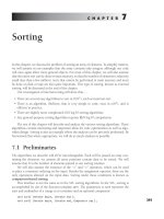
Viral classification
Viral classification is based on the nucleotides in the virus, its mode
of replication, the structure and symmetry of the structural proteins (capsids) and the presence or absence of an envelope.
Genetic material and replication
DNA viruses
• Double-stranded DNA viruses include poxviruses, herpesviruses, adenoviruses, papovaviruses and polyomaviruses.
• Single-stranded DNA viruses include parvoviruses.
Rhabdoviridae
–ss RNA
+ss RNA
New
virus
Host
membrane
Viral
proteins
Enveloped
virus
–ss RNA
New
virus
Viral
proteins
DNA viruses usually replicate in the nucleus of host cells by
producing a polymerase that reproduces viral DNA. Viral DNA
is not usually incorporated into host chromosomal DNA.
RNA viruses
RNA viruses possess a single strand of RNA and adopt different
reproductive strategies:
• RNA sense (positive) may serve directly as mRNA and be translated into structural protein and an RNA-dependent RNA
polymerase.
Medical Microbiology and Infection at a Glance, Fourth Edition. Stephen H. Gillespie, Kathleen B. Bamford. © 2012 John Wiley & Sons, Ltd.
64 Published 2012 by John Wiley & Sons, Ltd.
• RNA antisense (negative) contains an RNA-dependent RNA
polymerase that transcribes the viral genome into mRNA. Alternatively, the transcribed RNA can act as a template for further
viral (antisense) RNA.
• Retroviruses have single-stranded sense RNA that cannot act as
mRNA. This is transcribed into DNA by reverse transcriptase and
incorporated into host DNA. The subsequent transcription to
make mRNA and viral genomic RNA is under the control of host
transcriptase enzymes.
Capsid symmetry
Viral nucleic acid is covered by a protein coat of repeating units
(capsids), with either icosahedral (spherical) or helical (arranged
around a rotational axis) symmetry.
Repeating units reduce the number of genes devoted to production of the viral coat and simplify the process of viral assembly.
Envelope
A lipid envelope derived from host cell or nuclear membrane surrounds some viruses. The host membrane may incorporate viralencoded antigens that may act as receptors for other host cells.
Enveloped viruses are sensitive to substances that dissolve the lipid
membrane (e.g. ether).
Antiviral therapy
The intracellular location of viruses and their use of host cell
systems pose a challenge to the development of antiviral therapy.
Drugs may work at different stages of viral replication.
Uncoating
Amantadine/rimantidine prevents uncoating and release of influenza RNA but resistance arises readily. Pleconaril inhibits uncoating of picornaviruses and is active against enteroviruses and
rhinoviruses; it is absorbed orally and clinical trials suggest it
shortens clinical symptoms.
Nucleoside analogues
Chain termination
Aciclovir is selectively converted into acyclo-guanosine monophosphate (acyclo-GMP) by viral enzymes, then into a potent
inhibitor of viral DNA polymerase by host enzymes. The acycloGMP causes viral DNA chain termination. Resistance occurs
through the development of deficient thymidine kinase production
or alteration in the viral polymerase gene. The drug can be taken
orally and crosses the blood–brain barrier. Other agents (e.g. ganciclovir) work in a similar way.
Reverse transcriptase inhibition
Lamivudine inhibits the reverse transcriptase of hepatitis B and
HIV (see below). Nucleoside and nucleotide inhibitors are being
developed as alternative treatments for hepatitis B; these include
adefovir, entecavir, tenofovir, telbivudine and clevudine.
Ribavirin is a guanosine analogue that inhibits several steps in
viral replication including capping and elongation of viral mRNA.
It is active against respiratory syncytial virus, influenza A and B,
parainfluenza virus, Lassa fever, hantavirus and other
arenaviruses.
Nucleoside reverse transcriptase inhibitors
Nucleoside reverse transcriptase inhibitors (NRTIs) inhibit reverse
transcriptase by being incorporated as faulty nucleotides. Examples include the longest established antiretroviral drug zidovudine
(AZT), plus lamivudine (3TC), stavudine (d4T), tenofovir, didanosine (ddI) zalcitabine (ddC) and abacavir (see Chapter 46).
Non-nucleoside reverse transcriptase inhibitors
Non-nucleoside reverse transcriptase inhibitors (NNRTIs) inhibit
reverse transcriptase directly; examples include nevirapine, efavirenz, delavirdine and etravirine. They have been shown to be
effective agents in combination regimens. As resistance occurs
after a single mutation, they are used in maximally suppressive
regimens only.
Protease inhibitors
Protease inhibitors target the HIV-encoded protease. They are
highly effective antiretroviral compounds that cause significant
falls in viral load. They include atazanavir, indinavir, lopinavir,
ritonavir and saquinavir. Ruprintrivir acts in the same way against
human rhinovirus 3C protease. It is administered by nasal spray
and appears to have useful activity in rhinovirus infection.
Fusion inhibitors
Enfuvirtide inhibits binding with gp134; maraviroc inhibits binding
to CCR5 preventing fusion. Both agents are used for salvage
therapy in AIDS (see Chapter 46).
Release inhibitors
Neuraminidase inhibitors including zanamivir and oseltamivir
inhibit the final stage in the release of virus from the host cell.
Integrase inhibitors
These agents are being developed to block the insertion of the HIV
viral genome into the DNA of the host cell.
Other agents
Infections with hepatitis B and hepatitis C can also be treated with
α-interferon, a host cytokine.
Virus structure, classification and antiviral therapy Virology 65
30
Herpesviruses I
β
CMV
Viral load by NAAT
Specimens
– Blood
– Urine
– Saliva
Epithelial cells
Virus shed in
urine & pharynx
Diagnosis
– Immunofluorescence
– NAAT
– Culture
Treatment
– Ganciclovir
– Foscarnet
Severe disease in
immunocompromised
– Pneumonia
– Retinitis
– Enteritis
Herpesviruses are enveloped, double-stranded DNA viruses (120–
240 kb) encoding for more than 35 proteins. After an acute
infection, lifelong latency follows with the potential for relapse
to occur later in life, especially if the individual becomes
immunocompromised.
Classification
Herpesviruses are divided into three groups:
• α-herpesviruses are fast-growing cytolytic viruses that establish
latent infections in neurones (e.g. herpes simplex and varicella
zoster);
• β-herpesviruses are slow-growing viruses that become latent in
secretory glands and kidneys (e.g. cytomegalovirus [CMV], HHV6
and 7);
• γ-herpesviruses are latent in lymphoid tissues (e.g. Epstein–Barr
virus [EBV], HHV-8).
β
HHV-6
Exanthem subitum
HHV-7
Fever in immunocompromised
HHV-8
Kaposi sarcoma
EBV
γ
B-lymphocytes
& epithelial cells
Post-transplant lymphoma
Nasopharyngeal carcinoma
Burkitt's lymphoma
Infectious
mononucleosis
Congenital infection
– Fetal death
– Hearing loss
– Ocular disease
– Cerebral damage
Specimens
– Serum
– Throat gargle
Diagnosis
– Serology
– NAAT
– Culture
Clinical features
• Neonatal infection can be severe (see Chapter 45), or may be
initially be asymptomatic, later leading to the development of
deafness and/or to developmental milestone delay.
• Postnatal infection is usually mild.
• Immunocompromised patients, especially those with HIV infection or who have undergone organ transplantation, may develop
severe pneumonitis, retinitis or gut infection through reactivation
of latent infection or infection from the donor organ.
Diagnosis
• Diagnosis is usually by nucleic acid amplification test (NAAT)
on blood, urine or respiratory samples.
• Monitoring of viral load is important to identify patients with
severe disease who require treatment.
• The virus is readily cultured.
Cytomegalovirus
Treatment and prevention
• Transmitted vertically or by close contact.
• Infection occurs later in life with increasing wealth.
• Approximately 50% of adults in the UK have been infected.
• Infection may be transmitted to the fetus before or after birth.
• Infection may also be acquired from blood transfusion or organ
transplantation.
• Severe infections that threaten life or sight should be treated
with ganciclovir, together with immunoglobulin in the case of
pneumonitis.
• Valganciclovir, the ester of ganciclovir, is an oral preparation
used for initial treatment and maintenance.
• Alternatives, all of which are more toxic, include foscarnet and
cidofovir, a DNA polymerase chain inhibitor.
Epidemiology and pathogenesis
Medical Microbiology and Infection at a Glance, Fourth Edition. Stephen H. Gillespie, Kathleen B. Bamford. © 2012 John Wiley & Sons, Ltd.
66 Published 2012 by John Wiley & Sons, Ltd.
• Appropriate screening of donor organs and blood products can
reduce the risk of transmission.
Epstein–Barr virus
Epidemiology and pathogenesis
As with CMV, infection is generally found in the very young in
developing countries and in adults in industrialized countries.
Gaining entry via the pharynx, the virus infects B cells and disseminates widely. EBV is capable of immortalization of B cells
causing neoplasia: Burkitt’s lymphoma (found in sub-Saharan
Africa in association with malaria); nasopharyngeal carcinoma (in
China); and lymphoma (in immunocompromised patients including transplant recipients).
Clinical features
• Infection is characterized by fever, malaise, fatigue, sore throat,
lymphadenopathy and, occasionally, by hepatitis.
• Symptoms usually last about 2 weeks.
• Persistent symptoms may develop in a few patients.
• EBV infection is associated with tumours (see above).
Diagnosis
• Rapid slide agglutination technique.
• Definitive diagnosis is by detection of specific IgM to EBV viral
capsid antigen.
• NAAT-based diagnosis can now also be used.
• The pattern of immune response to Epstein–Barr nuclear antigen
complex (EBNA), latent membrane protein, terminal protein, the
membrane antigen complex and the early antigen (EA) complex
allow the stage of infection to be determined.
Human herpesviruses 6 and 7
• The sole member of the Roseolovirus genus, herpesvirus 6
(HHV-6) has two subtypes, A and B, which infect human T cells.
• Transmission is probably through infected saliva; almost all
individuals are infected by the end of their second year.
• The infection, known as ‘exanthem subitum’, is characterized by
a 3 to 5-day febrile illness that settles as the rash appears.
• Asymptomatic infection is common.
• It may be associated with febrile convulsions and encephalitis,
although the latter is rare.
• Hepatitis is another rare complication.
• An IgG enzyme immunoassay (EIA) is available and a quantitative NAAT may be helpful in the diagnosis.
• Infection with HHV-7 is almost universal by the age of 5, but
there is no clear association with disease.
• Diagnosis is with paired sera to detect antibody levels.
Human Kaposi sarcomavirus or human
herpesvirus 8
The human Kaposi sarcomavirus (HHV-8) is a γ-herpesvirus.
Transmission can be vertical from mother to child, and in the
young is by mucosal (non-sexual) contact. Initial infection is characterized by infectious mononucleosis-like syndrome. Later,
immunocompromised patients, especially those with AIDS, may
develop Kaposi sarcoma. Diagnosis is principally by NAAT in
suspect tissues. Serological tests using EIA and indirect fluorescence are available.
Herpesviruses I Virology 67
31
Herpesviruses II
Specimens
Relapse
Latent virus
in dorsal
root ganglion
– Vesicle fluid
– CSF
– Serum
Diagnosis
Latent virus
in dorsal
root ganglion
Acute
encephalitis
Treatment
– Aciclovir
– Famciclovir
Adult
infection
severe
Oral or
genital
lesions
Skin
lesions
α
Herpes simplex
Skin lesions
Encephalitis
Multi-organ
disease
Pneumonia
(high mortality)
Recurrent cold sores
or keratoconjunctivitis
– NAAT
– Immunofluorescence
– EM
– Culture
– Serology
VZV
Neonatal
herpes
Neonatal
infection
Relapse causes
shingles &
postherpetic
neuralgia
Specimens
– Vesicle fluid
– Skin scraping
– Serum
Diagnosis
– NAAT
– Immunofluorescence
– Culture
– Serology
Treatment
– Aciclovir
Vaccine available
Primary
Herpes simplex
Pathogenesis and epidemiology
• Transmitted by direct contact.
• Invades skin locally producing skin vesicles by its cytolytic
activity.
• Remains latent in the sensory ganglia.
• Reactivation is triggered by physical factors (e.g. infection, sunlight), or psychological stress.
• Cell-mediated immunity controls infection, therefore immunocompromised patients are at risk of reactivation and severe
infection.
Clinical features
• Herpes simplex virus 1 (HSV-1) is often asymptomatic, but
young children commonly develop fever, vesicular gingivostomatitis and lymphadenopathy.
• Adults with infection may exhibit pharyngitis and tonsillitis.
• Primary eye infection produces severe keratoconjunctivitis;
recurrent infection may result in corneal scarring.
• Primary skin infection (herpetic whitlow) usually occurs in traumatized skin (e.g. on fingers).
• Severe encephalitis may occur (see Chapter 49).
• Mother-to-child transmission perinatally may result in a generalized neonatal infection including encephalitis.
• HSV-2 infection causes painful genital ulceration that lasts up
to 3 weeks and is associated with recurrence.
• Genital herpes is an important cofactor in the transmission of
HIV.
• Meningitis is an uncommon complication of primary type 2
infection.
Diagnosis
A nucleic acid amplification test (NAAT) of vesicle fluid, genital
or mouth swabs is the standard diagnostic method, although the
virus grows readily and can be visualized by electron microscopy
(EM). The ratio between serum and CSF antibody may indicate
local production and can help in the diagnosis of HSV encephalitis. MRI or CT scans of the brain may detect temporal lobe lesions
that are typical of herpes encephalitis.
Treatment
Topical, oral and intravenous preparations of aciclovir and other
agents with better oral absorption, including valaciclovir and famci
clovir, are available. Encephalitis is treated with intravenous aciclovir.
Medical Microbiology and Infection at a Glance, Fourth Edition. Stephen H. Gillespie, Kathleen B. Bamford. © 2012 John Wiley & Sons, Ltd.
68 Published 2012 by John Wiley & Sons, Ltd.
Varicella zoster virus
Varicella zoster virus (VZV), which has only one serological type,
causes the acute primary infection known as chickenpox and its
recurrence, which is called shingles.
Pathogenesis and epidemiology
• VZV is found in the vesicle and transmission is by contact and
airborne spread from patients with vesicles.
• The attack rate in non-immune individuals is very high (>90%).
• The incubation period is 14–21 days.
• Infection is commonest in children aged 4–10 years.
• Recovery provides lifelong immunity.
• The virus remains latent in the posterior root ganglion and in
20% of patients will reactivate with lesions in the related dermatome, causing shingles.
• Shingles lesions contain VZV and are infectious to non-immune
individuals who are at risk of developing chickenpox.
• It is impossible to contract shingles directly from chickenpox or
other cases of shingles.
Clinical features
• The discomfort of chickenpox comes from the rash.
• Systemic symptoms are mild.
• Lesions, which appear in crops usually 2 or 3 days apart, affect
all parts of the body, including the oropharynx and genitourinary
tract, and progress through macules and papules to vesicular eruptions which, following rupture, develop a crust and spontaneously
heal.
• The rash lasts for 7–10 days, but complete resolution may take
as long again.
• Haemorrhagic skin lesions that can be life-threatening may
occur.
• Secondary infection with Staphylococcus aureus or Streptococcus
pyogenes may also require treatment.
• VZV pneumonia is more common in adults, especially in immunocompromised individuals, and has a high mortality; survivors
may recover completely or may have respiratory impairment.
• Postinfectious encephalitis, which is usually minor, can occur,
but there is also a rare fatal form.
• Maternal transmission through contact with vaginal lesions
during birth can result in severe neonatal infection.
• Shingles is a painful condition that usually affects older people
or immunocompromised individuals.
• Ocular damage may follow the involvement of the ophthalmic
division of the trigeminal nerve.
• Up to 10% of shingles episodes will be followed by postherpetic
neuralgia, a very painful condition that may last for many years
and can be associated with suicide.
Diagnosis
• Both chickenpox and shingles are usually diagnosed clinically.
• Laboratory diagnosis is by NAAT.
• Staining of fluid from a vesicle may show characteristic giant
cells.
• VZV may be visualized by EM or cultured.
• Serology is important to determine the immune status of patients
and staff in outbreaks.
Treatment and prevention
• Aciclovir or valaciclovir may be used for both adult chickenpox
and shingles.
• Postherpetic neuralgia may be reduced by early treatment.
• Pain may be severe and require referral to a pain clinic.
• A live attenuated-virus vaccine is available and recommended
for non-immune healthcare workers.
• Zoster immune globulin (ZIG) is given to those in close contact
with infection who are at risk of serious disease (e.g. neonates,
pregnant women and immunocompromised individuals).
Herpesviruses II Virology 69
32
DNA viruses: adenovirus, parvovirus and poxvirus
Adenovirus
Spectrum of adenovirus infection
Specimens
Pharyngoconjunctival fever
Haemorrhagic cystitis
Keratoconjunctivitis
Pharyngitis
Gastroenteritis
Acute respiratory infection
– Nasopharyngeal
– Eye exudates
– Stool
– Urine
– Biopsy
Diagnosis
– Immunofluorescence
– NAAT
– EM
– Culture
– EIA
Spectrum of parvovirus infection
Parvovirus
Specimens
Diagnosis
– Blood
– Cord blood
– Nasal/throat washings
– Amniotic fluid
– NAAT
– EM
– Hybridization
Adenovirus
Adenoviruses are unenveloped, icosahedral, double-stranded
DNA viruses that possess species-specific, group-specific and typespecific antigens. There are more than 50 serotypes of human
adenoviruses, which are divided into six groups (A–F) on the basis
of their genomic homology.
Hydrops fetalis and
fetal death
Slapped cheek
syndrome
Chronic bone-marrow
suppression in
immunocompromised
Aplastic crisis
Genetically modified adenoviruses and adeno-associated viruses
are increasingly being explored as vectors for gene therapy.
Diagnosis
Diagnosis is usually made by nucleic acid amplification test
(NAAT) but culture, serology and electron microscopy (EM) diagnosis are available.
Epidemiology and clinical features
• Transmitted by direct contact and faecal–oral route.
• Pharyngoconjunctival fever is caused by serotypes 3 and 7.
• Acute febrile pharyngitis is caused by serotypes 1–7.
• Serotypes 40 and 41 cause enteric infection.
• Serotypes 8, 19 and 37 cause conjunctivitis.
• Serotypes 4, 17 and 14 cause respiratory infection.
• Haemorrhagic cystitis is caused by serotypes 11 and 21.
• Immunocompromised patients may suffer severe pneumonia
(serotypes 1–7), urethritis (serotype 37) and hepatitis in liver
allografts.
• The clinical spectrum may vary depending on the site of
infection.
Prevention and control
Outbreaks must be managed according to infection control practices (both respiratory and contact). Outbreaks of ocular infection
at swimming pools are prevented by adequate chlorination. Transmission between patients undergoing ophthalmic examination can
be prevented by single-use equipment, adequate decontamination
of equipment and appropriate hygiene by healthcare staff.
Parvovirus
Parvoviruses are small, unenveloped, icosahedral, single-stranded
DNA viruses with one serotype, B19, known to cause human
disease and given the genus name Erythrovirus.
Medical Microbiology and Infection at a Glance, Fourth Edition. Stephen H. Gillespie, Kathleen B. Bamford. © 2012 John Wiley & Sons, Ltd.
70 Published 2012 by John Wiley & Sons, Ltd.
Epidemiology
Smallpox
Infection is found worldwide and throughout the year. Transmission is by the respiratory route. It may cause outbreaks of erythema infectiosum in schools. Seroprevalence increases with age
with more than 60% of adults possessing antibody.
Once a major cause of death worldwide this has now been eradicated but there are concerns that smallpox may become a bioterrorism weapon, which have prompted some countries to produce
stocks of vaccine.
Pathogenesis and clinical features
Monkeypox
• Parvovirus B19 invades red blood cells through globoside P
replicating in immature erythrocytes.
• It produces erythema infectiosum, a mild febrile disease that
typically occurs in young children who may exhibit a ‘slapped
cheek’ appearance.
• A symmetrical, small-joint arthritis may also develop, especially
in adults.
• Red cell production is arrested by infection, which may cause
severe anaemia in patients with a high red blood cell turnover (e.g.
aplastic crises in patients with sickle cell disease).
• The risk of infection in pregnancy is low, but it may lead to
hydrops fetalis and fetal death, although there is no evidence that
parvovirus causes congenital abnormalities.
• Infection during the first 20 weeks of pregnancy results in 10%
fetal loss.
A zoonotic infection in rainforest areas of Central and West Africa
that is similar to smallpox. The case fatality rate can reach 10% in
Africa, but was much lower in the USA where there was an outbreak associated with infected prairie dogs. Diagnosis is by EM
or NAAT.
Diagnosis
• Diagnosis is usually made clinically, but NAAT is the test of
choice.
• Detection of IgM is also used.
• Blood, nasal or throat washings, cord blood and amniotic fluid
can be examined by EM.
Prevention and control
No specific treatment or vaccine is available at present.
Respiratory precautions should prevent transmission in the hospital environment.
Papillomavirus
These are small, enveloped, double-stranded DNA viruses with
more than 100 types. Some are responsible for common warts and
genital warts. Types 16 and 18 predominate in cervical neoplasia;
they are transmitted by close contact, including by the sexual
route. Diagnosis of a common wart is clinical; cervical neoplasm
is diagnosed by cytology and NAAT. A vaccine against types 6,
11, 16 and 18 is now in use.
Orf
A zoonotic, pustular dermatitis originating in sheep and goats that
is characterized by a single vesicular lesion, which is typically
found on the finger and resolves spontaneously after a few weeks.
Diagnosis is usually clinical on the basis of appearance and a
history of exposure.
Molluscum contagiosum
• A common condition, especially in children, with crops of small,
regular, papular, ‘pearl-like’ skin lesions, usually occurring on the
face, arms, buttocks and back.
• It may be transmitted sexually, by direct contact or on fomites.
• Steroid therapy and/or infection with HIV increase the extent of
disease.
• The microscopic appearance is of epidermal hypertrophy that
extends into the dermis, and cells with inclusion bodies that are
seen in the prickle-cell layer.
• Diagnosis is usually clinical and can be confirmed by EM examination of lesion scrapings.
• The rash may last 1 year in immunocompetent individuals and
may become a chronic problem for patients with HIV infection.
Traditional treatment – by prodding the lesions with a sharp
implement – promotes healing.
Tanapox
Tanapox is a febrile illness usually associated with a single nodular
skin lesion that may ulcerate and heal spontaneously. Infection
is acquired in central and east Africa; the diagnosis is usually
suggested by the travel history and can be confirmed by EM
or NAAT.
Poxvirus
Poxviruses are double-stranded DNA viruses with complex symmetry and a shape that resembles a ball of wool.
DNA viruses: adenovirus, parvovirus and poxvirus Virology 71
33
Measles, mumps and rubella
Measles virus
Measles
Virus shedding
Specimens
Disease
– Serum
– Nasopharyngeal secretion
Prodrome
Infection
Koplik's spots
Diagnosis
Rash
7
0
14
21
SSPE
Days
Mumps virus
Rubella virus
Adults
Infection
Mumps
Children
Specimens
– Serum
– Saliva
– CSF
Diagnosis
Primary viraemia
Salivary glands
– parotitis
– Culture
– IgM
– NAAT detection
Testes
Ovaries
Pancreas
Meningitis
Mild infection
Fever & rash
Measles is due to an enveloped RNA virus, known as a Morbillivirus, with a single serotype. The virus encodes six structural proteins that facilitate attachment to the host cell and viral entry,
which includes two transmembrane glycoproteins: fusion (F) and
haemagglutinin (H). Antibodies to F and H are protective.
Pathogenesis and epidemiology
• Initially the virus infects epithelial cells of the upper respiratory
tract.
• It then invades neighbouring lymphoid tissue, which results in
primary viraemia and involvement of the reticuloendothelial
system.
• This is followed by a secondary viraemia and dissemination
throughout the body, which coincides with the onset of clinical
symptoms.
• It is transmitted by the airborne route, with a high attack rate.
• The incubation period is 9–12 days – individuals are infectious
for 3 days before the rash emerges.
Mild disease &
postinfection arthritis
Congenital transmission
– Cataracts
– Deafness
– Hepatitis
– Thrombocytopenia
Rubella
Specimens
– Serum
Diagnosis
– Serology (IgM)
Secondary viraemia
Measles
– IgM
– Antigen detection
– NAAT detection
• Natural infection is followed by lifelong immunity.
• Mortality is rare except in patients who have HIV infection,
are immunocompromised or malnourished (especially those with
vitamin A deficiency); mortality rates are highest in children under
2 years of age.
• Measles is rare in countries with a vaccination programme
but 90% coverage is required to ensure the disease does not
re-emerge.
Clinical features
• A prodromal 2 to 4-day coryzal illness occurs, during which
small white papules (Koplik’s spots) are found on the buccal
mucosa near the first premolars.
• A morbilliform rash appears, first behind the ears, then spreading centrifugally and becoming brownish.
• Secondary pneumonia, otitis media and croup are common
complications.
• Acute postinfectious encephalitis is a rare and serious
complication.
Medical Microbiology and Infection at a Glance, Fourth Edition. Stephen H. Gillespie, Kathleen B. Bamford. © 2012 John Wiley & Sons, Ltd.
72 Published 2012 by John Wiley & Sons, Ltd.
• Subacute encephalitis, a chronic progressive disease, occurs
mainly in children with leukaemia.
• Subacute sclerosing panencephalitis (SSPE) is a rare, progressive, fatal encephalitis that develops more than 6 years after
infection.
Diagnosis
• Diagnosis is usually clinical, but may be confirmed by salivary
IgM-specific enzyme immunoassay (EIA).
• SSPE is diagnosed by detection of virus-specific antibody that is
being synthesized in the CSF (e.g. specific IgM).
• A nucleic acid amplification test (NAAT) and molecular characterization of the virus by sequencing are also available.
Mumps
A member of the Paramyxovirus genus, the mumps virus is a pleomorphic, enveloped, antisense RNA virus with one serotype.
Epidemiology
• Mumps usually occurs in childhood but many adults are susceptible as it has a relatively low attack rate.
• The incubation period is 14–24 days.
• Subclinical infection is common, especially in children.
• It is transmitted readily by the aerial route.
• Infection creates lifelong immunity.
• Epidemics can re-emerge if vaccination coverage falls.
Clinical features
• Common features include fever, malaise, myalgia and parotid
gland inflammation.
• Meningitis occurs in up to 15% of patients with parotitis.
• Complete recovery is almost invariable, although rare fatal
forms and postmeningitis deafness may occur.
• Complications include orchitis (20%), oophoritis (5%) or pancreatitis (5%) usually in older individuals.
Diagnosis
• Diagnosis is usually clinical, but may be confirmed by specific
salivary or serum IgM.
• NAAT for diagnosis is also available.
Rubella
Rubella (rubivirus), which is a member of the Togaviridae family,
is an icosahedral, pleomorphic, enveloped, positive-strand RNA
virus with a single serotype.
Epidemiology
• Rubella is rare in countries with a vaccination programme.
• Transmission is by aerial droplets.
• Patients are infectious from 7 days before the rash appears until
14 days after the rash.
• Natural infection is followed by solid immunity.
Clinical features
Rubella is associated with fever, a fine, red, maculopapular rash
and lymphadenopathy. During the prodrome red pinpoint lesions
occur on the soft palate. Arthritis (more common in females) and
self-limiting encephalitis are complications.
Maternal infection may cause fetal death or severe abnormalities, such as deafness, central nervous system deficit, cataract,
neonatal purpura and cardiac defects, in up to 60% of cases; the
risk being highest during the first trimester.
Diagnosis
• Diagnosis is by detection of IgM and IgG antibodies in serum
or saliva.
• Congenital disease is diagnosed by finding specific IgM persistent antibodies (>6 months) in an infant, or viral detection by
culture or NAAT.
Prevention of measles, mumps and rubella
• A live attenuated combined vaccine (the MMR) is given between
13 and 15 months, with a booster dose given at school entry.
• Further booster doses of measles vaccine may be required.
• The rapid antibody response to measles vaccine can be used to
protect susceptible individuals exposed to measles.
• Women attending for contraceptive advice should be screened
for rubella antibodies and vaccinated if not pregnant.
• MMR should not be given to immunocompromised individuals.
Measles, mumps and rubella Virology 73
34
Influenza viruses
Matrix protein
Influenza
Nucleoprotein (A, B or C defines
type and does not change)
Antigenic Haemagglutinin (H)
variation Neuraminidase (N)
Diagnosis
• Nasopharyngeal aspirate
– Direct immunofluorescence
– Culture
– NAAT detection
• Serum
– Serology
Treatment + prevention
No. of
cases
• Amantadine
• Zanamivir
• Oseltamivir
• Vaccination
N.B.
Secondary bacterial infection
H1N1
Main immune response
to haemagglutinin and
neuraminidase
Drift
Shift
Time
Subtle yearly
changes
(antigenic
drift)
Yearly
epidemics
Abrupt
changes to
N and/or
N antigen
H2N2
Genetic reassortment
with human strains
Occasional transmission
to humans
Influenza virus
Virology and epidemiology
Influenza virus is an enveloped orthomyxovirus (100 nm) that contains a negative single-stranded RNA genome divided into eight
segments. This structure facilitates genetic re-assortment, which
allows the virus to change its surface antigens and the influenza
virus will take up genetic material from avian and pig influenza
strains. The virus expresses seven proteins, three of which are
responsible for RNA transcription. The nucleoprotein has three
antigenic types that designate the three main virus groups, influenza A, B and C. Of the three types, influenza A and, more rarely,
influenza B undergo genetic shift. The matrix protein forms a shell
under the lipid envelope with haemagglutinin and neuraminidase
proteins expressed as 10-nm spikes on the envelope, which interact
with host cells. Virus immunity is directed against the haemagglutinin (H) and neuraminidase (N) antigens.
Epidemic/pandemic ’flu
Annual epidemics of influenza are possible because the H and N
antigens change, known as antigenic drift. This means that there
are a sufficiently large number of individuals without immunity
for the virus to circulate and, in some years, for an epidemic to
occur. The virus may also undergo major genetic change, which is
often due to gene re-assortment, known as antigenic shift. When
this happens, as there are very few individuals with immunity, a
Avian strains
Lipid envelope
Pandemic
Drift
High attack rate
Enhanced morbidity
and mortality
worldwide pandemic may develop. Pandemics occur every 10–40
years, often originating in the Far East then circulating westwards.
Such novel strains can often be traced to infected birds, poultry or
pigs. Pandemic influenza A strains have a high attack rate and are
associated with increased morbidity and mortality: 20 million
people died in the ‘Spanish ’flu’ epidemic of 1919. The most recent
pandemic virus, which arose in Mexico and was designated ‘swine
’flu’, was an H1N1 virus and had a high attack rate in the young.
Viral pneumonia was most common in pregnant women and
patients who were immunocompromised, but the global mortality
rate was low. The risk of a pandemic is high when there are epizootics of avian ’flu circulating in domestic birds (e.g. H5N1) and
genetic re-assortment occurs. Serotypes B and C are exclusively
human pathogens that do not cause pandemics.
Avian ’flu
Avian strains are of great concern to poultry farmers, as avian ’flu
may cause high mortality in their flocks. Infection can be transmitted to poultry from migratory wild birds. The virus can spread to
humans and may be associated with high mortality (e.g. in the case
of the H5N1 virus). Person-to-person spread is uncommon.
Clinical features
The incubation period lasts 1–4 days and patients are infectious
for approximately 3 days, starting from 1 day before symptoms
Medical Microbiology and Infection at a Glance, Fourth Edition. Stephen H. Gillespie, Kathleen B. Bamford. © 2012 John Wiley & Sons, Ltd.
74 Published 2012 by John Wiley & Sons, Ltd.
emerge. Headache, myalgia, fever and cough last for 3–4 days.
Complications, which are more common in elderly people and
patients with cardiopulmonary disease, include primary viral or
secondary bacterial pneumonia.
Diagnosis
Most diagnoses are made clinically. Rapid laboratory diagnosis is
by direct immunofluorescence that can detect influenza A/B or C.
Nucleic acid amplification tests (NAATs) are more sensitive and
can identify the specific serotype, which can indicate whether a
patient is infected with the pandemic strain. Public health laboratory services responding to pandemics must develop these novel
tests quickly to track the progress of a new epidemic or pandemic
strain. Virus isolation is still required for vaccine design, a process
that is coordinated nationally by public health services and internationally by the WHO.
Treatment, prevention and control
Treatment is usually symptomatic; secondary bacterial infections
require appropriate antibiotics. Inactivated viral vaccines are prepared from the currently circulating viruses each year. Vaccination
provides 70% protection and is recommended for individuals at
risk of severe disease, such as those with cardiopulmonary disease
or asthma. Influenza can be treated with the neuraminidase inhibitors zanamivir and oseltamivir, which shorten the duration of
symptoms. They are indicated for patients who are at risk of severe
complications and may have value in slowing the progression of
a pandemic and reducing the associated mortality. Recent developments utilizing molecular cloning techniques have shortened
the time taken to produce novel vaccines in response to a pandemic, which proved useful in the swine ’flu pandemic. Research
continues to find a vaccine antigen that is effective but is not
variable.
Influenza viruses Virology 75
35
Parainfluenza and other respiratory viruses
PARAMYXOVIRUS
RSV
• Children < 3 years
• Winter epidemics
• Coryza
• Croup
• Coryza
• Bronchitits (older children)
• Bronchiolitis (younger
children)
• Radiological hyperinflation
• Secure LRTI in developing
countries
Rare
• Bronchitis
• Bronchiolitis
• Epiglottitis
CORONAVIRUS
METAPNEUMOVIRUS
• Coryza
• Zoonotic infection (SARS)
caused by severe LRTI
• Respiratory infection
(older children)
• Normally mild
• Winter epidemics
• Up to 10% of viral cases
Parainfluenza virus
This is a fragile, enveloped paramyxovirus (150–300 nm) containing a single strand of negative-sense RNA (15 kb). It has four types
that share antigenic determinants.
Pathogenesis and epidemiology
tion (croup). Rarely, bronchiolitis, bronchopneumonia or acute
epiglottitis may develop, signalled by reduced air entry and
cyanosis.
Diagnosis and treatment
The virus attaches to host cells, where the envelope fuses with the
host cell membrane. The virus multiplies throughout the tracheobronchial tree. Infection, which is transmitted by the respiratory
route, peaks in the winter, with the highest attack rates occurring
in children under 3 years old.
Diagnosis is clinical. Direct immunofluorescence gives rapid
results; viral isolation and reverse transcriptase nucleic acid amplification tests (NAATs) are available as part of a respiratory virus
screen. Treatment is symptomatic (e.g. paracetamol and humidification). Severe infection can be treated with ribavirin and humidified oxygen.
Clinical features
Respiratory syncytial virus
In this common, self-limiting condition, which usually lasts 4–5
days, children are distressed, coryzal and febrile. In young children, hoarse coughing often alternates with hoarse crying and is
associated with inspiratory stridor secondary to laryngeal obstruc-
This enveloped paramyxovirus (120–300 nm) containing a single
strand of negative-sense RNA attaches to host cells by 12-nm
glycoprotein spikes. There is antigenic variation within the two
types, designated A and B.
Medical Microbiology and Infection at a Glance, Fourth Edition. Stephen H. Gillespie, Kathleen B. Bamford. © 2012 John Wiley & Sons, Ltd.
76 Published 2012 by John Wiley & Sons, Ltd.
Epidemiology
Prevention
Respiratory syncytial virus (RSV) is found worldwide, infecting
children during the first 3 years of life. There are yearly epidemics
in the winter months in temperate countries and in the rainy season
in tropical countries. RSV spreads readily in the hospital environment. Patients who are elderly and frail, and those with a compromised respiratory tract can develop serious infection.
There is no currently available vaccine.
Clinical features
Coryza develops after a 4 to 5-day incubation period. In 40% of
cases bronchitis develops in older children and bronchiolitis in
the very young. Severe disease can develop quickly but, with intensive care, mortality is very low. Children with bronchiolitis are
febrile and tachypnoeic, with chest hyperinflation, wheezing and
crepitations. Cyanosis is rare. The radiological appearances are
variable and include hyperinflation and increased peribronchial
markings.
Diagnosis and treatment
Direct immunofluorescence or enzyme immunoassay (EIA) of
nasopharyngeal secretions is rapid. Many laboratories use
reverse transcriptase NAAT for diagnosis. The virus can be
cultivated.
Treatment for RSV infection is based on symptomatic relief and
humidification. Severe cases may require hospitalization and
humidified oxygen. Severely ill, immunocompromised patients
may benefit from aerosolized ribavirin.
Coronavirus
This is a spherical enveloped virus (80–160 nm) with positive-sense
linear single-stranded RNA (27 kb); the envelope contains widely
spaced club-shaped spikes. Coronaviruses cause a coryza-like
illness similar to that of rhinovirus. The virus has been observed
in the faeces of patients with diarrhoeal disease and asymptomatic
subjects. Diagnosis is by serology using a complement fixation test
(CFT) or EIA, by detection of coronavirus-specific antigens or by
electron microscopy.
A coronavirus that emerged in China was associated with severe
pneumonia (SARS). It was transmitted by the respiratory and oral
route; mortality was approximately 10%, but higher in elderly
people and patients who were immunocompromised. Healthcare
workers were vulnerable to infection, so stringent precautions were
required to prevent hospital transmission. Coordinated infection
control has permitted eradication of the virus.
Metapneumovirus
Human metapneumovirus, a paramyxovirus, has recently been
identified from children with acute respiratory tract infections. It
accounts for just under 10% of cases that occur in the winter
months, causing a clinical syndrome that is similar to RSV infection. Dual infection with RSV is associated with severe disease.
Diagnosis is by reverse transcriptase NAAT.
Parainfluenza and other respiratory viruses Virology 77
36
Enterovirus and viruses that infect the
gastrointestinal tract
Myocarditis
ENTEROVIRUS CONTROL
– Polio vaccination
– Hygiene
Echovirus 1, 6, 9, 19
Coxsackie A4, 16, B1–5
Hand, foot &
mouth disease
Coxsackie
5, 9, 10, 16
Herpangina
Coxsackie
2, 6, 8, 10
Pharynx
Diarrhoea
Picornavirus
Poliovirus
1–3
Specimens
Rhinovirus
> 100 serotypes
Coxsackie 24
Enterovirus 70
Paralysis
Diagnosis
– Culture
– NAAT
Enterovirus
Echovirus
Coxsackie
Skin
ENTEROVIRUS DIAGNOSIS
– Rectal swab
– Throat swab
– Nasal wash
– CSF
– Blood
– Urine
– Vesicle fluid
Meningitis &
encephalitis
Common cold
Ocular haemorrhagic
conjunctivitis
Enterovirus
OTHER VIRUSES
CAUSING DIARRHOEA
• Rotavirus
• Norovirus
• Astrovirus
• Adenovirus
Epidemiology
Enteroviruses are picornaviridae with three serotypic groups:
poliovirus, coxsackievirus and enteric cytopathic human orphan
(ECHO) virus (echovirus). Later isolates have been designated
with a number (e.g. enterovirus 68–72).
Enteroviruses are unenveloped, icosahedral, positive-sense
RNA viruses that encode for four proteins.
• Enteroviruses are spread by the faecal–oral route.
• In developing countries infection occurs early in life; it occurs
later in industrialized countries.
• Infection can occur in parents and carers of infants who have
received the live vaccine.
Pathogenesis
Polio may present as a minor illness (abortive polio), as aseptic
meningitis (non-paralytic polio), with lower motor neurone
damage and paralysis (paralytic polio), or as a late recrudescence
of muscle wasting that occurs sometimes decades after the initial
paralytic polio (progressive postpoliomyelitis muscle atrophy). In
paralytic polio, muscle involvement is maximal within a few days
after commencement of the paralysis; recovery may occur within
6 months.
• Aseptic meningitis (see Chapter 49) and, rarely, severe focal
encephalitis or general infection may present in neonates.
• The virus enters cells by a specific receptor that differs for different virus types, therefore defining tissue tropism.
• The virus is usually acquired via the intestinal tract, causing
subsequent viraemia and invasion of reticuloendothelial cells.
• Secondary viraemia leads to invasion of target organs (e.g.
meninges, spinal cord, brain or myocardium).
• Poliovirus appears to spread along nerve fibres; if significant
multiplication occurs within the dorsal root ganglia, the nerve fibre
may die, with resultant motor paralysis.
Clinical features
Medical Microbiology and Infection at a Glance, Fourth Edition. Stephen H. Gillespie, Kathleen B. Bamford. © 2012 John Wiley & Sons, Ltd.
78 Published 2012 by John Wiley & Sons, Ltd.
• Herpangina, a self-limiting, painful, vesicular pharyngeal infection, is caused by some types of coxsackievirus.
• Coxsackie B causes acute myocarditis (see Chapter 48).
• Hand, foot and mouth disease is characterized by a vesicular
rash of the palms, mouth and soles that heals without crusting.
Diagnosis and treatment
• Diagnosis is usually by nucleic acid amplification test (NAAT)
of CSF, throat swab and faecal specimen.
• Culture is available.
• The multiplicity of serotypes makes serological diagnosis
impractical.
• Treatment is supportive care but pleconaril shows benefit in the
treatment of enteroviral meningitis. Artificial ventilation may be
required in the case of polio.
poor sodium and glucose absorption by the immature cells that
replace the damaged enterocytes.
Epidemiology
Rotaviruses are the main cause of viral diarrhoea, occurring
usually in children between 6 months and 2 years of age. Morbidity
is highest in the young in developing countries. There are seasonal
peaks in the winter in temperate countries. Antibody to the virus
does not confer immunity to further infection.
Diagnosis
• Diagnosis by reverse transcriptase NAAT is most sensitive.
• Antigen can be detected by enzyme immunoassay (EIA).
• The virus can be visualized by electron microscopy (EM).
Treatment and prevention
Prevention
Two vaccines are available: the oral live attenuated Sabin and the
killed parenteral Salk vaccine. Now that polio is limited to a few
countries, the inactivated poliovirus vaccine (IPV) is used.
Treatment is symptomatic and supportive. The risk of infection
can be reduced by provision of adequate sanitation. Vaccines have
been introduced into countries where rotavirus morbidity and
mortality are high.
Rhinovirus
Norovirus and astrovirus
• Rhinovirus is responsible for the common cold.
• More than 100 serological types exist.
• It has a short incubation period (2–4 days).
• The virus is excreted whilst symptoms are present.
• Transmission is by contact.
The virus infects the upper respiratory tract, invading only the
mucosa and submucosa. The primary symptoms of headache,
nasal discharge, upper respiratory tract inflammation and fever
may be followed by secondary bacterial infections such as otitis
media and sinusitis. Infection occurs worldwide with a peak incidence occurring in the autumn and winter. Immunity after infection is poor because of the multiplicity of serotypes. Ruprintrivir
given by nasal spray has been shown to shorten symptoms in clinical trials. A vaccine is impractical.
Noroviruses are caliciviruses that cause outbreaks of acute diarrhoea and vomiting in hospitals and care homes, on cruise liners
and in other confined communities. Infection is transmitted by the
faecal–oral and aerosol routes with symptoms developing after a
short incubation period (24–48 h). The viruses can be divided into
five genogroups. Astroviruses are small spherical particles; more
than five serotypes have been recognized.
Virus replication occurs in the mucosal epithelium of the small
intestine, which results in broadening and flattening of the villi and
hyperplasia of crypt cells.
• Infection usually causes a self-limiting, acute diarrhoeal illness.
• It can present with sudden-onset, projectile vomiting and explosive diarrhoea.
• Sudden outbreaks of norovirus infection may occur in institutions, requiring the units to close to new admissions.
• Diagnosis is made by NAAT.
• Sequencing is required for epidemiological purposes and to
monitor the design of future NAAT detection assays.
• Prevention is by isolation, ward closure and good hand-washing
technique.
Rotavirus
Rotaviruses are unenveloped viruses that contain 11 doublestranded RNA segments coding for nine structural proteins and
several core proteins.
Pathogenesis
Rotaviruses infect small-intestinal enterocytes; damaged cells are
sloughed into the lumen, releasing viruses. Diarrhoea is caused by
Enterovirus and viruses that infect the gastrointestinal tract Virology 79
37
Hepatitis viruses
HEPATITIS A
Diagnosis
Raised transaminases
– IgM EIA
Jaundice
Treatment
IgM
Virus in blood
– Supportive
Control
Virus in faeces
– Vaccination
0
14
28
42
56
Days
Hepatitis A virus
Faecal–oral spread
Incubation 14–45 days
HEPATITIS C
Parenteral
Sexual
Treatment
– Pegylated IFNα
– Ribavirin
– Screening blood donation
Diagnosis
Parenteral
Sexual
Congenital
Raised transaminases
Virus in blood
Symptoms
HBsAg
Anti-HBc
Anti-HBs
HBeAg
Incubation
50–180 days
Anti-HBe
1
– NAAT detection
– Genotyping and viral load
measurement
Control
Incubation
40–120 days
HEPATITIS B
0
Diagnosis
2
3
4
5
6
7
8 Months
Hepatitis A
Hepatitis A virus (HAV) is a Hepatovirus related to the Enteroviruses (see Chapter 36) with four genotypes.
Transmission is by the faecal–oral route. Institutional outbreaks
are associated with summer and point-source outbreaks follow
faecal contamination of water or food (e.g. oysters). Seroprevalence is highest in individuals of lower socioeconomic groups.
Anicteric infection is more common in the young; the risk of
symptomatic disease increasing with age. Infection is characterized
by a ’flu-like illness followed by jaundice, with most patients
making an uneventful recovery. Virus is shed in stool before jaundice appears.
– EIA HBsAg, HBcAg
– EIA anti-HBs, anti-HBc
– NAAT detection
– Measurement of viral load
Treatment
– Suppress viral load using
pegylated IFNα and antivirals including lamivudine
Control
Hepatitis B
virus
– Screen blood donation
– Instrument sterilization
– Vaccination
Diagnosis
• Anti-HAV IgM is diagnostic appearing before jaundice develops
and persisting for 3 months.
• IgG antibodies determine a patient’s immune status.
• HAV RNA can be detected in the blood and stool during the
acute phase of infection by nucleic acid amplification test (NAAT).
Treatment and prevention
Treatment is symptomatic and chronic hepatitis does not occur.
Adequate sanitation and good personal hygiene will reduce the
transmission of HAV. Vaccination against HAV is recommended
for travellers to high-risk areas, patients with chronic liver infec-
Medical Microbiology and Infection at a Glance, Fourth Edition. Stephen H. Gillespie, Kathleen B. Bamford. © 2012 John Wiley & Sons, Ltd.
80 Published 2012 by John Wiley & Sons, Ltd.
tion and individuals with high-risk occupations (e.g. healthcare
workers, sewage workers). Passive immunity can be provided
using human immunoglobulin.
who have received unscreened transfusions. Healthcare workers
are at risk. Sexual transmission and vertical transmission do occur
but are uncommon.
Hepatitis B
Clinical features
Hepatitis B (HBV), a hepadnavirus, is an enveloped virus that
contains partially double-stranded DNA encoding surface antigen
(HBsAg), core antigen (HBcAg), pre-core protein (HBeAg), a
large active polymerase protein and transactivator protein. The
virus replicates through a reverse transcriptase. HBV is transmitted by parenteral, congenital and sexual routes. A quarter of the
global population is infected.
Infection may cause a mild acute hepatitis but many cases are
asymptomatic; fulminant disease is rare. HCV infection persists in
up to 80% of patients; up to 35% of these develop cirrhosis, liver
failure and hepatocellular carcinoma between 10 and 30 years
later. This occurs because frequent virus mutation results in immunologically distinct ‘quasi-species’, which allow the organism to
escape immunological control.
Clinical features
Diagnosis
• HBV infection has a long incubation period (up to 6 months).
• Acute hepatitis of variable severity develops insidiously.
• Fulminant disease carries a 1–2% mortality and 10% of patients
develop chronic hepatitis complicated by cirrhosis or hepatocellular carcinoma.
• Congenital infection carries a high risk of hepatocellular
carcinoma.
• HCV cannot be cultured.
• Diagnosis is by antibody and antigen detection.
• A NAAT is available.
• Sequencing to determine genotype defines the likelihood of
response to therapy (see below).
• Treatment is monitored by measurement of viral load.
Treatment and prevention
Diagnosis
• Immunoassays for HBsAg, HBeAg, HBcAg and associated antibodies enable the diagnosis of acute infection and previous exposure (see Figure).
• Viral load can be measured by NAAT and sequencing for resistance mutations allows monitoring of therapy and directs drug
choice.
• Ribavirin and pegylated α-interferon.
• Response is best in patients with genotypes 1 and 2 and those
with low initial viral load, but up to 80% will clear the virus.
• Liver fibrosis or necrotic inflammation from HCV infection is
an indication for liver transplantation.
• Preventive measures are similar to those employed against HBV.
• There is no vaccine.
Treatment and prevention
Hepatitis D
• Pegylated α-interferon.
• Lamivudine, adefovir, entecavir, tenofovir, telbivudine and clevudine have antiviral efficacy. Emtricitabine and valtorcitabine are
nearing clinical introduction.
• Therapy should be considered in chronic infection as responders
have a reduced risk of liver damage and liver cancer in the longterm. HBeAg seroconversion is often taken as a mark of treatment
success.
• Those at high risk should be immunized with recombinant HBV
vaccine.
• Vaccine and specific immunoglobulin should be administered to
neonates of infected mothers to reduce transmission.
• Blood donations must be effectively screened.
• Needle-exchange programmes for drug misusers and sexualhealth education schemes can help to reduce transmission.
Hepatitis C
Hepatitis C (HCV) is a sense RNA virus encoding a single polypeptide. Transmission is mainly through infected blood. Seroprevalence is approximately 1% in healthy blood donors, higher in
developing countries and highest in high-risk groups, such as those
This defective RNA virus is surrounded by an HBsAg envelope
and is transmitted with and in the same way as hepatitis B virus
or as a super-infection in an HBV carrier. Although asymptomatic
infection may occur, hepatitis D (HDV) is associated with severe
hepatitis and an accelerated progression to carcinoma. A real-time
NAAT is the most rapid method of making the diagnosis but
antigen detection or IgM antibody detection by enzyme immunoassay (EIA) can also provide confirmation. Preventive measures
for HBV also protect against HDV.
Hepatitis E
• Hepatitis E is a small, single-strand, non-enveloped RNA virus.
• Transmission is by the faecal–oral route.
• Outbreaks occur after contamination of water supplies or food.
• It is found in Asia, Africa and Central America.
• It usually causes a self-limiting hepatitis of varying severity.
• Diagnosis is by IgM or NAAT.
• Infection is prevented by hygiene measures.
Viral hepatitis can also be caused by other viruses (e.g. cytomegalovirus [CMV], herpes simplex and Epstein–Barr virus [EBV]).
Hepatitis viruses Virology 81
38
Tropical, exotic or arbovirus infections
Transmission
Eastern, Western
encephalitis
Yellow fever
Dengue
Central European tick-borne
Crimean haemorrhagic
Kyasanur forest
Louping ill
Colorado tick
Hantavirus
Ebola
Marburg
Lassa
Rabies
Contact
Dog/cat bite
Virus
examples
Clinical
syndromes
Encephalitis:
Rabies
Central European
tick-borne
Eastern, Western
encephalitis
Haemorrhagic fever:
Lassa
Kyasanur
Dengue
Crimean
More than 100 viruses can cause encephalitis or haemorrhagic
fever. Almost all are zoonoses, where the human is an accidental
host that has come into contact with the natural life cycle. They
are transmitted by direct contact with blood and body fluids or by
the bite of arthropods, such as mosquitoes, ticks and sandflies.
Some infections are associated with a high mortality.
Rabies
Rabies is a rhabdovirus infection that, once symptoms develop,
causes a fatal encephalomyelitis.
• It is a bullet-shaped, negative-sense RNA enveloped virus.
• It infects warm-blooded animals worldwide.
• The virus is found in saliva and is transmitted to humans through
the bite of an infected animal.
• Two epidemiological patterns exist: urban rabies, which is transmitted by feral and domestic dogs; and sylvatic rabies, which is
endemic in small carnivores in the countryside. Dog-bites are
responsible for most infections.
• Bats, raccoons and skunks are an important reservoir and vector
of infection in the Americas; the red fox is the reservoir of infection
in Europe.
• The virus enters via the motor endplates, spreading up the axons
to enter the brain. Sites with short neural connections to the
central nervous system have the shortest incubation period (7
days), whereas a bite on the foot may have an incubation period
of 100 days.
Yellow fever
Acute febrile
illness:
Colorado tick
Dengue
Haemorrhagic
pulmonary
syndrome:
Hantavirus
• Bite depth and viral inoculum also influence the incubation
period.
A prodromal fever, nausea and vomiting precede disease, which
takes one of two forms: furious rabies (hyperexcitability, hyperreactivity, hydrophobia) or dumb rabies (an ascending paralysis).
Disease is progressive and inevitably fatal. Diagnosis is based on
the clinical and epidemiological features, confirmed by specific
fluorescence in corneal scrapings, by brain biopsy, or by the finding
specific rabies antibody.
The disease may be prevented by pre-exposure vaccination, wound
care, local antiserum, systemic hyperimmunoglobulin and a postexposure vaccination course with the human diploid cell vaccine. Preexposure vaccination is reserved for those in a high-risk group (e.g.
vets and travellers to remote regions of endemic countries).
Yellow fever
Yellow fever virus is a flavivirus, an enveloped positive-sense RNA
virus, transmitted by Aedes aegypti. Yellow fever is a zoonosis in
which humans are an accidental host (sylvatic disease), but an
urban cycle results in periodic human epidemics.
Clinical features
• Infection may be asymptomatic or may cause acute hepatitis and
death from hepatic necrosis.
• A short incubation period is followed by fever, nausea and vomiting, and later by jaundice.
Medical Microbiology and Infection at a Glance, Fourth Edition. Stephen H. Gillespie, Kathleen B. Bamford. © 2012 John Wiley & Sons, Ltd.
82 Published 2012 by John Wiley & Sons, Ltd.
• Haemorrhagic manifestations may develop and vomitus may be
black because of digested blood (vomito negro).
• The mortality rate is high, but patients who recover do so
completely.
Diagnosis is clinical, supported by nucleic acid amplification test
(NAAT), culture and serology. Disease prevention is by mosquito
control and vaccination with the live attenuated vaccine.
Dengue
• Dengue is a flavivirus related to yellow fever virus with four
serotypes.
• It is transmitted by Aedes mosquitos.
• The incubation period is 2–15 days.
• It is found throughout the tropics and the Middle East.
• Epidemics occur when a new serotype enters the community or
a large number of susceptible individuals move into an endemic
area. Urban epidemics can be explosive and severe.
• Common features include a sudden onset of fever and chills, and
headache with pains in the bones and joints. The fever may be
biphasic and a mild rash may also be present.
• Dengue haemorrhagic syndrome causes severe shock and bleeding with mortality of 5–10%.
• Diagnosis is by NAAT, serology or culture.
• Prevention is by mosquito control.
• Treatment is symptomatic.
Japanese B encephalitis
• This is a mosquito-borne flavivirus infection that causes encephalitis with a high mortality.
• The natural reservoir is in pigs.
• It causes abrupt-onset fever and severe headache, nausea and
vomiting. Convulsions can occur.
• There may be permanent cranial nerve or pyramidal tract
damage.
• Prevention is by vaccination.
West Nile virus
This is a flavivirus infection from Africa, which has been found in
North America since 1999 and has spread across the continent into
Canada, Latin America and the Caribbean, that causes an
encephalitis-like syndrome.
• It is endemic in West Africa.
• It is transmitted from house rats to humans and from person to
person by contact.
• Patients present with fever, mouth ulcers, myalgia and haemorrhagic rash.
• Diagnosis is clinical and depends on exposure history.
• Confirmation is by NAAT or serology.
• Ribavirin improves outcome if given early and may be given as
postexposure prophylaxis to contacts.
• Special isolation is required in hospital.
Ebola and Marburg virus
• These viruses are found in Africa and are transmitted to humans
from primates or from a rodent reservoir.
• They cause haemorrhagic disease with high fever and
mortality.
• They may be transmitted in the hospital environment.
• Treatment is supportive and with hyperimmune serum.
• Control is not possible as the reservoir is not confirmed.
• Special isolation is required in hospital.
• A vaccine using vesicular stomatitis virus encoding Ebola and
Marburg antigens is in development.
Hantavirus
This bunyavirus infection is transmitted to humans from rodents
and causes either a haemorrhagic fever with renal failure or hantavirus pulmonary syndrome. The disease occurs widely throughout
the world. Person-to-person spread does not appear to take place.
The incubation period is 2–3 weeks, followed by fever, headache,
backache and injected conjunctiva and palate. Hypotension, shock
and oliguric renal failure follow. The mortality rate is about 5%.
Diagnosis is based on NAAT, serology and culture.
Nipah and Hendra virus
Nipah virus, a paramyxovirus, causes severe disease in humans
and animals. It is found in South Asia and causes febrile encephalitis with a high mortality rate. The reservoir is probably fruit bats,
with human infection from contact with bats or an intermediate
animal host such as pigs. Person-to-person spread occurs. The
related, rarer Hendra virus is also acquired from bats and causes
an influenza-like syndrome or encephalitis.
Lassa fever
• Lassa fever is a severe haemorrhagic fever caused by an
arenavirus.
Tropical, exotic or arbovirus infections Virology 83
39
Yeast infections
Candida spp.
Predisposition
– Serum
– Antibiotics
– HIV
Ocular infection
Meningitis
Predisposition
Pharyngitis
Oesophagitis
Pneumonia
& fungaemia
Treatment
– Imidazoles
– Amphotericin
– Echinocandins
Cryptococcus neoformans
– HIV
– Steroids
– Bird contact
Diagnosis
– Culture
– Antigen detection
Treatment
Vaginitis
Chronic paronychia
Chronic
pulmonary
infection
Disseminated
infection
Fungi cause a wide range of diseases, ranging from cutaneous
dermatophyte infections to invasive infection in the severely
immunocompromised patient. They may have a yeast-like morphology (see below), or be filamentous (see Chapter 40).
Candida spp.
– Amphotericin
– Imidazole maintenance
Systemic mycoses
– Histoplasma capsulatum
– Coccidioides immitis
– Paracoccidioides brasiliensis
yngitis and oesophagitis can be severe; the associated dysphagia
may lead to weight loss and is an AIDS-defining illness. Systemic
invasion is common in neutropenic patients. Candida spp. may
also cause systemic and line-associated infection following broadspectrum antimicrobial therapy in intensive care patients.
Candida spp. are widely distributed in the environment. They form
part of the normal commensal population of the skin, gastrointestinal tract and female genital tract. Following the use of broadspectrum antibacterial agents, fungal overgrowth may develop
into infection. Patients with immunodeficiencies are particularly
susceptible to this progression. Most infections are caused by
Candida albicans. Infection with other species such as C. tropicalis,
C. parapsilosis, C. glabrata and C. pseudotropicalis are a problem
in immunocompromised patients because they may be resistant to
the antifungal agents used in therapy or prophylaxis.
Laboratory diagnosis
Pathogenesis
Cryptococcus neoformans
Although these organisms possess melanin, adhesins and extracellular lipases and proteinases, they have only modest capacity to
invade. Infection occurs when the natural resistance provided by
the normal bacterial flora is altered by antibiotics, or where there
is a severe loss of immune function.
Cryptococcus neoformans is a saprophyte and animal commensal;
the composition of pigeon faeces favours its growth. It is a rare
cause of chronic lymphocytic meningitis in patients with lymphoma, those taking steroid or cytotoxic therapy and those with
intense exposure, such as pigeon fanciers. Cryptococcus is an
important pathogen in patients with T-cell deficiency.
Diagnosis is by microscopy, culture or nucleic acid amplification test
(NAAT). The significance of each isolate is determined in relation
to the overall clinical picture. Species identification is by biochemical
testing or increasingly by sequencing the 18S rRNA gene.
Antifungal susceptibility
Candida spp. are susceptible to amphotericin, with the exception
of C. lusitaniae. They are usually susceptible to the imidazoles (e.g.
fluconazole) and to 5-flucytosine.
Clinical features
Candida spp. cause pain and itching with creamy curd-like plaques
on mucosal surfaces that bleed when removed. Skin and nail-bed
infections are common. In immunocompromised patients, phar-
Pathogenesis
The pathogenicity depends on an antiphagocytic capsule, melanin
production and several lytic enzymes.
Medical Microbiology and Infection at a Glance, Fourth Edition. Stephen H. Gillespie, Kathleen B. Bamford. © 2012 John Wiley & Sons, Ltd.
84 Published 2012 by John Wiley & Sons, Ltd.
Clinical features
Infection usually presents as subacute meningitis, although pneumonia and fungaemic shock are recognized. In patients with
AIDS, relapses are common and lifelong suppressive therapy is
necessary.
P450 and sterol 14α-demethylase. This latter enzyme allows the
incorporation of 14-methyl sterols into the fungal membrane,
instead of ergosterol. Resistance can develop during long-term
treatment.
Clotrimazole and miconazole are frequently used as topical
preparations for minor infections.
Laboratory diagnosis
Infection is diagnosed by microscopy in CSF using Gram stain or
India ink, or by detection of the capsular polysaccharide antigen
by latex-agglutination. It can be cultured and identified by biochemical tests or 18S rRNA sequencing.
Treatment
Liposomal amphotericin is the treatment of choice and flucytosine
and fluconazole may also be used.
Pityriasis versicolor
Malassezia furfur infects the stratum corneum, causing brown,
scaly macules. Patients with AIDS may develop severe dermatitis.
Topical application of antifungal agents is usually successful.
Systemic yeast infections
Five main species of yeast are associated with systemic
infection: Histoplasma capsulatum, H. capsulatum var. duboisii,
Blastomyces dermatitidis, Coccidioides immitis and Paracoccidioides brasiliensis.
These organisms have a defined geographical distribution:
south-west USA, South America and Africa. Infection is acquired
by the respiratory route. Severe disease is more likely in patients
with reduced cell-mediated immunity.
Clinical features
Although usually asymptomatic or self-limiting, pulmonary or
cutaneous infection may disseminate in infants or immunocompromised patients, causing severe illness.
Laboratory diagnosis
These infections are diagnosed by microscopy and culture of
blood, sputum, CSF, urine or pus. The organisms are hazardous
and should be handled in a specialized containment facility.
Treatment
Fluconazole
Fluconazole can be given orally, topically and parenterally. It is
widely distributed, crosses the blood–brain barrier and is active
against Candida and Cryptococcus but not against filamentous
fungi. It is used for the prophylaxis and treatment of cryptococcal
infections and treatment of superficial and systemic candidiasis.
Although well tolerated, it may cause liver enzyme abnormalities
and has significant drug interactions, increasing the serum concentration of phenytoin, ciclosporin and oral hypoglycaemic agents
and reducing the rate of warfarin metabolism.
Itraconazole
In addition to being effective against Candida spp., C. neoformans
and Histoplasma, itraconazole also displays activity against filamentous fungi, including Aspergillus and the dermatophytes. It is
indicated in the treatment of invasive candidiasis, cryptococcosis,
aspergillosis, superficial mycoses and pityriasis versicolor. Resistance is rare. It is well absorbed and can be given orally, achieving
high tissue concentrations.
Voriconazole and posoconazole
Voriconazole is a broad-spectrum triazole that is active against
many yeasts and moulds including Aspergillus. It has been reported
to have a better success rate in proven invasive Aspergillus infection than amphotericin, but treatment is associated with transient
visual disturbance. Posoconazole has a wide spectrum of activity.
Further agents are in development.
Flucytosine
This synthetic fluorinated pyrimidine inhibits Candida spp., C.
neoformans and some moulds. The drug disrupts protein synthesis.
It is well absorbed orally and can be given intravenously. Adverse
events include bone marrow suppression, thrombocytopenia and
abnormal liver function tests. Resistance develops rapidly with
monotherapy.
Patients with severe disease may be treated with amphotericin B.
Antifungal compounds
Azoles
The azole group of compounds (clotrimazole, miconazole, fluconazole and itraconazole) act by blocking the action of cytochrome
Yeast infections Mycology 85
40
Filamentous fungi
Ocular infection
Animal
dermatophyte
Tinea capitis
Sinus infection
– Skin & nail cultures
Treatment
Bronchospasm
– Terbinafine
– Imidazole
– Griseofulvin
Bronchus
Antigen/antibody
complex type III
– Trichophyton
– Microsporum
– Epidermophyton
Diagnosis
Type I hypersensitivity
Mast cell
DERMATOPHYTES
Tinea
corporis
Fibrosis
Aspergilloma
Invasive disease in
immunocompromised
Colonization of
lung cavity
Tinea
cruris
Nail infection
ASPERGILLUS SPP.
Diagnosis
– Culture
– Antigen detection
– NAAT detection
Treatment
– Amphotericin
– Itraconazole
– Caspofungin
Aspergillus spp.
Aspergillus spp. are ubiquitous, free-living, saprophytic organisms;
A. fumigatus, A. niger, A. flavus and A. terreus are associated with
human infection.
Pathology and Clinical features
Inhalation of Aspergillus spores can provoke a type III hypersensitivity reaction with fever, dyspnoea and progressive lung fibrosis
(farmer’s lung). Colonization by Aspergillus can provoke a type
I hypersensitivity reaction that results in intermittent airway
obstruction (bronchopulmonary allergic aspergillosis).
Tinea pedis
Healed tuberculosis cavities or bronchiectasis can become colonized with Aspergillus, creating a ‘fungus ball’ or aspergilloma. In
neutropenic patients, Aspergillus infection typically begins in the
lungs and may be followed by fatal disseminated disease. Rarely
the paranasal sinuses, skin, central nervous system and eye may
become infected, often with a poor prognosis.
Laboratory diagnosis
Sputum culture is of limited value. An isolate from bron
choalveolar lavage is diagnostic (98% specificity) but lacks
sensitivity.
Medical Microbiology and Infection at a Glance, Fourth Edition. Stephen H. Gillespie, Kathleen B. Bamford. © 2012 John Wiley & Sons, Ltd.
86 Published 2012 by John Wiley & Sons, Ltd.
Antibody detection may confirm bronchopulmonary aspergillosis and farmer’s lung, but immunocompromised patients rarely
produce an antibody response. Enzyme immunoassay (EIA) to
detect galactomannan in serial samples is useful. Nucleic acid
amplification tests (NAATs) are a useful adjunct to diagnosis.
Treatment and prevention
Bronchopulmonary aspergillosis requires treatment of the airway
obstruction with bronchodilators and steroids. Invasive aspergillosis requires treatment with amphotericin B. Itraconazole has
activity against Aspergillus; voriconazole improves outcome in
pulmonary aspergillosis. Surgery may be beneficial in some cases
of pulmonary infection. Patients with farmer’s lung should avoid
further exposure. Neutropenic patients should be managed in
rooms with filtered air and infection should be aggressively treated
(see Chapter 41).
Other infections
Mucor, Rhizopus and Absidia may infect severely immunocompromised patients. Sinus infection may spread to the eyes and brain.
Pulmonary disease may be complicated by dissemination. These
infections are difficult to treat and have a poor prognosis.
Dermatophytes
Three genera of filamentous fungi are implicated in dermatophytosis: Epidermophyton, Microsporum and Trichophyton. Dermatophytes are also grouped according to their reservoir and host
preference: anthrophilic (mainly human pathogens); zoophilic
(mainly infect animals); and geophilic (found in soil and able to
infect animals or humans). Anthrophilic species spread by close
contact (e.g. within families, enclosed communities). Transmission
of geophilic species is rare. Close contact with animals may give
rise to zoophilic infection (e.g. pet owners, farmers and vets).
Clinical features
Dermatophyte infection in the form of ringworm presents as itchy,
red, scaly, patch-like lesions that spread outwards leaving a pale,
healed centre; chronic nail infection produces discoloration and
thickening; scalp infection is often associated with hair loss and
scarring. Clinical diagnostic labels are based on the site of infection
(e.g. tinea capitis, head and scalp; tinea corporis, trunk lesions).
Lesions are rarely painful, but zoophilic species produce an
intense inflammatory reaction with pustular lesions or an inflamed
swelling (kerion).
Laboratory diagnosis
Infection of skin and hair by some species may demonstrate a
characteristic fluorescence when examined under Wood’s light.
Skin scrapings, nail clippings and hair samples should be sent
dry to the laboratory. Typical branching hyphal elements may be
demonstrated by microscopy in a potassium hydroxide preparation. Dermatophytes take up to 4 weeks at 30 °C to grow on
Sabouraud’s dextrose agar.
Identification is based on colonial morphology, microscopic
appearance (lactophenol blue mount), biochemical tests and
sequencing of the 18S rRNA gene.
Treatment
Dermatophyte infections may be treated topically with imidazoles
(e.g. miconazole, clotrimazole, tioconazole or amorolfine). Some
infections require oral terbinafine for several weeks.
Antifungal compounds
Terbinafine
Terbinafine inhibits squalene epoxidase with resultant accumulation of aberrant and toxic sterols in the cell wall. It is indicated for
the oral treatment of superficial dermatophyte infections that have
failed to respond to local therapy. Reported adverse effects include
Stevens–Johnson syndrome, toxic epidermal necrolysis and hepatic
toxicity. Treatment should be continued for up to 6 weeks for skin
infections and 3 months or longer for nail infections.
Griseofulvin
Griseofulvin is active only against dermatophytes by inhibition of
fungal mitosis. Given orally, it is incorporated into the stratum
corneum or nail where it inhibits fungal invasion of new skin and
nail. Treatment must be continued until uninfected tissue grows.
It is now rarely used.
Polyenes
There are two polyene cyclic macrolides in clinical use, nystatin
and amphotericin B, which are active against almost all fungi.
Polyenes bind ergosterol in the fungal membrane forming a pore
that leads to leakage of the intracellular contents and cell death.
Resistance is rare.
Nystatin is used for topical treatment and the prevention of
fungal infection in immunocompromised patients. It has no value
for the treatment of dermatophyte infections. Amphotericin is
given parenterally; liposomal formulations overcome much of
the toxicity of earlier compounds enabling higher doses to be
given safely.
Echinocandins
The echinocandins act by inhibiting the synthesis of 1,3β-glucan,
a homopolysaccharide in the cell wall of many pathogenic fungi.
They are active against both Candida and Aspergillus. New agents
in this class, which are now entering clinical use (e.g. caspofungin),
are well tolerated and offer a useful alternative for refractory
infections.
Filamentous fungi Mycology 87
41
Intestinal protozoa
TREATMENT
• Giardia & E. histolytica
– Metronidazole
• Cyclospora & Isospora
– Co-trimoxazole
– Nitazoxanide
• Microsporidia
– Albendazole for
some species
DIAGNOSIS
Ingestion
of cysts
• Stool concentration
and direct exam
• Z–N or auramine for
Cryptosporidium & Isospora
• Fast trichrome for
microsporidia
• NAAT detection
Giardia trophozoites
multiplying by binary
fission
Adhering to epithelium
Isospora
Amoebae can disseminate
to liver, brain or peritoneum
Cyclospora
Cryptosporidium
E. histolytica
trophozoites
Microsporidia
Dysentery
Giardia
cysts
Forms adapted
E. histolytica for survival
cysts
outside body
Isospora
cysts
Excretion into
the environment
Entamoeba histolytica
Entamoeba histolytica infects the large intestine and is found
mainly in developing countries. It is transmitted by the faecal–oral
route. The organism causes disease through production of cysteine
protease and amoebapore, an epithelial cytotoxin. It is morphologically identical to E. dispar, which does not cause disease.
• Serology – may detect liver abscess, but not intestinal
infection.
Treatment
• Metronidazole for intestinal infection and abscess.
• Diloxanide furoate or paromomycin kills the chronic cyst stage.
Clinical features
Prevention and control
The onset is insidious with little systemic upset: the patient is
ambulant but passes frequent small-volume, offensive, bloody
stools. Abscesses may develop in the liver or, more rarely, abdomen,
lung or brain.
Steps to ensure that water is boiled and food adequately cooked
will reduce the risk of amoebic infection.
Diagnosis
• Rectal ulceration is seen on sigmoidoscopy.
• Trophozoites are demonstrated in ulcer biopsies.
• Three stool specimens for microscopy, antigen detection and
nucleic acid amplification test (NAAT).
• CT and ultrasound for abscesses.
Giardia lamblia
Giardia lamblia infection occurs worldwide where poor sanitation
allows water supplies or food to be contaminated with cysts from
human or animal faeces.
Pathogenesis
Trophozoites multiply in the jejunum and attach to the intestinal
wall by a sucking disk. The mechanism for Giardia diarrhoea is
Medical Microbiology and Infection at a Glance, Fourth Edition. Stephen H. Gillespie, Kathleen B. Bamford. © 2012 John Wiley & Sons, Ltd.
88 Published 2012 by John Wiley & Sons, Ltd.