Ebook Human anatomy physiology (1st edition) Part 1
Bạn đang xem bản rút gọn của tài liệu. Xem và tải ngay bản đầy đủ của tài liệu tại đây (34.34 MB, 380 trang )
Brief Contents
UNIT
1 Introduction to Anatomy and Physiology
1
2Introduction to the Organ Systems
15
3Chemistry29
4The Microscope
47
5The Cell
61
6Histology81
7The Integumentary System
103
8Introduction to the Skeletal System
119
9The Axial Skeleton
137
1 0 The Appendicular Skeleton
171
1 1 Joints199
1 2 Introduction to the Muscular System:
2 3
2 4
Anatomy of Blood Vessels
459
Circulatory Pathways and the Physiology
of Blood Vessels
487
2 5
2 6
2 7
2 8
2 9
3 0
3 1
3 2
3 3
The Lymphatic System
505
Anatomy of the Respiratory System
523
Physiology of the Respiratory System
539
Anatomy of the Digestive System
557
Physiology of the Digestive System
585
Anatomy of the Urinary System
599
Physiology of the Urinary System
617
The Reproductive System
635
Embryonic Development and Heredity
661
Muscle Tissue
217
1 3
1 4
1 5
Gross Anatomy of the Muscular System
241
Introduction to the Nervous System
281
The Central Nervous System: Brain
and Spinal Cord
301
1 6
The Peripheral Nervous System:
Nerves and Autonomic Nervous System
331
1 7
1 8
1 9
2 0
2 1
2 2
General Senses
355
Special Senses
365
The Endocrine System
389
Blood409
Anatomy of the Heart
427
INDEX I-1
Physiology of the Heart
443
CAT DISSECTION EXERCISES
1 Exploring the Muscular System of the Cat
2 Exploring the Spinal Nerves of the Cat
3 Exploring the Respiratory System of the Cat
4 Exploring the Digestive System of the Cat
5 Exploring the Cardiovascular System of the Cat
6 Exploring the Urinary System of the Cat
7 Exploring the Reproductive System of the Cat
C-1
C-21
C-27
C-33
C-41
C-49
C-53
Human Anatomy
& Physiology
Laboratory Manual
MAKING CONNECTIONS
C AT V E R S I O N
Catharine C. Whiting
University of North Georgia
With contributions by
Karen L. Keller
Frostburg State University
Editor-in-Chief: Serina Beauparlant
Senior Acquisitions Editor: Gretchen Puttkamer Roethle
Project Manager: Caroline Ayres
Project Editor: Kari Hopperstead
Development Editor: Alan Titche
Art Development Editors: Kelly Murphy and Elisheva Marcus
Editorial Assistant: Arielle Grant
Director of Development: Barbara Yien
Art Development Manager: Laura Southworth
Program Management Team Lead: Mike Early
Project Management Team Lead: Nancy Tabor
Production Management: S4Carlisle Publishing Services
Copyeditor: Lorretta Palaji
Compositor: S4Carlisle Publishing Services
Design Manager: Marilyn Perry
Interior Designer: tani hasegawa
Cover Designer: Side By Side Design
Illustrators: Imagineering
Rights & Permissions Project Manager: Donna Kalal
Rights & Permissions Management: Rachel Youdelman
Photo Researcher: Maureen Spuhler
Senior Manufacturing Buyer: Stacey Weinberger
Senior Marketing Manager: Allison Rona
Senior Anatomy & Physiology Specialist: Derek Perrigo
Media Content Producer: Nicole Tache
Cover Photo Credit: Peathegee, Inc./Blend Images/Corbis
Copyright ©2016 Pearson Education, Inc. All Rights Reserved. Printed in the United States
of America. This publication is protected by copyright, and permission should be obtained
from the publisher prior to any prohibited reproduction, storage in a retrieval system, or
transmission in any form or by any means, electronic, mechanical, photocopying, recording, or otherwise. For information regarding permissions, request forms and the appropriate contacts within the Pearson Education Global Rights & Permissions department, please
visit www.pearsoned.com/permissions/.
Acknowledgements of third party content appear on page inside back cover, which constitutes an extension of this copyright page.
®
PEARSON, ALWAYS LEARNING, MasteringA&P , A&P Flix™, Practice Anatomy Lab™
(PAL™), and Interactive Physiology are exclusive trademarks in the U.S. and/or other
countries owned by Pearson Education, Inc. or its affiliates.
®
Unless otherwise indicated herein, any third-party trademarks that may appear in this
work are the property of their respective owners and any references to third-party trademarks, logos or other trade dress are for demonstrative or descriptive purposes only. Such
references are not intended to imply any sponsorship, endorsement, authorization, or promotion of Pearson’s products by the owners of such marks, or any relationship between the
owner and Pearson Education, Inc. or its affiliates, authors, licensees or distributors.
Library of Congress Control Number
Library of Congress Cataloging-in-Publication Data is available upon request.
1 2 3 4 5 6 7 8 9 10—V364—18 17 16 15 14
www.pearsonhighered.com
0-321-78700-5 (Student edition)
978-0-321-78700-2 (Student edition)
0-13-397580-0 (Instructor’s Review Copy)
978-013-397580-2 (Instructor’s Review Copy)
About the Author
Catharine C. Whiting, University of North Georgia
Cathy Whiting began her college career at Waycross Junior College before
transferring to the University of Georgia and earning a B.S. in biology. She
earned both an M.S.T. and a Ph.D. at the University of Florida, training under
an extraordinary mentor, Dr. Louis J. Guillette, a brilliant researcher, author,
and educator who taught her how to do science and, more importantly, how to
teach. With 20 years of college teaching experience, Whiting seeks to engage
her students through active learning in order to facilitate the development of
critical-thinking and problem-solving skills. She has discovered that passionate
teaching leads to passionate learning. The recipient of several teaching awards
including Faculty Member of the Year, Advisor of the Year, and Master Teacher,
she considers her greatest reward to be the privilege of teaching and impacting the
lives of students.
Contributor
Karen L. Keller, Frostburg State University
Karen Keller earned both her B.S. and M.S. degrees in biology from Frostburg
State University and her Ph.D. in physiology from the University of Georgia,
College of Veterinary Medicine. She has taught at community college and
four-year college levels and has extensive experience teaching introductory
biology, anatomy and physiology, musculoskeletal anatomy, microbiology,
comparative vertebrate anatomy, histology, and parasitology courses. In addition,
she advises students interested in pursuing careers in the health professions and
is a member of the American Association of Anatomists, the Human Anatomy
and Physiology Society, and the Northeast Association of Advisors for the Health
Professions.
iii
This page intentionally left blank
Preface
Why Did I Write This Lab
Manual?
I have been teaching in a wide variety of settings since I graduated from the University of Georgia—as a laboratory assistant,
as a high school teacher, as a graduate assistant, as a tutor/
mentor for college athletes, as an assistant professor of biology
at a small liberal arts university, and, currently, as a professor of
biology at the University of North Georgia. Regardless of the
setting, I have always regarded teaching as an incredible opportunity and a great privilege. Through the years, I have learned
that effective teaching requires much hard work, dedication,
and enthusiasm. It involves a life-long pursuit of both content
knowledge and understanding how students learn. It involves
challenging students to develop critical-thinking and problemsolving skills. Most importantly, it involves building relationships with students and investing in their lives. As a matter of
fact, it was a late afternoon conversation with a group of students after lab in the fall of 2009 that inspired me to pursue
writing a lab manual.
I set out to write a lab manual that was first and foremost
a tool of engagement. In my experience, engaging students in
an active learning environment is the key to student success
in both the lecture and laboratory settings. When students
are engaged, exciting things happen. Attendance improves.
Students enjoy being in class. Grades soar! Students begin
to focus on learning instead of worrying about what is going
to be on the test. My hope is that instructors will be able to
use and adapt the activities in this manual to cultivate their
own active learning environment and to experience the joy of
watching students fully engage in the learning process. Imagine having to run students out of the lab so that the next lab
can get started. You will be amazed at what your students can
accomplish when they are engaged, challenged, and inspired!
How Is This Lab Manual
Different?
Human Anatomy & Physiology Laboratory Manual: Making
Connections distinguishes itself from other A&P lab manuals
by focusing heavily on addressing the three biggest teaching
challenges for A&P lab instructors: getting students to engage
in the lab, to prepare for the lab, and to apply concepts in the lab.
Getting Students Engaged
in the Lab
For many instructors this is the #1 teaching problem in the lab
course. The whole active-learning approach of Human Anatomy & Physiology Laboratory Manual: Making Connections is
centered around getting students engaged in the lab and asking questions. We achieve this by including a rich variety
of hands-on activities that use different learning modes
including labeling, sketching, touching, dissecting, observing, conducting experiments, interacting with groups, and
making predictions.
This lab manual includes many tried and true lab activities but also has some unique activities to help facilitate active
learning, including those listed in the table below.
Examples of Active Learning in This Lab Manual
Unit
Activity
How it facilitates active learning
Unit 2
Introduction to Organ
Systems
Activity 3—Studying
Homeostasis and Organ
System Interactions
Students work together to research and explain how organ systems interact during the patellar
reflex; high engagement factor; challenging task that requires students to think critically and
discuss their ideas with lab group members
Unit 6
Histology
Activity 4—Tissue
Identification Concept Map
Students must interact (discuss, question, argue, etc.) to determine the best set of questions to
identify the assigned tissue types; encourages students to think about tissues rather than to just
memorize them; high engagement and high energy; demands critical-thinking and problemsolving skills
Unit 10
The Appendicular
Skeleton
Activity 2—Identifying
Bones-in-a-Bag
Students identify bones and their features by touch only; high engagement and interaction as
students discuss and review the assigned features of each bone as it is pulled out of the bag
Unit 13
Gross Anatomy of the
Muscular System
Activity 1—Determining
How Skeletal Muscles Are
Named
Students complete an interactive overview activity that helps them understand how skeletal
muscles are named; this activity teaches students a very useful approach to learning specific
skeletal muscles (origin, insertion, innervation, and action) and prepares them for the remaining
activities in the unit; actively engages students as they perform various muscle actions and
locate muscles on different anatomical models throughout the lab
(continued)
v
vi
P r e fA C e
Examples of Active Learning in This Lab Manual (continued)
Unit
Activity
How it facilitates active learning
Unit 15
The Central Nervous
System: Brain and
Spinal Cord
Activity 3—Identifying
the Meninges/Ventricles
and Tracing the flow of
Cerebrospinal fluid
Students engage in a high-energy, interactive cerebrospinal fluid “dance” as they learn about
the production, flow, and return of CSf to venous circulation
Unit 19
The endocrine System
Activity 3—Investigating
endocrine Case Studies:
Clinician’s Corner
Mini case studies encourage students to apply the information that they have learned in
Activity 1 and Activity 2; builds critical-thinking and problem-solving skills
Unit 24
Blood Vessel Physiology
Activity 1—Tracing Blood
flow—General Systemic
Pathways
Students use their knowledge of heart and blood vessel anatomy obtained in previous units
along with anatomical models to trace the pathway of blood from the left ventricle to four
peripheral sites (eye, forearm, abdomen, and leg) and back to the right atrium; they work
together to diagram, label, and explain the exchange of materials at the capillary bed
Unit 25
The Lymphatic System
Activity 4—Using a
Pregnancy Test to
Demonstrate Antigen–
Antibody reactions
An interactive “wet lab” that engages students as they perform an enzyme-linked
immunosorbent assay (eLISA) to detect the presence of an antigen (human chorionic
gonadotropin) in unknown samples
Unit 28
Anatomy of the
Digestive System
Activity 3—examining
the Histology of Selected
Digestive Organs
Interactive question set encourages student engagement and challenges students to make
predictions and draw conclusions concerning the relationship between structure and function
at the histological level
Unit 31
Physiology of the
Urinary System
Activity 2—Simulating the
events of Urine Production
and Urine Concentration
Hands-on activity using beads to simulate renal function; a question set takes students through
a step-by-step process with increasingly challenging questions to help them better understand
the role of the kidneys in maintaining homeostasis, as well as to further identify structure/
function relationships
Key features of Human Anatomy & Physiology Laboratory Manual: Making Connections that help facilitate active
learning include:
• Lab Boosts
invite students
to do hands-on
demonstrations
of key concepts.
• Quick Tips provide hints for performing activities or
mnemonics for remembering key terms.
LabBOOST
Anatomy of the Renal Corpuscle
Understanding the anatomy of the renal corpuscle can be
confusing. Here is a trick to help you learn the anatomy of
the visceral layer of the glomerular capsule. Draw or tape
a “nucleus” to the back of each of your hands. Your hands
represent podocytes. Now, wiggle your fingers. Your fingers
represent pedicels which are foot-like processes of the podocytes. Bring your fingers together so that they interdigitate
(palms facing you). Note the slit-like openings between your
fingers. These openings represent filtration slits. This visceral
layer of the glomerular capsule overlies the glomerulus and its
fenestrations to form the renal corpuscle.
• Making Connections charts within activities encourage students to apply previously learned concepts.
• Guided questions within activities help students think
about the relevant concepts and how they apply to the
activity.
• Clinical Connection boxes highlight relevant dis-
eases or conditions and help reinforce learning of key
concepts.
P r e fA C e
vii
Getting Students to Prepare for Lab
Getting Students to Apply Concepts
This manual helps address this problem by providing extensive pre-lab assignments that include pre-lab activity questions for each activity in the unit. These pre-lab questions
are intended to get the student to peruse the lab activities
before lab. Assignable pre-lab assessments are also available
in MasteringA&P.
A third challenge and goal in the lab course is to get students
to see the connections between concepts learned in lecture
and their application in the lab. This manual fosters students’
ability to make these connections with unique Think About
It questions that begin each unit and Making Connections
charts within activities. Post-lab Assignments also include
Bloom’s Level II Review Questions and Concept Mapping.
PRE-LAB ASSIGNMENTS
Pre-lab quizzes are also assignable
in
To maximize learning, BEFORE your lab period carefu
carefully read this entire lab unit
te
and complete these pre-lab assignments using your textbook,
lecture notes, and
prior knowledge.
P R E - L A B Activity 1: Identifying the Structural Components of a Skeletal Muscle
1. Use the list of terms provided to label the accompanying illustration of a skeletal muscle; check off
each term as you label it.
□ endomysium
□ fascicle
□ muscle
□ epimysium
□ perimysium
□ muscle fiber
a
b
c
Tendon
B. Concept Mapping
Bone
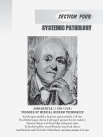
d outlining the white blood cell types.
1. Fill in the blanks to complete this concept map
f
e
eosinophil
lymphocyte
22. Use the list of terms provided to label the accompanying illustration
ustration of a skeletal muscle fiber
fiber;
check off each term as you label it.
□ mitochondrion
□ sarcolemma
□ terminal cisternae
□ myofibrils
□ sarcoplasmic reticulum
um
□ triad
□ nucleus
□ T-tubule
macrophage
mast cell
neutrophil
WHITE BLOOD CELL TYPES
Granulocytes
d
Agranulocyte
e
a
f
b
g
Basophil
Also called
polymorphonuclear
leukocyte
Monocyte
Moves into tissues
and becomes
Moves into tissues
and becomes
h
c
218
2. Construct a unit concept map to show the relationships among the following set of
terms. Include all of the terms in your diagram. Your instructor may choose to assign
additional terms.
agglutination
eosinophil
mast cell
antibody
hemoglobin
neutrophil
antigen
hormone
plasma
anucleate
diapedesis
lymphocyte
macrophage
plasma membrane
spectrin
viii
P r e fA C e
Other Key Features
Superb Art from
Amerman Textbook
Human Anatomy & Physiology Laboratory
Manual: Making Connections features a rich
and varied art program and integration of key
media and equipment used in the lab.
The art from the Amerman textbook
includes anatomical illustrations, photos, histology photomicrographs, and
physiology sequence figures.
Companion Lab Manual
to Erin Amerman’s Human
Anatomy & Physiology
This lab manual reflects the terminology and
explanations found in the Amerman textbook.
Table 13-1
Action(s)
Origin/Insertion/Nerve(s)
Concept Figures
Frontalis
Raises eyebrows;
wrinkles
skin of forehead
horizontally
O: Epicranial aponeurosis
I: Skin of eyebrows
N: Facial nerve
Frontalis
Occipitalis
Pulls scalp posteriorly
O: Occipital bone
I: Epicranial aponeurosis
N: Facial nerve
Corrugator
supercilii
Pulls eyebrows inferiorly
and medially (as in
squinting)
O: Medial supraorbital margin of frontal
bone
I: Skin of medial eyebrows
N: Facial nerve
Orbicularis
oculi
Closes eye; pulls skin
around the eyes, as in
blinking and winking
O: Orbital portions of the frontal bone
and maxilla
I: Skin of the orbital area and eyelids
N: Facial nerve
Levator labii
superioris
Elevates the upper lip;
everts and furrows upper
lip (as in sneering)
O: Zygomatic and upper maxilla near
orbit
I: Skin and muscle of the upper lip
N: Facial nerve
Zygomaticus
minor
Raises lateral portion of
the upper lip to expose
upper teeth (as in
smiling)
O: Zygomatic
I: Skin and muscle of the lateral upper lip
N: Facial nerve
Zygomaticus
major
Pulls the angle of the
mouth superiorly and
laterally (as in smiling or
laughing)
O: Zygomatic
I: Lateral muscle fibers of corner/angle of
mouth
N: Facial nerve
Risorius
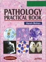
Glomerulus (glomerular
capillaries covered by
podocytes)
Orbicularis
oris
Depressor
anguli oris
Depressor
labii inferioris
Proximal
tubule
Muscles of Facial Expression
Muscle
Mentalis
Buccinator
(a) The renal corpuscle
Figure 30-9 The renal corpuscle.
Platysma
Occipitalis
Corrugator supercilii
Levator labii superioris
Zygomaticus
minor
Zygomaticus
major
Risorius
O: Connective tissue anterior to the ear
Pulls the angle of the
Glomerular
I: Modiolus*
mouth laterally
to makecapsule:
Podocyte Filtration slits Capillary Pedicels
Squamous
epithelium
N: Facial nerve
a closed-mouth
smile
(parietal layer)
O: Maxilla and mandible
Closes and protrudes
Capsular space
I: Skin and connective tissue of the lips
lips (as in puckering the
N: Facial nerve
lips for a kiss) Podocyte
(visceral layer)
Orbicularis oris
O: Lower body of mandible
Draws corners of the
I: Modiolus*
mouth inferiorly
N: Facial nerve
(unhappy face)
Efferent
O: Medial mandible near mental foramen
arteriole
I: Skin and connective tissue of lower lip
Afferent
N: Facial nerve
arteriole
O: Anterior mandible
Protrudes the lower lip
I: Skin of the chin near lower lip
and chin for drinking and
N: Facial nerve
“doubtful” expression
Orbicularis oculi
Depressor anguli oris
Protrudes lower lip
(sad or pouting
expressions)
alis
Depressor
Mentalis
labii inferioris
SEM (92,000×)
Helps manipulate food
during chewing and
expels air through
pursed lips (as in
blowing a trumpet)
O: Molar regions of maxilla and mandible
SEM
capillary surrounded
by podocytes
I: Orbicularis(b)
oris
andofconnective
tissue
of cheek/lips
N: Facial nerve
Lowers lower lip
and opens mouth
by depressing the
mandible
O: Connective tissue of deltoid and
pectoralis major
I: Mandible; skin and connective tissue
below mouth
N: Facial nerve
Note: Colors of actions and/or directions of action in Action(s) column match colors of directional
arrow(s) in Concept Figures.
*Mix of muscle and connective tissue at the corners of the mouth.
Platysma
Buccinator
P r e fA C e
PhysioEx™ 9.1
Additional Photos
of Lab Specimens
This lab manual contains additional images not found in the
Amerman textbook, including photos of anatomical models,
cadaver images, and histology photomicrographs.
i
a
b
c
j
k
d
e
f
l
m
n
o
g
p
h
f
a
g
b
h
c
i
d
j
e
ix
PhysioEx™ 9.1 is an easy-to-use physiology
lab simulation program that allows students to
repeat labs as often as they like, perform experiments without animals, and conduct experiments
that are difficult to perform in a wet lab environment because of
time, cost, or safety concerns. Every exercise includes an overview and every activity includes objectives, an introduction, a
pre-lab quiz, the experiment, a post-lab quiz, review sheet questions, and a lab report that students can save as a PDF and print
and/or email to their instructor. The online format with easy
step-by-step instructions includes everything students need in
one convenient place.
Each exercise and activity is referenced in the lab
manual where students are directed to access PhysioEx in
MasteringA&P. Pre-lab and post-lab quizzes and review sheets
for PhysioEx are assignable in MasteringA&P.
PhysioEx 9.1 includes 12 exercises containing a total of
63 physiology lab activities. The program features:
• Input data variability allows students to change variables and test various hypotheses for the experiments.
• Step-by-step instructions put everything students need
to do to complete the lab in one convenient place. Students gather data, analyze results, and check their understanding, all on screen.
• Stop & Think Questions and Predict Questions help
students think about the connections between the activities and the physiological concepts they demonstrate.
• Greater data variability in the results reflects more
realistically the results that students would encounter in
a wet lab experiment.
• Pre-lab and Post-lab Quizzes and short-answer
Review Sheets are offered to help students prepare for
and review each activity.
• Students can save their Lab Report as a PDF, which
they can print and/or email to their instructor.
• A Test Bank of assignable pre-lab and post-lab quizzes for use with TestGen or its course management system is provided for instructors.
• Seven videos of lab experiments demonstrate the actual experiments simulated on screen, making it easy
for students to understand and visualize the content of
the simulations. Videos demonstrate the following experiments: Skeletal Muscle, Blood Typing, Cardiovascular Physiology, Use of a Water-Filled Spirometer, Nerve
Impulses, BMR Measurement, and Cell Transport.
x
P r e fA C e
• Exercise 8: Chemical and Physical Processes of Digestion.
PhysioEx 9.1 topics include:
• Exercise 1: Cell Transport Mechanisms and Permeabil-
ity. Explores how substances cross the cell membranes.
Topics include: simple and facilitated diffusion, osmosis, filtration, and active transport.
• Exercise 2: Skeletal Muscle Physiology. Provides insights
into the complex physiology of skeletal muscle. Topics
include: electrical stimulation, isometric contractions,
and isotonic contractions.
• Exercise 3: Neurophysiology of Nerve Impulses. Investigates stimuli that elicit action potentials, stimuli that
inhibit action potentials, and factors affecting the conduction velocity of an action potential.
• Exercise 4: Endocrine System Physiology. Investigates
the relationship between hormones and metabolism;
the effect of estrogen replacement therapy; the diagnosis of diabetes; and the relationship between the levels
of cortisol and adenocorticotropic hormone and a variety of endocrine disorders.
• Exercise 5: Cardiovascular Dynamics. Allows students
to perform experiments that would be difficult if not
impossible to do in a traditional laboratory. Topics include: vessel resistance and pump (heart) mechanics.
• Exercise 6: Cardiovascular Physiology. Examines variables influencing heart activity. Topics include: setting
up and recording baseline heart activity, the refractory
period of cardiac muscle, and an investigation of factors
that affect heart rate and contractility.
• Exercise 7: Respiratory System Mechanics. Investigates
physical and chemical aspects of pulmonary function.
Students collect data simulating normal lung volumes.
Other activities examine factors such as airway resistance and the effect of surfactant on lung function.
Examines factors that affect enzyme activity by manipulating (in compressed time) enzymes, reagents, and
incubation conditions.
• Exercise 9: Renal System Physiology. Simulates the
function of a single nephron. Topics include: factors influencing glomerular filtration, the effect of
hormones on urine function, and glucose transport
maximum.
• Exercise 10: Acid-Base Balance. Topics include: respiratory and metabolic acidosis/alkalosis, and renal and respiratory compensation.
• Exercise 11: Blood Analysis. Topics include: hematocrit
determination, erythrocyte sedimentation rate determination, hemoglobin determination, blood typing, and
total cholesterol determination.
• Exercise 12: Serological Testing. Investigates antigen–
antibody reactions and their role in clinical tests used to
diagnose a disease or an infection.
Note: In addition to being available in MasteringA&P,
PhysioEx 9.1 is also available as a CD-ROM packaged with
this lab manual for no additional charge. Please contact your
Pearson representative for ordering information.
Biopac®
Activities that utilize the Biopac Student Labs® data
acquisition system are included in Unit 12, Introduction to the
Muscular System: Muscle Tissue; Unit 15, The Central Nervous
System: Brain and Spinal Cord; Unit 22, Physiology of the Heart;
and Unit 27, Physiology of the Respiratory System.
Instructions for other data acquisitions systems including
iWorx, Intellitool, and PowerLab are available in the
Instructor Resources in MasteringA&P.
Practice Anatomy Lab™ (PAL™) 3.0
™
Practice Anatomy Lab 3.0 (PAL) correlations
are indicated by the PAL logo and presented
as optional activities. These direct students
to related content in the PAL 3.0 software in
MasteringA&P.
Note: In addition to being available in MasteringA&P, Practice
Anatomy Lab 3.0 is also available as a DVD packaged with
this lab manual for no additional charge. Please contact your
Pearson representative for ordering information.
P r e fA C e
Assignable Content
in
• Drag-and-Drop
Based Questions
xi
Art Labeling Activities and Art
MasteringA&P is an online learning and assessment system
proven to help students learn. It helps instructors maximize
lab time with customizable, easy-to-assign, automatically
graded assessments that motivate students to learn outside
of class and arrive prepared for lab. The powerful gradebook
provides unique insight into student and class performance.
Instructors can easily assign the following:
• Pre-lab and Post-lab Quizzes for each activity in the
lab manual
• Clinical
Coaching Activities for select units that
include a brief clinical scenario with Bloom’s Level II
questions with feedback and hints
• Quizzes and Lab Practicals from PAL 3.0 Test Bank
• Pre-lab
and Post-lab Quizzes and Review Sheets
for PhysioEx 9.1
• Bone and Dissection Video Coaching Activities help
students identify bones and learn how to do organ
dissections
xii
P r e fa c e
• A&P Flix™ Animationsare3Dmovie-qualityanatomy
animations that include self-paced tutorials and gradable quizzes. Students learn structures and functions
fromtwosetsofanatomytopics:
• Origins,insertions,actions,andinnervations(over
60animations)
• Groupmuscleactionsandjoints(over50animations)
Study Tools in
Students get quick access to the following study tools in
MasteringA&P:
• Pre-lab and Post-lab Quizzes are provided for each
activity.
• Bone and Dissection videos
aidreviewofkeybonesand
organ dissections found in
thelabmanual.
• Dynamic
• Clinical
Case Study Coaching Activities increase
problem-solving skills and prepare students for future careers in allied health. Corresponding Teaching Strategies, available in the Instructor Resources in
MasteringA&P, enable instructors to “flip” the classroom by providing valuable tips on when and how to
use case studies. The worksheets and case studies are
alsoavailableintheStudyAreaofMasteringA&P.
• Learning
™
Catalytics is
a “bring your own device”
student engagement, assessment, and classroom
intelligence system. With
thisclassroomlecturetool,
instructors can flip the
classroom and a ssess students in real time using
open-ended tasks to probe
student
understanding.
Students use their smartphone, tablet, or laptop to
respond to questions in
class.
Study Modules
are designed to enable
studentstostudyeffectively
on their own, and to help
them quickly access and
learn the concepts they
need to be more successful on quizzes and exams.
These flashcard-style questions adapt to the student’s
performance and i nclude
art and explanations from
this lab manual to cement
thestudent’sunderstanding.
• Practice Anatomy Lab™ (PAL™) 3.0isanindispensable
virtual anatomy study and practice tool that gives students24/7accesstothemostwidelyusedlabspecimens
including human cadaver, anatomical models, histology, cat, and fetal pig. PAL 3.0 is easy to use and
includesbuilt-inaudiopronunciations,rotatablebones,
multiple-choicequizzes,andsimulatedfill-in-the-blank
lab practical exams. PAL 3.0 is also accessible on
mobiledevices.
P r e fA C e
• PhysioEx™
9.1 is easy-to-use physiology laboratory
simulation software. Every exercise includes an overview
and every activity includes objectives, an introduction,
a pre-lab quiz, the experiment, a post-lab quiz, review
sheet questions, and a lab report that students can save
as a PDF and print and/or email to their instructor.
xiii
Customization Options
An enhanced custom program allows instructors to pick
and choose content to tailor the lab manual at the activity
level, selecting only those activities they assign. Each activity
includes relevant background information, full-color figures,
tables, and charts.
For information on creating a custom version of this
manual, visit www.pearsonlearningsolutions.com, or contact
your Pearson representative for details.
Additional Instructor
Resources
Instructor Guide
• Videos of lab experiments
• A&P Flix animations
• Clinical Case Studies with worksheets
• Terminology Challenge worksheets
• Histology Atlas
• eText also available in MasteringA&P with eText
Three Versions
Human Anatomy & Physiology Laboratory Manual: Making
Connections is available in three versions for your students:
Main, Cat, and Fetal Pig. The Cat and Fetal Pig versions are
identical to the Main version except that they include seven
additional cat dissection exercises and nine additional fetal
pig dissection exercises, respectively, at the back of the lab
manual.
Main Version
Cat Version
0-13-395247-9 /
0-321-78700-5 /
978-0-321-78700-2 978-013-395247-6
Fetal Pig Version
0-13-399679-4 /
978-013-399679-1
0-13-405738-4 / 978-013-405738-5
This guide includes detailed instructions for setting up the
laboratory, time allotments for each activity, and answers to
the pre-lab assignments, activity questions, and post-lab assignments. Additionally, it describes strategies that encourage
active learning, including sample concept maps and an
overview of using concept mapping to increase student engagement. Finally, it discusses helpful hints for running an
effective lab, ways to avoid common pitfalls, and extension activities that can be used to expand activities when time allows.
Instructor Resources in
These resources include: editable pre-lab and post-lab quizzes, the Instructor’s Guide, instructions for each PhysioEx
activity, Terminology Challenge Worksheets, Clinical Case
Studies and Teaching Strategies for each case, A&P Flix (anatomy) in PPT, A&P Flix (anatomy) in MPEG, and instructions
for other data acquisition systems including iWorx, Intellitool, and Powerlab.
xiv
P r e fA C e
Student Supplements
Practice Anatomy Lab 3.0 Lab Guide
NEW! A Photographic Atlas
for Anatomy & Physiology
0-321-84025-9 / 978-0-321-84025-7 (standalone)
0-321-85767-4 / 978-0-321-85767-5 (with PAL 3.0 DVD)
by Ruth Heisler, Nora Hebert,
Jett Chinn, Karen Krabbenhoft,
Olga Malakhova
Written to accompany PAL 3.0,
the new Practice Anatomy Lab
3.0 Lab Guide contains exercises that direct the student to
select images and features in
PAL 3.0, and then assess their
understanding with labeling,
matching, short-answer, and fillin-the-blank questions. Exercises
cover three key lab specimens in
PAL 3.0—human cadaver, anatomical models, and histology.
0-321-86925-7 / 978-0-321-86925-8
by Nora Hebert, Ruth E. Heisler,
Jett Chinn, Karen M. Krabbenhoft,
Olga Malakhova
This brand new photo atlas is the
perfect lab study tool that helps
students learn and identify key
anatomical structures. Featuring
photos from Practice Anatomy
Lab™ 3.0 and other sources, the
Atlas includes over 250 cadaver
dissection photos, histology photomicrographs, and cat dissection
photos plus over 50 photos of anatomical models from leading manufacturers such as 3B Scientific®, SOMSO®, and Denoyer-Geppert Science Company.
Practice Anatomy Lab™ (PAL™) 3.0
0-321-68211-4 / 978-0-321-68211-6 (DVD)
by Nora Hebert, Ruth E. Heisler,
Jett Chinn, Karen Krabbenhoft,
Olga Malakhova
An indispensable virtual anatomy study and practice tool that
gives students 24/7 access to the
most widely used lab specimens
including human cadaver, anatomical models, histology, cat,
and fetal pig. PAL 3.0 also includes multiple-choice quizzes
and practice fill-in-the-blank lab
practicals.
The Anatomy Coloring Book,
Fourth Edition
0-321-83201-9 / 978-0-321-83201-6
by Wynn Kapit and Lawrence M. Elson
For more than 35 years, The
Anatomy Coloring Book has been
the best-selling human anatomy
coloring book! A useful tool for
anyone with an interest in learning anatomical structures, this
concisely written text features
precise, extraordinary handdrawn figures that were crafted
especially for easy coloring and
interactive study. The Fourth
Edition features user-friendly
two-page spreads with enlarged
art, clearer, more concise text descriptions, and new boldface
headings that make this classic coloring book accessible to a
wider range of learners.
Acknowledgments
A project of this magnitude is truly a team effort and I
have been a part of an amazing team. I have so many people to thank. I will be forever grateful to Acquisitions Editor
Gretchen Puttkamer for bringing me onto the team, for helping me to create a vision for this project, and for having the
patience to coach me through those rough beginnings. I owe
my deepest gratitude to the outstanding editorial, production,
and marketing teams at Pearson. A heartfelt thanks to Serina
Beauparlant, Editor-in-Chief, Kari Hopperstead, Project Editor, and Alan Titche, Development Editor, for their unending
support, encouragement, and direction. Their hard work and
dedication to this project inspired me to give this project my
all and to keep my eyes on the finish line. Kudos also to Allison
Rona, Marketing Manager, and Derek Perrigo, Senior Anatomy and Physiology Specialist, for their market guidance. I am
also grateful to Media Content Producer Nicole Tache for her
excellent work spearheading MasteringA&P for this manual.
The production of this book was a herculean task expertly
managed by Caroline Ayres, Project Manager at Pearson, and
Norine Strang, Senior Project Manager at S4Carlisle Publishing Services. Many thanks to Art Development Editors Kelly
Murphy and Elisheva Marcus, and Project Managers Alicia
Elliot and Lima Colati, who all provided expert guidance to
the amazing team of illustrators at Imagineering Art. Thanks
also to Maureen Spuhler for her excellent photo research, and
to Lorretta Palagi for her eagle-eyed copyediting.
I want to thank contributor Karen Keller of Frostburg
State University for her superb job of writing several units.
Many thanks go to Patricia Wilhelm of Johnson & Wales
University for her wonderful job of writing the cat dissection
unit, and to Kerrie Hoar of University of Wisconsin–
La Crosse for the beautiful cat dissection photographs. Thanks
also to Sarah Matarese of St. George’s School who contributed
the Biopac activities and to Wendy Rappazzo of Harford
Community College who authored clinical questions for
MasteringA&P. I am grateful to Sheri Boyce of Messiah College
and Anna Gilletly of Central New Mexico Community College
for their assistance in editing and preparing units. A huge
thank you to Carolyn Lebsack of Linn-Benton Community
College, Steve Leadon of Durham Technical Community
College, Kerrie Hoar of University of Wisconsin–La Crosse,
Michelle Gaston of Northern Virginia Community College,
and Bert Atsma of Union County College for their meticulous
accuracy checks. I owe very special thanks to Erin Amerman
for writing an outstanding textbook for this lab manual to
accompany. Erin is a gifted writer with incredible insight into
how students learn.
I would like to thank several of my colleagues at the University of North Georgia for their help, support, and valuable
insights. A special thanks to John Hamilton for his expert
photography. I owe JB Sharma a debt of gratitude for being
an endless source of encouragement and a model of teaching
excellence. I want to thank Lynn Berdanier for listening to
my wild ideas and for her willingness to try them out in her
labs. A special thanks to Mary Mayhew who, despite serving as a biology department head during a challenging time
of consolidation, was always available to me and continues
to be one of my greatest sources of support, encouragement,
and guidance. Finally, to Malynde Weaver, my friend, my colleague, and teacher/advisor extraordinaire—I am blessed to
serve alongside you.
To my current and former students, you are the inspiration for this project. Your passion for learning motivates me
to be the best teacher that I can be. You are the reason that
I teach!
To my mentor, my professor, and my friend, Dr. Louis J.
Guillette, thank you for investing in my life and for teaching
me the art of being an educator. You believed in me so many
years ago and that belief not only changed the direction of my
life but it also instilled in me a confidence in my abilities that
has taken root and enabled me to pursue my dreams.
I am deeply grateful to my husband, Mark, and to our
three incredible children—Jesse, Eli, and Ashton. Mark, this
project would have never happened without you. You are the
love of my life—an incredible husband and father—and I
am blessed beyond measure. You remind me daily with your
words and actions what is really important in life and you
help me keep my priorities in order. Jesse, Eli, and Ashton,
thank you for being so patient (most of the time) when mom
needed to write, to talk to Kari, Serina, and Alan, or to take a
nap. I know that you never thought this day would come, but
it is finished. Mom and Dad owe you an awesome vacation!
xv
xvi
ACkNOwLeDGMeNTS
Text and Media Reviewers
Pius Aboloye, North Lake College
Michele Alexandre, Durham Technical Community College
Chris Allen, College of the Mainland
Emily Allen, Rowan College of Gloucester County
Marcia Anglin, Miami Dade College–North Campus
Verona Barr, Heartland Community College
Dena Berg, Tarrant County College–Northwest Campus
Sheri Boyce, Messiah College
Ron Bridges, Pellissippi State Community College
Carol Britson, University of Mississippi
Geralyn Caplan, Owensboro Community & Technical College
Maria Carles, Northern Essex Community College
Carol Carr, John Tyler Community College
Ellen Carson, Florida State College–Jacksonville
Peter Charles, Durham Technical Community College
Teresa Cowan, Baker College
Ken Crane, Texarkana College
Mary Dettman, Seminole Community College
Karen Dunbar-Kareiva, Ivy Tech Community College
Kathryn Englehart, Kennebec Valley Community College
Sondra Evans, Florida State College–Jacksonville
Jill Feinstein, Richland Community College
Tracy Felton, Union County Community College
Christine Foley, Southwest Texas Junior College–Del Rio
Campus
Lori Frear, Wake Tech Community College
Kim Fredricks, Viterbo University
Lynn Gargan, Tarrant County College–Northeast Campus
Lori Garrett, Parkland College
Michelle Gaston, Northern Virginia Community College
Carol Gavareski, Bellingham Technical College
Anna Gilletly, Central New Mexico Community College
Miriam Golbert, College of the Canyons
Joanna Greene, Ivy Tech Community College–Anderson
Juan Guzman, Florida Gateway College
Bill Hanna, Massasoit Community College
Lesleigh Hastings, Wake Tech Community College
Stephanie Havemann, Alvin Community College
Heidi Hawkins, College of Southern Idaho
D.J. Hennager, Kirkwood Community College
Charmaine Henry, Baker University
Julie Huggins, Arkansas State
Jody Johnson, Arapahoe Community College
Karen Keller, Frostburg State University
Suzanne Kempke, St. Johns River Community College
Christine Kisiel, Mount Wachusett Community College
Ellen Lathrop-Davis, Community College of Baltimore County
Steven Leadon, Durham Technical Community College
Carolyn Lebsack, Linn-Benton Community College
Stephen Lebsack, Linn-Benton Community College
Jeffrey Lee, Essex Community College
Leona Levitt, Union County College
Christine Maney, Salem State College
Bruce Maring, Daytona State College
Robert Marino, Capital Community College
Sarah Matarese, St. George’s School
Cherie McKeever, Montana State University–
Great Falls College
Jaime Mergliano, John Tyler Community College
Justin Moore, American River College
Howard Motoike, LaGuardia Community College
Regina Munro, Chandler Gilbert Community College
Karen Murch-Shafer, University of Nebraska–Omaha
Zvi Ostrin, Hostos Community College
Ellen Ott-Reeves, Blinn College–Bryan Campus
Debbie Palatinus, Roane State Community College
Kevin Ragland, Nashville State Community College
Wendy Rappazzo, Harford Community College
Jean Revie, South Mountain Community College
Travis Robb, Allen Community College–Burlingame
Fredy Ruiz, Miami Dade College
Tracy Rusco, East Central College
Amy Ryan, Clinton Community College
Linda Schams, Viterbo University
Jeff Schinske, De Anza College
Steven Schneider, South Texas College
Maureen Scott, Norfolk State University
George Steer, Jefferson College of Health Sciences
James Stittsworth, Florida State College–Jacksonville
Deborah Temperly, Delta College
Terry Thompson, Wor-Wic Community College
Carlene Tonini-Boutacoff, College of San Mateo
Liz Torrano, American River College
Lisa Welch, Weatherford College
Deb Wiepz, Madison Area Technical College
Darrellyn Williams, Pulaski Technical College
Brief Contents
UNIT
1
2
3
4
5
6
7
8
9
10
11
12
13
14
15
16
17
18
19
20
21
22
Introduction to Anatomy and Physiology
Introduction to the Organ Systems
Chemistry
The Microscope
The Cell
Histology
The Integumentary System
Introduction to the Skeletal System
The Axial Skeleton
The Appendicular Skeleton
Joints
Introduction to the Muscular System:
Muscle Tissue
Gross Anatomy of the Muscular System
Introduction to the Nervous System
The Central Nervous System: Brain
and Spinal Cord
The Peripheral Nervous System:
Nerves and Autonomic Nervous System
General Senses
Special Senses
The Endocrine System
Blood
Anatomy of the Heart
Physiology of the Heart
1
15
29
47
61
81
103
119
137
171
199
217
241
281
301
331
355
365
389
409
427
443
23
24
25
26
27
28
29
30
31
32
33
Anatomy of Blood Vessels
Circulatory Pathways and the Physiology
of Blood Vessels
The Lymphatic System
Anatomy of the Respiratory System
Physiology of the Respiratory System
Anatomy of the Digestive System
Physiology of the Digestive System
Anatomy of the Urinary System
Physiology of the Urinary System
The Reproductive System
Embryonic Development and Heredity
459
487
505
523
539
557
585
599
617
635
661
CAT DISSECTION EXERCISES
1
2
3
4
5
6
7
Exploring the Muscular System of the Cat
Exploring the Spinal Nerves of the Cat
Exploring the Respiratory System of the Cat
Exploring the Digestive System of the Cat
Exploring the Cardiovascular System of the Cat
Exploring the Urinary System of the Cat
Exploring the Reproductive System of the Cat
INDEX
C-1
C-21
C-27
C-33
C-41
C-49
C-53
I-1
xvii
This page intentionally left blank
Contents
UNIT 1
INTRODUCTION TO ANATOMY
AND PHYSIOLOGY 1
P R E - L A B Assignments 2
Activity 1: Identifying Body Regions
and Exploring Surface Anatomy 6
Activity 2: Identifying Body Cavities
and Abdominopelvic Regions 7
Activity 3: Demonstrating and Identifying Body Planes
of Section 9
Activity 4: Assisting the Coroner 10
P O S T - L A B Assignments 11
UNIT 2
INTRODUCTION TO THE ORGAN
SYSTEMS 15
Activity 4: Perceiving Depth of Field 55
Activity 5: Caring for the Microscope 55
P O S T - L A B Assignments 57
THE CELL
UNIT 5
P R E - L A B Assignments
61
62
LabBOOST Organelles 66
Activity 1: Identifying Cell Components in a Wet
Mount 67
Activity 2: Identifying Cell Structures 67
Activity 3: Examining the Possible Role of Osmosis in
Cystic Fibrosis 68
Activity 4: Identifying the Stages of the Cell Cycle 72
Activity 5: Exploring Cellular Diversity 73
Exercise 1: Cell Transport Mechanisms
and Permeability 74
P O S T - L A B Assignments 75
™
P R E - L A B Assignments 16
Activity 1: Locating and Describing Major Organs and
Their Functions 18
Activity 2: Using Anatomical Terminology to Describe
Organ Locations 20
Activity 3: Studying Homeostasis and Organ System
Interactions 22
P O S T - L A B Assignments 25
UNIT 3
CHEMISTRY
29
P R E - L A B Assignments 30
Activity 1: Exploring the Chemical Properties
of Water 34
Activity 2: Determining pH and Interpreting the
pH Scale 36
Activity 3: Observing the Role of Buffers 38
LabBOOST Protein Structure 41
Activity 4: Analyzing Enzymatic Activity 41
P O S T - L A B Assignments 43
UNIT 4
THE MICROSCOPE
47
P R E - L A B Assignments 48
Activity 1: Identifying the Parts of the Microscope 50
Activity 2: Using the Microscope to View Objects 51
Activity 3: Determining Field Diameter and Estimating Size
of Objects 53
LabBOOST Metric Conversions 55
UNIT 6
HISTOLOGY
81
P R E - L A B Assignments 82
Activity 1: Examining Epithelial Tissue 84
Activity 2: Characterizing Connective Tissue 92
Activity 3: Exploring Nervous Tissue and Muscle
Tissue 95
Activity 4: Tissue Identification Concept Map 95
P O S T - L A B Assignments 97
UNIT 7
THE INTEGUMENTARY SYSTEM
103
P R E - L A B Assignments 104
Activity 1: Identifying and Describing Skin Structures 110
Activity 2: Examining the Histology of the Skin 111
Activity 3: Determining Sweat Gland Distribution 113
P O S T - L A B Assignments 115
UNIT 8
INTRODUCTION TO THE SKELETAL
SYSTEM 119
P R E - L A B Assignments 120
Activity 1: Reviewing Skeletal Cartilages 122
Activity 2: Classifying and Identifying the Bones
of the Skeleton 127
xix
xx
CO NT eN TS
LabBOOST Visualizing Sliding Filaments 227
Activity 3: Stimulating Muscle Contraction in Glycerinated
Skeletal Muscle Tissue 229
Activity 4: Electromyography in a Human Subject
Using
230
Exercise 2: Skeletal Muscle Physiology 234
P O S T - L A B Assignments 235
Activity 3: Examining the Gross Anatomy
of a Long Bone 129
LabBOOST Osteon Model 131
Activity 4: Exploring the Microscopic Anatomy
of Compact Bone—The Osteon 131
Activity 5: Examining the Chemical Composition
of Bone 132
P O S T - L A B Assignments 133
UNIT 9
THE AXIAL SKELETON
™
UNIT 13
137
P R E - L A B Assignments 138
Activity 1: Studying the Bones of the Skull 148
Activity 2: Examining the Fetal Skull 153
Activity 3: Studying the Bones of the Vertebral Column
and Thoracic Cage 159
Activity 4: Identifying Bones-in-a-Bag 164
P O S T - L A B Assignments 165
UNIT 10
THE APPENDICULAR SKELETON
P R E - L A B Assignments
171
172
LabBOOST The Pelvic Bones 177
Activity 1: Studying the Bones of the Appendicular
Skeleton 182
Activity 2: Identifying Bones-in-a-Bag 190
P O S T - L A B Assignments 191
UNIT 11
JOINTS
199
P R E - L A B Assignments 200
Activity 1: Identifying and Classifying Joints 205
Activity 2: Demonstrating Movements Allowed
by Joints 207
Activity 3: Comparing and Contrasting the Structure and
Function of Selected Synovial Joints 211
P O S T - L A B Assignments 213
UNIT 12
INTRODUCTION TO THE
MUSCULAR SYSTEM: MUSCLE
TISSUE 217
P R E - L A B Assignments 218
Activity 1: Identifying the Structural Components
of a Skeletal Muscle 221
Activity 2: Examining the Microscopic Anatomy
of Skeletal Muscle Tissue and the Neuromuscular
Junction 225
GROSS ANATOMY OF THE MUSCULAR
SYSTEM 241
P R E - L A B Assignments 242
Activity 1: Determining How Skeletal Muscles
Are Named 245
Activity 2: Mastering the Muscles of the Head and Neck 254
Activity 3: Mastering the Muscles of the Trunk 260
Activity 4: Mastering the Muscles of the Upper Limb 266
Activity 5: Mastering the Muscles of the Lower Limb 272
P O S T - L A B Assignments 273
UNIT 14
INTRODUCTION TO THE NERVOUS
SYSTEM 281
P R E - L A B Assignments 282
Activity 1: Calculating Reaction Time 285
Activity 2: Investigating the Motor Neuron 289
Activity 3: Investigating the Chemical Synapse 291
Activity 4: Exploring the Histology of Nervous Tissue
Exercise 3: Neurophysiology of Nerve
Impulses 293
P O S T - L A B Assignments 295
292
™
UNIT 15
THE CENTRAL NERVOUS SYSTEM:
BRAIN AND SPINAL CORD 301
P R E - L A B Assignments
302
LabBOOST Visualizing the Brain 308
Activity 1: Exploring the Functional Anatomy of the
Brain 308
Activity 2: Electroencephalography in a Human Subject
Using
312
Activity 3: Identifying the Meninges/Ventricles and Tracing
the Flow of Cerebrospinal Fluid 317
Activity 4: Examining the Functional Anatomy
of the Spinal Cord 320
Activity 5: Analyzing a Spinal Reflex 321
Activity 6: Dissecting a Sheep Brain and Spinal Cord 322
P O S T - L A B Assignments 325
CON T e NTS
UNIT 16
THE PERIPHERAL NERVOUS SYSTEM:
NERVES AND AUTONOMIC NERVOUS
SYSTEM 331
P R E - L A B Assignments
332
LabBOOST Learning the Cranial Nerves 338
Activity 1: Learning the Cranial Nerves 339
Activity 2: Evaluating the Function of the Cranial
Nerves 340
Activity 3: Identifying the Spinal Nerves and Nerve
Plexuses 344
Activity 4: Exploring the Autonomic Nervous System
P O S T - L A B Assignments 349
UNIT 17
GENERAL SENSES
355
SPECIAL SENSES
™
BLOOD
409
P R E - L A B Assignments 410
Activity 1: Exploring the Formed Elements of Blood 413
Activity 2: Performing a Hematocrit 415
Activity 3: Performing a Differential White Blood Cell
Count 416
Activity 4: Determining Coagulation Time 418
Activity 5: Determining Blood Types 420
Exercise 11: Blood Analysis 422
P O S T - L A B Assignments 423
™
P R E - L A B Assignments 356
Activity 1: Identifying General Sensory Receptors 358
Activity 2: Examining the Microscopic Structure
of General Sensory Receptors 359
Activity 3: Performing a Two-Point Discrimination
Test 360
P O S T - L A B Assignments 361
UNIT 18
Activity 3: Investigating Endocrine Case Studies: Clinician’s
Corner 403
Exercise 4: Endocrine System
Physiology 404
P O S T - L A B Assignments 405
UNIT 20
346
xxi
365
P R E - L A B Assignments 366
Activity 1: Exploring the Gross Anatomy of Olfactory
and Gustatory Structures and Demonstrating the Effect
of Olfaction on Gustation 370
Activity 2: Examining the Gross Anatomy of the Eye 375
Activity 3: Dissecting a Mammalian Eye 377
Activity 4: Performing Visual Tests 378
Activity 5: Examining the Gross Anatomy
of the Ear 382
Activity 6: Performing Hearing and Equilibrium Tests 383
P O S T - L A B Assignments 385
UNIT 21
ANATOMY OF THE HEART
427
P R E - L A B Assignments 428
Activity 1: Examining the Functional Anatomy
of the Heart 432
Activity 2: Dissecting a Mammalian Heart 434
Activity 3: Reviewing the Microscopic Structure
of Cardiac Muscle Tissue 436
Activity 4: Tracing Circulatory Pathways 438
P O S T - L A B Assignments 439
UNIT 22
PHYSIOLOGY OF THE HEART
443
P R E - L A B Assignments 444
Activity 1: Recording and Interpreting an
Electrocardiogram 448
Activity 2: Auscultating Heart Sounds 449
Activity 3: Electrocardiography in a Human Subject
Using
450
Exercise 6: Cardiovascular Physiology 454
P O S T - L A B Assignments 455
™
UNIT 19
THE ENDOCRINE SYSTEM
389
P R E - L A B Assignments 390
Activity 1: Exploring the Organs of the Endocrine
System 397
Activity 2: Examining the Microscopic Anatomy of the
Pituitary Gland, Thyroid Gland, Parathyroid Gland,
Adrenal Gland, and Pancreas 400
LabBOOST Microscopic Anatomy of the Adrenal
Cortex 402
UNIT 23
ANATOMY OF BLOOD VESSELS
P R E - L A B Assignments
459
460
LabBOOST Blood Vessel Pathways 463
Activity 1: Identifying the Major Arteries That Supply
the Head, Neck, Thorax, and Upper Limbs 468
Activity 2: Identifying the Major Arteries That Supply the
Abdominopelvic Organs and the Lower Limbs 469
xxii
CO NTe N TS
Activity 3: Identifying Veins That Drain into the Venae
Cavae 475
Activity 4: Examining the Histology of Arteries
and Veins 477
P O S T - L A B Assignments 479
UNIT 24
CIRCULATORY PATHWAYS AND
THE PHYSIOLOGY OF BLOOD
VESSELS 487
P R E - L A B Assignments 488
Activity 1: Tracing Blood Flow—General Systemic
Pathways 493
Activity 2: Tracing Blood Flow—Specialized Systemic
Pathways 494
Activity 3: Tracing Blood Flow—Pulmonary
Circulation 496
Activity 4: Tracing Blood Flow—Fetal Circulation 498
Activity 5: Measuring Blood Pressure
and Examining the Effects of Body Position
and Exercise 499
Exercise 5: Cardiovascular
Dynamics 500
P O S T - L A B Assignments 501
™
UNIT 25
THE LYMPHATIC SYSTEM
505
P R E - L A B Assignments 506
Activity 1: Exploring the Organs of the Lymphatic
System 512
Activity 2: Examining the Histology of a Lymph Node, a
Tonsil, and the Spleen 513
Activity 3: Tracing the Flow of Lymph through the
Body 514
Activity 4: Using a Pregnancy Test to Demonstrate
Antigen–Antibody Reactions 516
Exercise 12: Serological Testing 518
P O S T - L A B Assignments 519
™
UNIT 27
P R E - L A B Assignments 540
Activity 1: Analyzing the Model Lung and Pulmonary
Ventilation 545
Activity 2: Measuring Respiratory Volumes in a Human
Subject Using
545
Activity 3: Determining Respiratory Volumes and
Capacities at Rest and Following Exercise 548
Activity 4: Investigating the Control of Breathing 550
Exercise 7: Respiratory System Mechanics 552
P O S T - L A B Assignments 553
™
UNIT 28
ANATOMY OF THE RESPIRATORY
SYSTEM 523
P R E - L A B Assignments 524
Activity 1: Exploring the Organs of the Respiratory
System 530
Activity 2: Examining the Microscopic Anatomy
of the Trachea and Lungs 532
Activity 3: Examining a Sheep Pluck 534
P O S T - L A B Assignments 535
ANATOMY OF THE DIGESTIVE
SYSTEM 557
P R E - L A B Assignments 558
Activity 1: Exploring the Organs of the Alimentary
Canal 564
Activity 2: Exploring the Accessory Organs
of the Digestive System 568
Activity 3: Examining the Histology of Selected
Digestive Organs 575
P O S T - L A B Assignments 579
UNIT 29
PHYSIOLOGY OF THE DIGESTIVE
SYSTEM 585
P R E - L A B Assignments 586
Activity 1: Analyzing Amylase Activity 589
Activity 2: Analyzing Pepsin Activity 590
Activity 3: Analyzing Lipase Activity 592
Activity 4: Tracing Digestive Pathways 592
Exercise 8: Chemical and Physical Processes
of Digestion 594
P O S T - L A B Assignments 595
™
UNIT 30
UNIT 26
PHYSIOLOGY OF THE RESPIRATORY
SYSTEM 539
ANATOMY OF THE URINARY
SYSTEM 599
P R E - L A B Assignments 600
Activity 1: Exploring the Organs of the Urinary
System 606
Activity 2: Dissecting a Mammalian Kidney 608
Activity 3: Examining the Microscopic Anatomy
of the Kidney, Ureter, and Urinary Bladder 611
LabBOOST Anatomy of the Renal Corpuscle 612
P O S T - L A B Assignments 613
CON Te NTS
UNIT 31
PHYSIOLOGY OF THE URINARY
SYSTEM 617
P R E - L A B Assignments 618
Activity 1: Demonstrating the Function of the Filtration
Membrane 622
Activity 2: Simulating the Events of Urine
Production and Urine Concentration 623
Activity 3: Using the Results of a Urinalysis to Make
Clinical Connections 627
LabBOOST Understanding Tonicity 627
Exercise 9: Renal System Physiology 629
Exercise 10: Acid–Base Balance 630
P O S T - L A B Assignments 631
™
™
UNIT 32
THE REPRODUCTIVE SYSTEM
635
P R E - L A B Assignments 636
Activity 1: Examining Male Reproductive Anatomy 641
Activity 2: Examining Female Reproductive Anatomy 644
Activity 3: Modeling Meiosis 647
Activity 4: Comparing Spermatogenesis and Oogenesis 653
P O S T - L A B Assignments 655
UNIT 33
Embryonic Development and
Heredity 661
P R E - L A B Assignments 662
Activity 1: Exploring Fertilization and the Stages of Prenatal
Development 667
Activity 2: Examining the Placenta 670
xxiii
Activity 3: Learning the Language of Genetics 672
Activity 4: Exploring Dominant-Recessive
Inheritance 674
Activity 5: Exploring Other Patterns of Inheritance 677
P O S T - L A B Assignments 679
Cat Dissection Exercises
Dissection 1: Exploring the Muscular System
of the Cat C-1
Dissection 2: Exploring the Spinal Nerves
of the Cat C-21
Dissection 3: Exploring the Respiratory System
of the Cat C-27
Dissection 4: Exploring the Digestive System
of the Cat C-33
Dissection 5: Exploring the Cardiovascular System
of the Cat C-41
Dissection 6: Exploring the Urinary System
of the Cat C-49
Dissection 7: Exploring the Reproductive System
of the Cat C-53
INDEX
I-1