Ebook Atlas of anatomic pathology Part 2
Bạn đang xem bản rút gọn của tài liệu. Xem và tải ngay bản đầy đủ của tài liệu tại đây (31.98 MB, 135 trang )
4
Neurogenic Tumors
Neurogenic tumors of the mediastinum are relatively rare
and are most often encountered in the pediatric-age population. They most commonly arise from structures in the posterior mediastinum, although they can originate from all
three mediastinal compartments. Neurogenic tumors are, in
fact, the most common tumors of the posterior mediastinum
in both children and adults. Neurogenic tumors can be of
neuroblastic origin or may arise from peripheral nerve sheath
elements (Table 4.1).
Table 4.1 Neurogenic tumors of the mediastinum
Neuroblastic tumors
Ganglioneuroma
Ganglioneuroblastoma
Neuroblastoma
Peripheral nerve sheath tumors
Schwannoma
Neurofibroma
Malignant peripheral nerve sheath tumor
4.1
Neuroblastic Neoplasms
Neuroblastic neoplasms (Figs. 4.1, 4.2, 4.3, 4.4, 4.5, 4.6, 4.7,
4.8, 4.9, 4.10, 4.11, 4.12, 4.13, 4.14, 4.15, 4.16, 4.17, 4.18,
4.19, 4.20, 4.21, 4.22, 4.23, 4.24, 4.25, 4.26, 4.27, 4.28, 4.29,
4.30, 4.31, and 4.32) arise from primitive precursor cells of
the sympathetic nervous system. They are the most common
solid tumors in children under 1 year of age, although they
can also occur in older children and in adults. Some cases
can be associated with neurofibromatosis (NF1). They show
a histologic spectrum that ranges from very welldifferentiated and mature neuronal elements to tumors composed of primitive and poorly differentiated cells, mimicking
the entire spectrum of neuroblastic maturation.
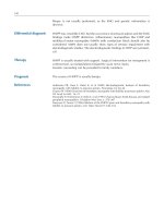
Fig. 4.1 Neuroblastic tumors are most often large and solid, well circumscribed, and surrounded by a fibrous capsule. They usually show a
smooth outer surface. This image is an example of a ganglioneuroma,
the most common benign tumor originating from the thoracic sympathetic nerves
S. Suster (ed.), Atlas of Mediastinal Pathology, Atlas of Anatomic Pathology,
DOI 10.1007/978-1-4939-2674-9_4, © Springer Science+Business Media, LLC 2015
89
90
4
Neurogenic Tumors
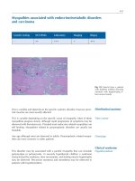
Fig. 4.2 Ganglioneuromas are benign neurogenic tumors composed of
mature neural elements admixed in varying proportions. They are often
paucicellular and show a variously collagenized stroma on scanning
magnification
Fig. 4.4 Schwannian stroma-poor ganglioneuroma shows a focus
(center) containing large ganglion cells with abundant cytoplasm and
round nuclei that are intimately admixed in various proportions with the
spindle cells and collagenous stroma
Fig. 4.3 On higher magnification, ganglioneuromas are composed
mainly of loose, fibrocollagenous stroma admixed with bland-appearing
fibroblastic or schwannian spindle cells. The stroma may vary from
loose and edematous to densely collagenized. Based on the extent of the
schwannian component, they have been classified into schwannian
stroma dominant (“mature” ganglioneuroma) and schwannian stroma
poor (“maturing” ganglioneuroma)
Fig. 4.5 Higher magnification from the field in Fig. 4.4 shows a cluster
of large ganglion cells with abundant granular eosinophilic cytoplasm
and large, round nuclei
4.1
Neuroblastic Neoplasms
Fig. 4.6 Schwannian stroma-rich mediastinal ganglioneuroma shows
clusters of large ganglion cells with small, round nuclei surrounded by
abundant granular eosinophilic cytoplasm embedded in a dense collagenous stroma containing abundant schwannian spindle cells
Fig. 4.7 Higher magnification of schwannian stroma-rich ganglioneuroma showing large ganglion cells with eccentric nuclei
91
Fig. 4.8 Higher magnification of ganglion cells in ganglioneuroma
showing characteristic small nuclei surrounded by abundant pink granular cytoplasm. The number of ganglion cells in these tumors may vary;
if they are very sparse, proper identification may require a diligent
search
Fig. 4.9 Ganglioneuroblastoma is a tumor characterized by an admixture in various proportions of both mature schwannian and ganglionic
elements, as well as primitive neuroblastic elements that form discrete
nests or islands of tumor cells. The tumors are grossly well circumscribed, with a fleshy, tan-white cut surface with focal areas of cystic
degeneration and foci of calcification
92
Fig. 4.10 Histologically, ganglioneuroblastoma shows areas that are
indistinguishable from ganglioneuroma in association with foci of neuroblastic elements. The neuroblastic elements are arranged in discrete,
small nests of primitive neuroblastic cells (lower right) that merge with
the schwannian-rich stroma (left)
Fig. 4.11 Higher magnification from the field in Fig. 4.10 shows
bland-appearing schwannian spindle cells (left) percolating between
small nests of small neuroblastic cells (right)
4
Neurogenic Tumors
Fig. 4.12 Schwannian stroma-poor ganglioneuroblastoma shows a
paucicellular, collagenous stroma merging imperceptibly with islands
and nests of primitive, small neuroblastic cells admixed with larger ganglionic cells
Fig. 4.13 Higher magnification of schwannian stroma-poor ganglioneuroblastoma shows a mixed cell population composed of small, primitive neuroblastic cells admixed with larger ganglionic cells
4.1
Neuroblastic Neoplasms
Fig. 4.14 Another example of ganglioneuroblastoma shows a few
small nests containing neuroblastic elements surrounded by a
schwannian-rich spindle-cell stroma
Fig. 4.15 Higher magnification of ganglioneuroblastoma showing a
mixed population of neuroblastic small cells admixed with larger ganglionic cells with abundant eosinophilic cytoplasm
93
Fig. 4.16 Ganglioneuroblastoma with foci of dystrophic calcifications. Neuroblastoma elements occasionally undergo dystrophic calcification that replaces the tumor cells. The presence of these foci of
calcification is an indication of a neuroblastic component in the tumor
and should prompt search of additional sections
Fig. 4.17 Neuroblastoma is the most common malignant neoplasm of
the posterior mediastinum in children, particularly under the age of 1
year. Grossly, the tumors are well circumscribed and encapsulated, with
a multinodular outer surface
94
Fig. 4.18 Cut surface of a neuroblastoma shows grayish white, soft
tissue with areas of hemorrhage and necrosis and foci of calcification
Fig. 4.19 Histologically, neuroblastoma is composed of a proliferation of small, round blue cells showing various degrees of organization.
Neuroblastomas have been classified into differentiating, poorly differentiated, and undifferentiated, depending on their degree of maturation
and organization. The well-differentiated tumors (differentiating neuroblastoma) overlap with ganglioneuroblastoma but show a predominance of neuroblastic elements over ganglionic cells and may also
contain a minor schwannian component. The poorly differentiated variants show no evidence of ganglionic or schwannian differentiation and
grow as sheets of small tumor cells that often present a nested appearance, as in this example
4
Neurogenic Tumors
Fig. 4.20 Higher magnification from Fig. 4.19 shows well-defined
islands or nests of tumor cells separated by a vascular stroma
Fig. 4.21 Higher magnification showing well-defined nests of tumor
cells with abundant fibrillary eosinophilic material (neuropil) in the
background. The fibrillary material (neuropil) is a distinctive feature of
neuroblastic neoplasms and helps in the differential diagnosis with
other small, round cell tumors
4.1
Neuroblastic Neoplasms
95
Fig. 4.22 Another area in the same tumor shows a peculiar linear,
single-file arrangement of neuroblastic cells embedded in abundant
fibrillary matrix, a feature often observed in these tumors
Fig. 4.24 Higher magnification in calcifying neuroblastoma shows
irregular deposits of calcific material surrounded by proliferation of
primitive, small, round blue cells
Fig. 4.23 Extensive stromal calcification is a common feature in neuroblastoma. The calcifying process can be so extensive as to obscure the
underlying neoplastic proliferation; recognition of the tumor cells may
then require extensive sampling
Fig. 4.25 Higher magnification shows small nests of tumor cells
admixed with calcific stromal deposits. The tumor cells show a primitive appearance with small, round nuclei with tiny nucleoli and no discernible cytoplasm
96
4
Neurogenic Tumors
Fig. 4.26 Another example of differentiating neuroblastoma on scanning magnification shows a lobular growth pattern separated by wellvascularized stroma and foci of stromal calcifications
Fig. 4.28 High power shows a scant amount of tumor cells displaying
various degrees of maturation, floating in abundant eosinophilic fibrillary matrix (neuropil)
Fig. 4.27 Higher magnification shows a lobule of tumor cells with
abundant background neuropil and a mixed cell population, including
ganglionic-type large cells and scattered smaller neuroblastic cells
Fig. 4.29 This neuroblastoma of an “undifferentiated” type shows
almost solid sheets of small, round, primitive cells with only a vague
hint of nesting
4.1
Neuroblastic Neoplasms
Fig. 4.30 Higher magnification from the same case as Fig. 4.29 shows
a primitive population of small, round, blue cells growing as sheets with
scattered mitoses. The tumor cells have small, irregular nuclei with
scant nuclear detail and small nucleoli
Fig. 4.31 Higher magnification shows the presence of Flexner-type
rosettes, another distinctive feature sometimes seen in poorly differentiated neuroblastoma
97
Fig. 4.32 Immunohistochemistry can aid in the differential diagnosis
of neuroblastoma, particularly the poorly differentiated and undifferentiated forms. Neuron-specific enolase (pictured) and other neuronal
markers (synaptophysin, chromogranin, neurofilament protein, etc.) are
generally positive in the tumor cells, to the exclusion of other specific
markers of differentiation (keratins, desmin, actin, S-100 protein, etc.).
Molecular genetics also plays a role, as alterations such as overexpression of MYCN and chromosome 1p deletion have been associated with
more aggressive behavior and can serve as markers for the more poorly
differentiated tumors
98
4.2
4
Peripheral Nerve Sheath Tumors
Peripheral nerve sheath tumors (PNSTs) are the most commonly encountered tumors in the posterior mediastinum.
These can also show a wide spectrum of differentiation ranging from benign, fully differentiated neural tumors (schwannoma, Figs. 4.33, 4.34, 4.35, 4.36, 4.37, 4.38, 4.39, 4.40,
4.41, 4.42, 4.43, 4.44, 4.45, 4.46, 4.47, 4.48, 4.49, 4.50, 4.51,
Fig. 4.33 Schwannoma is the most common type of peripheral nerve
sheath tumor (PNST) of the mediastinum. Although nearly all are seen
in the posterior mediastinum, they can also present as anterior mediastinal masses. Grossly, the tumors are encapsulated and well circumscribed. The cut surface is characterized by tan, homogenous, somewhat
lobulated tissue with areas of cystic degeneration
Fig. 4.34 Schwannomas are characterized by bland-appearing
spindle-cell proliferation that is completely surrounded by a fibrous
capsule
Neurogenic Tumors
4.52, 4.53, 4.54, 4.55, 4.56, 4.57, 4.58, and 4.59) to tumors
showing a prominent fibroblastic component (neurofibroma,
Figs. 4.60, 4.61, 4.62, and 4.63) to poorly differentiated
malignant neural neoplasms (malignant PNSTs, Figs. 4.64,
4.65, 4.66, 4.67, 4.68, 4.69, 4.70, 4.71, 4.72, 4.73, 4.74, 4.75,
and 4.76). The tumors most often affect young to middleaged adults and are commonly seen in a paravertebral location. Some tumors may adopt a dumbbell configuration and
penetrate into the spinal canal.
Fig. 4.35 The spindle cells in schwannoma form fascicles that can
intersect at right angles. Two growth patterns are recognized: (1) Antoni
type A (illustrated), characterized by closely packed tumor cells, and
(2) Antoni type B, characterized by loosely arranged spindle cells separated by abundant myxoid or edematous stroma
Fig. 4.36 Higher magnification from Antoni type A area of schwannoma shows tightly packed spindle cells forming fascicles that appear
to cross at right angles
4.2
Peripheral Nerve Sheath Tumors
99
Fig. 4.37 Higher magnification from Antoni type A area of schwannoma shows small spindle cells with dispersed chromatin and tapered
nuclei surrounded by dense collagenous stroma. The cells tend to adopt
a “wavy” appearance as they weave through the surrounding collagen
fibers
Fig. 4.39 Higher magnification from an Antoni type B area in mediastinal schwannoma shows areas of stromal collagenization alternating
with areas displaying prominent stromal edema
Fig. 4.38 This example of an Antoni type B area in mediastinal
schwannoma shows a well-circumscribed and encapsulated tumor with
strikingly edematous stroma
Fig. 4.40 Higher magnification from an Antoni type B area in mediastinal schwannoma shows a sparse spindle-cell proliferation separated
by abundant edematous and myxoid stroma
100
4
Neurogenic Tumors
Fig. 4.41 High-power view of the field in Fig. 4.40 shows small spindle to stellate cells devoid of cytologic atypia floating in a lightly myxoid stroma
Fig. 4.43 Another striking feature of Antoni type A schwannomas is
the focal presence of so-called Verocay bodies, composed of a striking
palisading of the spindle-cell nuclei around areas of densely eosinophilic basement membrane deposition
Fig. 4.42 Schwannomas can contain admixtures of Antoni type A and
Antoni type B areas within the same tumor
Fig. 4.44 Foci of Antoni type A with Verocay bodies can also be randomly scattered within otherwise typical Antoni type B areas in
schwannomas
4.2
Peripheral Nerve Sheath Tumors
101
Fig. 4.45 Higher magnification from Fig. 4.44 shows nicely formed
Verocay bodies in a small focus of Antoni type A schwannoma
Fig. 4.47 Perivascular hyalinization is most prominently seen in
Antoni type B areas of schwannoma, where it can affect groups of vessels adopting a plexiform appearance
Fig. 4.46 Another important feature of schwannoma is perivascular
cuffing of hyalinized collagen around the vessels, which can be
observed in both Antoni type A and type B areas
Fig. 4.48 Higher magnification from the field in Fig. 4.47 shows detail
of a vessel surrounded by a cuff of dense, fibrinous eosinophilic
material
102
4
Neurogenic Tumors
Fig. 4.49 Another striking feature of schwannoma is cystic degeneration of the tumor, resulting in a multilocular cystic appearance. This
appearance is more frequently seen in Antoni type B areas, but it also
may affect Antoni type A tumors
Fig. 4.51 Another common feature of schwannomas is a heavily
lipidized stroma. Numerous large, foamy macrophages are seen interspersed with the spindle cells in the stroma
Fig. 4.50 Higher magnification of a cystic area in mediastinal schwannoma shows cystically dilated space containing bland spindle cells in
its walls
Fig. 4.52 Higher magnification from the field in Fig. 4.51 shows abundant foamy macrophages (xanthoma cells) scattered between the spindle cells
4.2
Peripheral Nerve Sheath Tumors
Fig. 4.53 A common feature in schwannomas is the occasional presence of degenerating, atypical cells with enlarged, hyperchromatic, and
sometimes multilobulated nuclei that can harbor intranuclear inclusions. Such cells are the result of a degenerative process (so-called
“ancient” schwannoma) and should not be construed as evidence of
malignancy
Fig. 4.54 Schwannomas can also show striking cellularity, with mild
to moderate cytologic atypia raising the suspicion for malignancy. Such
tumors have been designated “cellular” schwannoma and may be very
difficult to separate from a low-grade malignant schwannoma. A helpful clue to the diagnosis is the good circumscription and complete
encapsulation of the tumor; malignant neoplasms are almost always
infiltrative and will not show a complete, well-formed capsule surrounding the lesion. This example shows a thick, fibrous capsule completely surrounding a fleshy, hemorrhagic tumor
103
Fig. 4.55 Scanning magnification of a cellular schwannoma shows
dense spindle-cell proliferation surrounded by a thick, fibrous capsule
Fig. 4.56 Higher magnification of cellular schwannoma shows
densely packed, hyperchromatic spindle cells with scattered mitoses
(center). Mitotic activity of up to 4 mitoses per 10 high-power fields can
be seen in these tumors. A diagnosis of cellular schwannoma requires
the absence of tumor cell necrosis and capsular invasion
104
Fig. 4.57 Schwannomas in the mediastinum can also be associated
with heavy pigment deposition (“melanotic schwannomas”). An
unusual form of melanotic schwannoma associated with the familial
complex of cardiac myxomas and Cushing’s syndrome has been designated as “psammomatous melanotic schwannoma.” The tumors are
characterized by a schwannian spindle-cell proliferation with heavy
cytoplasmic melanin pigment deposition, scattered psammoma bodies
and scattered mature adipocytes
Fig. 4.58 Psammomatous melanotic schwannoma of the posterior
mediastinum in a patient with Carney’s complex shows large, concentric foci of stromal calcifications. It is important to identify patients
with this condition, as melanotic schwannomas are associated with
malignant behavior in approximately 10 % of patients with this
syndrome
4
Neurogenic Tumors
Fig. 4.59 Typical schwannomas are usually straightforward and easy
to diagnose. Unusual cases may require immunohistochemistry. The
schwannoma in this image shows nuclear and cytoplasmic positivity
with S-100 protein and is negative for most other differentiation antigens, such as desmin, smooth muscle antigen (SMA), cytokeratins, and
glial fibrillary acidic protein (GFAP). S-100 protein positivity is lost
with progressive loss of differentiation, so strong positivity for this
marker strongly suggests a tumor is benign, even if it is highly cellular
or has marked cytologic atypia
Fig. 4.60 Neurofibromas in the posterior mediastinum are usually
associated with nerve trunks and can cause a fusiform expansion of the
involved nerves. Unlike schwannomas, they may not always have a capsule but usually present as well-circumscribed lesions. Multiple or
plexiform, multinodular lesions are associated with neurofibromatosis
(NF1) and have a high potential for malignant transformation. The cut
surface is a homogeneous tan-white color with a striking plexiform or
multinodular appearance
4.2
Peripheral Nerve Sheath Tumors
Fig. 4.61 Histologically, neurofibromas are composed of an admixture of Schwann cells and fibroblastic cells embedded in a collagenous
matrix. The scanning magnification in this example shows a striking
plexiform pattern. The stroma can show prominent myxoid changes,
but perivascular hyalinization, cystic degeneration, and palisading of
nuclei are not features seen in these tumors
Fig. 4.62 Higher magnification of mediastinal neurofibroma. The
tumor cells show oval to spindled nuclei with dispersed chromatin and
absence of nucleoli or mitotic activity. The stroma characteristically
displays scattered, short bundles of collagen fibers that resemble shredded carrots
105
Fig. 4.63 Neurofibromas can show prominent myxoid stromal changes
characterized by deposition of abundant light-staining mucosubstances
in the interstitium. Staining of the tumor cells for S-100 protein can be
helpful in distinguishing these tumors from other spindle-cell neoplasms with prominent myxoid changes
Fig. 4.64 Malignant peripheral nerve sheath tumors (PNSTs) are the
malignant counterparts of schwannoma and neurofibroma (“malignant
schwannoma” and “neurogenic sarcoma” of the old literature). Grossly,
they are characterized by large, bulky, and infiltrative tumors that may
or may not retain remnants of a preexisting capsule. On the cut surface,
the tumors are hemorrhagic, with areas of necrosis and cystic
degeneration
106
Fig. 4.65 Histologically malignant PNSTs are characterized by a fascicular spindle-cell proliferation that shows a “marbled” appearance:
fascicles composed of tightly packed spindle cells (dark areas) alternate with fascicles containing a more sparse spindle-cell population
(light areas)
Fig. 4.66 On higher magnification, malignant PNSTs show an atypical spindle-cell population characterized by enlarged, dark nuclei with
a condensed chromatin pattern and scattered mitotic figures (center).
Nuclear pleomorphism can vary widely, from minimal to striking with
pleomorphic and anaplastic tumor cells
4
Neurogenic Tumors
Fig. 4.67 Immunohistochemistry may be of very limited value
because S-100 protein will stain only scattered, isolated tumor cells, as
in this image. In fact, strong and diffuse positivity for S-100 protein in
a spindle-cell sarcoma must be regarded as suspect when trying to
establish a diagnosis of malignant PNST; it may indicate a cellular
schwannoma
Fig. 4.68 Another distinctive feature of malignant PNSTs is a welldeveloped herringbone pattern of growth, characterized by thin, elongated fascicles of tumor cells from which shorter branches of spindle
cells emanate at 45° angles
4.2
Peripheral Nerve Sheath Tumors
107
Fig. 4.69 Higher magnification of the herringbone pattern in a malignant PNST shows tightly wound fascicles of atypical spindle cells that
appear to be emanating from central spines. This pattern can also be
commonly observed in monophasic synovial sarcoma, which represents
the main histologic differential diagnosis for malignant PNSTs
Fig. 4.71 Another distinctive feature commonly observed in malignant PNST is prominent cuffing of small vessels in the tumor by the
tumor cells. The spindle tumor cells are layered circumferentially
around the adventitia, and atypical epithelioid cells replace the intima
and the vessel wall
Fig. 4.70 Higher magnification of the herringbone pattern in a malignant PNST shows cytologically atypical spindle cells with a dense chromatin pattern and mild nuclear pleomorphism with scattered mitotic
figures
Fig. 4.72 Malignant PNSTs are frequently accompanied by extensive
areas of necrosis. The areas of necrosis are irregular (“geographic”) and
can occupy large portions of the tumor
108
4
Neurogenic Tumors
Fig. 4.73 The areas of necrosis in malignant PNSTs often adopt a
peritheliomatous distribution: small nests of tumor cells surround a
small vessel and are separated from the rest of the tumor by intervening
areas of necrosis
Fig. 4.75 Higher magnification of epithelioid malignant PNST shows
concentric layering of epithelioid tumor cells around a small vessel,
with some retraction artifact from the surrounding stroma
Fig. 4.74 An unusual histologic variant of malignant PNST is characterized by the epithelioid appearance of the tumor cells. Epithelioid
malignant schwannomas usually display a lobular, plexiform pattern of
growth on scanning magnification
Fig. 4.76 High-power magnification of epithelioid malignant PNST
shows a rather monotonous population of round to oval tumor cells with
vesicular chromatin and prominent nucleoli surrounded by abundant
lightly eosinophilic cytoplasm. These tumors tend to display stronger
and more diffuse positivity for S-100 protein than the more conventional, spindle-cell form of malignant PNST
4.3
4.3
Primitive Neuroectodermal Tumor (Extraskeletal Ewing’s Sarcoma)
Primitive Neuroectodermal Tumor
(Extraskeletal Ewing’s Sarcoma)
Primitive neuroectodermal tumors (PNETs, Figs. 4.77, 4.78,
4.79, and 4.80) represent the extraosseous, soft tissue coun-
109
terpart of Ewing’s sarcoma of bone. They may rarely occur
in the mediastinum. The tumors are characterized by a recurrent, balanced translocation involving the EWRS1 gene on
chromosome 22; they are members of the ETS family of transcription factors.
Fig. 4.77 Histologically, primitive neuroectodermal tumors (PNETs)
are composed of sheets of primitive, small, round, blue cells that can
show varying degrees of neuroectodermal differentiation
Fig. 4.79 On higher magnification, the tumor cells of PNET show uniform small, round nuclei with a dense chromatin pattern and scant
eosinophilic cytoplasm, which is often periodic acid-Schiff (PAS) positive. The tumor cells in some cases may be larger, with oval nuclei and
prominent nucleoli. Distinction from undifferentiated neuroblastoma is
best accomplished with molecular studies to demonstrate absence of
MYCN amplification and the presence of an EWRS1 gene translocation
with detection of one of the characteristic fusion partners
Fig. 4.78 The tumor cells show lack of organization, but can sometimes form small pseudorosettes (circle) simulating neuroblastoma
Fig. 4.80 Immunohistochemical staining also can be helpful in the
diagnosis of PNET by demonstrating strong cytoplasmic or membranous positivity for CD99. Other markers that can be focally or sporadically expressed in these tumors include cytokeratin, neuron-specific
enolase (NSE), CD57, and synaptophysin
110
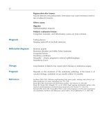
Suggested Reading
Adam A, Hochholzer L. Ganglioneuroblastoma of the posterior mediastinum: a clinicopathologic review of 80 cases. Cancer.
1981;47:373–81.
Ambros IM, Ambros PF, Strehl S, Kovar H, Gadner H, Salzer-Kuntschik
M. MIC2 is a specific marker for Ewing’s sarcoma and peripheral
primitive neuroectodermal tumors. Evidence for a common histogenesis of Ewing’s sarcoma and peripheral primitive neuroectodermal tumor from MIC2 expression and specific chromosomal
aberration. Cancer. 1991;67:1886–93.
Cardillo G, Carleo F, Khalil MW, Carbone L, Treggiari S, Salvadori L,
et al. Surgical treatment of benign neurogenic tumours of the mediastinum: a single institution report. Eur J Cardiothorac Surg.
2008;34:1210–4.
Carney JA. Psammomatous melanotic schwannoma: a distinctive
heritable tumor with special associations, including cardiac myxomas and Cushing’s syndrome. Am J Surg Pathol. 1990;14:
206–22.
Chalmers AH, Armstrong P. Plexiform mediastinal neurofibromas. A
report of two cases. Br J Radiol. 1977;50:215–7.
Chatten J, Shimada H, Sather HN, Wong KY, Siegel SE, Hammond
GD. Prognostic value of histopathology in advanced neuroblastoma: a report from the Childrens Cancer Study Group. Hum Pathol.
1988;19:1187–8.
Dehner LP. Primitive neuroectodermal tumor and Ewing’s sarcoma.
Am J Surg Pathol. 1993;17:1–13.
Cataldo D. Mediastinal ganglioneuroma: a rare and often asymptomatic
tumor. Chir Ital. 2005;53:403–5.
Ducatman BS, Scheithauer BW, Piepgras BG, Reiman HM, Ilstrup
DM. Malignant peripheral nerve sheath tumors. A clinicopathologic
study of 120 cases. Cancer. 1986;57:2006–21.
Hirschfeld K, Woodward W. Neurofibromata of the anterior mediastinum. Aust N Z J Surg. 1963;33:76–7.
Inoue M, Mitsudomi T, Osaki T, Oyama T, Haratake J, Yasumoto
K. Malignant transformation of an intrathoracic neurofibroma in
von Recklinghausen’s disease. Scand Cardiovasc J. 1998;32:
173–5.
Johnson MD, Glick AD, Davis BW. Immunohistochemical evaluation
of Leu-7, myelin basic protein, S-100 protein, glial fibrillary acidic
protein, and LN3 immunoreactivity in nerve sheath tumors and sarcomas. Arch Pathol Lab Med. 1988;112:155–60.
Joshi VV, Cantor AB, Altshuler G, Larkin EW, Neill JS, Shuster JJ,
et al. Recommendations for modification of terminology of neuroblastic tumors and prognostic significance of Shimada classification. A clinicopathologic study of 213 cases from the Pediatric
Oncology Group. Cancer. 1992;69:2183–96.
King RM, Telander RL, Smithson WA, Banks PM, Han MT. Primary
mediastinal tumors in children. J Pediatr Surg. 1982;17:
512–20.
4
Neurogenic Tumors
Koezuka S, Hata Y, Sato F, Otsuka H, Makina T, Tochigi N, Iyoda
A. Malignant peripheral nerve sheath tumor in the anterior mediastinum: a case report. Mol Clin Oncol. 2014;2:987–90.
Manduch M, Dexter DF, Ellis PM, Reid K, Isotalo PA. Extraskeletal
Ewing’s sarcoma/primitive neuroectodermal tumor of the posterior
mediastinum with t(11;22)(q24;q12). Tumori. 2008;94:888–91.
Marchevsky AM. Mediastinal tumors of peripheral nervous system origin. Semin Diagn Pathol. 1999;16:65–78.
Pekmeczi M, Reuss DE, Hirbe AC, Dahiya S, Gutmann DH, von
Deimling A, et al. Morphologic and immunohistochemical features
of malignant peripheral nerve sheath tumors and cellular schwannomas. Mod Pathol. 2014. doi:10.1038/modpathol.2014.109. [Epub
ahead of print].
Schweigert M, Meyer C, Wolf F, Stein HJ. Peripheral primitive neuroectodermal tumor of the thymus. Interact Cardiovasc Thorac Surg.
2011;12:303–5.
Shields TW, Reynolds M. Neurogenic tumors of the thorax. Surg Clin
North Am. 1988;68:645–68.
Shirakusa T, Tsutsui M, Montonaga R, Takata S, Yoshomine K, Kondo
K, Yoshida T. Intrathoracic tumors arising from the vagus nerve.
Review of resected tumors in Japan. Scand J Thorac Cardiovasc
Surg. 1989;23:173–5.
Steffanson K, Wollmann R, Jerkovic M. S-100 protein in soft tissue
tumors derived from schwann cells and melanocytes. Am J Pathol.
1982;106:261–8.
Sugio K, Inoue T, Inoue K, Tateishi M, Ishida T, Sugimachi
K. Neurogenic tumors of the mediastinum originated from the
vagus nerve. Eur J Surg Oncol. 1995;21:214–6.
Suster S. Recent advances in the application of immunohistochemical
markers for the diagnosis of soft tissue tumors. Semin Diagn Pathol.
2000;17:225–35.
Takeda S, Miyoshi S, Minami M, Matsuda H. Intrathoracic neurogenic
tumors–50 years’ experience in a Japanese institution. Eur J
Cardiothorac Surg. 2004;26:807–12.
Torres-Mora J, Dry S, Li X, Binder S, Amin M, Folpe AL. Malignant
melanotic schwannian tumor: a clinicopathologic, immunohistochemical, and gene expression profiling study of 40 cases, with a
proposal for the reclassification of “melanotic schwannoma”. Am J
Surg Pathol. 2014;38:94–105.
Turc-Carel C, Aurias A, Mugneret F, Lizard S, Sidaner I, Volk C, et al.
Chromosomes in Ewing’s sarcoma. I. An evaluation of 85 cases of
remarkable consistency of t(11;22)(q24;q12). Cancer Genet
Cytogenet. 1988;32:229–38.
Weitzner S. Adjacent malignant schwannoma and neurofibroma of
intrathoracic vagus. Am Surg. 1976;42:866–70.
Wick MR, Swanson PE, Scheithauer BW, Manivel JC. Malignant
peripheral nerve sheath tumor. An immunohistochemical study of
62 cases. Am J Clin Pathol. 1987;87:425–33.
Woodruff JM, Godwin TA, Erlandson RA, Susin M, Martini N. Cellular
schwannoma: a variety of schwannoma sometimes mistaken for a
malignant tumor. Am J Surg Pathol. 1981;5:733–44.
5
Soft Tissue Tumors
of the Mediastinum
Mesenchymal tumors of the mediastinum are relatively rare,
yet virtually all types of mesenchymal neoplasms have been
described in this location (Table 5.1).
Table 5.1 Mesenchymal tumors of the mediastinum
Category
Vascular tumors
Fibroblastic/fibrohistiocytic
tumors
Myogenic tumors
Lipomatous tumors
Bone and cartilaginous tumors
Tumors of unknown etiology
S. Suster (ed.), Atlas of Mediastinal Pathology, Atlas of Anatomic Pathology,
DOI 10.1007/978-1-4939-2674-9_5, © Springer Science+Business Media, LLC 2015
Tumor type
Hemangioma
Lymphangioma
Epithelioid
hemangioendothelioma
Angiosarcoma
Solitary fibrous tumor
Malignant solitary fibrous
tumor
Malignant fibrous histiocytoma
(undifferentiated sarcoma)
Leiomyoma
Leiomyosarcoma
Rhabdomyosarcoma
Malignant “triton” tumor
Lipoma
Atypical lipomatous tumor/
well-differentiated liposarcoma
Dedifferentiated liposarcoma
Myxoid/round cell liposarcoma
Pleomorphic liposarcoma
Chondrosarcoma
Chordoma
Extraskeletal mesenchymal
chondrosarcoma
Myxoid chondrosarcoma
Extraskeletal osteosarcoma
Malignant rhabdoid tumor
Synovial sarcoma
Alveolar soft part sarcoma
111
112
5.1
5
Vascular Tumors
The majority of tumors originating from vascular endothelium in the mediastinum are benign conditions that most
likely represent malformative or congenital processes occurring during childhood or adolescence (Figs. 5.1, 5.2, 5.3, 5.4,
5.5, 5.6, 5.7, 5.8, 5.9, 5.10, 5.11, 5.12, 5.13, 5.14, 5.15, 5.16,
5.17, 5.18, 5.19, 5.20, 5.21, 5.22, 5.23, 5.24, 5.25, 5.26, 5.27,
5.28, 5.29, 5.30, 5.31, 5.32, 5.33, 5.34, 5.35, 5.36, 5.37, 5.38,
5.39, 5.40, 5.41, 5.42, and 5.43). Benign, low-grade, and
Soft Tissue Tumors of the Mediastinum
malignant vascular neoplasms are extremely rare in the
mediastinum and tend to occur more often in the pediatric
population. Paradoxically, one of the rarest forms of lowgrade malignant vascular neoplasm, epithelioid hemangioendothelioma (Figs. 5.7, 5.8, 5.9, 5.10, 5.11, 5.12, 5.13, 5.14,
5.15, 5.16, 5.17, 5.18, 5.19, 5.20, 5.21, 5.22, 5.23, 5.24, 5.25,
5.26, 5.27, 5.28, 5.29, 5.30, 5.31, 5.32, 5.33, 5.34, 5.35, 5.36,
and 5.37), represents the vascular malignancy most commonly encountered in the mediastinum in adults.
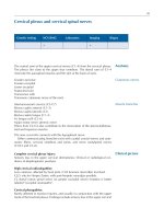
Fig. 5.1 Gross appearance of cystic lymphangioma (cystic hygroma)
of the mediastinum in a child shows a smooth and shiny outer surface
distended by fluid
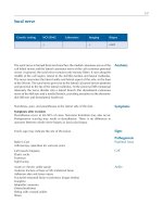
Fig. 5.3 On higher magnification, cystic lymphangiomas will disclose
small, rudimentary valves (arrows) in charge of regulating the lymph
flow
Fig. 5.2 Cystic lymphangiomas are composed of multiple, cystically
dilated lymphatics that often contain dense, lymphoid aggregates in
their walls. The dilated empty spaces may contain occasional small
lymphocytes but are devoid of red blood cells
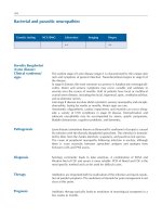
Fig. 5.4 Mediastinal hemangiomas are most commonly of the cavernous type and are characterized by multiple dilated vessels distended
with red blood cells
5.1
Vascular Tumors
Fig. 5.5 At higher magnification, the mediastinal hemangioma shows
thin-walled vessels lined by a single layer of endothelial cells and containing red blood cells in their lumen
Fig. 5.6 Mediastinal hemangiomas are most likely hamartomatous
malformations that often contain other components admixed with the
vascular elements, such as smooth muscle, fat, and cartilage. This
example shows a well-developed smooth muscle component surrounding the vessels, along with mature stromal fat
113
Fig. 5.7 Epithelioid hemangioendothelioma is the most common
malignant vascular neoplasm of the mediastinum, where they are characterized by relatively low-grade malignant behavior and are associated
with a very good survival rate following complete surgical excision.
The cut surface often shows a fleshy, multinodular appearance with
congestion and foci of hemorrhage. Gritty foci of calcifications are also
often noted
Fig. 5.8 Histologically, epithelioid hemangioendothelioma is characterized by a proliferation of large epithelioid cells with enlarged nuclei
and abundant cytoplasm that can closely resemble an epithelial malignancy. The nuclei show dispersed chromatin and prominent eosinophilic nucleoli; intranuclear cytoplasmic pseudoinclusions can be seen.
Despite the atypical appearance of the cells, mitoses are inconspicuous