Clinical brain mapping
Bạn đang xem bản rút gọn của tài liệu. Xem và tải ngay bản đầy đủ của tài liệu tại đây (9.88 MB, 301 trang )
Clinical Brain Mapping
Notice
Medicine is an ever-changing science. As new research and clinical experience
broaden our knowledge, changes in treatment and drug therapy are required.
The authors and the publisher of this work have checked with sources believed
to be reliable in their efforts to provide information that is complete and generally in accord with the standards accepted at the time of publication. However,
in view of the possibility of human error or changes in medical sciences, neither
the authors nor the publisher nor any other party who has been involved in the
preparation or publication of this work warrants that the information contained
herein is in every respect accurate or complete, and they disclaim all responsibility for any errors or omissions or for the results obtained from use of the information contained in this work. Readers are encouraged to confirm the information contained herein with other sources. For example and in particular, readers
are advised to check the product information sheet included in the package of
each drug they plan to administer to be certain that the information contained in
this work is accurate and that changes have not been made in the recommended
dose or in the contraindications for administration. This recommendation is
of particular importance in connection with new or infrequently used drugs.
Clinical Brain Mapping
Daniel Yoshor, MD
Associate Professor
Department of Neurosurgery
Baylor College of Medicine
Chief of Neurosurgery
St. Luke’s Episcopal Hospital
Houston, Texas
Eli M. Mizrahi, MD
Chair, Department of Neurology
Professor of Neurology and Pediatrics
Baylor College of Medicine
Chief of Neurophysiology
St. Luke’s Episcopal Hospital
Houston, Texas
New York Chicago San Francisco Lisbon London Madrid Mexico City
Milan New Delhi San Juan Seoul Singapore Sydney Toronto
Copyright © 2012 by The McGraw-Hill Companies, Inc. All rights reserved. Except as permitted under the United States Copyright Act of 1976, no part of this
publication may be reproduced or distributed in any form or by any means, or stored in a database or retrieval system, without the prior written permission of
the publisher.
ISBN: 978-0-07-180596-4
MHID: 0-07-180596-6
The material in this eBook also appears in the print version of this title: ISBN: 978-0-07-148441-1,
MHID: 0-07-148441-8.
All trademarks are trademarks of their respective owners. Rather than put a trademark symbol after every occurrence of a trademarked name, we use names in
an editorial fashion only, and to the benefit of the trademark owner, with no intention of infringement of the trademark. Where such designations appear in this
book, they have been printed with initial caps.
McGraw-Hill eBooks are available at special quantity discounts to use as premiums and sales promotions, or for use in corporate training programs. To contact
a representative please e-mail us at
TERMS OF USE
This is a copyrighted work and The McGraw-Hill Companies, Inc. (“McGraw-Hill”) and its licensors reserve all rights in and to the work. Use of this work is
subject to these terms. Except as permitted under the Copyright Act of 1976 and the right to store and retrieve one copy of the work, you may not decompile,
disassemble, reverse engineer, reproduce, modify, create derivative works based upon, transmit, distribute, disseminate, sell, publish or sublicense the work or
any part of it without McGraw-Hill’s prior consent. You may use the work for your own noncommercial and personal use; any other use of the work is strictly
prohibited. Your right to use the work may be terminated if you fail to comply with these terms.
THE WORK IS PROVIDED “AS IS.” McGRAW-HILL AND ITS LICENSORS MAKE NO GUARANTEES OR WARRANTIES AS TO THE ACCURACY,
ADEQUACY OR COMPLETENESS OF OR RESULTS TO BE OBTAINED FROM USING THE WORK, INCLUDING ANY INFORMATION THAT
CAN BE ACCESSED THROUGH THE WORK VIA HYPERLINK OR OTHERWISE, AND EXPRESSLY DISCLAIM ANY WARRANTY, EXPRESS OR
IMPLIED, INCLUDING BUT NOT LIMITED TO IMPLIED WARRANTIES OF MERCHANTABILITY OR FITNESS FOR A PARTICULAR PURPOSE.
McGraw-Hill and its licensors do not warrant or guarantee that the functions contained in the work will meet your requirements or that its operation will be
uninterrupted or error free. Neither McGraw-Hill nor its licensors shall be liable to you or anyone else for any inaccuracy, error or omission, regardless of cause,
in the work or for any damages resulting therefrom. McGraw-Hill has no responsibility for the content of any information accessed through the work. Under no
circumstances shall McGraw-Hill and/or its licensors be liable for any indirect, incidental, special, punitive, consequential or similar damages that result from the
use of or inability to use the work, even if any of them has been advised of the possibility of such damages. This limitation of liability shall apply to any claim
or cause whatsoever whether such claim or cause arises in contract, tort or otherwise.
Dedication
To our parents, Shulamit and Joseph Yoshor, and Julia and Isaac D. Mizrahi, who encouraged and
sustained us; and to our patients who inspire and teach us.
v
This page intentionally left blank
Contents
Contributors. . . . . . . . . . . . . . . . . . . . . . . . . . . . . . . . . . . . . . . . . . . . . . . . . . . . . . . . . . . . . . . . . . . . . . . . . . . . . . . . . . . . . . . . . . . . . . . . . . . . . . . . . . . . . . . . . . . . . . . . . . . . . . . . . ix
Preface . . . . . . . . . . . . . . . . . . . . . . . . . . . . . . . . . . . . . . . . . . . . . . . . . . . . . . . . . . . . . . . . . . . . . . . . . . . . . . . . . . . . . . . . . . . . . . . . . . . . . . . . . . . . . . . . . . . . . . . . . . . . . . . . . . . . xiii
Acknowledgments . . . . . . . . . . . . . . . . . . . . . . . . . . . . . . . . . . . . . . . . . . . . . . . . . . . . . . . . . . . . . . . . . . . . . . . . . . . . . . . . . . . . . . . . . . . . . . . . . . . . . . . . . . . . . . . . . . . . . . . . . xv
SECTION I: TECHNIQUES
Chapter 1.
Surface Anatomy as a Guide to Cerebral Function . . . . . . . . . . . . . . . . . . . . . . . . . . . . . . . . . . . . . . . . . . . . . . . . . . . . . . . . . . . 3
Gareth Adams, Jared Fridley, and Daniel Yoshor
Chapter 2.
Structural Imaging for Identification of Functional Brain Regions. . . . . . . . . . . . . . . . . . . . . . . . . . . . . . . . . . . . . . . . . .13
Jean C. Tamraz and Youssef G. Comair
Chapter 3.
Functional MRI for Cerebral Localization: Principles and Methodology . . . . . . . . . . . . . . . . . . . . . . . . . . . . . . . . . . .31
Michael S. Beauchamp
Chapter 4.
Functional MRI: Application to Clinical Practice in Surgical Planning and
Intraoperative Guidance. . . . . . . . . . . . . . . . . . . . . . . . . . . . . . . . . . . . . . . . . . . . . . . . . . . . . . . . . . . . . . . . . . . . . . . . . . . . . . . . . . . . . . . . 45
Michael Schulder and Robin Wellington
Chapter 5.
Neuropsychological Testing: Understanding Brain–behavior Relationships . . . . . . . . . . . . . . . . . . . . . . . . . . . . . . .55
Mario F. Dulay, Corwin Boake, Daniel Yoshor, and Harvey S. Levin
Chapter 6.
The Wada Test: Intracarotid Injection of Sodium Amobarbital to Evaluate Language
and Memory . . . . . . . . . . . . . . . . . . . . . . . . . . . . . . . . . . . . . . . . . . . . . . . . . . . . . . . . . . . . . . . . . . . . . . . . . . . . . . . . . . . . . . . . . . . . . . . . . . . . . 79
Brian D. Bell, Bruce P. Hermann, and Paul Rutecki
Chapter 7.
Extraoperative Brain Mapping Using Chronically Implanted Subdural Electrodes. . . . . . . . . . . . . . . . . . . . . . . .93
David E. Friedman and James J. Riviello, Jr.
Chapter 8.
Brain Mapping in the Operating Room . . . . . . . . . . . . . . . . . . . . . . . . . . . . . . . . . . . . . . . . . . . . . . . . . . . . . . . . . . . . . . . . . . . . . .103
Sepehr Sani, Edward F. Chang, and Nicholas M. Barbaro
Chapter 9.
Anesthesia for Brain Mapping Surgery . . . . . . . . . . . . . . . . . . . . . . . . . . . . . . . . . . . . . . . . . . . . . . . . . . . . . . . . . . . . . . . . . . . . . . .109
Nicholas P. Carling, Chris D. Glover, Daryn H. Moller, and Ira J. Rampil
Chapter 10. Clinical Applications of Magnetoencephalography in Neurology and Neurosurgery . . . . . . . . . . . . . . . . . . .119
Panagiotis G. Simos, Eduardo M. Castillo, and Andrew C. Papanicolaou
Chapter 11. Optical Spectroscopic Imaging of the Human Brain—Clinical Applications . . . . . . . . . . . . . . . . . . . . . . . . . . . . .131
Hongtao Ma, Minah Suh, Mingrui Zhao, Challon Perry, Andrew Geneslaw, and Theodore H. Schwartz
vii
viii
CONTENTS
Chapter 12. Electrocorticographic Spectral Analysis . . . . . . . . . . . . . . . . . . . . . . . . . . . . . . . . . . . . . . . . . . . . . . . . . . . . . . . . . . . . . . . . . . . . . .151
Mackenzie C. Cervenka and Nathan E. Crone
Chapter 13. Pediatric Brain Mapping: Special Considerations . . . . . . . . . . . . . . . . . . . . . . . . . . . . . . . . . . . . . . . . . . . . . . . . . . . . . . . . . . .167
Robert J. Bollo, Chad Carlson, Orrin Devinsky, and Howard L. Weiner
SECTION II: SYSTEMS
Chapter 14. Mapping of the Sensorimotor Cortex. . . . . . . . . . . . . . . . . . . . . . . . . . . . . . . . . . . . . . . . . . . . . . . . . . . . . . . . . . . . . . . . . . . . . . . . .189
Roukoz Chamoun, Krishna Satyan, and Youssef G. Comair
Chapter 15. Mapping of Human Language . . . . . . . . . . . . . . . . . . . . . . . . . . . . . . . . . . . . . . . . . . . . . . . . . . . . . . . . . . . . . . . . . . . . . . . . . . . . . . . .203
Nitin Tandon
Chapter 16. Mapping of the Human Visual System . . . . . . . . . . . . . . . . . . . . . . . . . . . . . . . . . . . . . . . . . . . . . . . . . . . . . . . . . . . . . . . . . . . . . . .219
Muhammad M. Abd-El-Barr, Mario F. Dulay, Paul Richard, William H. Bosking, and Daniel Yoshor
Chapter 17. Mapping of Hearing. . . . . . . . . . . . . . . . . . . . . . . . . . . . . . . . . . . . . . . . . . . . . . . . . . . . . . . . . . . . . . . . . . . . . . . . . . . . . . . . . . . . . . . . . . . .241
Albert J. Fenoy and Matthew A. Howard
Chapter 18. Mapping of Memory . . . . . . . . . . . . . . . . . . . . . . . . . . . . . . . . . . . . . . . . . . . . . . . . . . . . . . . . . . . . . . . . . . . . . . . . . . . . . . . . . . . . . . . . . . .269
Jeffrey G. Ojemann and Richard G. Ellenbogen
Index . . . . . . . . . . . . . . . . . . . . . . . . . . . . . . . . . . . . . . . . . . . . . . . . . . . . . . . . . . . . . . . . . . . . . . . . . . . . . . . . . . . . . . . . . . . . . . . . . . . . . . . . . . . . . . . . . . . . . . . . . . . . . . . . . . . . . . 277
Contributors
Muhammad M. Abd-El-Barr, MD, PhD
Department of Neurosurgery
University of Florida
Gainesville, Florida
Nicholas P. Carling, MD
Department of Pediatrics (Anesthesiology)
Texas Children’s Hospital
Baylor College of Medicine
Houston, Texas
Gareth Adams, MD, PhD
Department of Neurosurgery
Baylor College of Medicine
Houston, Texas
Chad Carlson, MD
Comprehensive Epilepsy Center
Department of Neurology
New York University School of Medicine
New York, New York
Nicholas M. Barbaro, MD
Department of Neurological Surgery
Indiana University School of Medicine
Indianapolis, Indiana
Eduardo M. Castillo, PhD
Department of Pediatrics, Center for
Clinical Neurosciences
Departments of Neurosurgery and Neurology
University of Texas—Health Science Center at Houston
Houston, Texas
Michael S. Beauchamp, PhD
Department of Neurobiology & Anatomy
University of Texas Health Science Center
Houston, Texas
Mackenzie C. Cervenka, MD
Department of Neurology
The Johns Hopkins University School of Medicine
Baltimore, Maryland
Brian D. Bell, PhD
Department of Neurology
University of Wisconsin School of Medicine and
Public Health
Department of Neurology
W.S. Middleton Memorial Veterans Hospital
Madison, Wisconsin
Roukoz Chamoun, MD
Department of Neurosurgery
Baylor College of Medicine
Houston, Texas
Corwin Boake, PhD
Department of Physical Medicine & Rehabilitation
University of Texas Medical School
Houston, Texas
Edward F. Chang, MD, PhD
Department of Neurological Surgery
University of California
San Francisco, California
Robert J. Bollo, MD
Department of Neurosurgery
Baylor College of Medicine
Neurosurgery Service
Texas Children’s Hospital
Houston, Texas
Youssef G. Comair, MD
Department of Neurosurgery
American University of Beirut
Beirut, Lebanon
Nathan E. Crone, MD
Department of Neurology
The Johns Hopkins University School of Medicine
Baltimore, Maryland
William H. Bosking, PhD
Max Planck Florida Institute
Jupiter, Florida
ix
x
CONTRIBUTORS
Orrin Devinsky, MD
Comprehensive Epilepsy Center
Department of Neurology
New York University School of Medicine
New York, New York
Mario F. Dulay, PhD
Department of Neurosurgery
The Methodist Hospital Neurological Institute
Houston, Texas
Richard G. Ellenbogen, MD
Department of Neurological Surgery
University of Washington School of Medicine
Seattle, Washington
Hongtao Ma, PhD
Department of Neurosurgery
Weill Medical College of Cornell University
New York, New York
Eli M. Mizrahi, MD
Departments of Neurology and Pediatrics
Baylor College of Medicine
Houston, Texas
Daryn H. Moller, MD
Department of Anesthesiology
University at Stony Brook
Stony Brook, New York
Albert J. Fenoy, MD
Department of Neurosurgery
University of Texas Health Science Center
Houston, Texas
Jeffrey G. Ojemann, MD
Department of Neurological Surgery
University of Washington School of Medicine
Seattle, Washington
Jared Fridley, MD
Department of Neurosurgery
Baylor College of Medicine
Houston, Texas
Andrew C. Papanicolaou, PhD
Department of Pediatrics, Center for
Clinical Neurosciences
Departments of Neurosurgery and Neurology
University of Texas—Health Science Center at Houston
Houston, Texas
David E. Friedman, MD
Department of Neurosciences
Winthrop-University Hospital
Mineola, New York
Andrew Geneslaw
Department of Neurosurgery
Weill Medical College of Cornell University
New York, New York
Chris D. Glover, MD
Department of Pediatrics (Anesthesiology)
Texas Children’s Hospital
Baylor College of Medicine
Houston, Texas
Bruce P. Hermann, PhD
Department of Neurology
University of Wisconsin School of Medicine and
Public Health
Madison, Wisconsin
Matthew A. Howard, MD
Department of Neurosurgery
University of Iowa Hospitals and Clinics
Iowa City, Iowa
Harvey S. Levin, PhD
Departments of Physical Medicine & Rehabilitation,
Pediatrics, Neurosurgery, and Neurology
Baylor College of Medicine
Houston, Texas
Challon Perry, MD
Department of Neurosurgery
Weill Medical College of Cornell University
New York, New York
Ira J. Rampil, MD
Department of Anesthesiology
University at Stony Brook
Stony Brook, New York
Paul Richard, MD
Department of Neurological Surgery
University of Pittsburgh Medical Center
Pittsburgh, Pennsylvania
James J. Riviello, Jr., MD
Division of Pediatric Neurology
Department of Neurology
NYU Comprehensive Epilepsy Center
New York University
New York, New York
Paul Rutecki, MD
Department of Neurology
University of Wisconsin School of Medicine and
Public Health
Department of Neurology
W.S. Middleton Memorial Veterans Hospital
Madison, Wisconsin
CONTRIBUTORS
Sepehr Sani, MD
Department of Neurosurgery
Rush University Medical Center
Chicago, Illinois
Krishna Satyan, MD
Department of Neurosurgery
Baylor College of Medicine
Houston, Texas
Michael Schulder, MD
Department of Neurosurgery
North Shore-LIJ Health System
Manhasset, New York
Jean C. Tamraz, MD, PhD
Department of Neuroscience & Neuroradiology
Saint-Joseph University
Beirut, Lebanon
Nitin Tandon, MD
Department of Neurosurgery
University of Texas Medical School
Houston, Texas
Howard L. Weiner, MD
Department of Neurosurgery
Division of Pediatric Neurosurgery
Comprehensive Epilepsy Center
Department of Neurology
New York University School of Medicine
New York, New York
Theodore H. Schwartz, MD
Department of Neurosurgery
Weill Medical College of Cornell University
New York, New York
Robin Wellington, PhD
Department of Psychology
St. John’s University
Flushing, New York
Panagiotis G. Simos, PhD
Department of Psychology
University of Crete
Rethymno, Greece
Daniel Yoshor, MD
Department of Neurosurgery
Baylor College of Medicine
Neuroscience Center
St. Luke’s Episcopal Hospital
Houston, Texas
Minah Suh, PhD
Department of Neurosurgery
Weill Medical College of Cornell University
New York, New York
Mingrui Zhao, MD, PhD
Department of Neurosurgery
Weill Medical College of Cornell University
New York, New York
xi
This page intentionally left blank
Preface
The localization of cerebral function is a critical task
for both neurosurgeons and neurologists. In Clinical
Brain Mapping we have addressed these localization efforts from the perspectives of our different but, at times,
overlapping backgrounds and clinical interests. One of
us is a neurosurgeon (Daniel Yoshor) and the other a
neurologist (Eli M. Mizrahi). We began our discussions
about clinical brain mapping early in our careers as we
cared for adults and children with medically intractable
epilepsy who were being evaluated for epilepsy surgery.
We have worked together for several years at the Baylor
Comprehensive Epilepsy Center considering the issues
of cerebral localization and weighing relative risks and
benefits of resective surgery for potential seizure control, as well as in resective surgery for brain tumors. In
the course of our clinical practice, we realized the need
for a concise and practical, but comprehensive, guide to
clinical brain mapping. In addition to our efforts with
patients, through our interactions with colleagues and
trainees, we realized that such a volume would be of
value to them both for reference and training. This was
beginning of the current volume.
Although initially considered within the context of
epilepsy surgery, Clinical Brain Mapping addresses a
wide range of clinical concerns. It addresses the techniques and functional bases for all clinical situations that
may require cerebral localization for diagnosis and management. Most of the techniques described are now part
of clinical care, others are just now emerging technology
and not yet fully integrated into clinical practice, and
some techniques have their greatest utility in clinical research. It is meant as a reference for neurosurgeons, neurologists, neuroradiologists, neuropsychologists, clinical
neuroscientists and others actively involved in the care
of those with or who are at risk for neurological impairment through intervention.
We have organized the volume into two sections:
Techniques and Systems. The Techniques section consists of chapters considering specific methods of cerebral location: operative anatomy, structural neuroimaging, functional MRI, magnetoencephalography, optical
imaging, neuropsychological testing, Wada testing, special intraoperative mapping techniques, extraoperative
brain mapping with implanted electrodes, electrocorticographic spectral analysis, special brain mapping techniques for pediatric patients and anesthetic techniques
for intraoperative brain mapping. In the Systems section
there are discussions of somatomotor and somotosensory function, language, vision, hearing, and memory.
Each is written by experts in their respective fields.
This book is intended to serve two purposes. It has
been developed as a practical guide to brain mapping
in the clinical setting and it is also designed to present
the scientific basis of the cortical systems that we wish
to localize and preserve in the care of our patients.
Daniel Yoshor, MD
Eli M. Mizrahi, MD
xiii
This page intentionally left blank
Acknowledgments
Brain mapping is typically a collaborative effort in both
the clinical and research settings. The development of
Clinical Brain Mapping has also been collaborative.
We are grateful to those who have been instrumental in its production, including the 47 contributors to
this volume who have given generously of their time
and expertise to write timely, insightful, and instructive
manuscripts.
We have been fortunate to work in an enriched
environment that fosters expert patient care and excellence in research. The clinicians, scientists, trainees and
students at Baylor College of Medicine form an invigorating intellectual community, which has fostered our
interests and growth as researchers and clinicians. Similarly, St. Luke’s Episcopal Hospital has encouraged and
supported our work and has proved to be a unique institution which promotes outstanding clinical care and
clinical research.
We are also grateful to Robert G. Grossman, MD,
and the late Peter Kellaway, PhD. They initially taught
and mentored us, and then became professional colleagues and personal friends.
Dr. Yoshor acknowledges the influence of Nicholas
M. Barbaro, MD, Mitchel S. Berger, MD, and Raymond
L. Sawaya, MD, in developing his interest in applying
brain mapping to clinical practice, and of John H.R.
Maunsell, PhD, and Michael S. Beauchamp, PhD, in developing a research program that strives to use rigorous scientific methodology in studying human cortical
function.
Dr. Mizrahi is grateful to his long-time colleagues
and collaborators in the Peter Kellaway Section of Neurophysiology, Department of Neurology, Baylor College
of Medicine, particularly James D. Frost, Jr., MD, and
Richard A. Hrachovy, MD. They continue to provide
valuable insights into the neurophysiological aspects of
cortical mapping.
As with any collaborative effort, there are many
people who have contributed directly and indirectly to
Clinical Brain Mapping. We are most grateful to our
co-workers, clinical and research colleagues, technologists, trainees, and administrative staff for their diligence
and hard work on our behalf. In particular, Lisa Rhodes,
R EEG/EP T., CLTM, has provided outstanding technical support for brain mapping studies in our patients
for many years. Kathleen Pierson and May-Lin Basso
provided critical and expert administrative assistance. Finally, we express our sincere thanks to the editorial staff
at McGraw-Hill Medical Publishing, Anne Sydor, PhD,
Executive Editor, and Regina Y. Brown, Senior Project
Development Editor and to Tilak Raj, Project Manager,
Aptara Corporation, Inc.
xv
This page intentionally left blank
SECTION I
TECHNIQUES
1
This page intentionally left blank
Chapter 1
Surface Anatomy as a Guide to
Cerebral Function
Gareth Adams1 , Jared Fridley 1 , and Daniel Yoshor 1,2
1
Department of Neurosurgery, Baylor College of Medicine, Houston, Texas
2
Neuroscience Center, St. Luke’s Episcopal Hospital, Houston, Texas
᭤ INTRODUCTION AND
the localizationist theory, which held that brain functions are localized to specific areas of the brain. Further
understanding of the localization of human brain function was based on correlating the neurological deficits
in patients with specific cortical lesions defined on postmortem examinations.2 For example, by performing autopsies on patients with aphasia, Paul Broca was able
to localize the functional areas responsible for the production of speech to the pars triangularis and pars opercularis of the dominant frontal lobe. Carl Wernicke was
able to localize language comprehension to the posterior, superior temporal gyrus. Correlation of lesions in
the occipital lobes from shrapnel and penetrating trauma
with the visual field defects sustained by soldiers in
World War II combat provided further localization of
visual function in humans.3 Similarly, studies of subjects with cortical lesions and sensory and motor deficits
demonstrated that motor and sensory functions are localized around the central sulcus.3 Collectively over a period of decades, a crude understanding of the anatomic
location of functional regions emerged from these
studies.
Two other methods have greatly further extended
our understanding of the functional organization of
human cerebral cortex. Pioneering studies employing
direct cortical electrical stimulation in human neurosurgical patients demonstrated consistent relationships between cortical anatomy and cortical eloquence among
many different subjects. For example, Wilder Penfield
demonstrated through human cortical mapping during
planning for cortical excisions that motor and sensory
function is localized around the central sulcus, and was
able to map a motor and sensory homunculus to the
central area.2,4 More recently, the advent of structural
and functional MRI (fMRI) has had an explosive impact on our understanding of the consistent relationship between anatomy and regional function.5,6 Studies
that combine both electrical stimulation and fMRI mapping in individual subjects has further validated these
relationships.7−9
HISTORICAL PERSPECTIVE
Our existing knowledge of a number of consistent relationships between specific anatomic landmarks and
local cortical function allows the use of anatomy to predict function with considerable accuracy, even without
direct physiological confirmation in an individual subject. Defining these landmarks with noninvasive structural magnetic resonance imaging (MRI) is routinely
used to infer regional function, as described in Chapter 2, “Structural Imaging for Identification of Functional
Brain Regions.” Direct inspection of anatomic clues, in
particular examination of the exposed cortical anatomy
of the brain during craniotomy, can provide highly
useful clues to functional localization. Because intersubject variation in cortical anatomy and functional localization is not insignificant, and because local pathology
such as a brain tumor maybe obscure anatomic clues,
accurate identification of functional regions often requires physiological mapping through other techniques
presented in this book. But anatomic landmarks remain invaluable, both as a primary method and as
an adjunct to the physiological techniques described
in this book, for plotting regional brain function. This
chapter reviews anatomic methods for estimating regional functional by simple visual inspection. It is broken down into sections detailing anatomic techniques
for surface localization of underlying cortical anatomy,
and clues for localization of speech, motor and sensory
function, vision and hearing based on cortical surface
anatomy.
Historically, the understanding of the presence of
localized brain function has been based on experimentally created lesions in animals. During a prominent public lecture and scientific debate in 1881, Sir David Ferrier
convincingly showed that creating a lesion in a monkey’s
left posterior frontal cortex resulted in a right-sided hemiplegia, and that bilateral lesions in the superior temporal lobes resulted in deafness.1 This evidence buttressed
3
SECTION I
TECHNIQUES
50%
NE
LI
C
DI
AN
OL
T
E
S
VIA
YL I N N
O T
TE
PAR
I
M
PREAUR
POINT
75%
ET
Y LV
IAN
R
S
PORAL L
PREAUR
LINE
0%
NASION
N
LOB
L
A
P
FR
O
25%
ORB.
TEMP.
ANG.
UPPER ROLANDIC POINT
E
L I N LO
BE
EXT. PAR. OCC.
FIS
OC
CI
LO PITAL
BE
4
100%
INION
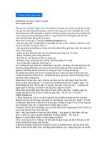
Figure 1–1. Taylor–Haughton lines.Method for approximating the central sulcus and
sylvian fissure using the Taylor–Haughton lines. (From Taylor and Haughton’s Some
Recent Researches on the Topography and Convolutions of the Brain.)
᭤ APPROXIMATE LOCALIZATION
OF CORTICAL STRUCTURAL
ANATOMY USING
EXTERNAL CRANIAL
LANDMARKS
The location of important cortical anatomic features,
such as the sylvian fissure and central sulcus, can be
approximated from the external anatomy of the skull.10
Taylor–Haughton lines (Fig. 1–1) can be simply constructed from external landmarks by drawing four lines
on the cranium. The baseline, or Frankfurt plane, is defined as a line passing the inferior margin of the orbit through the superior margin of the external auditory meatus. A second line is drawn from the nasion
to the inion across the top of the cranium and divided
into quarters. Two more lines are drawn perpendicular
to the baseline. The posterior ear line is perpendicular
to the baseline and passes through the mastoid process.
The condylar line is perpendicular to the baseline and
passes through the mandibular condyle.11
The location of the sylvian fissure can be approximated by drawing a line from the lateral canthus to
the three-quarter point along the Taylor–Haughton line
from the nasion to the inion. The central sulcus can be
approximated by multiple methods. One method to approximate the central sulcus is to connect a point 2 cm
posterior to the halfway point of the Taylor–Haughton
line across the top of the cranium with a point 5 cm
above the external auditory meatus. A second method is
to connect the point on the Taylor–Haughton line across
the top of the cranium where it is intersected by the
posterior ear line with the point on the approximated
sylvian fissure intersected by the condylar line.11 Other
techniques of localizing the sylvian fissure and central
sulcus based on external cranial landmarks have been
described, and like Taylor–Haughton lines, are also
quite accurate.12
᭤ ANATOMICAL LOCALIZATION OF
MOTOR AND SENSORY
FUNCTION IN THE EXPOSED
BRAIN
Motor and sensory functions are located in the Rolandic
cortex surrounding the central, or Rolandic, fissure.
Motor function is predominantly located in the anterior
CHAPTER 1
SURFACE ANATOMY AS A GUIDE TO CEREBRAL FUNCTION
wall of the central sulcus and in the precentral gyrus,
whereas sensory function is predominantly located in
the posterior wall of the central sulcus and in the postcentral gyrus. The first step in identifying the Rolandic
cortex is to identify the central sulcus. The position of
the central sulcus can be approximated on the surface of
the cranium using the techniques detailed in the previous section, and with the adjuvant use of image guidance
at surgery. However, definitively identifying the central
sulcus at surgery can be difficult even in the absence of
anatomic abnormalities such as tumors or dysplasia, particularly since much of the sulcal and gyral anatomy may
be obscured by the draining veins and the pial vessels.
The central sulcus separates the frontal and parietal lobes. Broca described the central sulcus containing
three curves and a superior and inferior genu. Superiorly
the central sulcus extends from the interhemispheric
fissure and often extends onto the mesial aspect of
the hemisphere. Inferiorly it is usually separated from
the sylvian fissure by the subcentral gyrus. It is rarely
interrupted.13−15 Localizing the central sulcus is possible
through its relationship to the sylvian fissure and to the
other surrounding sulci and gyri of the frontal, temporal,
and parietal lobes (Fig. 1–2).
Naidich et al. published a systematic method of
identifying the anatomic relationships of the low-middle
convexity in 1995.16 The first step is to identify the sylvian fissure, which separates the frontal and temporal lobes. It is composed of five rami, with the long
obliquely oriented section visible on the cortical surface
designated as the posterior horizontal ramus. The anterior horizontal ramus and anterior ascending ramus extend superiorly from the anterior section of the posterior
horizontal ramus forming a characteristic V or Y pattern.
These rami divide the inferior frontal gyrus from anterior
to posterior, into the pars orbitalis, pars triangularis, and
pars opercularis. The frontal convexity is divided into
superior, middle, and inferior gyri by the superior and
inferior frontal sulci. These sulci extend posteriorly and
fuse with the precentral sulcus. Anterior and parallel to
the precentral sulcus is the precentral gyrus. The middle
frontal gyrus often run into and fuses with the precentral
gyrus, forming a characteristic sideways capital T shape.
The postcentral gyrus lies posterior to the central
sulcus. It is typically narrower than the precentral sulcus. At its inferior border, it is bounded posteriorly by
the posterior subcentral sulcus, giving the inferior end of
the postcentral gyrus a characteristic widened appearance. The postcentral sulcus is located parallel to the
central sulcus immediately posterior to the postcentral
gyrus. It can be a single long sulcus or may be divided
into multiple segments. The parietal convexity is separated into the superior and inferior parietal lobules by
the intraparietal sulcus. The posterior ascending ramus
of the sylvian fissure hooks superiorly into the inferior
parietal lobule. The horseshoe-shaped gyrus in the anterior inferior parietal lobule superior to and surrounding
5
the termination of the posterior ascending ramus of the
sylvian fissure is the supramarginal gyrus. The superior
temporal gyrus runs parallel to the sylvian fissure, first
posteriorly then superiorly. It is capped by the angular
gyrus, another horseshoe-shaped gyrus making up the
posterior portion of the inferior parietal lobule. The characteristic roles of these areas in language-related function are described in a separate section later.
Primary motor and sensory function (Fig. 1–3), as
demonstrated by Penfield,4 are organized along the precentral and postcentral gyri in a somatotopic map, which
he represented with a homunculus superimposed onto
the cortex. This homunculus is positioned with its feet
within the interhemispheric fissure and its head extending toward the sylvian fissure, and represents a
somewhat crude, albeit useful, oversimplification of the
organization of motor cortex.
The cortical representation of motor hand function
is typically located in the superior portion of the precentral sulcus along the middle genu of the central sulcus.
The curve of the middle genu of the central sulcus becomes more pronounced in the depths of the central
sulcus, forming a knob or omega shape. This knob was
identified by Broca as the pli de passage moyen. Studies using fMRI have demonstrated that this area is the
cortical functional location of hand motor function, in
the precentral gyrus and on the anterior surface of the
central sulcus, and hand sensory function, in the posterior surface of the central sulcus and the postcentral
gyrus.14,17−19 This precentral knob is usually not visible
initially during surgery as it is obscured by the arachnoid
and bridging veins and is deep within the central sulcus.
The same area can be located intraoperatively by relying on other landmarks of the frontal lobe, as it is on the
central sulcus opposite the intersection of the superior
frontal sulcus with the precentral sulcus.
Tongue sensory function is located within the inferior widening of the postcentral gyrus immediately
above the sylvian fissure. Face sensory function is located in the narrow portion of the postcentral gyrus superior to the tongue functional region.20
While these anatomic landmarks do provide some
localization of motor and sensory cortical function, it can
be very difficult intraoperatively to identify the associated gyri and sulci, especially with a limited exposure.
These landmarks can provide initial localization allowing the targeting of further studies to verify the location
of the motor and sensory cortex, by phase reversal of somatosensory evoked potentials waveforms or by direct
cortical electrical stimulation.21
᭤ LOCALIZATION OF LANGUAGE-
RELATED FUNCTION
Language function is classically separated into two main
cortical areas. Wernicke’s area is involved in language
6
SECTION I
TECHNIQUES
A
B
C
D
E
F
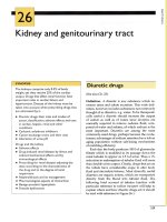
Figure 1–2. Identification of the central sulcus. A stepwise method for identifying the
central sulcus and the surrounding sulci and gyri. In panel A, the sylvian or lateral
fissure is identified with a (1). In panel B, the anterior horizontal ramus (2) and the
anterior ascending ramus (3) are identified in their typical Y shape. These rami define
the pars orbitalis (PObr), pars triangularis (PTr), and pars opercularis (POp). In panel C,
the precentral sulcus (4) is identified. In panel D, the central sulcus (5) and precentral
gyrus (PreC) are identified. In panel E, the postcentral sulcus (6) and the postcentral
gyrus (PostC) are identified. In panel F, the superior temporal sulcus (7), superior
temporal gyrus (STG), and middle temporal gyrus (MTG) are identified. (continued)
CHAPTER 1
SURFACE ANATOMY AS A GUIDE TO CEREBRAL FUNCTION
G
Figure 1–2. (Continued) In panel G, the intraparietal
sulcus (8), the supramarginal gyrus (SMG), and the
angular gyrus (AG) are identified.
comprehension, including both spoken and read language and is located in the posterior, superior temporal gyrus. Broca’s area is involved in the production
of speech and is located in the inferior frontal gyrus,
classically localized to the pars triangularis and pars opercularis. These two areas are connected by the arcuate fasciculus. In reality, there is significant variation in
the location of speech function between subjects.22−24
Surgical resections involving potential speech areas are
usually performed with the patient awake, which allows
cortical mapping of speech function through intraoperative stimulation. Recent studies have shown that speech
function has a much wider distribution across the frontal
lobe outside of the classical Broca’s area and is more dif-
A
7
fused across the temporal lobe outside of the classical
Wernicke’s area.24,25 However, it is still useful to be able
to identify the classical locations of these areas to serve
as a starting point for cortical mapping (Fig. 1–4).
Broca’s area is classically described as being located
in the pars triangularis and pars opercularis of the inferior frontal gyrus. The inferior frontal gyrus is bounded
by the sylvian fissure inferiorly and the inferior frontal
sulcus superiorly. The anterior horizontal ramus and anterior ascending sylvian ramus extend superiorly from
the sylvian fissure into the inferior frontal gyrus in a Y or
V shape.16 These two rami divide the inferior frontal sulcus into three parts, the pars orbitalis, pars triangularis,
and pars opercularis, from anterior to posterior. The inferior frontal gyrus is bounded posteriorly by the central
sulcus. Quinones-Hinojosa and colleagues used intraoperative mapping correlated with MRI to locate Broca’s
area in relation to the sulci defining the inferior frontal
gyrus.7 They proposed a method for localizing Broca’s
area based on the intersection of lines drawn from defined points in the inferior frontal gyrus. The first line is
drawn from the opercular tip posteriorly at a 45◦ angle
between the sylvian fissure and the anterior ascending
sylvian ramus. The second line is drawn superiorly, perpendicular from the sylvian fissure at the level of the precentral sulcus. The third line is drawn anteriorly, parallel
to the sylvian fissure at the level of the inferior tip of the
central sulcus. The intersection of these three lines provides an estimate of the location of Broca’s area. While
this technique does provide an estimated location for
Broca’s area, it is only an estimate and accurate localization of speech function is best determined with intraoperative or extraoperative cortical stimulation mapping.
B
Figure 1–3. Identification of motor and sensory cortex. A. In the left panel, primary
motor cortex is located in the precentral gyrus, with hand function (H) localized
perpendicular to the end of the superior frontal gyrus. B. In the right panel, primary
sensory cortex is located in the postcentral gyrus. Tongue sensory (T) is located in
the widened area of the postcentral gyrus close to the sylvian fissure. Face sensory
(F) is located in the narrow strip superior to tongue sensory, and hand sensory (H)
superior to face sensory.
8
SECTION I
TECHNIQUES
Figure 1–4. Localization of speech. Broca’s area (B) is classically located in the pars
triangularis and pars opercularis of the inferior frontal gyrus. Wernicke’s area (W) is
located in the posterior portion of the superior temporal gyrus and the supramarginal
gyrus. However, direct stimulation during awake craniotomy has demonstrated
speech function over a much wider area than the classical speech areas, as indicated
by the red outlines compared to the blue outlines of the classical speech areas.
Wernicke’s area is classically located in the posterior, superior temporal gyrus and in the supramarginal
gyrus of the inferior parietal lobule adjacent to the sylvian fissure. These areas can be identified by locating
the sylvian fissure. The superior temporal gyrus runs
between the sylvian fissure and the superior temporal
sulcus, which runs parallel to the sylvian fissure. The
posterior portions of both the sylvian fissure and the
superior temporal sulcus hook superiorly and terminate
in the inferior lobule of the parietal lobe. The supramarginal gyrus is the anterior portion of the parietal
lobe, which forms a horseshoe shape over the posterior end of the sylvian fissure. The angular gyrus forms
a similar horseshoe shape over the posterior end of
the superior temporal gyrus.13,16 Intraoperative mapping
during awake craniotomy has demonstrated that language function is highly variable between subjects and
can be widely dispersed over the temporal lobe and parietal lobe outside the classical Wernicke’s area.24
᭤ LOCALIZATION OF VISUAL
FUNCTION
Primary visual cortex (V1) is located in the occipital lobe
on the mesial surface both within the calcarine sulcus
and on the surrounding cortex. The visual cortex is organized in a retinotopic map with the fovea located posteriorly near the occipital pole. The vertical meridian is
located at the calcarine fissure and the horizontal meridian is deep within the calcarine fissure. Functional MRI
mapping of the visual cortex has demonstrated that the
V1 is located mostly within the folds of the calcarine
fissure. The fovea is located posteriorly near the occipital pole. Peripheral vision is located anteriorly. There is
significant magnification of the retinotopic map near the
fovea, with a much larger cortical area corresponding
to the area around the fovea. Other visual areas extend
superiorly and inferiorly from the calcarine fissure, corresponding to areas V2, V3, and V4.26−29 An area homologous to the middle temporal (MT) region is located at
the junction of the temporal, parietal, and occipital lobes
in humans (Fig. 1–5). This area is involved in processing
of movement.30−32
The calcarine sulcus is located on the mesial surface of the occipital lobe. It extends posteriorly from the
splenium of the corpus callosum to the occipital pole.
It is divided into an anterior and posterior portion by
the parietal-occipital sulcus. The posterior portion of the
calcarine sulcus splits into a Y shape as it approaches
the occipital pole, with the superior portion of the Y
sometimes extending onto the lateral surface of the occipital lobe. The calcarine sulcus ranges from 2.5 to 3 cm
deep.14,15 The MT region is located near the junction of
the temporal, parietal, and occipital lobes.30−32
᭤ LOCALIZATION OF AUDITORY
FUNCTION
Penfield and Rasmussen localized hearing function to
the superior temporal gyrus by direct electrical stimulation of the human cortex. Further studies using
positron emission tomography, fMRI, and direct cortical
recordings have demonstrated that the auditory cortex is