Development of the Heart
Bạn đang xem bản rút gọn của tài liệu. Xem và tải ngay bản đầy đủ của tài liệu tại đây (1.29 MB, 4 trang )
Development of the Heart
Development of the Heart
Bởi:
OpenStaxCollege
The human heart is the first functional organ to develop. It begins beating and pumping
blood around day 21 or 22, a mere three weeks after fertilization. This emphasizes
the critical nature of the heart in distributing blood through the vessels and the vital
exchange of nutrients, oxygen, and wastes both to and from the developing baby. The
critical early development of the heart is reflected by the prominent heart bulge that
appears on the anterior surface of the embryo.
The heart forms from an embryonic tissue called mesoderm around 18 to 19 days after
fertilization. Mesoderm is one of the three primary germ layers that differentiates early
in development that collectively gives rise to all subsequent tissues and organs. The
heart begins to develop near the head of the embryo in a region known as the cardiogenic
area. Following chemical signals called factors from the underlying endoderm (another
of the three primary germ layers), the cardiogenic area begins to form two strands
called the cardiogenic cords ([link]). As the cardiogenic cords develop, a lumen rapidly
develops within them. At this point, they are referred to as endocardial tubes. The two
tubes migrate together and fuse to form a single primitive heart tube. The primitive heart
tube quickly forms five distinct regions. From head to tail, these include the truncus
arteriosus, bulbus cordis, primitive ventricle, primitive atrium, and the sinus venosus.
Initially, all venous blood flows into the sinus venosus, and contractions propel the
blood from tail to head, or from the sinus venosus to the truncus arteriosus. This is a
very different pattern from that of an adult.
1/4
Development of the Heart
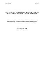
Development of the Human Heart
This diagram outlines the embryological development of the human heart during the first eight
weeks and the subsequent formation of the four heart chambers.
The five regions of the primitive heart tube develop into recognizable structures in a
fully developed heart. The truncus arteriosus will eventually divide and give rise to
the ascending aorta and pulmonary trunk. The bulbus cordis develops into the right
ventricle. The primitive ventricle forms the left ventricle. The primitive atrium becomes
the anterior portions of both the right and left atria, and the two auricles. The sinus
venosus develops into the posterior portion of the right atrium, the SA node, and the
coronary sinus.
As the primitive heart tube elongates, it begins to fold within the pericardium, eventually
forming an S shape, which places the chambers and major vessels into an alignment
similar to the adult heart. This process occurs between days 23 and 28. The remainder
of the heart development pattern includes development of septa and valves, and
remodeling of the actual chambers. Partitioning of the atria and ventricles by the
interatrial septum, interventricular septum, and atrioventricular septum is complete by
the end of the fifth week, although the fetal blood shunts remain until birth or shortly
after. The atrioventricular valves form between weeks five and eight, and the semilunar
valves form between weeks five and nine.
2/4
Development of the Heart
Chapter Review
The heart is the first organ to form and become functional, emphasizing the importance
of transport of material to and from the developing infant. It originates about day 18
or 19 from the mesoderm and begins beating and pumping blood about day 21 or
22. It forms from the cardiogenic region near the head and is visible as a prominent
heart bulge on the surface of the embryo. Originally, it consists of a pair of strands
called cardiogenic cords that quickly form a hollow lumen and are referred to as
endocardial tubes. These then fuse into a single heart tube and differentiate into the
truncus arteriosus, bulbus cordis, primitive ventricle, primitive atrium, and sinus
venosus, starting about day 22. The primitive heart begins to form an S shape within
the pericardium between days 23 and 28. The internal septa begin to form about day
28, separating the heart into the atria and ventricles, although the foramen ovale persists
until shortly after birth. Between weeks five and eight, the atrioventricular valves form.
The semilunar valves form between weeks five and nine.
Review Questions
The earliest organ to form and begin function within the developing human is the
________.
1.
2.
3.
4.
brain
stomach
lungs
heart
D
Of the three germ layers that give rise to all adult tissues and organs, which gives rise to
the heart?
1.
2.
3.
4.
ectoderm
endoderm
mesoderm
placenta
C
The two tubes that eventually fuse to form the heart are referred to as the ________.
1. primitive heart tubes
2. endocardial tubes
3. cardiogenic region
3/4
Development of the Heart
4. cardiogenic tubes
D
Which primitive area of the heart will give rise to the right ventricle?
1.
2.
3.
4.
bulbus cordis
primitive ventricle
sinus venosus
truncus arteriosus
A
The pulmonary trunk and aorta are derived from which primitive heart structure?
1.
2.
3.
4.
bulbus cordis
primitive ventricle
sinus venosus
truncus arteriosus
D
Critical Thinking Questions
Why is it so important for the human heart to develop early and begin functioning within
the developing embryo?
The human embryo is rapidly growing and has great demands for nutrients and oxygen,
while producing waste products including carbon dioxide. All of these materials must
be received from or delivered to the mother for processing. Without an efficient early
circulatory system, this would be impossible.
Describe how the major pumping chambers, the ventricles, form within the developing
heart.
After fusion of the two endocardial tubes into the single primitive heart, five regions
quickly become visible. From the head, these are the truncus arteriosus, bulbus cordis,
primitive ventricle, primitive atrium, and sinus venosus. Contractions propel the blood
from the sinus venosus to the truncus arteriosus. About day 23, the heart begins to form
an S-shaped structure within the pericardium. The bulbus cordis develops into the right
ventricle, whereas the primitive ventricle becomes the left ventricle. The interventricular
septum separating these begins to form about day 28. The atrioventricular valves form
between weeks five to eight. At this point, the heart ventricles resemble the adult
structure.
4/4