Khác biệt về biểu hiện và đáp ứng của suy tim mới so với đợt mất bù của suy tim mạn tính
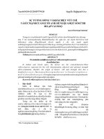
Bạn đang xem bản rút gọn của tài liệu. Xem và tải ngay bản đầy đủ của tài liệu tại đây (1.09 MB, 18 trang )
Differences in Presentations and
Responses to Management
of New Onset Heart Failure
versus Long Standing Heart Failure
Presenter: Nguyễn Văn Việt Thắng
BACKGROUND
Current definition of Heart Failure is vague and impractical
New classification of Heart Failure based on a
new test – the SEFV test.
Treatment according to the result of the test.
BACKGROUND
The SEFV test
Method
The result
Fluid distribution
Three
compartments
of the body
Intravascular
compartments
Intra-arterial
compartment
Extravascular
compartment
Intra-venous
compartment
New Non-Invasive SEFV test
Size and Expansion of the Femoral Vein test
• The SEFV is the ultrasound study examining the size of the
femoral vein and its expansion with cough.
• The ultrasound plane of the femoral artery and vein to be
checked is the coronal plane immediately proximal to the
bifurcation of the superficial and deep femoral artery.
SEFV
Size and Expansibility of the Femoral Vein test
SEFV
Here at the distal end of
the common femoral
artery, the coronal plane
of the artery is seen as a
single round structure
which pulsates. Next to
it is the femoral vein.
Principles of SEFV
The first principle: The volume of blood going
through the femoral artery and returning
through the common femoral vein should be the
same.
Principles of SEFV
The second principle: In the vascular system,
most of the circulating blood is in the veins. The
amount of blood in the arteries is small and the
size of the arteries does not change much due to
vascular tone in order to keep a fairly constant
blood pressure.
Normal expansion of femoral vein
(no fluid overload + no dehydration)
METHOD
Patients were enrolled and physical examination was recorded:
• Fluid overload in the venous system: presence of rales in the lung
and painful sensation with a minimal punch in the right lower rib
cage (fluid in the liver).
METHOD
• Fluid overload in the extravascular system: fluid
infiltration in the abdominal wall, edema at the ankle,
thigh, dependent areas (e.g. presacral area, etc).
METHOD
Low perfusion in the arterial compartment consists of
• Low blood pressure.
• Cerebral hypo-perfusion (causing dizziness or change of
mental status).
• Renal perfusion (causing pre-renal azotemia (increased blood
urea nitrogen).
• Distal peripheral arterial system perfusion (causing fatigue or
exercise intolerance)
Location of fluid overload
*Patients with more intravenous fluid overload
ACEI + BB + Short term fast acting loop diuretics
(eg: furosamide).
*Patients with more extravascular fluid overload
ACEI + BB + Long acting, lower dose diuretics
(eg: HCTZ).
RESULTS
50 patients were enrolled from January 2015 to April
2016. All came with shortness of breath and had a
diagnosis of HF in the emergency room. All the
patients were diagnosed with HF with low or
preserved EF
60% patients with long standing dilated
cardiomyopathy had more extravascular fluid
overload
Compared to only 30% patients of recent dilated
cardiomyopathy
RESULTS
The patients with new or recent onset of dilated
cardiomyopathy recovered faster (within 24 hours)
while the other patients took longer to recover.
These latter patients also needed more
medications
CONCLUSION
Based on the location of the fluid overload,
the patients with new onset of HF were faster
to recover with less times and lower need for
resources
REFERENCE
• Thach Nguyen, Advait Soni, Chau BL Vien, Ryan Phan,
Tung Mai. “Differences of Presentations and
Responses to Management of New Onset Heart
Failure versus Long Standing Heart Failure.”