DSpace at VNU: Activated platelet-rich plasma improves adipose-derived stem cell transplantation efficiency in injured articular cartilage
Bạn đang xem bản rút gọn của tài liệu. Xem và tải ngay bản đầy đủ của tài liệu tại đây (713.63 KB, 11 trang )
Van Pham et al. Stem Cell Research & Therapy 2013, 4:91
/>
RESEARCH
Open Access
Activated platelet-rich plasma improves
adipose-derived stem cell transplantation
efficiency in injured articular cartilage
Phuc Van Pham1*, Khanh Hong-Thien Bui2, Dat Quoc Ngo3, Ngoc Bich Vu1, Nhung Hai Truong1,
Nhan Lu-Chinh Phan1, Dung Minh Le1, Triet Dinh Duong2, Thanh Duc Nguyen2, Vien Tuong Le2
and Ngoc Kim Phan1
Abstract
Introduction: Adipose-derived stem cells (ADSCs) have been isolated, expanded, and applied in the treatment of
many diseases. ADSCs have also been used to treat injured articular cartilage. However, there is controversy
regarding the treatment efficiency. We considered that ADSC transplantation with activated platelet-rich plasma
(PRP) may improve injured articular cartilage compared with that of ADSC transplantation alone. In this study, we
determined the role of PRP in ADSC transplantation to improve the treatment efficiency.
Methods: ADSCs were isolated and expanded from human adipose tissue. PRP was collected and activated from
human peripheral blood. The effects of PRP were evaluated in vitro and in ADSC transplantation in vivo. In vitro, the
effects of PRP on ADSC proliferation, differentiation into chondrogenic cells, and inhibition of angiogenic factors
were investigated at three concentrations of PRP (10%, 15% and 20%). In vivo, ADSCs pretreated with or without
PRP were transplanted into murine models of injured articular cartilage.
Results: PRP promoted ADSC proliferation and differentiation into chondrogenic cells that strongly expressed
collagen II, Sox9 and aggrecan. Moreover, PRP inhibited expression of the angiogenic factor vascular endothelial
growth factor. As a result, PRP-pretreated ADSCs improved healing of injured articular cartilage in murine models
compared with that of untreated ADSCs.
Conclusion: Pretreatment of ADSCs with PRP is a simple method to efficiently apply ADSCs in cartilage
regeneration. This study provides an important step toward the use of autologous ADSCs in the treatment of
injured articular cartilage.
Keywords: Adipose tissue-derived stem cells, Articular cartilage injury, Joint failure, Mesenchymal stem cells,
Osteoarthritis, Platelet-rich plasma
Introduction
Platelet-rich plasma (PRP) has been widely used across many
clinical fields, especially for skincare and orthopedics. PRP
contains at least seven growth factors including epidermal
growth factor, platelet-derived growth factor, transforming
growth factor-beta, vascular endothelial growth factor
* Correspondence:
1
Laboratory of Stem Cell Research and Application, University of Science,
Vietnam National University, 227 Nguyen Van Cu, District 5, Ho Chi Minh City,
Vietnam
Full list of author information is available at the end of the article
(VEGF), fibroblast growth factor, insulin-like growth factor,
and keratinocyte growth factor. The therapeutic effect of PRP
occurs because of the high concentration of these growth factors compared with that in normal plasma [1,2]. Many of
these growth factors have important roles in wound healing
and tissue regeneration. PRP stimulates the expression of type
I collagen and matrix metalloproteinase-1 in dermal fibroblasts [3], and increases the expression of G1 cycle regulators,
type I collagen, and matrix metalloproteinase-1 to accelerate
wound healing [4].
In animal models, intra-articular PRP injection influences
cartilage regeneration in all severities of rabbit knee
© 2013 Van Pham et al.; licensee BioMed Central Ltd. This is an Open Access article distributed under the terms of the
Creative Commons Attribution License ( which permits unrestricted use,
distribution, and reproduction in any medium, provided the original work is properly cited.
Van Pham et al. Stem Cell Research & Therapy 2013, 4:91
/>
osteoarthritis [5]. In a porcine model, PRP attenuates arthritic changes as assessed histologically and based on protein
synthesis of typical inflammatory mediators in the synovial
membrane and cartilage [6]. Clinically, PRP can repair cartilage with focal chondral defects. Siclari and colleagues
performed this experiment on 52 patients (mean age: 44
years) with focal chondral defects in radiologically confirmed
nondegenerative or degenerative knees [7]. Defects were
coated with PRP-immersed polymer-based implant. Compared with the baseline and 3-month follow-up, the results
showed that the Knee injury and Osteoarthritis Outcome
Score showed clinically meaningful and significant improvement in all subcategories. Histological analysis of
biopsied tissue showed hyaline-like to hyaline cartilage repair tissue that was enriched with cells showing a chondrocyte morphology, proteoglycans, and type II collagen
(col-II) [7]. PRP injection with arthroscopic microfracture
also improves early osteoarthritic knees with cartilage
lesions in 40-year-old to 50-year-old patients, and the
indication of this technique could be extended to 50-year
-old patients [8]. In addition, PRP injection significantly
improves the Visual Analog Scale for Pain score and the
International Knee Documentation Committee score
[9,10]. In a recent study with a larger patient cohort (120
patients), Spakova and colleagues showed that autologous
PRP injection is an effective and safe method for the treatment of the initial stages of knee osteoarthritis [11]. In this
research, 120 patients with Grade 1, Grade 2, or Grade 3
osteoarthritis according to the Kellgren and Lawrence grading scale were enrolled. Patients were treated using three
intra-articular applications of PRP. Statistically significantly
better results in the Western Ontario and McMaster Universities Osteoarthritis Index and the Numeric Rating Scale
scores were recorded patients who received PRP injections
after 3-month and 6-month follow-up.
Stem cells from adipose tissue were isolated and differentiated in vitro into adipogenic, chondrogenic, myogenic, and
osteogenic cells in the presence of specific induction factors
[12]. These cells are termed adipose-derived stem cells
(ADSCs). ADSCs express surface markers as CD44, CD73,
CD90, and CD105, but are negative for CD14, CD34, and
CD45 [13-16]. This profile is similar to that of mesenchymal
stem cells (MSCs) that have been suggested by Dominici and
colleagues [17]. Compared with MSCs from bone marrow and
umbilical cord blood, MSCs from adipose tissue have many
advantages [18]. ADSCs are considered a suitable autologous
cell source. Moreover, ADSCs have been used to treat many
diseases such as liver fibrosis [19], nerve defects [20-22], ischemia [23,24], skeletal muscle injury [25], passive chronic immune thrombocytopenia [26], and infarcted myocardium [27]
in animals; and systemic sclerosis in human [28,29].
ADSCs have been extensively investigated in preclinical
studies for the treatment of cartilage injuries and osteoarthritis in animal models including dogs [30-32], rabbits
Page 2 of 11
[33], horses [34], rats [35], mice [36-38], and goats [39]. In
a recent study, Xie and colleagues showed that ADSCseeded PRP constructs develop into functional chondrocytes
that secrete cartilaginous matrix in rabbits at 9 weeks post
implantation [40]. These studies show evidence of functional
improvement, especially scores for lameness, pain, and
range of motion compared with control dogs [30-32], prevention of osteoarthritis and repair of defects in rabbit [33],
upregulation of glycosaminoglycans as well as col-II to promote osteochondral repair and osteoarthritis prevention in
rat [35], and protection against cartilage damage [36] as well
as anti-inflammatory and chondroprotective effects [37] in
mice following ADSC transplantation. These results have
prompted human clinical trials for the treatment of
osteoarthritis.
For example, Pak showed significant positive changes in
all patients transplanted with ADSCs [41]. Various phase I
and phase II clinical trials using ADSCs have been undertaken for osteoarthritis or degenerative cartilage
(NCT01300598, NCT01585857 and NCT01399749). More
importantly, in one clinical trial 18 patients underwent
ADSC and PRP transplantation. The results of this study
showed that intra-articular injection of ADSCs and PRP is
effective for reducing pain and improving knee function in
patients being treated for knee osteoarthritis [42].
In another study, however, ADSCs were considered to
inhibit cartilage regeneration. This conclusion was drawn
from experiments of ADSC transplantation in rats. This
study showed that ADSCs highly express and secrete
VEGF-A into the culture supernatant. The supernatant
inhibits chondrocyte proliferation, reduces Sox9, alcan,
and col-II mRNA levels, reduces proteoglycan synthesis,
and increases apoptosis. ADSCs have been implanted in
1 mm noncritical hyaline cartilage defects in vivo, and
showed inhibition of cartilage regeneration by radiographic
and equilibrium partitioning of an ionic contrast agent via
micro-computed tomography imaging. Histology revealed
that defects with ADSCs had no tissue ingrowth from the
edges of the defect [43].
Based on the above results, we considered that ADSC
transplantation in combination with PRP might improve
the efficiency of injured articular cartilage treatment.
We theorized that PRP affects ADSC proliferation and differentiation, especially chondrogenic differentiation. This
study therefore aimed to evaluate the effects of PRP on
ADSC proliferation and differentiation into chondrocytes
in vitro, and cartilage formation in vivo.
Materials and methods
Isolation of stromal vascular fraction cells from
adipose tissue
Stromal cells were first isolated from the abdominal adipose
tissue of 10 consenting healthy donors. From each patient,
approximately 40 to 80 ml lipoaspirate was collected in two
Van Pham et al. Stem Cell Research & Therapy 2013, 4:91
/>
50 ml sterile syringes. All procedures and manipulations
were approved by our Institutional Ethical Committee
(Laboratory of Stem Cell Research and Application,
University of Science, Vietnam National University, Ho Chi
Minh City, Vietnam) and the Hospital Ethical Committee
(Ho Chi Minh City Medicine and Pharmacy University
Hospital, Ho Chi Minh City, Vietnam). The syringes were
stored in a sterile box at 2 to 8°C and immediately transferred to the laboratory. The stromal vascular fraction
(SVF) was isolated using an ADSC Extraction kit
(GeneWorld, Ho Chi Minh City, Vietnam) according to the
manufacturer’s instructions. Briefly, 80 ml lipoaspirate was
placed into a sterile disposable 250 ml conical centrifuge
tube (2602A43; Corning 836, North Street Building,
Tewksbury, MA 01876, USA). The adipose tissue was
washed twice in PBS by centrifugation at 400 × g for
5 minutes at room temperature. Next, the adipose tissue
was digested using the SuperExtract Solution (1.5 mg
collagenase/mg adipose tissue) at 37°C for 30 minutes
with agitation at 5-minute intervals. The suspension
was centrifuged at 800 × g for 10 minutes, and the SVF
was obtained as a pellet. The pellet was washed twice
with PBS to remove any residual enzyme, and resuspended
in PBS to determine the cell quantity and viability using an
automatic cell counter (NucleoCounter; Chemometec,
Gydevang 43, DK-3450 Allerod, Denmark).
Platelet-rich plasma preparation
Human PRP was derived from the peripheral blood of the
same donor as the adipose tissue using a New-PRP Pro Kit
(GeneWorld) according to the manufacturer’s guidelines.
Briefly, 20 ml peripheral blood was collected into vacuum tubes
and centrifuged at 800 × g for 10 minutes. The plasma fraction
was collected and centrifuged at 1000 × g for 5 minutes to obtain a platelet pellet. Most of the plasma was then removed,
leaving 3 ml plasma to resuspend the platelets. This preparation was inactivated PRP. Finally, PRP was activated by
activating tubes containing 100 μl of 20% CaCl2.
Adipose-derived stem cell culture
SVF cells were cultured to expand the number of ADSCs.
SVF cells were cultured in DMEM/F12 (Sigma-Aldrich, St
Louis, MO, USA) containing 1× antibiotic–mycotic and
10% fetal bovine serum (FBS; Sigma-Aldrich) at 37°C with
5% CO2. The medium was changed twice per week. At 70
to 80% confluence, the cells were subcultured using 0.25%
trypsin/ethylenediamine tetraacetic acid (GeneWorld).
Cell proliferation assay
A total of 5 × 10³ ADSCs per well were cultured in 96-well
plates in 100 μl DMEM/F12 containing 10% PRP, 15% PRP,
20% PRP, or 10% FBS as the control.
Twenty microliters of MTT (5 g/l; Sigma-Aldrich) was
added to each well, followed by incubation for 4 hours and
Page 3 of 11
then addition of 150 μl DMSO/well (Sigma-Aldrich). Plates
were then agitated for 10 minutes until the crystals dissolved
completely. Absorption values were measured at a wavelength
of 490 nm and a reference wavelength of 630 nm using a DTX
880 microplate reader (Beckman Coulter, Krefeld, Germany).
Immunophenotyping
Third-passage ADSCs were examined for their immunophenotype by flow cytometry according to previously
published protocols [44]. Briefly, cells were washed twice
in Dulbecco’s PBS containing 1% BSA (Sigma-Aldrich).
Cells were stained for 30 minutes at 4°C with anti-CD14fluorescein isothiocyanate, anti-CD34-fluorescein isothiocyanate, anti-CD44-phycoerythrin, anti-CD45-fluorescein
isothiocyanate, anti-CD90-phycoerythrin, or anti-CD105fluorescein isothiocyanate mAb (BD Biosciences, Franklin
Lakes, NJ, USA). Stained cells were analyzed by a
FACSCalibur flow cytometer (BD Biosciences). Isotype
controls were used for all analyses.
Gene expression analysis
Third-passage ADSCs were evaluated for the effects of
PRP on their proliferation and differentiation. ADSCs
were cultured in six-well plates at 1 × 10⁵ cells/well in
DMEM/F12 with 10% FBS and 1% antibiotic–mycotic
for 24 hours. The medium was then replaced with
DMEM/F12 with 1% antibiotic–mycotic and 10% PRP,
15% PRP, 20% PRP, or 10% FBS as the control. ADSCs
were cultured under these conditions for 1 week with
two medium changes per week. ADSCs were then isolated
to evaluate their gene expression.
Total RNA was extracted as described elsewhere [44].
RNA was precipitated with 500 μl isopropyl alcohol at
room temperature for 10 minutes. ADSCs were analyzed
for the expression of chondrogenic markers including col-II,
Sox9, and aggrecan. Real-time RT-PCR was performed with
an Eppendorf gradient S thermal Cycler (EppendorfAG, Hamburg, Germany). The reaction mixture (25 μl)
contained 10 mM Tris–HCl, pH 8.3, 50 mM KCl, 1.5
mM MgCl2, 200 μM dNTP mix, 0.2 μM each primer,
and 1 U Taq DNA polymerase. Relative expression levels
were normalized to glyceraldehyde-3-phosphate dehydrogenase (GAPDH) and calculated using the 2–ΔCCt method.
All PCR primers have been described previously [45,46].
VEGF concentration measurement
To measure the concentration of VEGF secreted by ADSCs,
1.5 × 10⁶ viable ADSCs were seeded in 75 cm2 culture flasks
containing DMEM/F12 with 10% PRP, 15% PRP, 20% PRP,
or 10% FBS. These cells were incubated at 37°C with 5%
CO2 for 72 hours. The media were then replaced, and the
cells were incubated for a further 72 hours. The culture
supernatants were collected, centrifuged at 4,980 × g for 10
minutes and stored at −80°C until use. The concentration
Van Pham et al. Stem Cell Research & Therapy 2013, 4:91
/>
of VEGF was then determined by an ELISA kit (Abcam,
Cambridge, MA, USA). VEGF concentrations were also
measured in the fresh media. VEGF produced by ADSCs
was calculated by subtracting the values in culture supernatants from those in the fresh media.
Stem cell transplantation
To evaluate the effects of PRP on ADSC transplantation
in osteoarthritis, we used a mouse model of articular
cartilage injury. In this experiment, we compared the
efficiency of transplantation using ADSCs treated with
15% PRP (PRP15 group) or 10% FBS (FBS10 group), and
control PBS injection. All procedures were approved by
the Local Ethics Committee of the Stem Cell Research
and Application Laboratory, University of Science. Articular
cartilage injury was induced by joint destruction in the hind
limbs of NOD/SCID mice using a 32 G needle. Briefly, 12
mice were anesthetized using ketamine (40 mg/kg) and
then subjected to hind-limb joint destruction. An uninjured
mouse was used as a control. Injured mice were equally
divided into the PRP15 group (four mice), in which mice
were transplanted with ADSCs cultured with 15% PRP; the
FBS10 group (four mice), in which mice were transplanted
with ADSCs cultured with 10% FBS; and the negative control group (four mice), in which mice were injected with
PBS. The mice were then anesthetized and injected with
either ADSCs or PBS (negative control). In the treatment
groups, 2 × 10⁶ ADSCs of the PRP15 or FBS10 groups
suspended in 200 μl PRP were injected into the knee joint
via two doses with a 10-minute interval between injections.
For functional evaluation, hind-limb movement was then
evaluated daily. Mice were placed in water. The natural
response was a pedal response in water. We recorded the
pedal response of treated hind limbs. After 45 days, all mice
Page 4 of 11
were euthanized and their hind limbs were used for histological analysis and further experiments. The samples were
fixed in 10% formalin, decalcified, sectioned longitudinally,
and stained with H & E (Sigma-Aldrich). Using H &
E-stained sections, three parameters were examined
for the knee joints: the area of damaged cartilage (%),
the area of regenerated cartilage (%), and the number of
regenerated cartilage cell layers. The damaged cartilage area
was determined by mature cartilage that was lost compared
with that in the control.
Statistical analysis
All experiments were performed in triplicate. P ≤0.05
was considered significant. Data were analyzed using
Statgraphics software 7.0 (Statgraphics Graphics System,
Warrenton, VA, USA).
Results
ADSCs proliferate in vitro and maintain expression of
specific markers after several passages
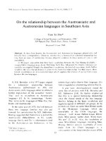
We successfully isolated the SVF from adipose tissue. A
total of 1.43 ± 0.15 × 10⁶ stromal cells with a viability
of 94.4 ± 3.54% were collected from 1 g adipose tissue
(n = 10). The cells were cultured with a 100% success rate
(10/10) without microorganism contamination. After 24
hours of incubation, fibroblast-like cells appeared in the
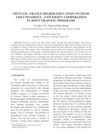
cultures (Figure 1A). From day 3, cells rapidly proliferated
and reached confluence on day 7 (Figure 1B). The cells
were subcultured three times before use in experiments.
After the third passage, the cells maintained a homogeneous fibroblastic shape (Figure 1C).
The cells expressed MSC-specific markers with >95% positive staining for CD44, CD73, and CD90 (Figure 1G, H, I),
and <4% of cells were positive for hematopoietic markers
Figure 1 Adipose-derived stem cell culture and marker confirmation. (A) At 24 hours after seeding, fibroblast-like cells adhered to the
surface of the flask, (B) proliferated and reached confluence after 1 week, and (C) became homogeneous after three subcultures. At the third
passage, adipose-derived stem cells expressed mesenchymal stem cell-specific markers including (G) CD44, (H) CD90, and (I) CD73, while (D)
CD14, (E) CD34, and (F) CD45 were negative.
Van Pham et al. Stem Cell Research & Therapy 2013, 4:91
/>
CD14, CD34 and CD45 (Figure 1D, E, F). Moreover,
they also hold potential differentiation into specific
cells. In fact, they were successfully differentiated into
adipocytes in previous published research [47]. These
cells were considered to be ADSCs and used for further
experiments.
Platelet-rich plasma efficiently stimulates ADSC
proliferation
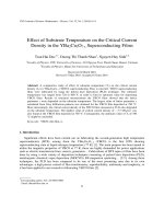
To investigate the effects of PRP on ADSC proliferation,
we performed cell proliferation assays. The results showed
that PRP could replace FBS in growth medium. In the mice
transplanted with ADSCs cultured with 10% PRP (PRP10
group), in the PRP15 group, and in the mice transplanted
with ADSCs cultured with 20% PRP (PRP20 group),
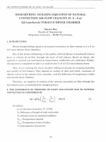
ADSCs adhered to the flask surface. Under a microscope,
ADSCs exhibited a normal shape (Figure 2A, B, C) similar
to that of FBS-cultured ADSCs (Figure 2D). In MTT assays,
we found that PRP strongly stimulated ADSC proliferation.
At the three concentrations of PRP, ADSC proliferation was
stimulated more strongly than that in medium containing
10% FBS (FBS10 group). After 3 days of PRP treatment,
ADSCs started to increase their proliferation rate compared
with that in the control (FBS10 group). The differences
Page 5 of 11
were statistically significant at day 7 in all three groups
treated with PRP (Figure 2E). Compared with 10% PRP
and 10% FBS, 15% PRP and 20% PRP stimulated ADSC
proliferation more strongly. However, the difference
between 15% PRP and 20% PRP was not significant.
We therefore concluded that 15% PRP was the optimal
concentration for robust proliferation of ADSCs.
Platelet-rich plasma does not change marker expression
but induces expression of genes related to chondrocytes
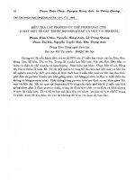
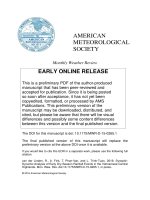
Figure 3 shows the percentages of ADSCs expressing
specific markers in the three groups. The percentages
of ADSCs expressing CD44, CD73, and CD90 were
98.32 ± 1.21%, 97.21 ± 3.21%, and 96.21 ± 1.22% for
CD44, 95.12 ± 2.12%, 96.27 ± 2.19%, and 95.54 ± 3.10% for
CD73, 98.81 ± 1.11%, 97.37 ± 1.27%, and 98.92 ± 2.01%
for CD90 in the PRP10, PRP15, and PRP20 groups, respectively. The percentages of ADSCs expressing CD14,
CD34, and CD45 were 2.13 ± 1.11%, 2.65 ± 1.21%, and
1.98 ± 0.45% for CD14, 0.21 ± 0.11%, 0.98 ± 0.09%, and
1.31 ± 0.89% for CD34, and 2.11 ± 0.87%, 1.63 ± 1.08%,
and 1.55 ± 0.51% for CD45 in the PRP10, PRP15, and
PRP20 groups, respectively (Figure 3A, B, C). Compared
with FBS (Figure 1D, E, F, G, H, I), these results showed
Figure 2 Adipose-derived stem cell proliferation in experimental groups. Adipose-derived stem cells (ADSCs) maintained the shape in four
different media: (A) 10% platelet-rich plasma (PRP10), (B) 15% PRP (PRP15), (C) 20% PRP (PRP20) and (D) 10% fetal bovine serum (FBS10). (E)
ADSC proliferation significantly increased in medium containing PRP at 10%, 15%, and 20% compared with that in medium containing 10% FBS.
OD, optical density.
Van Pham et al. Stem Cell Research & Therapy 2013, 4:91
/>
Page 6 of 11
Figure 3 Platelet-rich plasma does not change adipose-derived stem cell marker expression but changes chondrocyte-related gene
expression. The expression of CD14, CD34, CD44, CD45, CD73, and CD90 was changed in the (A) 10% platelet-rich plasma (PRP10), (B) 15% PRP
(PRP15), and (C) 20% PRP (PRP20) groups compared with the 10% fetal bovine serum (FBS10) group (Figure 1). (D) Expression of collagen type II
(COL-II), Sox9, and aggrecan was strongly promoted in the PRP10, PRP15, and PRP20 groups compared with that in the FBS10 group. GAPDH,
glyceraldehyde-3-phosphate dehydrogenase; SSC.
that the three concentrations of PRP did not affect
marker expression of ADSCs.
However, there were differences in the expression of
some genes including col-II, Sox9, and aggrecan. Compared
with the FBS10 group, ADSCs in the PRP10, PRP15 and
PRP20 groups showed increased expression of col-II,
Sox9, and aggrecan, all of which are important for chondrogenesis. As shown in Figure 3D, col-II expression
increased from 20.07 ± 5.13 (compared with GAPDH)
to 60.33 ± 11.68, 67.67 ± 23.80, and 69.00 ± 15.62 in the
FBS10, PRP10, PRP15, and PRP20 groups, respectively
(P ≤0.05). Similarly, expression of chondrogenic markers
Sox9 and aggrecan also increased in the PRP10, PRP15,
PRP20 groups compared with that in the FBS10 group.
Sox9 expression increased from 4.67 ± 2.08 in the FBS10
group to 41.33 ± 7.09, 54.33 ± 10.07, and 44.33 ± 6.03
(compared with GAPDH) in the PRP10, PRP15, and PRP20
groups, respectively (P ≤0.05). Aggrecan expression
also increased from 3.00 ± 1.00 in the FBS10 group to
27.67 ± 6.51, 45.00 ± 6.24, and 41.33 ± 5.86 in the PRP10,
PRP15 and PRP20 groups, respectively (P ≤0.05). These
data demonstrated that PRP changed the gene expression
of ADSCs toward the chondrogenic lineage but did not
change the surface marker expression of ADSCs.
Platelet-rich plasma-treated ADSCs secrete less VEGF-A
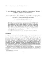
The results showed that ADSCs in the PRP10, PRP15, and
PRP20 groups produce less VEGF-A. The concentrations of
Van Pham et al. Stem Cell Research & Therapy 2013, 4:91
/>
VEGF were 536.67 ± 40.41 ng/ml, 336.67 ± 51.32 ng/ml,
380.0 ± 50 ng/ml, and 1,493.33 ± 143.64 ng/ml in the
PRP10, PRP15, PRP20, and FBS10 groups, respectively
(Figure 4). Compared with the FBS10 group, these
decreases were significant in the PRP10, PRP15, and
PRP20 groups. VEGF concentrations in the PRP15 and
PRP20 groups significantly decreased compared with that
in the PRP10 group, indicating that VEGF expression was
inhibited more efficiently at higher concentrations of PRP.
However, the reduction of VEGF was not significant when
increasing the concentration of PRP from 15% to 20%. Taken
together, PRP decreased VEGF-A expression by 2.78-fold,
4.44-fold, and 3.93-fold in the PRP10, PRP15, PRP20 groups
compared with that in the FBS10 group, respectively.
This result suggests that transplantation of PRP-treated
ADSCs may improve injured articular cartilage.
Articular cartilage regeneration by platelet-rich
plasma-treated ADSC transplantation
The results showed a significant difference among the
treatment and negative control groups, especially in terms
of the time until mice could control their hind-limb movement as well as regeneration of the joint cartilage. The time
until recovery of hind-limb movement decreased from
32.5 ± 7.5 days in negative control (PBS-injected) mice to
17.5 ± 3.5 days in the PRP15 group, but did not decrease
for the FBS10 group (30.5 ± 5.5 days). In the PRP15 mice,
histological analysis showed that the mean area of damaged
joint cartilage was 70% with 45% of regenerated cartilage
formed after 45 days. This regenerated cartilage layer had
about 12 layers of chondrocytes. However, in mice of the
FBS10 group the mean area of damaged joint cartilage was
70%, but there was only 30% regenerated cartilage formed
after 45 days and about eight layers of chondrocytes. In the
negative control mice, the mean area of damaged joint
Page 7 of 11
cartilage was 80%, but there was only 20% regenerated cartilage formed after 45 days and five layers of chondrocytes
(Figure 5).
Discussion
PRP is a natural source of growth factors. In this study,
we determined the effects of PRP on ADSC transplantation
in an injured articular cartilage model. To investigate
the physiological changes of ADSCs induced by PRP,
we successfully isolated ADSCs and PRP.
We isolated the SVF with good viability from adipose
tissue. From the SVF, we isolated ADSCs that expressed
some MSC characteristics including expression of CD44,
CD74, and CD90, and the absence of hematopoietic cell
lineage markers CD14, CD34, and CD45. These cells
differentiated into adipocytes in vitro. We also prepared
PRP with growth factors enriched by five to seven times
compared with those in normal plasma (data not shown).
Next, we evaluated the effects of PRP on ADSC proliferation. The results from MTT assays showed that PRP
strongly stimulated ADSC proliferation, demonstrating
that PRP contains growth factors that are essential for
ADSC proliferation. There are numerous important
growth factors, such as basic fibroblast growth factor
(bFGF), epidermal growth factor, and platelet-derived
growth factor, which stimulate stem cell proliferation
[48,49]. In previous studies, PRP efficiently stimulated
ADSC proliferation [50-53]. Kocaoemer and colleagues
showed that ADSCs rapidly proliferate in medium
supplemented with 10% human serum and 10% PRP
rather than 10% FBS [50]. However, in contrast to our
results showing that 15% PRP was the optimal concentration in medium to stimulate proliferation, Kakudo and colleagues showed that 5% activated PRP maximally promotes
ADSC proliferation, whereas 20% activated PRP does not
Figure 4 Vascular endothelial growth factor-A secretion is reduced in platelet-rich plasma-treated adipose-derived stem cells. Vascular
endothelial growth factor (VEGF)-A concentrations were significantly decreased in culture supernatants of the 10% platelet-rich plasma (PRP10),
15% PRP (PRP15), and 20% PRP (PRP20) groups compared with that in the 10% fetal bovine serum (FBS10) group.
Van Pham et al. Stem Cell Research & Therapy 2013, 4:91
/>
Page 8 of 11
Figure 5 Recovery of mouse knee joints. (A) The cartilage layer of 15% platelet-rich plasma (PRP)-cultured adipose-derived stem cell
(ADSC)-treated mice was similar to that in normal mice. There was evidence of regenerated cartilage formation at the articular cartilage margin in
the treated mice, and the thickness of the cartilage layer of the treated mice compared with (B) before treatment and (C) control. H & E-stained
articular cartilage sections of mice that received (D, E) 15% PRP-cultured ADSC transplantation, (F) 10% fetal bovine serum (FBS)-cultured ADSC
transplantation, or (C) PBS injections.
promote proliferation [53]. More importantly, PRP not only
stimulates ADSC proliferation but also preserves the differentiation potential of ADSC in vitro [51,52]. However,
Gharibi and Hughes recently showed that ADSCs treated
with bFGF, epidermal growth factor, platelet-derived growth
factor, and ascorbic acid show a loss of differentiation potential prior to reaching senescence [48], indicating that
PRP may induce differentiation into functional cells.
In our study, we considered that PRP not only stimulates ADSC proliferation but also differentiation into
chondrogenic cells. We therefore investigated the
changes of ADSC phenotype when cultured in medium
supplemented with PRP or FBS. PRP did not change
surface marker expression of ADSCs after culture in
PRP-containing medium for 1 week. However, there were
significant differences in the expression of chondrogenesisrelated genes.
ADSCs treated with PRP exhibited upregulated expression of chondrogenesis-related gene such as col-II,
Sox9, and aggrecan. We found that col-II gene expression increased by 3.01-fold, 3.37-fold, and 3.44-fold in
the PRP10, PRP15, and PRP20 groups, compared with that
in the FBS10 group, respectively. Similarly, expression of
other chondrogenic markers including Sox9 and aggrecan
also increased in the PRP10, PRP15, and PRP20 groups
compared with that in the FBS10 group. Sox9 expression
strongly increased in the PRP10, PRP15 and PRP20 groups
compared with that in the FBS10 group. These results demonstrated that PRP changed the gene expression of ADSCs
toward the chondrogenic lineage but did not change the
surface marker expression of ADSCs.
The secretion of certain growth factors, especially
VEGF-A from ADSCs, inhibits cartilage regeneration
[43]. VEGF enhances catabolic pathways in chondrocytes,
and VEGF overexpression is associated with progression
of osteoarthritis in articular cartilage [54,55]. In fact, VEGF
induces matrix metalloproteinase expression in immortalized chondrocytes [56]. We therefore considered
that PRP may not only promote ADSC differentiation
into chondrogenic cells but might also inhibit VEGF
secretion. For this reason, PRP-treated ADSCs may induce chondrocyte differentiation and regenerate cartilage. We confirmed that, after treatment with PRP for 1
week, ADSCs downregulated VEGF secretion into the
culture supernatant. PRP10, PRP15 and PRP20 ADSCs
downregulated VEGF expression by 2.78-fold, 4.44-fold,
and 3.93-fold compared with that in FBS10 ADSCs,
respectively. This observation indicates that PRP-treated
ADSCs may improve ADSC transplantation in injured
articular cartilage. In fact, Lee and colleagues improved
ADSC transplantation in cartilage regeneration by neutralizing VEGF with mAbs [43] .
PRP showed several beneficial effects on ADSCs
for chondrogenic differentiation in vitro. Similarly, in
muscle-derived stem cells, PRP promotes the expression of
bone morphogenic protein-4, promotes collagen synthesis,
suppresses chondrocyte apoptosis, and enhances the integration of transplanted cells in the repair process [57]. PRP
also increases cartilage catabolism in synoviocytes [58].
The effects of PRP are induced by growth factors of the
platelets. As indicated above, PRP contains several important growth factors that have effects on proliferation
and differentiation, such as bFGF and transforming growth
factor-beta. In fact, bFGF enhances the kinetics of MSC
chondrogenesis, leading to early differentiation, possibly
by a priming mechanism [59]. In addition, bFGF induces
ADSC chondrogenesis [60,61]. bFGF-treated bone marrowderived MSCs also undergo chondrogenic differentiation
[62]. Furthermore, transforming growth factor-beta stimulates chondrogenic differentiation of MSCs [63,64].
We also evaluated the role of PRP in chondrogenesis
in vivo. The results showed significantly different efficiencies
Van Pham et al. Stem Cell Research & Therapy 2013, 4:91
/>
of injured articular regeneration by transplantation of PRPtreated (PRP15 group) and untreated ADSCs (FBS10 group).
PRP15 ADSC transplantation efficiently reduced the recovery time of hind-limb movement compared with that of
ADSC transplantation alone. Importantly, ADSC transplantation showed an effect compared with that of the control
(PBS injection), but not significantly. Stimulation of cartilage
regeneration was also achieved in PRP15 ADSC transplantation. Compared with FBS10 ADSC transplantation and
PBS injection, PRP15 ADSCs efficiently stimulated cartilage
formation. ADSC transplantation also stimulated cartilage
formation compared with that of PBS injection but more
slowly and at a lower efficiency. These results showed that
PRP is an important factor that promotes both in vitro and
in vivo chondrogenesis of ADSCs. Previous studies have
performed co-transplantation of ADSCs and PRP in dogs
[30-32,35], and co-transplantation of the SVF and PRP in
humans [41,42,65] and mice [37,38], resulting in significant
improvements of injured articular cartilage. Transplantation
of ADSCs without PRP in rats [43] or SVF transplantation
without PRP in horses [34] inhibits cartilage regeneration
[43] or provides insignificant improvements [34].
Conclusion
Adipose tissue provides a rich source of MSCs. ADSCs
have been used to treat injured articular cartilage in recent
years. However, ADSC transplantation in injured articular
cartilage has caused controversy regarding the treatment efficiency and ADSC transplantation combined
with additional factors to induce chondrogenic differentiation. This study revealed that PRP is a suitable factor in
ADSC transplantation to treat injured articular cartilage.
PRP stimulates ADSC proliferation and induces ADSC
differentiation into chondrogenic cells with overexpression
of col-II, Sox9, and aggrecan. In particular, PRP reduces
VEGF expression that inhibits cartilage regeneration to
improve cartilage regeneration in vivo by PRP-treated
ADSC transplantation. PRP-treated ADSC transplantation significantly improves cartilage formation in murine
models compared with that of untreated ADSC transplantation. These results reveal a promising therapy of
injured articular cartilage by transplantation of ADSCs
combined with PRP.
Abbreviations
ADSC: Adipose-derived stem cell; bFGF: Basic fibroblast growth factor;
BSA: Bovine serum albumin; col-II: Type II collagen; DMEM: Dulbecco’s
modified Eagle’s medium; ELISA: Enzyme-linked immunosorbent assay;
FBS: Fetal bovine serum; GAPDH: Glyceraldehyde-3-phosphate
dehydrogenase; H & E: Hematoxylin and eosin; mAb: Monoclonal antibody;
MSC: Mesenchymal stem cell; PBS: Phosphate-buffered saline;
PCR: Polymerase chain reaction; PRP: Platelet-rich plasma; RT: Reverse
transcriptase; SVF: Stromal vascular fraction; VEGF: Vascular endothelial
growth factor.
Competing interests
The authors declare that they have no competing interests.
Page 9 of 11
Authors’ contributions
PVP carried out studies including primary culture, ADSC isolation and culture,
PRP preparation, and manuscript writing. KH-TB, TDD, TDN, and VTL collected
the adipose tissue and peripheral blood, and established animal models.
DQN carried out the histological analysis of cartilage. NBV, NHT performed
the stem cell transplantation in murine models, and evaluated injured
articular cartilage healing. DML and NL-CP performed gene expression
analyses and measured the VEGF-A concentrations. NKP revised the
manuscript, edited figures, and processed data. All authors read and
approved the final manuscript.
Acknowledgements
This work was funded by grants from GeneWorld Ltd, Ho Chi Minh City,
Vietnam.
Author details
Laboratory of Stem Cell Research and Application, University of Science,
Vietnam National University, 227 Nguyen Van Cu, District 5, Ho Chi Minh City,
Vietnam. 2University of Medical Center, Ho Chi Minh University of Medicine
and Pharmacy, 215 Hong Bang, District 5, Ho Chi Minh City, Vietnam.
3
Department of Pathology, University of Medicine and Pharmacy, 217 Hong
Bang, District 5, Ho Chi Minh City, Vietnam.
1
Received: 16 May 2013 Revised: 21 June 2013
Accepted: 16 July 2013 Published: 1 August 2013
References
1. Borrione P, Gianfrancesco AD, Pereira MT, Pigozzi F: Platelet-rich plasma in
muscle healing. Am J Phys Med Rehabil 2010, 89:854–861.
2. Yu W, Wang J, Yin J: Platelet-rich plasma: a promising product for
treatment of peripheral nerve regeneration after nerve injury.
Int J Neurosci 2011, 121:176–180.
3. Kim DH, Je YJ, Kim CD, Lee YH, Seo YJ, Lee JH, Lee Y: Can platelet-rich plasma
be used for skin rejuvenation? Evaluation of effects of platelet-rich plasma on
human dermal fibroblast. Ann Dermatol 2011, 23:424–431.
4. Cho JW, Kim SA, Lee KS: Platelet-rich plasma induces increased expression
of G1 cell cycle regulators, type I collagen, and matrix metalloproteinase-1
in human skin fibroblasts. Int J Mol Med 2012, 29:32–36.
5. Kwon DR, Park GY, Lee SU: The effects of intra-articular platelet-rich
plasma injection according to the severity of collagenase-induced knee
osteoarthritis in a rabbit model. Ann Rehabil Med 2012, 36:458–465.
6. Lippross S, Moeller B, Haas H, Tohidnezhad M, Steubesand N, Wruck CJ, Kurz
B, Seekamp A, Pufe T, Varoga D: Intraarticular injection of platelet-rich
plasma reduces inflammation in a pig model of rheumatoid arthritis of
the knee joint. Arthritis Rheum 2011, 63:3344–3353.
7. Siclari A, Mascaro G, Gentili C, Kaps C, Cancedda R, Boux E: Cartilage repair
in the knee with subchondral drilling augmented with a platelet-rich
plasma-immersed polymer-based implant. Knee Surg Sports Traumatol
Arthrosc 2013. Epub ahead of print.
8. Lee GW, Son JH, Kim JD, Jung GH: Is platelet-rich plasma able to enhance
the results of arthroscopic microfracture in early osteoarthritis and
cartilage lesion over 40 years of age? Eur J Orthop Surg Traumatol 2013,
23:581–587.
9. Jang SJ, Kim JD, Cha SS: Platelet-rich plasma (PRP) injections as an
effective treatment for early osteoarthritis. Eur J Orthop Surg Traumatol
2013, 23:573–580.
10. Patel S, Dhillon MS, Aggarwal S, Marwaha N, Jain A: Treatment with
platelet-rich plasma is more effective than placebo for knee
osteoarthritis: a prospective, double-blind, randomized trial.
Am J Sports Med 2013, 41:356–364.
11. Spakova T, Rosocha J, Lacko M, Harvanova D, Gharaibeh A: Treatment of
knee joint osteoarthritis with autologous platelet-rich plasma in
comparison with hyaluronic acid. Am J Phys Med Rehabil 2012, 91:411–417.
12. Zuk PA, Zhu M, Mizuno H, Huang J, Futrell JW, Katz AJ, Benhaim P, Lorenz
HP, Hedrick MH: Multilineage cells from human adipose tissue:
implications for cell-based therapies. Tissue Eng 2001, 7:211–228.
13. Zimmerlin L, Donnenberg VS, Rubin JP, Donnenberg AD: Mesenchymal
markers on human adipose stem/progenitor cells. Cytometry A 2013,
83:134–140.
Van Pham et al. Stem Cell Research & Therapy 2013, 4:91
/>
14. Zhu X, Du J, Liu G: The comparison of multilineage differentiation of
bone marrow and adipose-derived mesenchymal stem cells. Clin Lab
2012, 58:897–903.
15. Gaiba S, Franca LP, Franca JP, Ferreira LM: Characterization of human
adipose-derived stem cells. Acta Cir Bras 2012, 27:471–476.
16. Khan WS, Adesida AB, Tew SR, Longo UG, Hardingham TE: Fat pad-derived
mesenchymal stem cells as a potential source for cell-based adipose
tissue repair strategies. Cell Prolif 2012, 45:111–120.
17. Dominici M, Le Blanc K, Mueller I, Slaper-Cortenbach I, Marini F, Krause D,
Deans R, Keating A, Prockop D, Horwitz E: Minimal criteria for defining
multipotent mesenchymal stromal cells. The International Society for
Cellular Therapy position statemen. Cytotherapy 2006, 8:315–317.
18. Christodoulou I, Kolisis FN, Papaevangeliou D, Zoumpourlis V: Comparative
evaluation of human mesenchymal stem cells of fetal (Wharton’s jelly)
and adult (adipose tissue) origin during prolonged in vitro expansion:
considerations for cytotherapy. Stem Cells Int 2013, 2013:246134.
19. Harn HJ, Lin SZ, Hung SH, Subeq YM, Li YS, Syu WS, Ding DC, Lee RP, Hsieh DK,
Lin PC, Chiou TW: Adipose-derived stem cells can abrogate chemical-induced
liver fibrosis and facilitate recovery of liver function. Cell Transplant 2012,
21:2753–2764.
20. Gu JH, Ji YH, Dhong ES, Kim DH, Yoon ES: Transplantation of adipose
derived stem cells for peripheral nerve regeneration in sciatic nerve
defects of the rat. Curr Stem Cell Res Ther 2012, 7:347–355.
21. Liu G, Cheng Y, Guo S, Feng Y, Li Q, Jia H, Wang Y, Tong L, Tong X:
Transplantation of adipose-derived stem cells for peripheral nerve repair.
Int J Mol Med 2011, 28:565–572.
22. Santiago LY, Clavijo-Alvarez J, Brayfield C, Rubin JP, Marra KG: Delivery of
adipose-derived precursor cells for peripheral nerve repair. Cell Transplant
2009, 18:145–158.
23. Mazo M, Hernández S, Gavira JJ, Abizanda G, Araña M, López-Martínez T,
Moreno C, Merino J, Martino-Rodríguez A, Uixeira A, García de Jalón JA,
Pastrana J, Martínez-Caro D, Prósper F: Treatment of reperfused ischemia
with adipose-derived stem cells in a preclinical Swine model of
myocardial infarction. Cell Transplant 2012, 21:2723–2733.
24. Rigol M, Solanes N, Farré J, Roura S, Roqué M, Berruezo A, Bellera N,
Novensà L, Tamborero D, Prat-Vidal C, Huzman MA, Batlle M, Hoefsloot M,
Sitges M, Ramírez J, Dantas AP, Merino A, Sanz G, Brugada J, Bayés-Genís A,
Heras M: Effects of adipose tissue-derived stem cell therapy after
myocardial infarction: impact of the route of administration. J Card Fail
2010, 16:357–366.
25. Pecanha R, Bagno LL, Ribeiro MB, Robottom Ferreira AB, Moraes MO,
Zapata-Sudo G, Kasai-Brunswick TH, Campos-de-Carvalho AC, Goldenberg
RC, Saar Werneck-de-Castro JP: Adipose-derived stem-cell treatment of
skeletal muscle injury. J Bone Joint Surg Am 2012, 94:609–617.
26. Xiao J, Zhang C, Zhang Y, Zhang X, Zhao J, Liang J, Zhong X, Chen Y:
Transplantation of adipose-derived mesenchymal stem cells into a
murine model of passive chronic immune thrombocytopenia. Transfusion
2012, 52:2551–2558.
27. Yang JJ, Yang X, Liu ZQ, Hu SY, Du ZY, Feng LL, Liu JF, Chen YD:
Transplantation of adipose tissue-derived stem cells overexpressing
heme oxygenase-1 improves functions and remodeling of infarcted
myocardium in rabbits. Tohoku J Exp Med 2012, 226:231–241.
28. Riordan NH, Ichim TE, Min WP, Wang H, Solano F, Lara F, Alfaro M, Rodriguez
JP, Harman RJ, Patel AN, Murphy MP, Lee RR, Minev B: Non-expanded adipose
stromal vascular fraction cell therapy for multiple sclerosis. J Transl Med
2009, 7:29.
29. Scuderi N, Ceccarelli S, Onesti MG, Fioramonti P, Guidi C, Romano F, Frati L,
Angeloni A, Marchese C: Human adipose-derived stem cells for cell-based
therapies in the treatment of systemic sclerosis. Cell Transplant 2013,
22:779–795.
30. Black LL, Gaynor J, Adams C, Dhupa S, Sams AE, Taylor R, Harman S,
Gingerich DA, Harman R: Effect of intraarticular injection of autologous
adipose-derived mesenchymal stem and regenerative cells on clinical
signs of chronic osteoarthritis of the elbow joint in dogs. Vet Ther 2008,
9:192–200.
31. Black LL, Gaynor J, Gahring D, Adams C, Aron D, Harman S, Gingerich DA,
Harman R: Effect of adipose-derived mesenchymal stem and
regenerative cells on lameness in dogs with chronic osteoarthritis of the
coxofemoral joints: a randomized, double-blinded, multicenter,
controlled trial. Vet Ther 2007, 8:272–284.
Page 10 of 11
32. Guercio A, Di Marco P, Casella S, Cannella V, Russotto L, Purpari G, Di Bella S,
Piccione G: Production of canine mesenchymal stem cells from adipose
tissue and their application in dogs with chronic osteoarthritis of the
humeroradial joints. Cell Biol Int 2012, 36:189–194.
33. Toghraie FS, Chenari N, Gholipour MA, Faghih Z, Torabinejad S, Dehghani S,
Ghaderi A: Treatment of osteoarthritis with infrapatellar fat pad derived
mesenchymal stem cells in rabbit. Knee 2011, 18:71–75.
34. Frisbie DD, Kisiday JD, Kawcak CE, Werpy NM, McIlwraith CW: Evaluation of
adipose-derived stromal vascular fraction or bone marrow-derived
mesenchymal stem cells for treatment of osteoarthritis. J Orthop Res
2009, 27:1675–1680.
35. Lee JM, Im GI: SOX trio-co-transduced adipose stem cells in fibrin gel to
enhance cartilage repair and delay the progression of osteoarthritis in
the rat. Biomaterials 2012, 33:2016–2024.
36. ter Huurne MC, van Lent PLEM, Blom AB, Blattes R, Jeanson Y, Casteilla L,
Jorgensen C: A single injection of adipose-derived stem cells protects
against cartilage damage and lowers synovial activation in experimental
osteoarthritis. Arthritis Rheum 2011, 63:1784.
37. ter Huurne M, Schelbergen R, Blattes R, Blom A, de Munter W, Grevers LC,
Jeanson J, Noël D, Casteilla L, Jorgensen C, van den Berg W, van Lent PL:
Antiinflammatory and chondroprotective effects of intraarticular
injection of adipose-derived stem cells in experimental osteoarthritis.
Arthritis Rheum 2012, 64:3604–3613.
38. Van Pham P, Hong-Thien Bui K, Quoc Ngo D, Tan Khuat L, Kim Phan N:
Transplantation of nonexpanded adipose stromal vascular fraction and
platelet-rich plasma for articular cartilage injury treatment in mice
model. J Med Eng 2013, 2013:7.
39. Murphy JM, Fink DJ, Hunziker EB, Barry FP: Stem cell therapy in a caprine
model of osteoarthritis. Arthritis Rheum 2003, 48:3464–3474.
40. Xie X, Wang Y, Zhao C, Guo S, Liu S, Jia W, Tuan RS, Zhang C: Comparative
evaluation of MSCs from bone marrow and adipose tissue seeded in
PRP-derived scaffold for cartilage regeneration. Biomaterials 2012,
33:7008–7018.
41. Pak J: Regeneration of human bones in hip osteonecrosis and human
cartilage in knee osteoarthritis with autologous adipose-tissue-derived
stem cells: a case series. J Med Case Rep 2011, 5:296.
42. Koh YG, Jo SB, Kwon OR, Suh DS, Lee SW, Park SH, Choi YJ: Mesenchymal
stem cell injections improve symptoms of knee osteoarthritis.
Arthroscopy 2013, 29:748–755.
43. Lee CS, Burnsed OA, Raghuram V, Kalisvaart J, Boyan BD, Schwartz Z:
Adipose stem cells can secrete angiogenic factors that inhibit hyaline
cartilage regeneration. Stem Cell Res Ther 2012, 3:35.
44. Phuc PV, Nhung TH, Loan DT, Chung DC, Ngoc PK: Differentiating of
banked human umbilical cord blood-derived mesenchymal stem cells
into insulin-secreting cells. In Vitro Cell Dev Biol Anim 2011, 47:54–63.
45. Martin I, Jakob M, Schafer D, Dick W, Spagnoli G, Heberer M: Quantitative
analysis of gene expression in human articular cartilage from normal
and osteoarthritic joints. Osteoarthr Cartil 2001, 9:112–118.
46. Li Y, Tew SR, Russell AM, Gonzalez KR, Hardingham TE, Hawkins RE:
Transduction of passaged human articular chondrocytes with adenoviral,
retroviral, and lentiviral vectors and the effects of enhanced expression
of SOX9. Tissue Eng 2004, 10:575–584.
47. Van Pham P, Dang LT-T, Truong NH, Phan NK: Can activated platelet rich
plasma combined with adipose-derived stem cells be used to treat skin
wrinkles? a mechanism study. In Medical Advancements in Aging and
Regenerative Technologies: Clinical Tools and Applications. IGI Global
Germany; 2013:313–329.
48. Gharibi B, Hughes FJ: Effects of medium supplements on proliferation,
differentiation potential, and in vitro expansion of mesenchymal stem
cells. Stem Cells Transl Med 2012, 1:771–782.
49. Chieregato K, Castegnaro S, Madeo D, Astori G, Pegoraro M, Rodeghiero
F: Epidermal growth factor, basic fibroblast growth factor and
platelet-derived growth factor-bb can substitute for fetal bovine
serum and compete with human platelet-rich plasma in the ex vivo
expansion of mesenchymal stromal cells derived from adipose tissue.
Cytotherapy 2011, 13:933–943.
50. Kocaoemer A, Kern S, Kluter H, Bieback K: Human AB serum and
thrombin-activated platelet-rich plasma are suitable alternatives to
fetal calf serum for the expansion of mesenchymal stem cells from
adipose tissue. Stem Cells 2007, 25:1270–1278.
Van Pham et al. Stem Cell Research & Therapy 2013, 4:91
/>
Page 11 of 11
51. Zhang YS, He JH, Xiao GY, Li QM: Effect of platelet-rich plasma on the
proliferation and adipogenic differentiation of human adipose-derived
stem cells in vitro. Nan Fang Yi Ke Da Xue Xue Bao 2011, 31:525–528.
52. Li H, Liu D, Yu Y, Wu T: Experimental research of the promotion effect of
autogeneic PRP on osteogenic differentiation of human adipose-derived
stem cells in vitro. Zhongguo Xiu Fu Chong Jian Wai Ke Za Zhi 2009,
23:732–736.
53. Kakudo N, Minakata T, Mitsui T, Kushida S, Notodihardjo FZ, Kusumoto K:
Proliferation-promoting effect of platelet-rich plasma on human
adipose-derived stem cells and human dermal fibroblasts. Plast Reconstr
Surg 2008, 122:1352–1360.
54. Pfander D, Kortje D, Zimmermann R, Weseloh G, Kirsch T, Gesslein M,
Cramer T, Swoboda B: Vascular endothelial growth factor in articular
cartilage of healthy and osteoarthritic human knee joints. Ann Rheum Dis
2001, 60:1070–1073.
55. Enomoto H, Inoki I, Komiya K, Shiomi T, Ikeda E, Obata K, Matsumoto H,
Toyama Y, Okada Y: Vascular endothelial growth factor isoforms and their
receptors are expressed in human osteoarthritic cartilage. Am J Pathol
2003, 162:171–181.
56. Pufe T, Harde V, Petersen W, Goldring MB, Tillmann B, Mentlein R: Vascular
endothelial growth factor (VEGF) induces matrix metalloproteinase
expression in immortalized chondrocytes. J Pathol 2004, 202:367–374.
57. Mifune Y, Matsumoto T, Takayama K, Ota S, Li H, Meszaros LB, Usas A,
Nagamune K, Gharaibeh B, Fu FH, Huard J: The effect of platelet-rich
plasma on the regenerative therapy of muscle derived stem cells for
articular cartilage repair. Osteoarthr Cartil 2013, 21:175–185.
58. Browning SR, Weiser AM, Woolf N, Golish SR, SanGiovanni TP, Scuderi GJ,
Carballo C, Hanna LS: Platelet-rich plasma increases matrix
metalloproteinases in cultures of human synovial fibroblasts. J Bone Joint
Surg Am 2012, 94:e1721–e1727.
59. Cheng T, Yang C, Weber N, Kim HT, Kuo AC: Fibroblast growth factor 2
enhances the kinetics of mesenchymal stem cell chondrogenesis.
Biochem Biophys Res Commun 2012, 426:544–550.
60. Kabiri A, Esfandiari E, Hashemibeni B, Kazemi M, Mardani M, Esmaeili A:
Effects of FGF-2 on human adipose tissue derived adult stem cells
morphology and chondrogenesis enhancement in Transwell culture.
Biochem Biophys Res Commun 2012, 424:234–238.
61. Buckley CT, Kelly DJ: Expansion in the presence of FGF-2 enhances the
functional development of cartilaginous tissues engineered using
infrapatellar fat pad derived MSCs. J Mech Behav Biomed Mater 2012,
11:102–111.
62. Stewart AA, Byron CR, Pondenis H, Stewart MC: Effect of fibroblast growth
factor-2 on equine mesenchymal stem cell monolayer expansion and
chondrogenesis. Am J Vet Res 2007, 68:941–945.
63. Bhang SH, Jeon JY, La WG, Seong JY, Hwang JW, Ryu SE, Kim BS: Enhanced
chondrogenic marker expression of human mesenchymal stem cells by
interaction with both TGF-β3 and hyaluronic acid. Biotechnol Appl
Biochem 2011, 58:271–276.
64. Fan H, Zhang C, Li J, Bi L, Qin L, Wu H, Hu Y: Gelatin microspheres
containing TGF-beta3 enhance the chondrogenesis of mesenchymal
stem cells in modified pellet culture. Biomacromolecules 2008, 9:927–934.
65. Koh YG, Choi YJ: Infrapatellar fat pad-derived mesenchymal stem cell
therapy for knee osteoarthritis. Knee 2012, 19:902–907.
doi:10.1186/scrt277
Cite this article as: Van Pham et al.: Activated platelet-rich plasma
improves adipose-derived stem cell transplantation efficiency in injured
articular cartilage. Stem Cell Research & Therapy 2013 4:91.
Submit your next manuscript to BioMed Central
and take full advantage of:
• Convenient online submission
• Thorough peer review
• No space constraints or color figure charges
• Immediate publication on acceptance
• Inclusion in PubMed, CAS, Scopus and Google Scholar
• Research which is freely available for redistribution
Submit your manuscript at
www.biomedcentral.com/submit