DSpace at VNU: Luminescent nanomaterials containing rare earth ions for security printing
Bạn đang xem bản rút gọn của tài liệu. Xem và tải ngay bản đầy đủ của tài liệu tại đây (1.29 MB, 12 trang )
Int. J. Nanotechnol., Vol. 8, Nos. 3/4/5, 2011
Luminescent nanomaterials containing rare earth
ions for security printing
Tran Kim Anh*, Dinh Xuan Loc,
Tran Thu Huong and Nguyen Vu
Institute of Materials Science,
Vietnam Academy of Science and Technology,
18 Hoang Quoc Viet, Cau Giay distr., Hanoi, Vietnam
E-mail:
E-mail:
E-mail:
E-mail:
*Corresponding author
Le Quoc Minh
Institute of Materials Science,
Vietnam Academy of Science and Technology,
18 Hoang Quoc Viet, Cau Giay distr., Hanoi, Vietnam
and
College of Technology,
Vietnam National University of Hanoi,
144 Xuan Thuy, Hanoi, Vietnam
E-mail:
Abstract: The high-efficiency luminescent nanomaterials with different
emission wavelengths of red (YVO4:Eu3+), green (CePO4:Tb3+), ZnS:Mn2+ and
YVO4:Eu3+@SiO2 were successfully prepared with different concentrations of
Mn and rare earth ions as active centres by chemical synthesis. Structure
properties were studied. It was found that the particle size of our samples was
in the range of 10–30 nm. Photoluminescent properties were studied under 325,
337, 365 and 370 nm excitations in order to apply in luminescent labels. The
primary colour components are red and green emission making them very
convenient and attractive for screen security printing systems. Hundreds of
different labels with a size of 1–10 cm2 were prepared by screen-printing as
well as inkjet printing. By improving the Epson printer, commercial red, green
and blue inks were used in the printing application. Screen and inkjet printing
were deemed good methods for security printing. Our products were beautiful,
high resolution and withstood tropical weather.
Keywords: luminescent nanomaterial; Tb3+; Eu3+ security printer; coreshell.
Reference to this paper should be made as follows: Anh, T.K., Loc, D.X.,
Huong, T.T., Vu, N. and Minh, L.Q. (2011) ‘Luminescent nanomaterials
containing rare earth ions for security printing’, Int. J. Nanotechnol., Vol. 8,
Nos. 3/4/5, pp.335–346.
Copyright © 2011 Inderscience Enterprises Ltd.
335
336
T.K. Anh et al.
Biographical notes: Tran Kim Anh graduated from Hanoi University in 1970,
received her PhD in Physics from the Institute of Physics, Polish Academy of
Science, Warsaw, Poland, in 1988. She has been an Associate Professor since
1996. She has been the Director of Physics and Chemistry of Advanced Optical
Materials Laboratory since 1998. She received the National Award (group) on
Sciences and Technology in 2005. She has been a Director of Research since
2009. Her research interests include physics and technology of luminescent
nanomaterials containing rare earth ions and nano-structured semiconductors
including ZnO and ZnS as well as planar waveguide.
Dinh Xuan Loc graduated from Hanoi University in 1990, received Master’s
Degree in Chemistry in 1999 and has been a Senior Researcher since 2008.
He is currently doing his PhD thesis at the Institute of Materials Science,
Vietnam Academy of Science and Technology. His research interests include
nanomaterials containing rare earth and transition metal ions.
Tran Thu Huong received her Bachelor’s Degree in Chemistry. She did her
Master’s Degree at the International Training Institute of Materials Science in
1999. She received her Doctorate of Philosophy in Materials Science from the
Institute of Materials Science, Vietnam Academy of Science and Technology,
in 2007. Her research interests include luminescent nanomaterials containing
rare earth ions and planar waveguide. She received the Vallet Scholarship
2006–2007.
Nguyen Vu received his Master’s Degree from the International Training
Institute of Materials Science in 1998, and then he received the PhD in
Materials Science from the Institute of Materials Science, Vietnam Academy of
Science and Technology, in 2007. His research interests include the physical
chemistry of nanomaterials, optoelectronic and photonic application,
luminescent nanomaterials containing rare earth ions and up-conversion
nanomaterials.
Le Quoc Minh is an Associate Professor of Physics in the Institute of Materials
Science and the Director of Research, Vietnam Academy of Science and
Technology. He received his PhD in Physical Chemistry, Berlin, Germany. He
has been the Director of Laboratory of Photochemistry, Imaging and Photonics;
Lecturer at the University of Engineering and Technology, Vietnam National
University, Hanoi. He is the Vice President and Secretary of Society of
Materials Science of Vietnam. His research interests include materials science,
physics and chemistry of photoresponsive compounds, luminescence materials,
nanomaterials, hybrid and composite for optoelectronics and application in
biophotonics.
1
Introduction
Nanophotonics is an exciting field in nanoscience and nanotechnology. It allows
the opportunities for basic research and the development of new breakthrough solutions.
The interaction of light with matter on a nanometre-size scale has had many new
developments. Nanophotonics is an exciting frontier in nanoscience and technology for
chemists, physicists, engineers and biologists as well as biomedical researchers [1].
Some interesting topics include the novel synthetic routes and processing of
nanomaterials, self-assembled periodic, nanoscales, non-linear optical processes and
Luminescent nanomaterials containing rare earth ions for security printing 337
time-resolved and spectrally resolved studies. Many report, for example, about the
applications of nanoparticles in biophotonics [2], luminescent nanomaterials for
biological labelling [3], functionalised europium nanorods for in vitro imaging [4],
development of new ink materials based on luminescent nanomaterials for the security
labels of printing products such as passport and visa documents has encouraged research
in this direction [5]. We studied luminescence, energy transfer and up-conversion
mechanism of Y2O3 doped with Eu3+, Tb3+, Tm3+, Er3+ and Yb3+ ions [6], and security
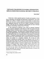
labels is attractive application. Figure 1 shows an image under UV exposure of a
European visa for corresponding author, in which security part was made with
luminescent materials.
This paper reports on the preparation of luminescent nanomaterials from YVO4:Eu3+,
ZnS:Mn2+, and CePO4:Tb3+ for two important spectral fields at red and green regions.
Their structures, nanoform and optical properties, especially luminescence behaviour,
were shown to depend on preparation conditions and allowed us to obtain a high
luminescence efficiency, nanosize and fine distribution in the liquid phase. We present
also the fabrication capacity of a new ink based on the obtained luminescent
nanomaterials for screen or inkjet printing technology.
Figure 1
2
Security printing for a European visa (see online version for colours)
Experiment
2.1 Sample preparation
Preparation of YVO4:Eu3+ nanoparticles by hydrothermal synthesis
YVO4:Eu3+ nanomaterials were prepared by hydrothermal method, which is described
in detail [7]. Y2O3, Eu2O3, HNO3, NH4VO3 and NaOH (analytical reagent of Merck)
were used as starting materials and ethanol (Prolabo Chemical reagent) as solvent
for the preparation and washing of the YVO4.Eu3+ nanocrystalline powders. Y(NO3)3 and
Eu(NO3)3 were prepared by dissolving Y2O3 and Eu2O3 in nitric acid. The process of the
synthesis of YVO4:Eu3+ nanoparticles powders is as follows: the Y(NO3)3.6H2O and
Eu(NO3)3.5H2O were dissolved in 15 ml of deionised water, and the solution was poured
into a Teflon vessel. During stirring, 15 ml of 1M NaOH solution were rapidly added to
the solution, which become a white suspension. To the well-stirred suspension,
Na3VO4.10H2O dissolved in 30 ml of deionised water was added. Finally, the reaction
mixture pH reached 12.5 by using 1M NaOH solution. The Teflon vessel containing the
suspension was capped and placed in an autoclave and heated at 200°C for 1 h.
338
T.K. Anh et al.
The suspension was centrifuged at 3000 rpm for 10 min. The supernatant was discarded.
The precipitate was suspended in 30 ml deionised water and centrifuged again for several
times. The powder of the nanoparticles was obtained by removing ethanol, which was
treated at 60°C for several hours.
Silica coating of the YVO4:Eu3+ nanoparticles
YVO4:Eu3+ nanomaterials were prepared by co-precipitate method. The synthesis
procedure of YVO4:Eu3+ nanoparticles is as follows: The Na3VO4 powder was
completely dissolved in 50 ml of deionised water. Y(NO3)3.6H2O, 99.8%, and
Eu(NO3)3.5H2O, 99.9%, were added to the solution in a 100 ml round-bottomed flask and
this was followed by a magnetic stirring for 120 min. The pH of the solution after the
reaction was in the range of 12–12.5 by using 1M NaOH solution. The content ratio of
Y3+ and Eu3+ were 0.9/0.1 moles, respectively. After that, the solution was poured into a
Teflon-lined stainless steel autoclave and heated at 200°C from 20 h to 40 h, and then
cooled down slowly to room temperature. The resulting products were collected, and
centrifuged at 5900 rpm, washed several times using water, and then air-dried at 60°C for
several hours.
5 ml of an aqueous solution of tetraethylorthosilicate (TEOS) and isopropanol
were mixed in a 100 ml round-bottomed flask. Then, YVO4:Eu3+ nanoparticles were
added to the same flask. The resulting solution was stirred at ambient temperature for 2 h.
The obtained mixture was then refluxed for 24 h. The grafted colloids were finally
purified by ultrafiltration.
Preparation of ZnS:Mn2+ nanoparticles
ZnS:Mn2+ nanomaterials were prepared by co-precipitate method from Zn(CH3COO)2,
Mn(CH3COO)2 and (NH4)2Sx with different concentration of Mn of 0.01, 0.025 and 0.05
at%. Polyvinyl alcohol (PVA) was used as the polymer matrix. Metal solution was mixed
with PVA solution with the weight ratio of metal acetate to PVA of 1/3 for about one
hour while being stirred at 80°C. Then, (NH4)2Sx solution was added drop-wise into the
solution above while stirring. The emulsion was vigorously agitated using a magnetic
stirrer for one hour more. The ZnS:Mn2+ nanoparticles were separated from the solution
by centrifugation and washed several times in hot water and ethanol. The powder
of the nanoparticles was obtained by removing the ethanol after being dried at 70–80°C
for 2–3 h.
Preparation of CePO4:Tb3+/LaPO4 core/shell nanoparticles
Recently, CePO4:Tb3+ nanorods and nanowires were studied [8,9]. In our laboratory,
CePO4:Tb3+ core nanoparticles were synthesised as follows: two aqueous solutions of
Ce(NO3)3 and Tb(NO3)3 were mixed in an appropriate molar ratio. The solution was
concentrated by heating at 80°C until the excess free water had evaporated. The mixture
was dissolved in methanol. Then, tris(2-ethylhexyl)phosphate (TEHP) was added to the
solution under a vacuum at 80°C, the water and the methanol ingredients contained
in the solution were distilled away. A freshly prepared solution of crystalline phosphoric
acid dissolved in a mixture of trioctylamine and TEHP was added into the metal solution.
The resulting microemulsion solution was transferred into a stainless Teflon-lined
autoclave and heated at 200°C for 4 h. The resulting suspension was naturally cooled
down to room temperature to get to the CePO4:Tb3+ core nanoparticles.
Luminescent nanomaterials containing rare earth ions for security printing 339
CePO4:Tb3+/LaPO4 core/shell nanoparticles were synthesised as follows: The solution
of La(NO3)3 in TEHP was prepared as same as Ce(NO3)3 solution in TEHP. And, the
freshly prepared solution of crystalline phosphoric acid dissolved in a mixture of
trioctylamine and TEHP was added into the above-mentioned La(NO3)3 solutions. Then,
this solution was added to one half of the CePO4:Tb3+ product. Finally, the resulting
microemulsion solution was transferred into a stainless Teflon-lined autoclave and heated
at 200°C for 4 h.
2.2 Characterisation
The YVO4:Eu3+, CePO4:Tb3+, and ZnS:Mn2+ nanoparticles were analysed by the D-5000
X-ray diffractometer (Siemens) with Cu Kα radiation. The morphology of the samples
was observed by using a transmission electron microscope (TEM, JEM-1010)
and a field emission scanning electron microscope (FE-SEM, Hitachi, S-4800).
The photoluminescent (PL) measurements were performed at room temperature by using
a cw He-Cd laser (325 nm) as the excitation source, a Spectrapro 2300i monochromator
(Acton) as the dispersive unit, and a Pixis 256 CCD (Acton) as the detector. PL spectra
of the nanoparticles were also measured by using a spectrometer system Horiba Jobin
Yvon IHR 550 after being excited at 370 nm by a diode. Different wavelengths of
254 nm, 277 nm and 365 nm were used for our studies and applications.
3
Results and discussion
3.1 Structural and morphological properties
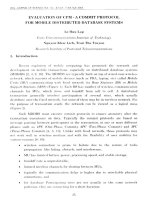
The X-ray diffraction pattern of typical ZnS:Mn2+ nanoparticles is given in Figure 2.
It can be noted that all of the diffraction peaks could be well indexed to the zinc-blende
crystal structure, having prominent (111), (220), and (311) d-spacings. In this Mn-doped
sample, no additional phase was observed, indicating that manganese was successfully
doped in the ZnS crystalline lattice. The width of the diffraction lines is broadened
because of the small size of the crystallites.
Figure 2
XRD pattern of ZnS:Mn2+ (see online version for colours)
340
T.K. Anh et al.
A TEM image of ZnS:Mn2+ is presented in Figure 3. Figure 4 shows a TEM image of the
YVO4:Eu3+ nanoparticles prepared by hydrothermal synthesis. It can be seen from the
figures that the YVO4:Eu3+ sample occurs through the aggregation of the nanocrystals
exhibiting sizes of about 20 nm.
Figure 3
TEM image of ZnS:Mn2+
Figure 4
TEM image of YVO4:Eu3+ nanoparticles prepared by hydrothermal synthesis
The mean diameter of a nanoparticle corresponds to the diameter of a spherical volume
from the FE-SEM image, with the diameter of the YVO4:Eu3+ nanoparticles being about
15–20 nm.
3.2 Photoluminescent (PL) properties
Synthesis and luminescence properties of Mn2+-doped ZnS nanocrystals are presented in
[10]. For ZnS:Mn2+, the 4T1-6A1 transition within the 3d5 configuration of the divalent
manganese ion has been studied, peak at 584 nm to compare with ZnS:Tb3+ nanoparticles
have two main peaks in the green emission at 517 and 541 nm (5D4-7Fj).
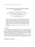
PL spectra of CePO4:Tb3+ nanoparticles and CePO4:Tb3+/LaPO4 core/shell
nanoparticles under 277 nm excitation are presented in Figure 5. The spectra show
narrow emission peaks resulting from 4f-4f transitions within Tb3+ ions, with the most
Luminescent nanomaterials containing rare earth ions for security printing 341
intense peak at 543 nm corresponding to the 5D4–7F5 transition. Three other peaks are
observed at 489 nm (5D4–7F6), 583 nm (5D4 –7F4), 618 nm (5D4–7F3), respectively.
Comparing PL intensity of CePO4:Tb3+ nanoparticles and CePO4:Tb3+/LaPO4 core/shell
nanoparticles, it is noted that the PL intensity of CePO4:Tb3+/LaPO4 core/shell sample is
about 20 times higher than that of CePO4:Tb3+ sample.
Figure 5
PL spectra of CePO4:Tb3+ nanoparticles and CePO4:Tb3+/LaPO4 core/shell nanoparticles
under 277 nm excitation (see online version for colours)
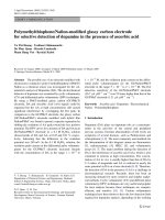
Figure 6 shows PL spectra under 370 nm excitation of YVO4:Eu3+ nanoparticles prepared
by hydrothermal synthesis. Under UV excitation, europium-doped YVO4 nanoparticles
exhibit strong red luminescence caused by a transition within the f-electron shell of the
europium ions. From the 5D0 level, there is a magnetic dipole level, which allowed
transitions to a sublevel of 7F1, two electric dipole level transitions to the sublevels of 7F2,
and three magnetic dipole transitions to sublevels of 7F3. The most intense luminescence
lines correspond to the 5D0–7F2 emission (619 nm) in YVO4:Eu3+ nanoparticles similar in
bulk materials. Figure 6 presents the comparison of the emission spectra change from 1 to
7 at.% Eu3+ in YVO4:Eu3+ nanoparticles. Similar to bulk materials, the quantum yield
shows a maximum at a doping level at about 5 at.% europium and decreases at higher
europium concentration (7 at.%). It is the quenching effect of the luminescence at high
concentrations in lanthanide-doped systems. This typical property is observed when the
distance between europium ions decreases below a critical value.
Figure 6
PL spectra of YVO4:Eu3+ nanoparticles for 5, 3, 7, 1 at.% from top to bottom under
370 nm excitation (see online version for colours)
342
T.K. Anh et al.
Similar PL spectra of YVO4:Eu3+ and YVO4:Eu3+@SiO2 nanoparticles are presented in
Figures 7 and 8.
Figure 7
PL spectrum of YVO4:Eu3+ nanoparticles at 200°C for 6h under an excitation
wavelength of 325 nm at room temperature (see online version for colours)
Figure 8
PL spectrum of YVO4:Eu3+@Silica nanoparticles under an excitation wavelength
of 325 nm at room temperature (see online version for colours)
The PL spectra of the YVO4:Eu3+ nanoparticles and the YVO4:Eu3+ with core-shell
structure nanoparticles displayed red emission with narrow peaks corresponding to the
intra-4f (5D0–7Fj, j = 1, 2, 3, 4) transitions of Eu3+ ions.
The peaks were found at 594 nm (5D0–7F1), 619 nm (5D0–7F2), 652 nm (5D0–7F3), and
702 nm (5D4–7F4), with the strongest emission at 619 nm. One can observe from the peak
at 619 nm that the PL intensity of the SiO2 coated sample is higher than that of the
uncoated one.
3.3 Application
Nano Y2O3 doped with Er3+, Yb3+ was used to prepare infrared cards [11]. Labelled
microplaces were printed using screen technique or inkjet technology [12]. The
CorelDraw, Photoshop and Colour system of RGB (Red, Green, Blue) and CMYK
printing system controlled the inkjet machine and produced the labelled images. Some
different printing samples demonstrate the application (Figure 9).
Luminescent nanomaterials containing rare earth ions for security printing 343
Figure 9
Lotus image of Vietnam Airline logo and VIETNAM letter using ZnS:Mn2+ /PVA
under UV lamp (see online version for colours)
Figure 10 shows solution of nanomaterials from CePO4:Tb3+ under a UV lamp. Figure 11
presents a Vietnamese passport and a European visa, printed using commercial inks.
The nanosolution is of CePO4:Tb3+ with a strong green emission under UV 254 nm
excitation. The comparison of our samples with commercial luminescent materials
shown that our nanomaterials could be used for security printing. The application
of our nanomaterials could be extended into the security field as well as biology
labelling.
Figure 10 Solution of nanomaterials from CePO4:Tb3+ under a UV lamp (see online version
for colours)
Figure 11 Photo images of the luminescent security on Vietnamese passport and a European visa
under UV lamp (see online version for colours)
344
T.K. Anh et al.
3.4 Future works
Our new results will lead to future studies on luminescent nanophotonics. We have some
results on nanomaterials containing rare earth ions [13,14], hybrid and composite with
nanostructures for photonic technology [15], preparation and photonic features of
erbium-activated silica-zirconia multilayer films derived from sol gel process [16],
preparation and infrared emission of silica–zirconia–alumina doped with erbium for
planar waveguide [17], luminescence and up-conversion mechanisms of photonic
materials doped with Eu3+, Er3+ and Yb3+ ions [18] and nanomaterials containing rare
earth ions for infrared card and planar waveguide application [19]. We will try to study
some new nanomaterials in possible papers on the subject of functionalised fluorescent
oxide nanoparticles: artificial toxins for sodium channel targeting and imaging at the
single-molecule level [20], inorganic phosphate nanorods as novel fluorescent labels in
cell biology [21], up-conversion properties of nanocrystalline ZrO2:Yb3+, Er3+ phosphors
[22], functionalised europium nanorods for in-vitro imaging [4], biological and clinical
aspects of lanthanide coordination compounds [23].
Our luminescent nanomaterials could be used for some application. Nanosolution of
YVO4:Eu3+ could be used to recognise viruses such as Rota virus. Our up-conversion of
Y2O3:Er3+ or Y2O3:Er3+, Yb3+ could be used as infrared cards or for security illuminated
by infrared laser 980 nm and to see the green or red colour depending on the
concentration of Er3+ and Yb3+.
4
Conclusion
YVO4:Eu3+, ZnS:Mn2+, and CePO4:Tb3+ nanomaterials were successfully prepared by
hydrothermal and co-precipitate methods. The crystal structure and surface of
nanomaterials were analysed by X-ray diffraction, TEM and photoluminescence
characters were studied by high-resolution photoluminescence with different excited
wavelengths. The X-ray diffraction and TEM results show that our samples are
nanomaterials with particle size about 10–30 nm. Photoluminescent spectra were studied
in detail. They have strong PL intensity and their colour can be modified by
concentration of Eu3+, Tb3+ and Mn2+. The promising material for imaging technology,
excited by He-Cd laser, YVO4:Eu3+, also be excited by a 370-nm diode. The optimal
concentration of Eu3+ is 5 at.%. ZnS:Mn2+ and YVO4:Eu3+ nanomaterials have been the
subject of much research due to their wide application in semiconductor-electronic
technologies. The influence of concentration has been discussed. ZnS:Mn2+, CePO4:Tb3+
and YVO4:Eu3+ were used to prepare red or green labels under 365-nm lamps to control
money or 370-nm diode to easily recognise green or red emissions.
Acknowledgements
We are very grateful to Prof. Acad Nguyen Van Hieu, National independent research
project 2/2/742 DTDL NN 2009-2012, the Vietnam Academy of Science and
Technology, Key Laboratory for Electronic Materials and Device, Institute of Materials
Science, Vietnam Television (VTV2), Prof. Pham Thi Minh Chau, Prof. Pham Hong
Duong, Cooperman laboratory and IMAG Corp.
Luminescent nanomaterials containing rare earth ions for security printing 345
References and Notes
1
2
3
4
5
6
7
8
9
10
11
12
13
14
15
16
17
18
Prasad, P.N. (2004) Nanophotonics, A John Wiley & Sons, New York.
Prasad, P.N. (2003) Introduction to Biophotonics, Wiley-Interscience, New York.
Wang, F., Tan, W.B., Zhang, Y., Fan, X. and Wang, M. (2006) ‘Luminescent nanomaterials
for biological labeling’, Nanotechnology, Vol. 17, pp.R1–R13.
Wong, K-L., Law, G-L., Murphy, M.B., Tanner, P.A., Wong, W-T., Lam, P.K-S. and
Lam, M.H-W. (2008) ‘Functionalized europium nanorods for in vitro imaging’, Inorg. Chem.,
Vol. 47, No. 12, pp.5190–5196.
www.blacklite/product/fluorescent
Anh, T.K., Benalloul, P., Barthou, C., Giang, L.T.K., Vu, N. and Minh, L.Q. (2007)
‘Luminescence, energy transfer and up – conversion mechanisms of Y2O3 nanomaterials doped
with Eu, Tb, Tm, Er and Yb ions’, J. Nanomater., Vol. 2007, Article ID 48247,
Doi: 10.1155/2007/48247.
Chau, P.T.M., Anh, T.K., Loc, D.X. and Vu, N. (2008) ‘Synthesis and characterization of
nanocrystalline YVO4:Eu3+’, Proceedings of the Eleventh Vietnamese German Seminar on
Physics and Engineering, Nha Trang, 31 March–05 April, pp.88–92.
Di, W., Zhao, X., Nie, Z., Wang, X., Lu, S., Zhao, H. and Ren, X. (2010) ‘Heat-treatmentinduced luminescence degradation in Tb3+- doped CePO4 nanorods’, J. Lumin., Vol. 130,
pp.728–732.
Bao, J., Yu, R., Zhang, J., Wang, D., Deng, J., Chen, J. and Xing, X. (2010) ‘Oxalate-induced
hydrothermal synthesis of CePO4:Tb nanowires with enhanced photoluminescence’,
Scr. Mater., Vol. 62, pp.133–136.
Tripathi, B. (2007) ‘Synthesis and luminescence properties of manganese-doped ZnS
nanocrystals’, Solid-State Electron., Vol. 51, pp.81–84.
Anh, T.K., Vu, N., Strek, W., Hui, D. and Minh, L.Q. (2007) ‘Preparation, optical properties
and application of nanomaterials doped with rare earth ions’, ICCE15, Hainan Island, China,
15–21 July, pp.967–970.
www.lumiglo.com
Anh, T.K., Minh, L.Q., Vu, N., Huong, T.T., Huong, N.T., Barthou, C. and Strek, W. (2003)
‘Nanomaterials containing rare earth ions Tb, Eu, Er and Yb: preparation, optical properties
and application potential’, J. Lumin., Vols. 102–103, pp.391–394.
Vu, N., Anh, T.K., Liem N.Q., Quan, N.H. and Khoi, N.T. (2008) ‘CePO4:Tb nanoparticles:
preparation, structure and optical properties’, J. Korean Phys. Soc., Vol. 52, No. 5,
pp.1514–1517.
Minh, L.Q., Binh, N.T., Huong, T.T., Huong, N.T., Thanh, N.T., Nghiem, V.T., Mien, V.D.,
Phong, P.V. and Anh, T.K. (2006) ‘Hybrid and composite with nanostructures for photonic
technology’, 1st IWOFM-3rd IWONN Conference, Halong, Vietnam, 3–10 December,
pp.480–484.
Huong, T.T., Anh, T.K., Kirchhof, J., Aichele, C. and Minh, L.Q. (2006) ‘Preparation and
photonic features of Erbium-activated Silica-Zirconia multilayer films derrived from sol gel
process’, International Conference on Engineering Physics ICEP 2006, Hanoi, 9–13 October,
pp.79–82.
Huong, T.T., Anh, T.K., Nam, M.H., Barthou, C., Strek, W. and Minh, L.Q. (2007)
‘Preparation and infrared emission of silica-zirconia-alumina doped with erbium for planar
waveguide’, J. Lumin., Vols. 122–123, pp.911–913.
Anh, T.K., Huong, T.T., Chi, T.T.K., Vu, N., Nam, M.H., Giang, L.T.K., Tuyen, L.D.,
Strek, W., Barthou, C. and Minh, L.Q. (2005) ‘Luminescence and up – conversion mechanism
of some photonic materials doped with Eu, Er and Yb ions’, Proceeding of the First
Vietnamese-Italian International Joint Workshop, Hanoi, 28–29 November, pp.63–70.
346
T.K. Anh et al.
19 Anh, T.K., Vu, N., Huong, T.T. and Minh, L.Q. (2004) ‘Nanomaterials containing rare earth
ions for infrared card and planar waveguide application’, The 2nd International Workshop on
Nanophysics and Nanotechnology (IWONN04), Hanoi, 22–23 October, pp.161–164.
20 Beaurepaire, E., Buissette, V., Sauviat, M.P., Giaume, D., Lahlil, K., Mercuri, A.,
Casanova, D., Huignard, A., Martin, J.L., Gacoin, T., Boilot, J.P. and Alexandrou, A. (2004)
‘Functionalized fluorescent oxide nanoparticles: artificial toxins for sodium channel targeting
and imaging at the single-molecule level’, Nano Lett., Vol. 4, No. 11, pp.2079–2083.
21 Patra, C.R., Bhattacharya, R., Patra, S., Basu, S., Mukherjee, P. and Mukhopadhyay, D.
(2006) ‘Inorganic phosphate nanorods are a novel fluorescent label in cell biology’,
J. Nanobiotech., Vol. 4, No. 11, pp.1–15.
22 Hyppänen, I., Hölsä, J., Kankare, J., Lastusaari, M. and Pihlgren, L. (2007) ‘Up conversion
properties of nanocrystalline ZrO2:Yb3+, Er3+ phosphors’, J. Nanomater., Article ID 16391,
pp.1–8.
23 Misra, S.N., Gagnani, M.A., Indira, D.M. and Shukla, R.S. (2004) ‘Biological and clinical
aspects of lanthanide coordination compounds’, Bioinorg. Chem. Appl., Vol. 2, Nos. 3–4,
pp.155–192.