DSpace at VNU: Fe3O4 o-Carboxymethyl Chitosan Curcumin-based Nanodrug System for Chemotherapy and Fluorescence Imaging in HT29 Cancer Cell Line
Bạn đang xem bản rút gọn của tài liệu. Xem và tải ngay bản đầy đủ của tài liệu tại đây (2.21 MB, 3 trang )
doi:10.1246/cl.2011.1264
Published on the web October 15, 2011
1264
Fe3O4/o-Carboxymethyl Chitosan/Curcumin-based Nanodrug System
for Chemotherapy and Fluorescence Imaging in HT29 Cancer Cell Line
Ha Phuong Thu,*1 Le Thi Thu Huong,2 Hoang Thi My Nhung,3 Nguyen Thi Tham,3 Nguyen Dac Tu,3
Ha Thi Minh Thi,4 Pham Thi Bich Hanh,5 Tran Thi Minh Nguyet,1 Nguyen Thi Quy,3
Pham Hong Nam,1 Tran Dai Lam,1 Nguyen Xuan Phuc,1 and Duong Tuan Quang*6
1
Institute of Materials Science, Ha Noi 844, Vietnam
2
Hanoi University of Agriculture, Ha Noi 844, Vietnam
3
Hanoi University of Science, Vietnam National University, Ha Noi 844, Vietnam
4
Hue University of Medicine and Pharmacy, Hue 8454, Vietnam
5
Institute of Chemistry, Vietnam Academy of Science and Technology, Ha Noi 844, Vietnam
6
Department of Chemistry, Hue University, Hue 8454, Vietnam
(Received August 19, 2011; CL-110696; E-mail: , )
A multifunctional nanodrug system containing Fe3O4,
o-carboxymethyl chitosan (OCMCs), and curcumin (Cur) has
been prepared and characterized by infrared and fluorescence
spectroscopy, X-ray diffraction (XRD), and field-emission
scanning electron microscopy (FE-SEM). The fluorescent
staining experiments showed that this system not only had no
effect on the cell internalization ability of curcumin but also
successfully led curcumin into the HT29 cells as expected. From
real-time cell analysis (RTCA), the effect of Fe3O4/OCMCs/
Cur on this cancer cell line was found to be much stronger than
that of pure curcumin. This system contained magnetic particles
and, therefore, could be also considered for hyperthermia
therapy in cancer treatment.
A great number of natural dietary compounds were
investigated to look for therapeutic modalities with no or
minimal side effects to normal organs in cancer treatment.
Among these, curcumin, a yellow compound isolated from
rhizomes of the herb curcuma longa, has received considerable
attention because of its putative cancer prevention and anticancer activities which are mediated through influencing multiple signaling pathways.14
Although curcumin possesses these remarkable features,
the extremely low solubility in aqueous solutions limits its
bioavailability and chemical efficacy.5,6 To deal with this
obstacle, a variety of methods including the incorporation of
curcumin into liposomes and into phospholipid vesicles are
being studied.79 More recently, the approach of biodegradable
polymer nanoparticles has been developed.1012 This offers
promising enhanced therapeutic performance of anticancer drugs
by increasing their bioavailability, solubility, and retention time.
These drug formulations are superior to traditional medicines
with respect to control release, targeted delivery, and therapeutic
impact.
OCMCs has a structure similar to chitosan, but the
o-hydroxy group of each monomer is substituted by a carboxymethyl group through ether bond formation. It is an amphiprotic
ether, exhibiting nontoxicity, biodegradability, biocompatibility,
and strong bioactivity and has, therefore, garnered increasing
interest in biomedical applications. More strikingly, it can load
hydrophobic anticancer drugs effectively.13,14
Furthermore, magnetic nanoparticles with proper surface
coatings have been widely developed because of their great
Chem. Lett. 2011, 40, 12641266
applications. They can be used not only as magnetic resonance
imaging contrast agents in medicinal diagnosis but also for
therapeutic purposes such as drug delivery and hyperthermia
treatment.1522
In this work, we present the preparation of a multifunctional
nanodrug system containing Fe3O4, OCMCs, and curcumin and
the effect of this system on the viability of HT29 cancer cell line.
First, 150 mg of Fe3O4 was synthesized by chemical
coprecipitation of Fe2+ and Fe3+ ions according to a procedure
in the literature.23 The Fe3O4 obtained was then ultrasonically
vibrated in 50 mL of distilled H2O to get 3 mg mL¹1 Fe3O4 fluid.
Next, OCMCs-coated Fe3O4 fluid was prepared using ex situgrafting. 10 mL of Fe3O4 fluid was mixed with 5 mL of
2 mg mL¹1 aqueous OCMCs solution, then ultrasonically vibrated for 1 h and stirred for 24 h to obtain an OCMCs-coated
Fe3O4 fluid. To this fluid, 7.5 mL of 4 mg mL¹1 ethanolic
curcumin solution was added. The resulting solution was stirred
for 48 h in a closed flask and then stirred in open air for further
24 h to evaporate ethanol completely. Subsequently, the solution
was magnetically deposited to obtain 5 mL of Fe3O4/OCMCs/
Cur fluid. It was dried at 60 °C to get a dark brown powder.
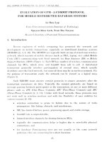
Figure 1 displays the FE-SEM images and XRD patterns of
Fe3O4, Fe3O4/OCMCs, and Fe3O4/OCMCs/Cur.
Figure 1. FE-SEM images of (a) Fe3O4, (b) Fe3O4/OCMCs,
(c) Fe3O4/OCMCs/Cur, and (d) their XRD patterns.
© 2011 The Chemical Society of Japan
www.csj.jp/journals/chem-lett/
Fe3O4 fluid contained aggregates, composed of spherical
particles with a size of 1020 nm. Fe3O4/OCMCs fluid was less
aggregated from fairly uniform-sized particles ranging from 20
to 25 nm. Upon the encapsulation of curcumin, Fe3O4/OCMCs/
Cur obtained had nearly the same size. However, different with
the others, Fe3O4/OCMCs/Cur was of isolated particles; in this
case the aggregation could not be observed.
From the XRD patterns of Fe3O4, Fe3O4/OCMCs, and
Fe3O4/OCMCs/Cur nanoparticles, it was clear that all six
diffraction peaks corresponded to faces of (200), (311), (400),
(422), (511), and (440), characteristic for crystalline Fe3O4,
which was the standard pattern for crystalline magnetite with a
spinel structure.24 The particle size of Fe3O4 calculated on the
basis of the Scherrer formula was in the range of 1020 nm,
consistent with that from FE-SEM image. As could be seen from
the diffraction patterns, after being encapsulated by OCMCs, the
crystallinity of Fe3O4 was almost unchanged. Thus, Fe3O4 was
apparently present in all samples under investigation.
The formation of Fe3O4/OCMCs/Cur nanoparticles was
also evidenced by IR (see Supporting Information25) and
fluorescence spectra. The peak at 584 cm¹1 in the IR spectrum
of Fe3O4, characteristic of FeOFe in the oxide,23 was shifted
to 564 cm¹1 for OCMCs/Fe3O4 and to 570 cm¹1 for Fe3O4/
OCMCs/Cur. Because of the complexation of curcumin with the
OCMCs, the wavenumbers corresponding to the characteristic
peaks of OCMCs was shifted. Comparing Fe3O4/OCMCs and
Fe3O4/OCMCs/Cur peak shifts were observed from 3440 to
3391 cm¹1 and 1637 to 1626 cm¹1. This data confirmed the
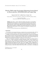
presence of curcumin in the OCMCs matrices. Curcumin is a
strongly fluorescent compound. Therefore, this component in the
Fe3O4/OCMCs/Cur system can be monitored by fluorospectrometry. Figure 2 showed fluorescence spectra of curcumin and
Fe3O4/OCMCs/Cur. The fluorescence maximum of the latter
was shifted by 27 nm compared with that of curcumin only. This
result further confirmed that the microenvironment of OCMCs/
Cur was changed after conjugation of OCMCs with curcumin.6,26 Moreover, the fluorescence intensity of Fe3O4/OCMCs/
Cur was also decreased probably due to the quenching effect of
the electron transfer from the excited curcumin to ferric ion.27
The influence of Fe3O4/OCMCs/Cur on the cell internalization ability of curcumin was investigated. In these experiments, 2 © 105 cells were seeded on a coverslip placed in each
well of the 24-well plate. After 24 h of culture, the cells were
incubated with Fe3O4/OCMCs/Cur at the final concentration of
10 ¯g mL¹1 for 15 h and then fixed with 4% PFA (Sigma).
Fluorescent staining was carried out to label actins with
Rhodaminephalloidin and nuclei with Hoechst (Invitrogen).
Coverslips were observed with an LSM 510 microscope (Carl
Zeiss).
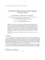
Fluorescent images taken by LSM 510 indicated the
presence of curcumin as the green signal inside HT29 cells
incubated with Fe3O4/OCMCs/Cur (Figure 3). The green signal
was due to the autofluorescence of curcumin when excited by an
argon laser. It, therefore, could not be seen in control cells. This
result demonstrated that the conjugation did not affect the cell
internalization ability of curcumin but also successfully led
curcumin into the cells as expected.
An in vitro cytotoxicity evaluation of materials was carried
out using an X-CELLigence system (Roche Inc.). The system
measures electrical impedance across interdigitated microelecChem. Lett. 2011, 40, 12641266
Fluorescence Intensity/arb unit
1265
Curcumin
Fe3O4 /OCMCs/Cur
20000
15000
10000
5000
0
400
500
600
700
800
Wavelength/nm
Figure 2. Fluorescence spectra of curcumin and Fe3O4/
OCMCs/Cur.
Figure 3. Fluorescent images of HT29 cells in normal culture
conditions (control) and incubated with conjugated curcumin for
15 h.
trodes integrated on the bottom of tissue culture E-plates. The
impedance measurement provides quantitative information of
cell number and viability. The real-time cell assay started with
the background reading by adding 50 ¯L of DMEM media
(Invitrogen) to each well of E-plate 96 and then monitored at
15 s intervals within 1 min. Next, 130 ¯L of DMEM media
containing 104 HT29 cells was seeded into each well of the
E-plate, and the cells were monitored every 15 min for 20 h
to obtain the growth baseline reading. At the time point of
treatment, 20 ¯L of conjugated curcumin or pure curcumin was
added into each well to get 5 concentrations of the range from
0.01 to 100 ¯g mL¹1. Dynamic cell proliferation of cells was
monitored in 30-min intervals from the time of treatment until
the end of the experiment (72 h). Cell Index values were
analyzed by RTCA software (Roche Inc.) to get IC50 and further
evaluation.
© 2011 The Chemical Society of Japan
www.csj.jp/journals/chem-lett/
1266
Figure 4. Dose-response curve of HT29 cell treated with pure
curcumin (a, red curve), conjugated curcumin (a, green curve)
and (b) Fe3O4/OCMCs.
The RTCA showed the cytotoxicity of Fe3O4/OCMCs/Cur
on HT29 cells with the IC50 of 0.36 ¯g mL¹1 (P < 0.05),
meanwhile the IC50 value of pure curcumin was 3.6 ¯g mL¹1.
Dose-response curve of HT29 cells treated with pure curcumin
was significantly higher than that with conjugated curcumin
(Figure 4). This apparently suggested that our conjugate
efficiently conducted curcumin and, therefore, enhanced its
biological activity in cancer cells. Indeed, it was curcumin but
not Fe3O4/OCMCs that determined the cytotoxicity of the
conjugate. Fe3O4/OCMCs had a negligible impact on cancer
cells (IC50 = 125.610 ¯g mL¹1, P < 0.05). Its cytotoxicity effect
was 350 times less than that of the whole conjugate. The
improvement of cytotoxicity was probably due to the water
solubility (curcumin in 1 mL of Fe3O4/OCMCs/Cur fluid was
found to be 6 mg) and cell internalization ability of the
conjugate.
Magnetic fluid hyperthermia is a promising tool in the
therapy of various cancers. This is because tumor cells are more
sensitive to temperatures in the range of 4246 °C than normal
tissue cells.19 In this work, we just proved that our conjugate
could satisfy criteria of temperature for hyperthermia therapy.
All the samples enabled the temperature to increase up to 42 °C
and even higher for 10 min (see Supporting Information25). The
temperature retention lasted 10 min and could prolong when the
heating conditions were held. Some experiments on magnetic
hyperthermia therapy are in progress.
In conclusion, a Fe3O4/OCMCs/Cur-based nanodrug system could be successfully prepared by ex situ grafting. This
system not only could be used as a tool for monitoring the drug
circulation by the fluorescence technique but also in cancer
treatment. The system was proven to successfully lead curcumin
into HT29 cells, and its effect on this cancer cell line was much
stronger than that of pure curcumin. It is promising to develop
this conjugate as a new smart nanomaterial for drug delivery.
This work was financially supported by the National
Foundation for Science and Technology development of
Vietnam-NAFOSTED under Grant No. 106.99-2010.42.
Chem. Lett. 2011, 40, 12641266
References and Notes
1 L. M. Huong, H. P. Thu, N. T. B. Thuy, T. T. H. Ha, H. T. M.
Thi, M. T. Trang, T. T. N. Hang, D. H. Nghi, N. X. Phuc,
D. T. Quang, Chem. Lett. 2011, 40, 846.
2 G. Sa, T. Das, Cell Div. 2008, 3, 14.
3 P. Anand, C. Sundaram, S. Jhurani, A. B. Kunnumakkara,
B. B. Aggarwal, Cancer Lett. 2008, 267, 133.
4 S. Karmakar, N. L. Banik, S. J. Patel, S. K. Ray, Neurosci.
Lett. 2006, 407, 53.
5 G. Liang, L. Shao, Y. Wang, C. Zhao, Y. Chu, J. Xiao, Y.
Zhao, X. Li, S. Yang, Bioorg. Med. Chem. 2009, 17, 2623.
6 L. D. Tran, N. M. T. Hoang, T. T. Mai, H. V. Tran, N. T.
Nguyen, T. D. Tran, M. H. Do, Q. T. Nguyen, D. G. Pham,
T. P. Ha, H. V. Le, P. X. Nguyen, Colloids Surf., A 2010,
371, 104.
7 M. Takahashi, D. Kitamoto, T. Imura, H. Oku, K. Takara, K.
Wada, Biosci., Biotechnol., Biochem. 2008, 72, 1199.
8 K. Sou, S. Inenaga, S. Takeoka, E. Tsuchida, Int. J. Pharm.
2008, 352, 287.
9 L. Li, F. S. Braiteh, R. Kurzrock, Cancer 2005, 104, 1322.
10 P. Anand, H. B. Nair, B. Sung, A. B. Kunnumakkara, V. R.
Yadav, R. R. Tekmal, B. B. Aggarwal, Biochem. Pharmacol.
2010, 79, 330.
11 M. M. Yallapu, B. K. Gupta, M. Jaggi, S. C. Chauhan,
J. Colloid Interface Sci. 2010, 351, 19.
12 M. M. Yallapu, M. Jaggi, S. C. Chauhan, Colloids Surf., B
2010, 79, 113.
13 Z. Aiping, L. Jianhong, Y. Wenhui, Carbohydr. Polym. 2006,
63, 89.
14 A. Anitha, S. Maya, N. Deepa, K. P. Chennazhi, S. V. Nair,
H. Tamura, R. Jayakumar, Carbohydr. Polym. 2011, 83, 452.
15 A. Kumar, P. K. Jena, S. Behera, R. F. Lockey, S. Mohapatra,
S. Mohapatra, Nanomedicine 2010, 6, 64.
16 Q. A. Pankhurst, N. K. T. Thanh, S. K. Jones, J. Dobson,
J. Phys. D: Appl. Phys. 2009, 42, 224001.
17 B. Koppolu, M. Rahimi, S. Nattama, A. Wadajkar, K. T.
Nguyen, Nanomedicine 2010, 6, 355.
18 T. K. Jain, S. P. Foy, B. Erokwu, S. Dimitrijevic, C. A. Flask,
V. Labhasetwar, Biomaterials 2009, 30, 6748.
19 J.-H. Park, K.-H. Im, S.-H. Lee, D.-H. Kim, D.-Y. Lee, Y.-K.
Lee, K.-M. Kim, K.-N. Kim, J. Magn. Magn. Mater. 2005,
293, 328.
20 J.-H. Lee, J.-t. Jang, J.-s. Choi, S. H. Moon, S.-h. Noh, J.-w.
Kim, J.-G. Kim, I.-S. Kim, K. I. Park, J. Cheon, Nat.
Nanotechnol. 2011, 6, 418.
21 A. Jordan, R. Scholz, P. Wust, H. Fähling, R. Felix, J. Magn.
Magn. Mater. 1999, 201, 413.
22 L.-Y. Zhang, H.-C. Gu, X.-M. Wang, J. Magn. Magn. Mater.
2007, 311, 228.
23 A. Zhu, L. Yuan, T. Liao, Int. J. Pharm. 2008, 350, 361.
24 Z. Ma, Y. Guan, H. Liu, J. Polym. Sci., Part A: Polym.
Chem. 2005, 43, 3433.
25 Supporting Information is available electronically on the
CSJ-Journal Web site, />index.html.
26 H. Yu, Q. Huang, Food Chem. 2010, 119, 669.
27 J. S. Kim, D. T. Quang, Chem. Rev. 2007, 107, 3780.
© 2011 The Chemical Society of Japan
www.csj.jp/journals/chem-lett/