DSpace at VNU: Gold-linked electrochemical immunoassay on single-walled carbon nanotube for highly sensitive detection of human chorionic gonadotropin hormone
Bạn đang xem bản rút gọn của tài liệu. Xem và tải ngay bản đầy đủ của tài liệu tại đây (720.56 KB, 6 trang )
Biosensors and Bioelectronics 42 (2013) 592–597
Contents lists available at SciVerse ScienceDirect
Biosensors and Bioelectronics
journal homepage: www.elsevier.com/locate/bios
Gold-linked electrochemical immunoassay on single-walled carbon
nanotube for highly sensitive detection of human chorionic
gonadotropin hormone
Nguyen Xuan Viet a,b, Miyuki Chikae a, Yoshiaki Ukita a, Kenzo Maehashi c,
Kazuhiko Matsumoto c, Eiichi Tamiya d, Pham Hung Viet e, Yuzuru Takamura a,n
a
School of Materials Science, Japan Advanced Institute of Science and Technology (JAIST), 1-1 Asahidai, Nomi City, Ishikawa 923-1292, Japan
Faculty of Chemistry, Hanoi University of Science, VNU, 19 Le Thanh Tong, Hoan Kiem District, Ha Noi, Vietnam
The Institute of Scientific and Industrial Research, Osaka University, 8-1 Mihogaoka, Ibaraki, Osaka 567-0047, Japan
d
Department of Applied Physics, Osaka University, 2-1 Yamadaoka, Suita, Osaka 565-0871, Japan
e
Research Center for Environmental Technology and Sustainable Development (CETASD), Hanoi University of Science, VNU, 334 Nguyen Trai, Thanh Xuan
District, Ha Noi, Vietnam
b
c
a r t i c l e i n f o
a b s t r a c t
Article history:
Received 19 August 2012
Received in revised form
13 November 2012
Accepted 14 November 2012
Available online 23 November 2012
A new sensitive gold-linked electrochemical immunoassay (GLEIA) for the detection of the pregnancy
marker human chorionic gonadotropin (hCG) has been developed using the direct electrochemical
detection of Au nanoparticles. We utilized single-walled carbon nanotube (SWCNT) microelectrodes; 24
SWCNT microelectrodes were arrayed on a single Si substrate 25 Â 30 mm2 in size, for the development
of a new GLEIA (SWCNT-GLEIA). This SWCNT-GLEIA provided convenient and cost-effective tests with
the required antibody and antigen sample volumes as small as 2.0 mL for a group of 4 SWCNT
microelectrodes. In addition, this assay also exhibited properties of high sensitivity and selectivity
benefitting from the intrinsic extraordinary features of SWCNTs. Using scanning electron microscopy,
we also observed Au nanoparticle-labeled antigen–antibody complexes immobilized on the surface of
the SWCNT microelectrodes. The concentration of the pregnancy marker (hCG) showed a linear
relationship with the current intensity obtained from differential pulse voltammetry measurements
with a limit of detection (LOD) of 2.4 pg/mL (0.024 mIU/mL) hCG. This LOD is 15 times more sensitive
than a previous GLEIA, which used screen-printed carbon electrodes.
& 2012 Elsevier B.V. All rights reserved.
Keywords:
Electrochemical immunoassay
Gold nanoparticles
Carbon nanotube electrode
Sandwiched type
Immunosensor
hCG
1. Introduction
An immunosensor, a type of biosensor, can be defined as a
compact analytical device incorporating antibodies or antigens or
their fragments, either integrated within or intimately associated
with a physicochemical transducer. Immunosensors provide sensitive and selective tools for determining the presence of proteins
on the basis of a specific reaction between an antibody and
antigen (Veetil and Ye, 2007). Immunosensors can help in directly
monitoring a molecular recognition event on the surface of a chip.
A large number of immunosensors have been developed using
different transducers that exploit changes in mass (Janshoff et al.,
2000; Ward and Buttry, 1990), heat (Luong et al., 1988), electrochemical (Dzantiev et al., 1996; Shah and Wilkins, 2003), or
optical properties (Brecht and Gauglitz, 1995; Haes and Van
Duyne, 2002; Morgan et al., 1996). Most of the reagents employed
n
Corresponding author.
E-mail address: (Y. Takamura).
0956-5663/$ - see front matter & 2012 Elsevier B.V. All rights reserved.
/>
in immunosensor, such as antibodies, enzymes, and fluorescence
labels are very expensive, and additionally, analytes such as blood
from a neonate or spinal fluid are precious commodities (Veetil
and Ye, 2007). Hence, miniaturization of diagnostic devices without affecting their sensitivity or limit of detection is highly
desirable. With advancements in the field of micro- and nanofabrication and lab-on-chip concepts, novel high-throughput
immunosensors that offer decreased analysis time and ease of
automation, integration, and portability are being explored.
Among the various immunosensor developed, electrochemical
immunosensor have become the predominant analytical technique for the quantitative detection of biomolecules due to their
simplicity, high sensitivity, low cost, fast analysis and ease of
miniaturization (Bakker, 2004; Privett et al., 2010; Skla´dal, 1997).
Moreover, sandwich-type electrochemical immunosensors have
gained much attention because of their high specificity and
sensitivity (Campbell et al., 2001; Chen et al., 2006; Idegami
et al., 2008).
It is well-known that, in conventional single-walled carbon
nanotube (SWCNT)-modified electrodes, such as SWCNT-modified
N. Xuan Viet et al. / Biosensors and Bioelectronics 42 (2013) 592–597
glassy carbon electrodes (Luo et al., 2001; Wang et al., 2001, 2002),
screen-printed carbon electrodes (SPCEs) (Lin et al., 2004; Sha
et al., 2006), and platinum electrodes (Okuno et al., 2007a, 2007b;
Tsujita et al., 2009, 2008), the electrochemical signals come from
both the SWCNTs and the supporting electrodes (carbon or
platinum, etc.) because the supporting electrodes are also exposed
to the electrolyte solutions. In most of these cases, SWCNTs exhibit
greatly enhanced electrochemical signals, so that the contribution
of the supporting electrodes is negligible. However, in some special
cases, such as when measuring electro-double layer charge currents, or in cases where the reactions are specifically enhanced on
plane supporting electrodes, this becomes a problem. In addition,
the nonspecific adsorption of protein on nanotubes is not desirable,
especially when using actual biological fluid samples that contain
many co-existing proteins (Nedelkov and Nelson, 2001; Tombelli
et al., 2005; Wang, 2002). More sophisticated sensors, therefore,
are needed to address issues such as target recognition enhancement, blockage of undesired interference (co-existing proteins,
nonspecific adsorption on the nanotube surfaces, etc.), and longterm storage. Nonspecific binding directly affects the selectivity
and sensitivity of devices.
In this paper, we describe a sandwich-type electrochemical
immunoassay for highly sensitive and selective detection of the
biomarker molecule hCG, which is used as a model of detection.
A SWCNT microelectrode (Viet et al., 2012) was used in this
electrochemical immunoassay instead of conventional electrodes
such as glassy carbon electrodes or SPCEs. Au nanoparticles were
used to label the antibody immunocomplex in this electrochemical immunoassay.
2. Experimental
2.1. Reagents
Monoclonal anti-human a-subunit of follicle-stimulating hormone (Mab-FSH) with an affinity constant of 2.8 Â 10 À 9 M À 1, and
monoclonal anti-human chorionic gonadotropin (Mab-hCG) with
an affinity constant of 4.9 Â 10 À 9 M À 1, were purchased from
Medix Biochemica (Kauniainen, Finland). The molecular weight
of recombinant human chorionic gonadotropin (hCG) was determined as 57.1 kDa using sodium dodecyl sulfate polyacrylamide
gel electrophoresis (SDS-PAGE), and its potency was measured as
10,000 IU/mg (Rohto Pharmaceutical Co., Ltd., Osaka, Japan).
A colloidal solution of Au nanoparticles of diameter 40 nm was
purchased from British Biocell International Ltd., (Cardiff, UK).
HCl, NaH2PO4 Á 2H2O, polyethylene glycol (PEG), KH2PO4 and
593
dimethylformamide (DMF) were purchased from Wako Pure
Chemical Industries (Osaka, Japan). Sodium azide (NaN3) was
purchased from Nakarai Tesque (Kyoto, Japan). 1-pyrenebutanoic
acid succinimidyl ester was purchased from Life Technologies
Corporation (Carlsbad, CA, USA). Bovine serum albumin (BSA) was
purchased from Sigma-Aldrich, (St. Louis, MO, USA). Polyethylene
glycol amine with a molecular weight of 5000 Da was purchased
from SUNBRIGHT (NOF Corporation, Tokyo, Japan). Male urine
solution was purchased from Lee Biosolution, Inc. Other reagents
were of analytical grade, and all solutions were prepared and
diluted using ultra-pure water (18.2 MO cm) from the Milli-Q
system (Millipore, Billerica, MA, USA).
2.2. Instrument
Scanning electron microscopy (SEM) images were obtained
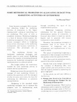
using Hitachi S-4100 with accelerating voltage 20 kV. Electrochemical measurements were performed on an ALS/CH Instruments
electrochemical analyzer, model 730C (Austin, Texas, USA) as
shown in Fig. 1, in which a 3-electrode system was used with a Pt
wire as the counter, an AgCl/Ag micro-electrode as the reference
(Microelectrodes Inc., Bedford, NH, USA), and a SWCNT microelectrode as the working electrode.
2.3. Sandwiched immunosensor procedure
a) Preparation of Au nanoparticle-labeled hCG antibody
(Au-Mab-hCG)
The preparation of Au-Mab-hCGs was performed by a similar
method as previously reported by our group (Idegami et al.,
2008; Nagatani et al., 2006; Tanaka et al., 2006) with a slight
modification. Briefly, an aliquot (200 microliter) of Mab-hCG
solution (50 mg/mL in 5 mM KH2PO4, pH 7.5) was mixed with
1.8 mL of 0.1% Au nanoparticle solution, and kept for 10 min at
room temperature. Then, 100 microliter of 1% PEG in 50 mM
KH2PO4 solution (pH 7.5) and 200 microliter of 10% BSA in
50 mM KH2PO4 solution (pH 9.0) were added to block the
uncoated surfaces of the Au nanoparticles. After the immobilization and blocking procedures, Au nanoparticle-conjugated
Mab-hCGs (Au-Mab-hCGs) were collected by centrifugal
operation (8000 g for 15 min at 4 1C). Au-Mab-hCGs were
suspended in 2 mL of the preservation solution (1% BSA,
0.05% PEG 20,000, 0.1% NaN3, and 150 mM NaCl in 20 mM
Tris-HCl buffer, pH 8.2), and collected again in the same
manner. For the stock solution, Au-Mab-hCGs were suspended
Fig. 1. Electrochemical measurement set-up with a SWCNT microelectrode as the working electrode (WE), a Pt wire as the counter electrode (CE), and an Ag/AgCl
microelectrode as the reference electrode (RE).
594
N. Xuan Viet et al. / Biosensors and Bioelectronics 42 (2013) 592–597
in the preservation solution and the optical density was
adjusted to an OD520 of 6.
b) Fabrication of immunosensor
An array of 24 SWCNT microelectrodes on a single Si substrate
was made following a procedure that has been previously
described and characterized in detail (Viet et al., 2012). Briefly,
an SWCNT network was synthesized by catalyst chemical
vapor deposition using ethanol as the carbon source on the
Si substrate. Next, a chromium layer (200 nm) was thermally
evaporated onto the SWCNT network using the plasma sputtering method. A photoresist layer with a thickness of 15 mm
(PMER photoresist) was subsequently spun over the chromium
layer. A disk-type pattern with a diameter of 180 mm was
formed inside the Pt contacts by exposing them to 458-nm
helium light for 30 s and then developing in PG-7 solution. The
exposed chromium layer was removed by chromium etchant
solution in 2 min. Then, a thermal SiO2 layer of 250 nm was
sputtered onto the exposed SWCNT network by the plasma
sputtering method. Finally, the residual disk-type pattern of
the photoresist layers and chromium layers was cleaned using
remover and chromium etchant solution, respectively. Note
that between 2 successive steps, the SWCNT microelectrodes
were washed by Milli-Q water for 1 min and then blown under
N2 gas to dry.
Next, SWCNT microelectrodes were incubated in 30 mL dry
DMF solution with 0.1 mM 1-pyrenebutanoic acid succinimidyl ester as a linker molecule for 30 min at room temperature,
followed by rinsing with DMF solvent to remove the unbound
molecules from the SWCNTs (Chen et al., 2001; Okuno et al.,
2007a). In order to covalently immobilize Mab-FSH on the
SWCNTs, SWCNT microelectrodes were exposed overnight to
400 mg/mL Mab-FSH in 10 mM phosphate-buffered saline
(PBS, pH 7.4) by dropping 2.0 mL of Mab-FSH on each group
of SWCNT microelectrodes. Following this, the excess antibodies were rinsed with PBS. To deactivate reactive groups and
suppress nonspecific binding, 4.0 mL of 10 mM PBS solution
containing 1% PEG–NH2 was added onto the resulting electrodes, and incubated for 1 h at room temperature. The array was
then rinsed with PBS.
c) Sandwich-type
immunoreaction
and
electrochemical
measurement
A scheme illustrating the principle of the gold-linked electrochemical immunoassay (GLEIA) on SWCNT microelectrodes
(SWCNT-GLEIA) is shown in Fig. 2. Different concentrations of
the hCG antigen solution were made by diluting the stock
solution in PBS containing 1% BSA for detection. In case of
detection of hCG in biological fluid, stock solution of hCG was
spiked in male urine solution to make different concentration.
For the detection of the antibody–antigen reaction, 2.0 mL of
the antigen solution was placed on a group of 4 SWCNT
microelectrodes for 1 h at room temperature. After rinsing
with PBS, 2.0 mL of Au-Mab-hCG solution was applied onto the
surface, and incubated for 30 min at room temperature.
Finally, the SWCNT microelectrodes were rinsed carefully
with PBS.
The direct redox reaction was performed in 0.1 M HCl solution
(30 mL) covering the entire three-electrode zone at room
temperature (as shown in actual photo of electrochemical
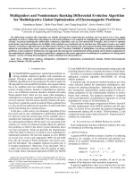
Fig. 2. Scheme illustrating the principle of the gold-linked electrochemical immunoassay on SWCNT microelectrodes.
Fig. 3. SEM images of (a) SWCNTs immobilized with Mab-FSH and blocking agents, (b) Au nanoparticle-labeled antigen–antibody complexes immobilized on the surface of
the SWCNT microelectrode with a hCG concentration of 1.0 ng/mL (10.0 mIU/mL). Scale bars ¼ 500 nm.
N. Xuan Viet et al. / Biosensors and Bioelectronics 42 (2013) 592–597
measurement in Fig. 1). The pre-oxidation of Au nanoparticles
was performed at a constant potential 1.2 V for 40 s, immediately followed by DPV, while scanning the potential range
from 1.0 V to 0.0 V with a step potential of 4.0 mV, pulse
amplitude of 50 mV, and a pulse period of 0.2 s. The potentials
were recorded against the Ag/AgCl microelectrode as the
reference (Idegami et al., 2008).
595
2012), when some Au nanoparticles nonspecifically adsorbed
onto the Si/SiO2 substrate and did not contact the SWCNTs, they
were not able to generate electrochemical signals. This should
improve the effect of depressing the background signal, resulting
in a lower limit of detection. This is the advantage of using a
SWCNT network over other CNT-modified electrodes.
3.2. Electrochemical operation of GLEIA using SWCNT
microelectrode
3. Results and discussion
3.1. SEM images of GLEIA using SWCNT microelectrodes
Fig. 3a shows the SEM image of the SWCNT network inside the
SWCNT microelectrode after immobilizing with Mab-FSH and
blocking agent. Fig. 3b is the SEM image of Au nanoparticlelabeled immunocomplexes immobilized on the surface of the
SWCNTs (see white arrows in Fig. 3b). Au nanoparticles were
distributed on the surface of the SWCNT network with a hCG
concentration of 1.0 ng/mL (10.0 mIU/mL). This shows that the
antigen, hCG, was successfully detected using the SWCNT microelectrode for GLEIA.
Because the surface of the SWCNT microelectrode was not
totally covered with SWCNTs (Dumitrescu et al., 2008; Viet et al.,
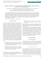
Fig. 4. Cyclic voltammogram of Au-Mab-hCG immobilized on a SWCNT microelectrode at 50 mV/s in 0.1 M HCl solution. The concentration of hCG was 100 ng/ml
(1.0 Â 103 mIU/mL).
Fig. 4 illustrates the cyclic voltammogram (CV) obtained from
the Au-Mab-hCG-immobilized immunosensor after the antigen–
antibody reaction (with 100 ng/mL hCG–1.0 Â 103 mIU/mL) in the
potential range from 0.0 to 1.4 V vs Ag/AgCl in 0.1 M HCl solution.
The reduction peak of Au ions could be observed at a potential of
around þ0.5 V, corresponding with reaction (1) in Fig. 4. The
positive shift of the gold reduction peak on SWCNT microelectrodes compared with SPCEs (from þ0.35 V (Idegami et al., 2008;
Quinn et al., 2005) on SPCEs to around þ0.5 V on SWCNTs) in the
CV curve illustrated that SWCNTs promote the reduction of Au
ions better than do SPCEs; one reason is the difference in the
environment of the reference electrode. In SPCEs, the reference
electrode is immersed directly in 0.1 M HCl solution and has a
potential of 0.2881 V compared with a normal hydrogen electrode
(NHE). On the other hand, in the case of SWCNT electrodes, the
reference electrode is immersed in 3.0 M KCl solution, thus it has
a potential of 0.21 V vs. the NHE (Bard 2001).
In the operation of the GLEIA, the reduction peak current of
DPV was used for the detection of Au nanoparticles in 0.1 M HCl
solution. This process involves the oxidation of Au nanoparticles
into Au ions before the Au ions are reduced on the electrode
surface to obtain a good electrochemical signal (Idegami et al.,
2008). The effect of the pre-oxidation potential on the current
densities of the DPV reduction peak of the Au ion was investigated. The pre-oxidation potentials were measured at 1.20, 1.50,
and 1.70 V vs Ag/AgCl with a pre-oxidation time of 40 s in the
presence of 250 pg/mL (2.5 mIU/mL) hCG, shown in Fig. s1 of
supplemental document. A rapid decrease in the reduction peak
current intensity was observed with increasing pre-oxidation
potential. This indicates that the loss of Au ions occurs more
easily at high pre-oxidation potential than at lower pre-oxidation
potentials. Therefore, in this electrochemical measurement,
1.20 V was the optimum pre-oxidation potential. Fig. 5a shows
DPV curves obtained from the Au-Mab-hCG-immobilized immunosensor with different concentrations of hCG (from 10.0 pg/mL
to 2.0 Â 103 pg/mL–0.1 mIU/mL to 20.0 mIU/mL) in PBS containing 1% BSA at an applied potential of 1.20 V. The reduction peaks
Fig. 5. (a) Differential pulse voltammograms of the Au-Mab-hCG on SWCNT microelectrodes in 0.1 M HCl solution. (b) Normalized calibration curves in GLEIA using
SWCNT microelectrodes (curve I and II), SWCNT-modified SPCEs (curve III), and SPCEs (curve IV) as the platform. The concentration of hCG ranged from 10.0 pg/mL to
2.0 Â 103 pg/mL (0.1 mIU/mL to 20.0 mIU/mL) in PBS containing 1% BSA.
596
N. Xuan Viet et al. / Biosensors and Bioelectronics 42 (2013) 592–597
were observed at approximately þ0.52 V, nearly equal to the CV
result of $ 0.5 V. The peak current intensity increased in proportion to increasing hCG concentration.
The analytical range and sensitivity of the immunosensor were
calculated by extracting the current intensity as a function of the
hCG concentration from Fig. 5a. The results are shown in Fig. 5b
(curve I). The reduction peak current intensity of Au ions
depended linearly on the hCG concentration in this concentration
range, and the correlation coefficient (R2) of the linear fitting
curve for this relationship was 0.9906. Under the above measured
conditions, an LOD of 2.4 pg/mL (0.024 mIU/mL) for hCG was
calculated as 3SD (where SD is the standard deviation of 5 measurements of blank samples). This value is 15 times lower than
the previous work of our lab using SPCEs (Idegami et al., 2008) as
platform for this immunosensor.
The LOD of this immunosensor increases to 53 pg/mL
(0.53 mIU/mL) in the male urine solution (curve II in Fig. 5b).
This value of LOD is around 20 times higher than that measuring
in PBS containing 1% BSA. For a comparison, we also conducted
the GLEIA using planar SPCE for same urine sample, and the result
got the LOD of 1.85 ng/mL (18.5 mIU/mL) (Fig. s2), which is 51
times higher than LOD in PBS containing 1% BSA using planar
SPCE. The LOD of SWCNT microelectrode increases 20 times in
urine sample and this is considered to be caused by the deviation
of signal due to non-specific binding of various bio-substances in
urine sample. The value of 20 is still less than the value of 51 for
the increase in SPCE. This fact indicates that our SWCNT microelectrode has better suppression property of non-specific binding
and better selectivity not only in PBS with 1% BSA but also in urine
sample than conventional planar SPCE. These values for SWCNT
microelectrode were obtained using the same described condition
above, and may be improved more by further optimization.
This sandwich-type immunosensor using Au nanoparticles as
label has several advantages over the use of enzyme as the label.
In the case of enzyme-based detection systems, the electrode
surface is covered with the immune-complexes and blocking
agents; these biomolecules remain on the surface during electrochemical measurement, and may disturb the performance of the
electrode. In our method, the pre-oxidation of Au nanoparticles at
a high potential and the denaturation of the biomolecules in
highly acidic conditions were carried out simultaneously. Thus,
the detachment of possible blocking molecules from the surface
provided a large electroactive area for oxidized Au ions to be
reduced again efficiently during the DPV scan. Additionally, the
loss of oxidized Au ions by diffusion was avoided because of the
negative charge of the chelated compounds with the high concentration of chloride ions in the acidic electrolyte. The constant
application of highly positive voltage rapidly attracted negatively
charged Au chelates and promoted their electrodeposition on the
carbon surface (Idegami et al., 2008).
3.3. Sensitivity of GLEIA using SWCNT microelectrodes, GLEIA on
SWCNT-modified SPCEs, and SPCEs
Fig. 5b shows the normalized calibration curves of GLEIA on a
platform of SWCNT microelectrodes (curve I) (this study), SPCEs
(curve IV) (Idegami et al., 2008), and SWCNT-modified SPCEs
(curve III), with hCG concentrations ranging from 10.0 pg/mL to
2.0 Â 103 pg/mL (0.1 mIU/mL to 20.0 mIU/mL). These normalized
curves determine the current density on each type of electrode
used for GLEIA. The procedure for GLEIA on SPCEs and SWCNTmodified SPCEs are similar with those described above for the
SWCNT microelectrode. The LOD of GLEIA on SPCEs and SWCNTmodified SPCEs were 36 pg/mL (0.36 mIU/mL) and 13 pg/mL
(0.13 mIU/mL), correspondingly. These results show that GLEIA
using the SWCNT microelectrode has the highest sensitivity.
The high sensitivity of this SWCNT-GLEIA was attributed to the
combination of the high performance of our SWCNT microelectrode with the ability to enhance electrochemical signals, reduce
nonspecific binding, and effectively detect the signals directly
from Au nanoparticles. The performance of GLEIA on SWCNTmodified SPCEs was better in comparison with GLEIA on SPCEs.
This comes from the enhancement of SPCE performance due to
the presence of SWCNTs. However, the performance of SWCNTmodified SPCEs was lower than that of SWCNT microelectrodes
because the SWCNTs using for modifying SPCEs underwent acid
treatment (Gooding et al., 2003), which leads to shortening, more
sidewall defects, and lower electrical conductivity than with asgrown SWCNTs (Zhang et al., 2004).
4. Conclusion
A new sensitive gold-linked electrochemical immunoassay for
the detection of the pregnancy marker, hCG, has been successfully
developed based on the sandwich-type immunosensor. This
SWCNT-GLEIA, based on microelectrodes that use an SWCNT
network directly grown on Si, exhibited the highest sensitivity
compared with those of GLEIAs conducted using SPCEs and
SWCNT-modified SPCEs. This SWCNT-GLEIA also showed good
selectivity when detecting hCG in male urine solution. The LOD of
SWCNT-GLEIA got the values of 2.4 pg/mL (0.024 mIU/mL) and
53 pg/mL (0.53 mIU/mL) hCG, when it was spiked in PBS containing 1% BSA and in male urine solution, correspondingly.
Acknowledgments
This work was partially supported by a Grant-in-Aid for
Scientific Research on Priority Areas (No. 19054011) and the
Cooperative Research Program of ‘‘Network Joint Research Center
for Materials and Devices’’ from the Ministry of Education,
Culture, Sports, Science and Technology of Japan.
Appendix A. Supporting information
Supplementary data associated with this article can be found in
the online version at />
References
Bakker, E., 2004. Analytical Chemistry 76 (12), 3285–3298.
Bard, A.J., Faulkner, L.R., 2001. Wiley.
Brecht, A., Gauglitz, G., 1995. Biosensors and Bioelectronics 10 (9–10), 923–936.
Campbell, C.N., Gal, D., Cristler, N., Banditrat, C., Heller, A., 2001. Analytical
Chemistry 74 (1), 158–162.
Chen, J., Yan, F., Du, D., Wu, J., Ju, H., 2006. Electroanalysis 18 (7), 670–676.
Chen, R.J., Zhang, Y., Wang, D., Dai, H., 2001. Journal of the American Chemical
Society 123 (16), 3838–3839.
Dumitrescu, I., Unwin, P.R., Wilson, N.R., Macpherson, J.V., 2008. Analytical
Chemistry 80 (10), 3598–3605.
Dzantiev, B.B., Zherdev, A.V., Yulaev, M.F., Sitdikov, R.A., Dmitrieva, N.M., Moreva,
I.Y., 1996. Biosensors and Bioelectronics 11 (1–2), 179–185.
Gooding, J.J., Wibowo, R., Liu, Yang W., Losic, D., Orbons, S., Mearns, F.J., Shapter,
J.G., Hibbert, D.B., 2003. Journal of the American Chemical Society 125 (30),
9006–9007.
Haes, A.J., Van Duyne, R.P., 2002. Journal of the American Chemical Society 124
(35), 10596–10604.
Idegami, K., Chikae, M., Kerman, K., Nagatani, N., Yuhi, T., Endo, T., Tamiya, E., 2008.
Electroanalysis 20 (1), 14–21.
Janshoff, A., Galla, H.-J., Steinem, C., 2000. Angewandte Chemie International
Edition 39 (22), 4004–4032.
Lin, Y., Lu, F., Wang, J., 2004. Electroanalysis 16 (1–2), 145–149.
Luo, H., Shi, Z., Li, N., Gu, Z., Zhuang, Q., 2001. Analytical Chemistry 73 (5),
915–920.
N. Xuan Viet et al. / Biosensors and Bioelectronics 42 (2013) 592–597
Luong, J.H.T., Mulchandani, A., Guilbault, G.G., 1988. Trends in Biotechnology 6
(12), 310–316.
Morgan, C.L., Newman, D.J., Price, C.P., 1996. Clinical Chemistry 42 (2),
193–209.
Nagatani, N., Tanaka, R., Yuhi, T., Endo, T., Kerman, K., Takamura, Y., Tamiya, E.,
2006. Science and Technology of Advanced Materials 7 (3), 270–275.
Nedelkov, D., Nelson, R.W., 2001. Biosensors and Bioelectronics 16 (9–12),
1071–1078.
Okuno, J., Maehashi, K., Kerman, K., Takamura, Y., Matsumoto, K., Tamiya, E.,
2007a. Biosensors and Bioelectronics 22 (9–10), 2377–2381.
Okuno, J., Maehashi, K., Matsumoto, K., Kerman, K., Takamura, Y., Tamiya, E.,
2007b. Electrochemistry Communications 9 (1), 13–18.
Privett, B.J., Shin, J.H., Schoenfisch, M.H., 2010. Analytical Chemistry 82 (12),
4723–4741.
Quinn, B.M., Dekker, C., Lemay, S.G., 2005. Journal of the American Chemical
Society 127 (17), 6146–6147.
Sha, Y., Qian, L., Ma, Y., Bai, H., Yang, X., 2006. Talanta 70 (3), 556–560.
Shah, J., Wilkins, E., 2003. Electroanalysis 15 (3), 157–167.
597
Skla´dal, P., 1997. Electroanalysis 9 (10), 737–745.
Tanaka, R., Yuhi, T., Nagatani, N., Endo, T., Kerman, K., Takamura, Y., Tamiya, E.,
2006. Analytical and Bioanalytical Chemistry 385 (8), 1414–1420.
Tombelli, S., Minunni, M., Luzi, E., Mascini, M., 2005. Bioelectrochemistry 67 (2),
135–141.
Tsujita, Y., Maehashi, K., Matsumoto, K., Chikae, M., Takamura, Y., Tamiya, E., 2009.
Japanese Journal of Applied Physics 48 (6) 06FJ02.
Tsujita, Y., Maehashi, K., Matsumoto, K., Chikae, M., Torai, S., Takamura, Y., Tamiya,
E., 2008. Japanese Journal of Applied Physics 47 (4), 2064–2067.
Veetil, J.V., Ye, K., 2007. Biotechnology Progress 23 (3), 517–531.
Viet, N.X., Ukita, Y., Chikae, M., Ohno, Y., Maehashi, K., Matsumoto, K., Viet, P.H.,
Takamura, Y., 2012. Talanta 91 (0), 88–94.
Wang, J., 2002. Analytica Chimica Acta 469 (1), 63–71.
Wang, J., Li, M., Shi, Z., Li, N., Gu, Z., 2001. Electrochimica Acta 47 (4), 651–657.
Wang, J., Li, M., Shi, Z., Li, N., Gu, Z., 2002. Analytical Chemistry 74 (9), 1993–1997.
Ward, M.D., Buttry, D.A., 1990. Science 249 (4972), 1000–1007.
Zhang, X., Sreekumar, T.V., Liu, T., Kumar, S., 2004. The Journal of Physical
Chemistry B 108 (42), 16435–16440.