DSpace at VNU: Three new dammarane glycosides from Betula alnoides
Bạn đang xem bản rút gọn của tài liệu. Xem và tải ngay bản đầy đủ của tài liệu tại đây (194.41 KB, 4 trang )
Phytochemistry Letters 4 (2011) 179–182
Contents lists available at ScienceDirect
Phytochemistry Letters
journal homepage: www.elsevier.com/locate/phytol
Three new dammarane glycosides from Betula alnoides
Minh Giang Phan a,*, Thi To Chinh Truong a, Tong Son Phan a, Katsuyoshi Matsunami b, Hideaki Otsuka b
a
b
Faculty of Chemistry, College of Natural Science, Vietnam National University, Hanoi, 19 Le Thanh Tong Street, Hanoi, Viet Nam
Graduate School of Biomedical Sciences, Hiroshima University, 1-2-3 Kasumi, Minami-ku, Hiroshima 734-8553, Japan
A R T I C L E I N F O
A B S T R A C T
Article history:
Received 26 December 2010
Received in revised form 16 February 2011
Accepted 25 February 2011
Available online 21 March 2011
Twenty compounds including three new dammarane glycosides, named betalnoside A (7), betalnoside B
(8), and betalnoside C (11) were isolated from Betula alnoides Buch. -Ham. ex D. Don (Betulaceae), of
which 13 (1–13) were from the leaves, seven (14–20) from the stem bark, and three (2, 16, and 17) from
the twigs. Their structures were determined using spectroscopic analyses.
ß 2011 Phytochemical Society of Europe. Published by Elsevier B.V. All rights reserved.
Keywords:
Betula alnoides
Betulaceae
Dammarane glycoside
1. Introduction
2. Results and discussion
The genus Betula (Betulaceae) comprises more than 35
scientifically recognized species found in temperate and boreal
zones of the northern hemisphere (Fuchino et al., 1995; Krasutsky,
2006). A comprehensive review (Krasutsky, 2006) revealed the
concentration of lupanes (major) and oleananes (minor) in the
outer bark of Betula plants. Dammarane triterpenoids were
reported predominantly from the leaves of Betula species such
as B. ermanii, B. platyphylla var. japonica, B. maximowicziana, B.
davurica, B. ovalifolia, and B. schmidtii (Fuchino et al., 1995, 1996a,b,
1998a,b,c). The great number of occurrences of lupanes–oleananes
in the outer bark and dammaranes in the leaves is considered to be
significant due to their chemosystematic relevance. There has been
also an increase of chemosystematic interest in the phenolic
(flavonoid, lignan, and diarylheptanoid) constituents of the leaves
(Keina¨nen et al., 1999; Keina¨nen and Julkune-Tiitto, 1998) and
inner bark (Fuchino et al., 1995, 1996a,b, 1998a,b) of Betula species.
B. alnoides Buch. -Ham. ex D. Don (Betulaceae) is the only Betula
species recorded in the Flora of Vietnam (Pham, 1993). The
expected constituents lupeol, 3-O-acetoxyoleanolic acid, betulinic
acid, and betulin were found in a previous report from the bark of B.
alnoides (Kamperdick et al., 1995). Our present study investigated
the distribution of triterpenoids and flavonoids in the leaves, twigs,
and stem bark of B. alnoides.
Twenty compounds were isolated from B. alnoides, of which 13
(1–13) were from the leaves, three (2, 16, and 17) from the twigs,
and seven (14–20) from the stem bark. Compounds 7, 8, and 11,
named betalnosides A–C, are new dammarane glycosides. The
structures of the known compounds, pentacosanoic acid (1), bsitosterol (2) (Goad and Akihisha, 1997), ovalifoliolide B (3)
(Fuchino et al., 1998b), rhamnocitrin (4) (Harborne, 1994),
chrysoeriol (5) (Agrawal, 1989), 1-O-(tetracosanoyl)glycerol (6)
(Sultana et al., 1999), b-sitosterol 3-O-b-D-glucopyranoside (9),
quercetin 3-O-b-D-glucopyranoside (10) (Harborne, 1994), rutin
(12) (Harborne and Mabry, 1982), quercetin (13) (Harborne, 1994),
taraxeryl acetate (14) (Jin et al., 2007), taraxerone (15) (Sakurai
et al., 1987), lupeol (16) (Fuchino et al., 1995), betulin (17) (Fuchino
et al., 1995), betulinic acid (18) (Jin et al., 2007), oleanolic acid (19)
(Fuchino et al., 1995), and ursolic acid (20) were determined by
comparing their spectroscopic data (EIMS, HRESIMS, 1H, and 13C
NMR) with the reported literature values or those of the authentic
samples.
Compound 7 was isolated as a white amorphous powder. Its
molecular formula was determined to be C35H60O7 by positive-ion
HRESIMS (m/z 615.4230 [M+Na]+). The IR spectrum showed a
hydroxyl absorption band at 3381 cmÀ1. Acid hydrolysis of 7 with
1 M HCl gave D-xylose, which was identified by HPLC analysis
(Matsunami et al., 2009). In the 1H NMR spectrum of 7 (Table 1)
signals for eight tertiary methyl groups, of which three were linked
to oxygenated carbons [dH 1.15 (3H, s), 1.17 (3H, s), and 1.19 (3H,
s)], two oxygenated methine groups [dH 3.15 (1H, dd, J = 12.0 Hz,
4.5 Hz) and 3.77 (1H, t, J = 7.5 Hz)], and protons of a sugar moiety
* Corresponding author. Tel.: +84 4 38351439.
E-mail address: (M.G. Phan).
1874-3900/$ – see front matter ß 2011 Phytochemical Society of Europe. Published by Elsevier B.V. All rights reserved.
doi:10.1016/j.phytol.2011.02.011
M.G. Phan et al. / Phytochemistry Letters 4 (2011) 179–182
180
Table 1
1
H (500 MHz) and
C/H
1
2
3
4
5
6
7
8
9
10
11
12
13
14
15
16
17
18
19
20
21
22
23
24
25
26
27
28
29
30
10
20
30
40
50
13
C NMR (125 MHz) spectroscopic data of 7, 8, and 11.
7 (CD3OD)a
8 (CD3OD)a
dc
dH (J in Hz)
dC
dH (J in Hz)
dC
dH (J in Hz)
40.3
27.4
90.7
40.4
57.7
19.3
36.5
41.7
52.2
38.1
22.7
26.8
44.2
51.0
33.1
27.3
51.2
16.8
16.8
87.8
23.6
36.5
28.4
84.8
72.9
26.2
25.4
28.4
16.0
16.9
b-Xylose
107.4
75.5
78.0
71.3
66.7
0.99
1.71
3.15
–
0.81
1.51
1.32
–
1.41
–
1.29
1.66
1.83
–
1.11
1.25
1.67
1.01
0.91
–
1.16
1.73
1.81
3.77
–
1.17
1.15
1.05
0.86
0.93
s
s
s
s
s
0.99
1.71
3.15
–
0.81
1.51
1.32
–
1.40
–
1.32
1.69
1.84
–
1.10
1.29
1.74
1.02
0.91
–
1.14
1.30
1.60
3.96
–
4.82
1.74
1.05
0.86
0.93
s
s
s
s
s
d (7.5)
m
t (9.0)
m
m, 3.84 dd (11.3, 5.5)
33.5
21.4
82.4
37.5
50.4
18.1
34.8
39.4
53.7
41.3
75.6
33.7
40.5
50.1
31.4
26.3
48.8
16.7
16.8
86.9
24.8
35.9
26.7
83.6
72.0
26.9
24.2
29.2
22.6
16.9
a-Arabinose
100.7
73.5
72.8
69.7
66.4
a-Arabinose
99.9
73.3
71.7
68.8
65.1
1.32
1.60
3.29
–
1.22
1.42
1.42
–
1.74
–
4.04
1.32
1.62
–
1.10
1.62
2.00
1.05
0.96
–
1.17
1.20
1.31
3.79
–
1.18
1.15
0.94
0.86
1.00
4.29
3.21
3.31
3.48
3.20
40.3
27.3
90.6
40.4
57.7
19.3
36.5
41.7
52.1
38.0
22.7
25.8
43.6
51.5
32.3
28.7
51.1
16.7
16.8
75.8
25.2
38.2
30.2
77.3
147.0
111.2
17.8
28.4
16.0
16.9
b-Xylose
107.4
75.5
77.9
71.3
66.7
4.26
3.55
3.45
3.74
3.45
d (7.5)
m
m
m
m, 3.95 m
4.28
3.55
3.52
3.84
3.25
d (7.5)
m
m
m
m, 3.95 m
m, 1.71 m
m, 1.82 m
dd (12.0, 4.5)
br d (11.4)
m (1.58)
m, 1.60 m
m
m, 1.52 m
m
m
m, 1.53 m
m, 1.89 m
m
s
s
s
m
m, 1.97 m
t (7.5)
100
200
300
400
500
a
11a,b
4.29
3.21
3.31
3.48
3.20
m, 1.71 m
m, 1.78 m
dd (12.0, 4.5)
br d (11.4)
m, 1.58 m
m, 1.60 m
m
m, 1.52 m
m
m
m, 1.49 m
m, 1.87 m
m
s
s
s
m, 2.07 m
m, 2.07 m
t (5.2)
br s, 4.93 br s
br s
s
s
s
d (7.5)
m
t (9.0)
ddd (10.0, 9.0, 5.5)
m, 3.84 dd (11.5, 5.5)
m
m, 1.75 m
br s
m
m
m, 2.30 br d (14)
m
ddd (10.5, 10.5, 5.5)
m, 2.48 m
m
m, 1.41 m
m, 1.95 m
m
s
s
s
m, 1.58 m
m, 1.85 m
t (7.0)
Assignments were based on DEPT, HSQC, and HMBC (compound 11) spectra.
H NMR was measured in CDCl3 and 13C NMR in CD3OD + CDCl3.
b 1
(dH 3.20–4.29) were observed. The 13C NMR spectrum of 7 (Table 1)
showed the presence of 35 signals. After subtraction of five carbons
for a xylopyranosyl moiety (dC 66.7, 71.3, 75.5, 78.0, and 107.4)
(Fuchino et al., 1998c) 30 signals left belonged to a tetracyclic
triterpenoid moiety containing an epoxide ring (dC 84.8 and 87.8).
On the basis of the NMR data the aglycone of 7 was identified as
ocotillol (Fu et al., 2005). The xylopyranosyl moiety of 7 was
determined to be linked to C-3 of the aglycone on the basis of the
significant downfield shift of C-3 (dC 90.7) on going from ocotillol
(dC 79.0). The chemical shift of C-3 was also indicative of the 3aH
orientation of 7 (Li et al., 2007); glycosidation of the 3a-hydroxyl
group caused a chemical shift to ca. dC 82–83 of C-3 (Fuchino et al.,
1996b). The coupling constant of the anomeric proton [dH 4.29 (1H,
d, J = 7.5 Hz)] indicated the b configuration at C-10 for the xylose.
Therefore 7 was determined to be 3-O-b-D-xylopyranosyl ocotillol
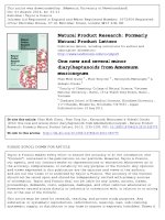
(Fig. 1) which was given a trivial name betalnoside A.
Compound 8 was isolated as a white amorphous powder. Its
molecular formula was determined to be C35H60O7 by positive-ion
HRESIMS (m/z 615.4229 [M+Na]+). The IR spectrum showed a
hydroxyl absorption band at 3385 cmÀ1. Acid hydrolysis of 8 with
1 M HCl gave D-xylose, which was identified by HPLC analysis
(Matsunami et al., 2009). The 1H and 13C NMR spectra of 8 (Table 1)
indicated that the structures of 8 and 7 differed only in the side
chain at C-24. The side chain, which contained a tertiary (dC 75.8)
and a secondary (dC 77.3) hydroxyl groups, two methylenes (dC
30.2 and 38.2), and an isopropenyl group (dC 17.8, 111.2, and
147.0), was determined as depicted in Fig. 1 (S2) by comparing its
NMR data with those of 20(S),24(S)-dihydroxydammara-25-en-3one (Malinovskaya et al., 1980) and notopanaxoside A (Komakine
et al., 2006). The absolute stereochemistries of C-20 and C-24 of 8
could not be determined in this study. Therefore 8 was determined
to be 3-O-b-D-xylopyranosyl 3b,20,24-trihydroxydammar-25-ene
which was given a trivial name betalnoside B.
Compound 11 was isolated as a white amorphous powder. Its
molecular formula was determined to be C40H68O12 by positive-ion
HRESIMS (m/z 763.4594 [M+Na]+). The IR spectrum showed a
hydroxyl absorption band at 3392 cmÀ1. Acid hydrolysis of 11 with
1 M HCl gave L-arabinose, which was identified by HPLC analysis
(Matsunami et al., 2009). The 13C NMR spectrum of 11 (Table 1)
showed the presence of 40 signals, of which 30 were assigned to an
ocotillone-type aglycone (Fu et al., 2005) and ten to two
arabinopyranosyl moieties (dC 66.4, 69.7, 72.8, 73.5, and 100.7;
and 65.1, 68.8, 71.7, 73.3, and 99.9) (Fu et al., 2001). Accordingly,
the signals observed at dC 82.4 and 75.6 were assigned to two
glycosylated methines at C-3 and C-11, respectively, by comparing
the 13C NMR spectroscopic data of 11 with those of 3-epi-ocotillol
[()TD$FIG]
M.G. Phan et al. / Phytochemistry Letters 4 (2011) 179–182
181
R
12
19
O
O
3
2
11
21
12
19
13
18
1
29
10
4
5
3
21
16
15
O
HO
OH
O
OH
7 R = S1
8 R = S2
S2 =
O
OH
30
6
29
28
14
OH
17
OH
O
HO
OH
O
11
23
O
6
28
7
26
25
24
S1 =
7 30
5
OH
27
14 15
8
4
O
OH 1'
8
22
16
9
2'
3'
18
10
3
O
5'
HO
HO
23
20
17
4'
13
9
2
26
25
24
O
1
OH
27
11
22
HO
20
Fig. 1. Chemical structures of compounds 3, 7, 8, and 11.
(Fuchino et al., 1996b) (A ring), 20(S),24(R)-epoxydammarane3a,11a,25-triol (Fuchino et al., 1995) (B, C, D rings, and the side
chain), and their analogous compounds (Fuchino et al., 1995, 1996b).
Based on the coupling constants of H-3 [dH 3.29 (1H, br s)] and H-11
[dH 4.04 (1H, ddd, J = 10.5 Hz, 10.5 Hz, 5.5 Hz)], H-3 and H-11 were
assigned as a-oriented (Fig. 1). On the basis of the NMR chemical
shifts (Fu et al., 2005; Sugimoto et al., 2009) the absolute
configurations at C-20 and C-24 of this ocotillone-type triterpenoid
were determined as S and R, respectively. The linkages of the sugar
moieties were confirmed by HMBC correlations between H-10 (dH
4.26) and C-3, between H-3 and C-10 (dC 100.7), and between H-100 (dH
4.28) and C-11 (Fig. 2). The a-anomeric configurations for the
arabinoses were determined by their 3J coupling constant (7.5 Hz)
between H-1 and H-2. Therefore 11 was determined to be 3,11-di-Oa-L-arabinopyranosyl 20(S),24(R)-epoxydammarane-3a,11a,25-triol which was given a trivial name betalnoside C.
Full 1H NMR assignments and the revised stereostructure of
ovalifoliolide B (3) based on 2D NMR techniques (1H–1H COSY,
HMQC, HMBC, and NOESY) were also reported by us. The
stereochemistry of the isopropenyl group at C-5 of 3 was revised
as b-oriented by the NOESY correlations between H-28a (dH 4.69)
and H3-19 (dH 1.13) and between H3-29 (dH 1.73) and H-1b (dH 1.79).
3. Experimental
3.1. General procedures
Optical rotations were determined using a Jasco P-1030 digital
polarimeter.
HRESIMS spectra were measured on a Thermo Fischer
[()TD$FIG]
OH
OH
O
HO
O
OH
OH
O
HO
O
OH
Fig. 2. HMBC (H ! C) correlations of 11.
O
Scientific LTQ Orbitrap XL mass spectrometer. 1H, 13C NMR, DEPT,
H–1H COSY, HSQC, HMBC, and NOESY spectra were recorded on a
Bruker Avance 500 NMR spectrometer. Silica gel Merck 60
(Darmstadt, Germany) and Diaion HP-20 (Mitsubishi, Japan) were
used for open-column chromatography (CC) and flash chromatography (FC). TLC was performed on precoated silica gel Merck 60
F254 plates.
1
3.2. Plant materials
The leaves, twigs, and stem bark of B. alnoides Buch. -Ham. ex D.
Don (voucher specimen: No. 426) were collected in June 2007 from
district Dong Van, province Ha Giang, Vietnam by Dr. Tran Ngoc
Ninh of the Institute of Biological Resources and Ecology, Vietnam
Academy of Science and Technology, Hanoi, Vietnam.
3.3. Extraction and isolation
The dried powdered leaves (580 g), the stem bark (270 g), and
the twigs (800 g) of B. alnoides were extracted separately with
MeOH at room temperature. The combined MeOH extracts were
concentrated and then successively partitioned between water and
organic solvents of increasing polarities to give n-hexane-, CH2Cl2-,
EtOAc-, and n-BuOH-soluble fractions.
The leaves. The n-hexane-soluble fraction (14 g) was subjected
to silica gel CC (n-hexane–acetone 15:1 to 2:1) to give 13 fractions.
Fr. 3 (2.06 g) was washed with n-hexane to yield 1 (18 mg). Fr. 6
(1.52 g) was subjected to silica gel CC (n-hexane–EtOAc 19:1, 9:1,
and 4:1) to give 2 (10.8 mg). Fr. 7 (1.74 g) was separated by silica
gel CC (n-hexane–acetone 9:1, 6:1, and 4:1) to give 3 (53.8 mg).
The CH2Cl2-soluble fraction (27.6 g) was chromatographed by
silica gel CC (n-hexane–acetone 15:1 to 2:1) to give five fractions.
Frs. 1 (0.95 g) and 2 (2.3 g) were washed with n-hexane or acetone,
respectively, to give 1 (35.2 mg), 2 (10.8 mg), and 3 (143.5 g). The
EtOAc-soluble fraction (9 g) was subjected to silica gel CC (CH2Cl2–
MeOH 29:1 to 3:1) to give three fractions. Fr. 2 (0.64 g) was
separated by silica gel CC (CH2Cl2–EtOAc 9:1 to 2:1) and then
purified by repeated silica gel FC (n-hexane–EtOAc, CH2Cl2–EtOAc,
or CH2Cl2–MeOH gradients) to give a mixture of 4 and 5 (5.3 mg), 6
(3.2 mg), 7 (3 mg), 8 (3.3 mg), and 9 (13.9 mg). Fr. 3 (7.62 g) was
chromatographed by silica gel CC (CH2Cl2–acetone 9:1 to 1:2) to
give 9 (28.3 mg), 10 (45.3 mg), and 11 (5.3 mg). The 1-BuOHsoluble fraction (5.5 g) was separated by Diaion HP-20 CC (MeOH–
H2O 20%, 40%, and 60%). Compounds 12 (5 mg) and 13 (8.1 mg)
were obtained from frs. 3 and 4 eluted with MeOH–H2O 40% and
MeOH–H2O 60%, respectively, by silica gel CC with CH2Cl2–MeOH
4:1 or 6:1.
182
M.G. Phan et al. / Phytochemistry Letters 4 (2011) 179–182
The stem bark. The CH2Cl2-soluble fraction (9.21 g) was
subjected to silica gel CC (n-hexane–acetone 29:1 to 4:1) to give
six fractions. Fr. 1 (0.15 g) was separated by silica gel CC (nhexane–CH2Cl2 29:1 and 19:1) to give 14 (16.2 mg) and 15
(22 mg). Fr. 2 (0.8 g) was washed with acetone to give 16 (3 mg). Fr.
5 (1.6 g) was chromatographed on silica gel (CH2Cl2–acetone 29:1
and 19:1) to give 17 (45.1 mg), 18 (20.9 mg), and a mixture of 19
and 20 (9.2 mg).
The twigs. The n-hexane-soluble fraction (2.4 g) was subjected
to silica gel CC (n-hexane–acetone 19:1 to 2:1) to give seven
fractions. Fr. 2 (0.77 g) was chromatographed by silica gel CC (nhexane–acetone 90:1) to give 16 (4 mg). Fr. 3 (0.36 g) was washed
with n-hexane to give 2 (40 mg). The CH2Cl2-soluble fraction
(7.87 g) was chromatographed twice by silica gel CC (CH2Cl2–
EtOAc 19:1 to 4:1) to give 17 (3.5 mg).
3.3.1. Ovalifoliolide B (3)
Colorless needles; ½a24
D +92.3 (c 0.2, CHCl3); mp 196–198 8C; IR
(film): nmax cmÀ1 3483, 1702, 1633, 1445, 1371, 1292, 1165, 1082,
1028; HRESIMS: m/z 495.34335 [M+Na]+, calc. for C30H48O4Na:
495.34448; 1H NMR (CDCl3): d 0.93 (3H, s, H3-18), 0.95 (3H, s, H330), 1.12 (3H, s, H3-26), 1.13 (6H, s, H3-19, H3-21), 1.13 (1H, m, H15a), 1.20 (3H, s, H3-27), 1.22 (1H, m, H-7a), 1.39 (1H, t, J = 13.5 Hz,
H-1a), 1.47 (1H, m, H-6a), 1.49 (1H, m, H-15b), 1.58 (1H, m, H-16a),
1.59 (1H, m, H-7b), 1.65 (1H, m, H-22a), 1.70 (1H, m, H-22b), 1.73
(2H, m, H-12a, H-13), 1.73 (3H, s, H3-29), 1.77 (2H, m, H-16b, H23a), 1.79 (1H, m, H-1b), 1.81 (1H, m, H-5), 1.82 (1H, m, H-9), 1.84
(1H, m, H-17), 1.86 (1H, m, H-23b), 1.91 (1H, m, H-6b), 2.38 (1H, t,
J = 15.5 Hz, H-12b), 2.40 (1H, dd, J = 15.5 Hz, 8.0 Hz, H-2a), 2.60
(1H, dd, J = 15.5 Hz, 12.5 Hz, H-2b), 3,72 (1H, t, J = 7,0 Hz, H-24),
4.51 (1H, q, J = 8.5 Hz, H-11), 4.69 (1H, s, H-28a), 4.86 (1H, s, H28b).
3.3.2. Betalnoside A (7)
White amorphous powder; ½a24
D +0.83 (c 0.06, CH3OH); IR
(film): nmax cmÀ1 3381, 1456, 1375, 1164, 1075, 1042; HRESIMS:
m/z 615.4230 [M+Na]+, calc. for C35H60O7Na 615.4231; 1H and 13C
NMR: see Table 1.
3.3.3. Betalnoside B (8)
White amorphous powder; ½a24
D +108 (c 0.03, CH3OH); IR (film):
nmax cmÀ1 3385, 1455, 1374, 1164, 1070, 1042; HRESIMS: m/z
615.4229 [M+Na]+, calc. for C35H60O7Na 615.4231; 1H and 13C
NMR: see Table 1.
3.3.4. Betalnoside C (11)
White amorphous powder; ½a24
D À4.58 (c 0.31, CH3OH); IR
(film): nmax cmÀ1 3392, 1456, 1385, 1160, 1071, 1041; HRESIMS:
m/z 763.4594 [M+Na]+, calc. for C40H68O12Na 763.4603; 1H and 13C
NMR: see Table 1.
3.4. Sugar analysis
Compound 7 (about 500 mg) was heated in 1 M HCl (1.0 ml) at
90 8C for 2 h. After cooling, the reaction mixture was extracted with
EtOAc and the aqueous layer was subjected to HPLC analysis
[column: Shodex Asahipak NH 2P-50 4E, F = 4.6 mm, L = 25 cm,
mobile phase: MeCN–H2O (4:1, v/v), detection: optical rotation
detector (JASCO 2090Plus), and flow rate: 1.0 ml/min] to detect Dxylose, which was identified by comparison of its retention time
with that of authentic sample, D-xylose (tR: 8.6 min, positive
optical rotation). HPLC analyses under the same conditions as
above revealed the presence of D-xylose for 8 and L-arabinose for
11. The sugars were identified by comparison of their retention
times with those of authentic samples, L-arabinose (tR: 8.4 min,
positive optical rotation).
Acknowledgement
This work was supported by the National Foundation for
Science and Technology Development (NAFOSTED, Hanoi, Vietnam).
References
Agrawal, P.K. (Ed.), 1989. Carbon-13 NMR of Flavonoids. Elservier, Amsterdam.
Fuchino, H., Satoh, T., Tanaka, N., 1995. Chemical evaluation of Betula species in
Japan I. Constituents of Betula ermanii. Chem. Pharm. Bull. 43, 1937–1942.
Fuchino, H., Konishi, S., Satoh, T., Yagi, A., Saitsu, K., Tatsumi, T., Tanaka, N., 1996a.
Chemical evaluation of Betula species in Japan II. Constituents of Betula platyphylla var. japonica. Chem. Pharm. Bull. 44, 1033–1038.
Fuchino, H., Satoh, T., Tanaka, N., 1996b. Chemical evaluation of Betula species in
Japan III. Constituents of Betula maximowicziana. Chem. Pharm. Bull. 44, 1748–
1753.
Fuchino, H., Satoh, T., Shimizu, M., Tanaka, N., 1998a. Chemical evaluation of Betula
species in Japan V. Constituents of Betula davurica. Chem. Pharm. Bull. 46, 166–
168.
Fuchino, H., Satoh, T., Shimizu, M., Yokochi, M., Tanaka, N., 1998b. Chemical
evaluation of Betula species in Japan. V. Constituents of Betula ovalifolia. Chem.
Pharm. Bull. 46, 169–170.
Fuchino, H., Satoh, T., Hida, J., Terada, M., Tanaka, N., 1998c. Chemical evaluation of
Betula species in Japan. VI. Constituents of Betula schmidtii. Chem. Pharm. Bull.
46, 1051–1053.
Fu, H., Koike, K., Zheng, Q., Mitsunaga, K., Jia, Z., Nikaido, T., Lin, W., Guo, D., Zhang, L.,
2001. Fargoside A–F, triterpenoid saponins from Holboellia fargesii. Chem.
Pharm. Bull. 49, 999–1002.
Fu, L., Zhang, S., Li, N., Wang, J., Zhao, M., Sakai, J., Hasegawa, T., Mitsui, T., Kataoka, T.,
Oka, S., Kiuchi, M., Hirose, K., Ando, M., 2005. Three new triterpenes from
Nerium oleander and biological activity of the isolated compounds. J. Nat. Prod.
68, 198–206.
Goad, L.J., Akihisha, T., 1997. Analysis of Sterols. Chapmann & Hall, London.
Harborne, J.B. (Ed.), 1994. The Flavonoids. Advances in Research since 1986.
Chapman & Hall, London.
Harborne, J.B., Mabry, T.J. (Eds.), 1982. The Flavonoids. Advances in Research.
Chapman & Hall, London.
Jin, W.J., Cai, X.F., Na, M.K., Lee, J.J., Bae, K.H., 2007. Triterpenoids and diarylheptanoids from Alnus hirsuta inhibit HIF-1 in AGS cells. Arch. Pharm. Res. 30, 412–
418.
Kamperdick, C., Thuy, T.T., Sung, T.V., Adam, G., 1995. Triterpenoids from Betula
alnoides. Planta Med. 61, 486.
Keina¨nen, M., Julkune-Tiitto, R., 1998. High performance liquid chromatographic
determination of flavonoids in Betula pendula and Betula pubescens leaves. J.
Chromatogr. 793, 370–377.
Keina¨nen, M., Julkunen-Tiitto, R., Rousi, M., Tahvanainen, J., 1999. Taxonomic
implications of phenolic variation in leaves of birch (Betula L.) species. Biochem.
Syst. Ecol. 27, 243–254.
Komakine, N., Okasaka, M., Takaishi, Y., Kawazoe, K., Murakami, K., Yamada, Y.,
2006. New dammarane-type saponin from roots of Panax notoginseng. J. Nat.
Med. 60, 135–137.
Krasutsky, P.A., 2006. Birch bark research and development. Nat. Prod. Rep. 23, 919–
942.
Li, Q., Yao, Z.H., Shi, Y.H., Liu, X., Yao, X.S., Ye, W.C., 2007. Determination of the threedimensional structure of gynoside A in solution using NMR and molecular
modelling. Molecules 3, 907–916.
Malinovskaya, G.V., Novikov, V.L., Denisenko, V.A., Uvarova, N.I., 1980. A new
triterpene from the leaves of Betula mandschurica. Chem. Nat. Compd. 16,
257–261.
Matsunami, K., Otsuka, H., Kondo, K., Shinzato, T., Kawahata, M., Yamaguchi, K.,
Takeda, Y., 2009. Absolute configuration of (+)-pinorenol 4-O-[600 -O-galloyl]-bD-glucopyranoside, macarangiosides E, and F isolated from the leaves of Macaranga tanarius. Phytochemistry 70, 1277–1285.
Pham, H.H., 1993. Illustrated Flora of Vietnam. Published by the author, Montreal.
Sakurai, N., Yaguchi, Y., Inoue, T., 1987. Triterpenoids from Myrica rubra. Phytochemistry 26, 217–219.
Sugimoto, S., Nakamura, S., Matsuda, H., Kitagawa, N., Yoshikawa, M., 2009. Chemical constituents from seeds of Panax ginseng: structure of new dammarane-type
triterpene ketone, panaxadione, and HPLC comparisons of seeds and flesh.
Chem. Pharm. Bull. 57, 283–287.
Sultana, N., Armstrong, J.A., Waterman, P.G., 1999. Benzopyran derivatives from the
aerial parts of Eriostemon rhomboideus. Phytochemistry 52, 895–900.