DSpace at VNU: Identification and characterization of putative stem cells in the adult pig ovary
Bạn đang xem bản rút gọn của tài liệu. Xem và tải ngay bản đầy đủ của tài liệu tại đây (1.38 MB, 10 trang )
© 2014. Published by The Company of Biologists Ltd | Development (2014) 141, 2235-2244 doi:10.1242/dev.104554
RESEARCH ARTICLE
STEM CELLS AND REGENERATION
Identification and characterization of putative stem cells in the
adult pig ovary
ABSTRACT
Recently, the concept of ‘neo-oogenesis’ has received increasing
attention, since it was shown that adult mammals have a renewable
source of eggs. The purpose of this study was to elucidate the origin of
these eggs and to confirm whether neo-oogenesis continues throughout
life in the ovaries of the adult mammal. Adult female pigs were utilized to
isolate, identify and characterize, including their proliferation and
differentiation capabilities, putative stem cells (PSCs) from the ovary.
PSCs were found to comprise a heterogeneous population based on
c-kit expression and cell size, and also express stem and germ cell
markers. Analysis of PSC molecular progression during establishment
showed that these cells undergo cytoplasmic-to-nuclear translocation of
Oct4 in a manner reminiscent of gonadal primordial germ cells (PGCs).
Hence, cells with the characteristics of early PGCs are present or are
generated in the adult pig ovary. Furthermore, the in vitro establishment
of porcine PSCs required the presence of ovarian cell-derived
extracellular regulatory factors, which are also likely to direct stem cell
niche interactions in vivo. In conclusion, the present work supports a
crucial role for c-kit and kit ligand/stem cell factor in stimulating the
growth, proliferation and nuclear reprogramming of porcine PSCs, and
further suggests that porcine PSCs might be the culture equivalent of
early PGCs.
KEY WORDS: Ovarian stem cells, Oogenesis, Kit ligand, Nuclear
reprogramming, Differentiation
INTRODUCTION
The question of ‘neo-oogenesis’ has received renewed attention since it
was shown that the mouse ovary has an unexpected ability to regenerate
immature oocytes after their destruction (Johnson et al., 2004). The
culture of cells attained from scrapings of the human ovarian surface
epithelium (OSE) resulted in the formation of large oocyte-like cells
(OLCs) expressing zona pellucida proteins (Bukovsky et al., 2005),
leading the authors to suggest that putative germ cells within the OSE of
the postnatal ovary differentiate from mesenchymal progenitors in
the ovarian tunica albuginea. In line with this possibility, small round
(2-4 μm diameter) c-kit/stage-specific embryonic antigen (SSEA)positive cells were isolated from human OSE cells. These cells
expressed early primordial germ cell (PGC) markers, including OCT4
(POU5F1), NANOG and SOX2 (Virant-Klun et al., 2008). The
1
Department of Animal Biotechnology, College of Animal Bioscience & Biotechnology,
2
Konkuk University, Seoul 143-701, Korea. Department of Biotechnology, School of
Biotechnology, International University, Vietnam National University, Ho Chi Minh
3
City 70000, Vietnam. School of Biotechnology, Tan Tao University, Long An 81000,
4
Vietnam. Department of Physiology, Catholic University of Daegu School of
Medicine, Daegu 705718, Korea.
*These authors contributed equally to this work
‡
Authors for correspondence (; )
Received 5 October 2013; Accepted 23 January 2014
isolated PGCs were similar to cells termed ‘very small embryonic-like
(VSEL) stem cells’, which have been found in a number of human and
other animal adult tissues (Ratajczak et al., 2008).
More recently, female germline stem cells (FGSCs) were shown to
be capable of producing oocytes, and the fertilized oocytes were in
turn capable of generating offspring in mice. The FGSCs were
identified at the ovarian surface as cells of ∼12-20 μm diameter.
These cells expressed germ cell markers but not early stem cell
markers (Zou et al., 2009), raising controversy as to their true nature
(Telfer et al., 2005; Zhang et al., 2012). Some stem cell biologists
assert that FGSCs appear after the PGC stage but before the formation
of true oogonia, and can be thus classified as ‘growth-arrested
oogonia’ (Abban and Johnson, 2009; Notarianni, 2011). However,
no evidence for the presence of oogonia was found in the human
ovary after their final clearing during the first 2 years of postnatal
development (Byskov et al., 2011), and therefore arguments persist as
to the origin of FGSCs (De Felici, 2010; Oatley and Hunt, 2012).
White et al. (2012) confirmed that the ovaries of reproductive age
adult humans possess rare, mitotically active germ cells that have the
capacity to generate oocytes. Furthermore, Hayashi et al. (2012)
reported that the transplantation of both female PGCs and
embryonic gonadal somatic cells underneath the ovarian bursa or
the kidney capsule of recipient mice resulted in the transformation
of induced embryonic stem cells (ESCs) into PGC-like cells. The
PGC-like cells then went on to contribute to the pool of OLCs in the
reconstituted ovaries. These studies jointly indicate the possibility
of reconstituting crucial aspects of human as well as murine female
germline cell development in vitro. However, important questions
remain regarding the origin, nature and potential roles of these germ
cells before any serious consideration of their application to human
medicine can be made.
Cell cultures derived from OSE scrapings were employed to show
convincingly that VSEL stem cells exist in the adult OSE of human
and other large mammals, and confirmed the in vitro development
of OLCs from OSE tissue (Bukovsky et al., 2005; Virant-Klun et al.,
2008; Parte et al., 2011). Although these data support the presence
of postnatal oogenesis in adult humans and other mammals, the
culture systems employed were very simple, and it remains
unknown whether the cells obtained in fact constitute genuine
proliferating populations.
In addition, in contrast to the wave of meiosis initiation observed
in fetal mouse ovaries, a radial gradient is observed in human fetal
ovaries. This suggests the existence of species-specific differences
in meiosis commencement cues, with local somatic cell interactions
versus diffusible signals operating in humans versus mice
(Gkountela et al., 2013). The procurement of mammalian models
of oogenesis other than the mouse is therefore essential for
understanding such mechanisms, as some of the events in mouse
oogenesis diverge widely from those in human oogenesis
(Anderson et al., 2007; Zayed et al., 2007). As such, the aim of
2235
DEVELOPMENT
Hong-Thuy Bui1,2,3,*,‡, Nguyen Van Thuan2,3,*, Deug-Nam Kwon1, Yun-Jung Choi1, Min-Hee Kang1,
Jae-Woong Han1, Teoan Kim4 and Jin-Hoi Kim1,‡
this study was to isolate, identify and characterize germline stem
cells from the ovary of adult pigs, to elucidate their origin, and
finally to investigate the regulation of their proliferation,
reprogramming and differentiation in vitro.
RESULTS
Cell culture media
MEM-Alpha, StemPro-34 and DMEM-F12 were initially used for
the optimization of putative stem cell (PSC) culture conditions.
Although this study also used culture supplements, such as GDNF,
bFGF (FGF2), EGF and LIF, that are essential for the maintenance of
spermatogonial stem cells (Kubota et al., 2004) and FGSCs (Zou
et al., 2009), these culture conditions were deemed insufficient for the
establishment of porcine PSCs (supplementary material Table S3).
Therefore, the utility of DMEM-F12 supplemented with 10% fetal
bovine serum (FBS) or 10% Knockout Serum Replacement (KSR)
(Invitrogen) was examined, as was that of DMEM supplemented with
B27 (Invitrogen) or various concentrations of stem cell factor (SCF;
also known as kit ligand) (0, 10, 20, 30, 40, 50 ng/ml; STEMCELL
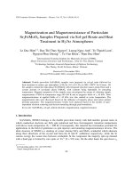
Technologies, Vancouver, Canada) (Fig. 1).
The results showed that supplementation with SCF significantly
enhanced the proliferation of PSCs in a concentration-dependent
manner. Supplementation with FBS stimulated the proliferation of
certain, morphologically flat ovarian somatic cells, and interfered
with the growth of the PSCs. Furthermore, PSCs cultured with KSR
readily reaggregated with ovarian somatic cells to form clumps, also
inhibiting PSC proliferation (Fig. 1A-C). Therefore, DMEM-F12
supplemented with B27 (DMEM-F12/B27) plus 40 ng/ml SCF was
considered the most effective medium for PSC growth (Fig. 1D).
Development (2014) 141, 2235-2244 doi:10.1242/dev.104554
Ovarian cell-derived regulatory factors are crucial for the
establishment of PSCs
Primary ovarian cells formed spherical colonies comprising
compact clusters of small round PSCs (5-7 μm in diameter) 1 day
after culture in DMEM-F12/B27 plus SCF, interspersed with a few
red blood cells (RBCs) (Fig. 2Aa,b). The PSC clusters appeared
dark and shiny, with constituent cells that were smaller or similar in
size to RBCs (6-8 μm). The PSCs could easily be distinguished
from the RBCs at 1 day because the latter were of the typical
biconcave disc shape (Fig. 2Ab). The PSCs had completely round
nuclei that took up almost the entire volume of the cell, as evidenced
by DAPI staining (Fig. 2Ba), as has been described for VSEL stem
cells in the adult human ovary (Parte et al., 2011). However, the
PSCs were either not detected or only weakly detected by MayGrunwald-Giemsa staining (Fig. 2Bb).
After 1 week, the PSCs increased in number and size, and some
grew to ∼10-12 μm (Fig. 2Ac; supplementary material Fig. S1).
Most of the PSCs were 10-12 μm in diameter after 10 days in culture,
forming groups of cells that clustered around the ovarian cell
colonies (Fig. 2Ca,b). At this time, the colonies and the surrounding
PSCs were treated with 0.05% trypsin-EDTA for 2 min to disperse
the PSCs, while leaving most of the colonies intact. Then, the cells
were passed through a 40-μm filter to remove all of the remaining
colonies, which contained ovarian cells such as theca stem cells and
granulosa cells (Honda et al., 2007; Kossowska-Tomaszczuk et al.,
2009). The filtered cells were cultured on laminin-coated dishes or
on a mitomycin C-treated mouse embryonic fibroblast (MEF) feeder
layer. After 1 month in culture under these conditions, with one
passage per week, the proliferation of the PSCs was reduced.
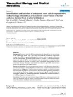
Fig. 1. Comparison of culture media and culture supplements for the establishment of PSCs. (A) Proliferation of PSCs after 1 week of culture in MEM-Alpha
(a), StemPro-34 (b) and DMEM-F12 (c) medium. After 1 month in culture, DMEM-F12 exhibited a significant effect on PSC proliferation (f, compared with d,e).
(B) Spontaneously differentiated oocytes appeared after subculture in DMEM-F12. (C) Effect of KSR and serum-free B27 supplementation on PSC proliferation
(n=6). PSCs were cultured for 7 days on gelatin-coated dishes. Note the improved growth of PSCs in DMEM-F12 supplemented with B27 (DMEM-F12/B27) versus
KSR. (D) Effect of SCF on PSC proliferation (n=6). PSCs were cultured for 7 days on gelatin-coated dishes with DMEM-F12/B27 supplemented with various
concentrations of SCF (10, 20, 30, 40 or 50 ng/ml). PSC proliferation was considerably improved in the presence of 40 ng/ml SCF. Error bars indicate s.e.m.
2236
DEVELOPMENT
RESEARCH ARTICLE
RESEARCH ARTICLE
Development (2014) 141, 2235-2244 doi:10.1242/dev.104554
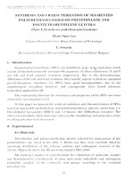
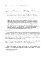
Fig. 2. Development of PSCs. (A) After isolation from the ovary, PSCs in culture appeared dark and shiny and were easily distinguished from RBCs, which
had a typical biconcave disc morphology (asterisks). The PSCs gathered in hollows formed by ovarian epithelial cells after 4 days in culture (b), or were trapped
within the theca stem cell colonies. The PSCs increased in number and size after 1 week (c). (B) The small PSCs (5-7 μm in diameter) were similar in size to RBCs
and round in appearance, but unlike RBCs they had a high nucleus-to-cytoplasm ratio, and the nuclei were stained by DAPI. PSCs were weakly detected by
May-Grunwald-Giemsa staining, whereas all of the RBCs were stained red or blue. (C) PSCs grew to a uniform size (10-12 μm) after 10 days in culture, forming
groups of cells that clustered around theca stem cell colonies (a,b). PSCs were maintained for 1 month on a layer of ovarian somatic cells (c,d). (D) Flow
cytometric characterization of PSCs after 1 week in culture demonstrated that 25% of the cells were small (5-7 μm) and 75% were large (10-12 μm). Vasa-positive
cells comprised 1.79% of the small PSCs and 5.71% of the large PSCs (a). Some PSCs were also positive for other germ and stem cell markers, such as
Fragilis, Thy-1, SSEA4 and c-kit (b). After 2 weeks in culture, the PSCs became uniform in size and made up an increasing percentage of the total cell
population (c). Scale bar: 50 μm.
mitomycin C-treated MEF feeder layers after 1 month for long-term
culture, as described in the scheme for the establishment of PSCs
(supplementary material Fig. S3A).
PSCs undergo molecular progression during establishment
Flow cytometry analysis revealed abundant PSC proliferation after
isolation and culture for 1 week. Of these, 4.65% of the cells were
positive for the germ cell marker Vasa and some of the cells
were also positive for additional germ and stem cell markers, such as
Fragilis, Thy-1, SSEA4 and c-kit (Fig. 2Da,b). At this time, two
populations of PSCs were observed: one with a cell diameter of
5-7 μm and one with a cell diameter of 10-12 μm (Fig. 2Da). The
cells became identical in size after 2 weeks in culture, at 10-12 μm,
with an increasing percentage of cells positive for germ and stem
cell markers (Fig. 2Dc).
About 2.8% of all mouse testicular cells are c-kit positive
(Kanatsu-Shinohara et al., 2004) and have the capacity to become
multipotent germline stem cells, whereas c-kit-negative cells go on
to become spermatogonial stem cells (Izadyar et al., 2008). We
similarly observed two distinct subsets of cells (c-kit positive versus
c-kit negative) within the PSC population. This finding was
strengthened by immunofluorescence analysis showing that, after
1 month in culture, most of the PSCs expressed high levels of the
reprogramming factor Oct4, whereas only 22% of the PSCs
expressed high levels of c-kit (Fig. 3Aa-d,B).
2237
DEVELOPMENT
Furthermore, the cells changed their morphology from round to
adherent, and somatic cell types appeared (supplementary material
Fig. S2A,B). These observations indicate that the present culture
conditions were not suitable for the establishment and long-term
maintenance of PSCs.
Because the PSCs tended to gather in hollows formed by the
primary ovarian cells (Fig. 2A), and because extracellular secreted
factors play essential roles in stem cell-niche interactions, we
hypothesized that ovarian cells might provide an appropriate
in vitro microenvironment for the establishment, maintenance and
proliferation of PSCs. Thus, we generated PSC cultures containing
ovarian cells. After 10 days in culture, the colonies and the
surrounding PSCs were treated with 0.25% trypsin-EDTA for
3 min. This treatment dispersed most of the cells, including the
ovarian cell colony-derived cells. The dispersed cells were then
passed through a 40-μm filter to remove only the largest clumps
of theca stem cells, followed by culture on dishes coated with gelatin
(1:1 dilution).
Under these conditions, PSCs formed clusters or grew as
dispersed cells on top of flat layers of epithelial and somatic
ovarian cells. The cells required passage at confluence every
5-7 days, with cultures being split at a 1:2 dilution. Although the
PSCs continued to grow, most of the remaining theca stem cells and
the flat cell layers gradually disappeared after more than 1 month in
culture (Fig. 2Cc,d). Therefore, the PSCs were transferred onto
RESEARCH ARTICLE
Development (2014) 141, 2235-2244 doi:10.1242/dev.104554
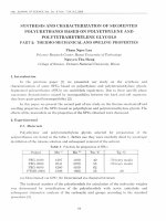
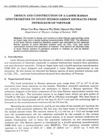
Fig. 3. SCF improves the reprogramming
of porcine PSCs during establishment.
PSCs were isolated and cultured in medium
without and with 40 ng/ml SCF for 1, 2, 3
and 4 weeks. They were then collected for
the detection of Oct4 and c-kit by
immunostaining. (A) Representative
immunofluorescence detection of Oct4 and
c-kit expression in PSCs after 4 weeks in
culture; DAPI (a,e). (B) Quantification of
c-kit-positive PSCs after 1, 2, 3 and 4 weeks
in culture. (C) Quantification of nuclear
versus cytoplasmic localization of Oct4 in
PSCs after 1, 2, 3 and 4 weeks in culture.
2238
PSCs share characteristics with epiblast-derived PGCs
We next investigated the developmental origin of porcine PSCs. In
normal development, c-kit, SSEA1 and SSEA4 are expressed by the
majority of pregonadal PGCs and are progressively downregulated
when PGCs enter into meiosis in the embryonic ovary (Kerr et al.,
2008). By contrast, Vasa protein is detectable only when PGCs enter
the gonadal ridges and remains elevated in human fetal and postnatal
oocytes (Castrillon et al., 2000). VASA (DDX4)-negative VSEL
stem cells (2-4 μm) isolated from the human OSE express genes
typical of ESCs, such as NANOG and SOX2, thereby indicating their
undifferentiated status. After culture for 3 weeks under differentiation
conditions, VASA-negative cells are transformed into OLCs
expressing VASA and ZP2, a marker for oocytes (Virant-Klun
et al., 2008). In the present study, small Vasa-positive porcine PSCs
(5-7 μm in diameter) began to reduce their expression of Nanog,
Sox2 and Rex1 after 1 week in culture (Fig. 4E), indicating their
transformation to a differentiating status. Previous investigations
showed that Vasa-positive VSEL stem cells isolated from adult
organs express several characteristic markers of early PGCs,
including fetal-type alkaline phosphatase, Oct4, SSEA-1, CXCR4,
Stella, Fragilis, Nobox and Hdac6 (Ratajczak et al., 2008). Because
the porcine PSCs described herein similarly express a number of
typical, early PGC markers (Figs 2 and 4), these findings might
indicate a close association of PSCs with Vasa-positive VSELs and
epiblast-derived PGCs.
In addition, the strong expression of ESC markers (e.g. Nanog,
Sox2, Rex1, cMyc and KLF4) in porcine PSCs after 4 weeks in
culture demonstrates that the PSCs can dedifferentiate under
appropriate conditions (Fig. 4E). We have occasionally observed
small, amoeboid process-bearing PSCs, which are probably
counterparts to gonadal PGCs, that still retain their motile capability
to wander throughout the ovarian tissue (Motta et al., 1997)
(supplementary material Movie 1). Taken together with the
observed molecular progression of PSCs, our results suggest that
Vasa-positive cells with the characteristics of early PGCs are present
or are generated in the adult pig ovary, and that these small Vasapositive PSCs are probably derived from VSEL stem cells in the OSE.
DEVELOPMENT
Interestingly, when PSCs were cultured without SCF, the
percentage of c-kit-positive PSCs was significantly decreased
relative to culture with SCF (Fig. 3A,B). In addition, SCF treatment
significantly affected the expression of Oct4 (Fig. 3A,C). PSCs
cultured in the presence of SCF exhibited intense cytoplasmic
staining for Oct4 after 1 week in culture (Fig. 4B), whereas Oct4
expression was reduced in the cytoplasm and augmented in the
nucleus after 2 weeks in culture (Fig. 4Ce). Furthermore, SCF
treatment significantly increased the number of large PSCs
expressing Oct4 in the nucleus after 1 month in culture (Fig. 3C).
A similar phenomenon has been described in PGCs undergoing
nuclear reprogramming over the course of fetal development in mice
and humans (Anderson et al., 2007; Gkountela et al., 2013). Hence,
c-kit and SCF are crucial to the nuclear reprogramming required for
the establishment of porcine PSCs.
After 1 week in culture, small PSCs with a cell diameter of
5-7 μm demonstrated cytoplasmic localization of the germ cell
markers Vasa, Stella and SSEA4 (Fig. 4A,B; supplementary
material Fig. S4A). In addition, Oct4 protein expression was
found throughout entire colonies of ovarian cells, whereas Stella
was only found in small PSCs gathered around the colonies
(Fig. 4Be,f ). This result confirmed that the ovarian cell colonies
contained theca stem cells or somatic cells, as they do not express
any germ cell markers (Honda et al., 2007).
After 2 weeks in culture, the PSCs became much larger and
abundant in the cytoplasm, adhering loosely to the ovarian cell
colonies and maintaining their expression of germ cell markers
(Fig. 4Ca-d). Sohlh1 protein, which is detected in germ cell cysts,
was also detected in PSCs at 2 weeks (Fig. 4Cf ). Although all of
the small PSCs expressed germ cell markers after 1 week (Fig. 4D),
the expression levels of stem cell markers (e.g. Oct4, Nanog,
Sox2, Rex1, cMyc and KLF4) showed substantial cell-to-cell
variation (Fig. 4E). After 4 weeks, all of the PSCs were 10-12 μm
in diameter and strongly expressed stem and germ cell markers
at both the protein and mRNA level (Fig. 4Ch,i,D,E). The oocyte
markers SCP3 and ZP were not detected in the cells during culture
(Fig. 4D).
RESEARCH ARTICLE
Development (2014) 141, 2235-2244 doi:10.1242/dev.104554
Maintenance of PSCs in vitro and induced differentiation into
OLCs
Newly established PSCs were expanded in vitro for at least 6 months
and passaged 30 times without loss of proliferative potential
(Fig. 5A). Moreover, the cells maintained expression of the
identifying germline markers (Fig. 5B; supplementary material
Fig. S4B). The estimated cell doubling time was 48-72 h (Fig. 5C).
After that, although differentiated cells increased among PSCs after
long-term culture, they retained high proliferation as shown by large
numbers of PSCs double positive for BrdU and Oct4 or Vasa
(Fig. 5D,E). Live cell imaging showed that the germinal granules
were equally separated into daughter PSCs after cell division
(Fig. 5F, arrows). These cytoplasmic structures are characteristically
observed in germline cells, becoming discernible at later stages of
germ cell differentiation (Chuma et al., 2009). These results
demonstrate that live PSCs undergo mitosis in culture, providing the
clearest evidence of in vitro oogenesis.
In addition, the PSCs showed positive alkaline phosphatase
staining, and the intensity of the staining was stronger in the
germinal granules than in any other region of the cell (Fig. 5G).
Cytogenetic analysis also showed that the PSCs had a normal
karyotype of 38, XX (Fig. 5H). Transplantation of PSCs into
immunodeficient mice failed to result in teratoma formation,
indicating that these cells are not pluripotent stem cells (Fig. 5I).
To confirm the presence of in vitro oogenesis, we transduced a
transgene encoding EGFP into porcine PSCs that had been cultured
for more than 6 months to create EGFP-PSCs. The EGFP-PSCs
reaggregated with dispersed adult pig ovarian cortical tissue (OCT)
cells at a ratio of one EGFP-PSC to five OCT cells (Fig. 6Aa). After
2 days in culture, numerous clumps of aggregated cells formed that
contained both EGFP-PSCs and OCT cells (Fig. 6Ab). After 2 weeks
in culture, many primordial OLCs were observed that consisted of
both EGFP-positive OLCs derived from the EGFP-PSCs, and EGFPnegative OLCs derived from the OCT cells (Fig. 6Ac,d). Hence,
OLCs were spontaneously generated from PSCs reaggregated with
ovarian tissues, consistent with earlier reports from mouse and human
models (Pacchiarotti et al., 2010; White et al., 2012).
To study the differentiation potential of OLCs further, the PSCs,
after 3 weeks of isolation (supplementary material Fig. S4C), were
cultured under differentiation conditions for 4 weeks. During this
time, some of the PSCs grew large in size (∼50 μm in diameter) and
aggregated with others to form oocyte-cumulus complex (OCC)like structures (Fig. 6Bb, arrows). Although all of the PSCs were
exposed to the same culture medium, only ∼0.1% developed into
OCC-like structures (supplementary material Fig. S5A). This is
similar to the situation in the ovary, where a high somatic cell to
oocyte ratio is required to provide the requisite microenvironment
for oocyte growth and differentiation.
2239
DEVELOPMENT
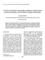
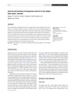
Fig. 4. PSCs undergo molecular progression during establishment. (A,B) After 1 week in culture, small PSCs showed cytoplasmic localization of Vasa, Stella
and Oct4. Compact colonies were surrounded by small PSCs and contained theca stem cells or somatic ovarian cells (Bg, arrows). (C) After 2 weeks, the
PSCs became larger and maintained their expression of Fragilis, Stella, Oct4 and Sohlh1. Oct4 protein expression was reduced in the cytoplasm and became
localized in the nuclei of PSCs at this time (e). After 4 weeks in culture, most of the PSCs were large (10-12 μm) and maintained their expression of the germ cell
markers DAZL and Blimp1. The flat layer of epithelial and somatic cells did not express any germ cell markers (g,j, arrows). (D,E) mRNA expression levels
of oocyte-specific (ZP and SCP3), germ cell-specific (Fragilis, Blimp1, Vasa, c-kit and DAZL) and stem cell-specific (Oct4, Nanog, Sox2, Rex1, cMyc and KLF4)
markers in PSCs. β-actin mRNA was used as the normalization control. Ov, ovarian tissue; RT-, control (water); 1, 2, small PSC samples #1 and #2 after 1 week
in culture; 3, PSCs after 4 weeks in culture. (F) Alexa Fluor 488 (anti-rabbit NC1; anti-mouse NC2) and Alexa Fluor 568 (anti-rabbit NC3; anti-mouse NC4)
were used as negative controls. Scale bars: 10 μm.
RESEARCH ARTICLE
Development (2014) 141, 2235-2244 doi:10.1242/dev.104554
Gene expression analysis showed that OLCs expressed many of
the same germ cell markers as PSCs (Fig. 6C). However, the oocyte
markers ZP, ZPC, SCP3 and GDF9 were only found in OLCs after
2 weeks of differentiation. After 3-4 weeks of differentiation, these
oocyte markers reached expression levels in OLCs that were similar
to those in normal germinal vesicle (GV)-stage oocytes (Fig. 6C), as
summarized in the procedure for the differentiation of PSCs
(supplementary material Fig. S3B).
Immunostaining clearly showed that the germ cell markers
Blimp1 and DAZL were expressed in all of the PSCs, whereas the
OLCs alone exhibited positive staining for the oocyte markers
GDF9 and LHX8 (Fig. 7Aa-c; supplementary material Fig. S5B).
In addition, the OLCs exhibited positive staining for Vasa,
c-kit, DAZL, Stella, SCP3 and GDF9, whereas the adjacent
somatic cells were negative, indicating specific expression of
these germ cell markers in OLCs (Fig. 7A,B). As with normal
primordial oocytes, the PSC-generated OLCs contained many
cytoplasmic germinal granules (Fig. 7C). After 2 weeks in
culture, ∼10% of the PSCs grew sufficiently large to approximate
the size of fully grown oocytes (>100 μm; Fig. 7D). The cells
also expressed oocyte and germ cell markers (supplementary
material Fig. S5C,D).
To elucidate whether the oocytes generated were truly derived
from mitotically active PSCs, and did not instead represent oocytes
2240
derived from primary ovarian cells, we isolated and purified PSCs
by SSEA4-based magnetic bead sorting, as small SSEA4-positive
cells from human ovarian cell cultures are reportedly related to
ESCs and cells of the germinal lineage (Virant-Klun et al., 2013),
and small porcine PSCs showed cytoplasmic expression of SSEA4
(supplementary material Fig. S4A). Cell sorting resulted in the
collection of 759±46 (s.e.m. for three replicate experiments) cells
from ten different ovaries. The SSEA4-positive cells were then
transfected with EGFP. Owing to the important role of ovarian
cell-derived regulatory factors in the establishment of porcine
PSCs, the GFP-positive SSEA cells were aggregated with
dispersed adult pig OCT cells as described above and cultured
for more than 1 month.
Finally, EGFP-positive SSEA cells were differentiated into OLCs
in vitro and transplanted into immunodeficient female mice. The
further in vitro differentiation of OLCs provided direct evidence for
EGFP-positive live oocytes (Fig. 7E). The dual immunofluorescencebased detection of EGFP in vivo, along with detection of either
the oocyte-specific transcription factor LHX8 or the early ovarian
follicle-specific growth and differentiation factor GDF9, identified
many GFP/LHX8 or GFP/GDF9 double-positive cells distributed
throughout the xenograft (Fig. 7F, arrows). These results convincingly
demonstrate the differentiation capacity of PSCs into oocytes, both
in vitro and in vivo.
DEVELOPMENT
Fig. 5. Characterization and maintenance of PSCs. (A) Maintenance of PSCs after long-term culture on MEF feeder cells. (B) PSCs could be expanded in vitro
for months without the loss of germ cell markers. (C) Selected cell lines were frozen/thawed and propagated for at least 6 months, with an estimated cell doubling
time of 48-72 h. (D,E) BrdU incorporation together with Oct4 (D) and Vasa (E) expression was detected in the PSCs after long-term culture, whereas the feeder
cells were negative for these markers (see merge with DAPI image). Arrow indicates a dividing PSC. (F) The presence of actively dividing PSCs was
demonstrated by live cell imaging [with photos taken from the beginning (a) until the end of cell division (f )]. Arrows indicate germinal granules. (G) PSCs stained
positive for alkaline phosphatase (a). High magnifications (b,c) show PSCs in M phase (large; right) and in S phase (small; left). (H) PSCs showed a normal
karyotype (38, XX). (I) Teratoma formation was assessed after the transplantation of PSCs into the testes of immunodeficient mice. No tumors were found at
5 months after PSC transplantation, whereas control murine ESCs formed tumors at 1 month after transplantation (asterisk). Scale bars: 10 μm.
RESEARCH ARTICLE
Development (2014) 141, 2235-2244 doi:10.1242/dev.104554
DISCUSSION
The current study has shown that cells with characteristics of early
PGCs are present or are generated in the adult pig ovary.
Moreover, porcine PGC-like PSCs continue to maintain their
germ stem cell identity in vitro and can differentiate into OLCs
under appropriate culture conditions. In addition, experimental
evidence showed that PGC-like PSCs are probably generated from
Vasa-positive VSEL stem cells in vitro. Finally, we demonstrated
the important role of ovarian cell-derived regulatory factors
and the proximal stem cell niche in the establishment of porcine
PSCs.
Our results are consistent with those of other investigators
suggesting that PSCs in the OSE originate from VSELs, and that
PSCs might support neo-oogenesis. However, whether VSELs can
proliferate in vitro or in vivo has yet to be elucidated. The selfrenewal and differentiation of stem cells in the body must be
properly controlled by the specialized microenvironment of the
stem cell niche (Morrison and Spradling, 2008), and secreted factors
(e.g. extracellular matrix molecules, cytokines) produced by niche
cells are known to play essential roles in stem cell-niche
interactions. However, the biological, molecular and functional
nature of the OSC niche remains largely unknown.
The present study suggests that co-culture with ovarian cells is
necessary for the establishment of PGC-like PSCs. Communication
between germline and somatic cells is indispensable for stem cell
maintenance, as well as for germ cell proliferation and differentiation.
Importantly, human and bovine OSE-derived cells co-express SCF
and c-kit, implying that SCF can act as an autocrine factor in the
normal OSE (Parrott et al., 2000). Interestingly, we demonstrated that
SCF increased not only the proliferation of PSCs, but also the
proportion of c-kit-positive PSCs. SCF also mediated alterations in
the cytoplasmic-to-nuclear translocation of Oct4 after 2 weeks in
culture. Therefore, SCF stimulated the growth, proliferation and
nuclear reprogramming of porcine PSCs.
The function of the OSE during the mammalian postnatal period
remains elusive. Whether germline stem cells exist in the adult
mammalian ovary and, if they do exist, whether they can generate
oocytes, need to be precisely addressed. A recent study indicated that
oogonia fail to stain with pluripotent immunohistochemical markers
after 2 years of age in human (Byskov et al., 2011). However, these
findings do not rule out the possibility of de novo transformation of
OSE cells into multipotent stem-like cells in the postnatal human
ovary. On the other hand, Kerr et al. (2012) found no evidence for the
regeneration of primordial follicles after chemical- or γ-radiationmediated depletion. We demonstrated in an earlier study that
busulfan treatment is cytotoxic to murine oocytes, stimulating
follicular apoptosis and disrupting folliculogenesis (Park et al.,
2013). Nonetheless, the finite number of oocytes formed during the
fetal period does not rule out the possibility of neo-folliculogenesis.
In an effort to ascertain the existence of FGSCs in postnatal mouse
ovaries, adult mouse ovaries were recently shown to be capable of
supporting the formation of new follicles when provided with
transplanted premeiotic female PGCs and companion pre-follicular
cells. The transplanted PGCs were, however, only able to form
follicles with their own pre-follicular cells, and the transplanted prefollicular cells could only form follicles with the transplanted PGCs
(Zhang et al., 2012). Although the authors concluded that neooogenesis does not normally occur in adult mouse ovaries, these
results nevertheless provide an answer to the important question
of whether the adult ovary can support neo-oogenesis from
transplanted PGCs. Taken together, we suggest that germline stem
cells per se might not persist in postnatal and adult mammalian
ovaries, but that progenitor cells/small PSCs in the ovary can instead
differentiate into germline stem cells under appropriate conditions.
Notably, our observations indicate that early PGC-like PSCs are
found in the adult pig ovary. These PGC-like PSCs might correspond
to PGCs that survive into adulthood, rather than to the large (∼1520 μm) migrating PCGs. Although PGC reprogramming has not yet
2241
DEVELOPMENT
Fig. 6. Induced differentiation of PSCs into OLCs. (A) Expression of EGFP-positive cells was observed throughout the clumps of PSCs reaggregated with
dispersed adult pig OCT cells (a,b). Primordial EGFP-positive OLCs derived from EGFP-positive PSCs and EGFP-negative OLCs derived from EGFP- negative
OCT cells were both observed after 2 weeks in culture (c,d). (B) After culture under differentiation conditions for 2-4 weeks, some of the PSCs formed primordial
OLCs (30-35 μm in diameter; a, inset), and some of the PSCs proceeded to form OLCs (50 μm in diameter; b, inset) or OCC-like structures (b, arrows).
(C) mRNA expression levels of oocyte-specific (ZP, ZPC, SCP3 and GDF9b) and germ cell-specific (Vasa, Blimp1, Fragilis and c-kit) markers in differentiated
cells. β-actin mRNA was used as the normalization control. PSCs, control PSCs at 3 weeks after isolation; 1, 2, 3, 4, PSCs that differentiated into OLCs
after 1, 2, 3 and 4 weeks, respectively; GV, oocyte derived from pig ovary. Scale bars: 10 μm.
RESEARCH ARTICLE
Development (2014) 141, 2235-2244 doi:10.1242/dev.104554
Fig. 7. Characteristics of OLCs generated from PSCs. (A,B) OLCs exhibited positive staining for GDF9, Blimp1, Vasa, c-kit, DAZL, Stella and SCP3,
whereas the adjacent somatic cells were negative for these markers (see in merged image c,f ). (C) As with normal primordial oocytes, the PSC-generated OLCs
contained many cytoplasmic germinal granules. (D) Under differentiation, OLCs grew as large as growing oocyte-like cells (a) or fully grown oocyte-like cells (b).
(E) In vitro differentiation of OLCs provided direct evidence for EGFP-positive living oocyte-like cells. (F) Dual immunofluorescence analysis of EGFP expression
(green) and either LHX8 or GDF9 expression (red) in murine xenografts following EGFP-PSC injection for 2 weeks (a,b). EGFP-positive oocytes were not
detected in the pig ovarian tissue in control xenografts, whereas GDF9 was detected in all oocytes (c). Arrows indicate injected EGFP-PSCs in the OCT. Scale
bars: 20 μm.
2242
undifferentiated cells with stem cell characteristics, which, under
suitable conditions, can undergo proliferation and differentiation.
VSELs isolated from adult tissues might epitomize an ‘allpowerful’ stem cell for regenerative medicine applications, as
suggested by Ratajczak et al. (2008). Like ESCs, VSELs are
pluripotent with maximum regenerative potential, but unlike ECSs
they do not form teratomas. The question of whether pluripotent
stem cells that appear during the culture of mammalian ovarian
tissue originate from unipotent germ stem cells will probably be
resolved in due course, but perhaps more important are our findings
showing that it is in fact possible to derive and expand autologous
stem cells from ovarian tissue. The isolation and characterization of
human PSCs will contribute considerably to the prospect of using
stem cells to produce developmentally competent oocytes in vitro,
with clear clinical potential. Our work also supports further inquiry
into a myriad of health parameters in premenopausal woman, with
applications in tissue repair and restoration.
MATERIALS AND METHODS
Ethics statement
The treatment of the pigs used in this research followed guidelines of the
Institutional Animal Care and Use Committee of the National Institute of
Animal Science, Suwon, South Korea (approval no. 2009-004, D-grade).
Isolation and purification of PSCs
Ovaries (10-12 for each experiment) were collected from prepubertal gilts at
a local slaughterhouse. Cortical slices (0.1-0.5 mm thick) were cut from the
ovarian surface using a surgical blade (No. 21, Feather Safety Razor, Osaka,
DEVELOPMENT
been reported in the pig, studies on PGC reprogramming in the human
fetal ovary and the testis showed nuclear localization of Oct4 during
the first trimester, with intense cytoplasmic expression during the
second trimester. At week 17 of fetal development, Oct4 is again
identified in the nucleus (Bhartiya et al., 2010; Gkountela et al., 2013).
We also found that PSCs undergo similar cytoplasmic-to-nuclear
reprogramming of Oct4 expression, with localization of Oct4 detected
in the nucleus of large PSCs. Although the significance of cytoplasmic
Oct4 expression is unknown, it is notably coincident with major global
epigenetic changes, such as the wholesale epigenetic loss of
H3K27me3 and H2A.Z in PGCs, followed by the expression of
Oct4 in the cytoplasm (Gkountela et al., 2013).
Why porcine PGCs should be maintained in the postnatal ovary is
still a matter of controversy. Recent investigations suggest the
presence of two distinct PGC populations in human fetal gonads.
While Vasa-positive PGCs enter meiosis in the fetal ovary, the fate of
c-kit-positive PGCs remains unclear (Gkountela et al., 2013). The
authors propose that c-kit-positive PGCs persisting in the second
trimester gonad represent a more primitive PGC population than
Vasa-positive cells, an idea supported by their maintenance of a core
germ cell gene expression signature at the single-cell level. The work
of Gkountela and colleagues also raises questions about the lineage
relationships and fates of the c-kit-positive cells. As Laird (2013)
discusses, will they be culled in a wave of apoptosis or, as their
transcriptome suggests, will they enter meiosis and be conserved in
the ovary? Although these issues require further investigation, we
maintain that the adult mammalian ovary contains a small number of
RESEARCH ARTICLE
Development (2014) 141, 2235-2244 doi:10.1242/dev.104554
EGF, 0.05 IU follicle-stimulating hormone (Sigma-Aldrich), 0.03 IU
luteinizing hormone (Sigma-Aldrich), 0.01 mM dibutyryl cAMP (SigmaAldrich) (Cayo-Colca et al., 2011) and 1% polyvinylpyrrolidone (PVP) 360
(Sigma-Aldrich) (Hashimoto et al., 2007). The aggregated cells were cultured
for 2 weeks, replacing half the medium every 2-3 days.
Japan) (Bui et al., 2007) and dissociated by mincing, followed by a two-step
enzymatic digestion involving a 15 min incubation with 1 mg/ml
collagenase (type IV, Sigma-Aldrich) dissolved in Hank’s Balanced Salt
Solution (HBSS) and 10 min with 0.25% trypsin-EDTA at 38.5°C. Trypsin
was neutralized by adding 10% fetal bovine serum (FBS), and tissues
dispersed into single cells by gentle pipetting. The dispersed cells were
passed through a 40-μm filter and the dissociated cells were allocated to
60 mm gelatin-coated tissue culture dishes and incubated overnight.
To prepare the primary ovarian cells, fibroblasts were allowed to attach to
the bottom of a gelatin-coated culture plate, while the floating cells were
passaged onto a secondary culture plate after vigorous pipetting. The cells
were maintained at 38.5°C in an atmosphere of 5% CO2 in air. After
selection, 1-2×10⁴ cells were plated in one well of a 24-well gelatin-coated
plate (Corning). Half of the culture medium was changed every other
day, and the primary ovarian cells were passaged further as described in
the Results.
PSCs were then isolated based on their expression of SSEA4 via magnetic
bead sorting. After a two-step enzymatic digestion, the ovarian cells were
incubated with anti-SSEA4 antibody for 30 min on ice. After rinsing and
resuspending in HBSS, mouse anti-IgG magnetic beads (Miltenyi Biotec)
were added to the cell suspension and incubated for a further 30 min on ice.
After one additional wash, the cell preparations were loaded onto MACS
Cell Separation columns and separated according to the manufacturer’s
specifications (Miltenyi Biotec). Small (5-7 μm diameter) SSEA4-positive
PSCs were obtained and transfected with enhanced green fluorescent protein
(EGFP) as described below.
Twenty-four pig OCT pieces (2×2×1 mm) were individually injected with
∼1×10³ EGFP-PSCs using a 10 μl NanoFil syringe with a 35-gauge
bevelled needle (World Precision Instruments). Recipient nude female mice
were anesthetized and a small incision was made along the dorsal flank for
subcutaneous insertion of the pig ovarian tissue (four grafts per mouse).
Xenografts were removed 1-2 weeks after transplantation, fixed in 4%
paraformaldehyde, paraffin embedded and serially sectioned (6 μm) for
immunohistochemical analysis using a mouse monoclonal antibody against
GFP. High-temperature antigen retrieval was first performed using 0.01 M
sodium citrate buffer ( pH 6.0). After cooling, sections were incubated for
10 min with 3% hydrogen peroxide in methanol to block endogenous
peroxidase activity as per the manufacturer’s protocol (Vector Laboratories).
Sections were then blocked for 1 h using 1% normal goat serum and
incubated with GFP antibody for immunostaining. Negative controls (the
xenografted tissues that received vehicle injections) were run in parallel and
did not show a positive signal. To confirm and extend these observations,
dual immunofluorescence-based detection of GFP and either GDF9 or
LHX8 in xenografted human ovarian tissues was performed with DAPI
counterstaining.
Transduction of the EGFP transgene into PSCs
Karyotyping and teratoma formation
An HIV-1-based self-inactivating lentiviral vector plasmid ( pLV-EGFP)
was constructed as described (Ikawa et al., 2003). For lentiviral vector
transduction, a single-cell suspension of PSCs (1-2×10⁶ cells) was mixed
with the lentiviral vector in 100 ml for 6 h (107 U final concentration). After
washing with PSC culture medium, transduced cells were cultured on a layer
of MEF feeder cells.
Cells were prepared and treated as described previously (Bui et al., 2012).
Immunohistochemistry
Acknowledgements
Cells and tissues were fixed and treated, and then quantitative analysis was
conducted as described (Bui et al., 2010). Antibodies and the dilutions
employed are summarized in supplementary material Table S1.
We are especially grateful to Professors Takashi Miyano (Kobe University, Japan)
and Teruhiko Wakayama (Yamanashi University, Japan) for valuable discussions.
Bromodeoxyuridine (BrdU) incorporation assay
PSCs were cultured in medium containing BrdU (50 μg/ml; Sigma-Aldrich)
for 5 days. Detection of DNA synthesis was performed as described
previously (Bui et al., 2010).
Flow cytometry and reverse transcription PCR (RT-PCR)
Cells were prepared and treated as described previously (Bui et al., 2012).
Synthesized cDNAs were subjected to RT-PCR using the specific primers
listed in supplementary material Table S2.
Intraovarian PSC injection and xenografting
Statistical analysis
Each experiment was repeated at least five times. More than 50
immunostained samples were examined in each group. Results are
presented as mean±s.e.m. Data were analyzed by applying Student’s t-test.
Competing interests
The authors declare no competing financial interests.
Author contributions
H.-T.B., N.V.T. and J.-H.K. designed the experiments, analyzed and discussed the
results. H.-T.B. and D.-N.K. performed the experiments. T.K. provided GFP
transgenes for FGSCs. Y.-J.C., M.-H.K. and J.-W.H. contributed new reagents/
analytic tools. H.-T.B. wrote the manuscript.
Funding
This work was supported by a Woo Jang-Choon project grant [PJ007849] from the
Research and Development Agency (RDA) and Institute of Planning & Evaluation
for Technology (IPET) [111047-5] of the Republic of Korea.
Differentiation of PSCs into OLCs
Supplementary material
Supplementary material available online at
/>
References
Abban, G. and Johnson, J. (2009). Stem cell support of oogenesis in the human.
Hum. Reprod. 24, 2974-2978.
Anderson, R. A., Fulton, N., Cowan, G., Coutts, S. and Saunders, P. T. K. (2007).
Conserved and divergent patterns of expression of DAZL, VASA and OCT4 in the
germ cells of the human fetal ovary and testis. BMC Dev. Biol. 7, 136.
Bhartiya, D., Kasiviswanathan, S., Unni, S. K., Pethe, P., Dhabalia, J. V.,
Patwardhan, S. and Tongaonkar, H. B. (2010). Newer insights into premeiotic
development of germ cells in adult human testis using Oct-4 as a stem cell marker.
J. Histochem. Cytochem. 58, 1093-1106.
Bui, H.-T., Van Thuan, N., Kishigami, S., Wakayama, S., Hikichi, T., Ohta, H.,
Mizutani, E., Yamaoka, E., Wakayama, T. and Miyano, T. (2007). Regulation of
chromatin and chromosome morphology by histone H3 modifications in pig
oocytes. Reproduction 133, 371-382.
2243
DEVELOPMENT
A two-stage culture system was established for (1) PSC differentiation and (2)
PSC growth. First, PSCs were plated at 1×10⁴ cells per well of a 24-well tissue
culture plate (Corning) that was treated with poly-D-lysine (0.05 mg/ml;
Sigma-Aldrich) and laminin (0.005 mg/ml; Sigma-Aldrich). Cells were
maintained at 38.5°C in an atmosphere of 5% CO2 in air in differentiation
medium containing DMEM (Invitrogen), penicillin/streptomycin (Invitrogen),
5% FBS (Invitrogen), 5% porcine follicular fluid (Sigma-Aldrich),
0.23 mM sodium pyruvate (Sigma-Aldrich), 0.1 mM non-essential amino
acids (Invitrogen), 2 mM L-glutamine (Millipore) and 0.1 mM
β-mercaptoethanol (Millipore). One half of the culture medium was
replaced every 2-3 days. A number of aggregates containing large cells
formed after 3-4 weeks.
Next, for PSC growth, the aggregates were collected and transferred to
growth medium containing TCM199 (Invitrogen), 3 mg/ml BSA (SigmaAldrich), 5 μl/ml insulin/transferrin/selenium A (Invitrogen), 0.23 mM
sodium pyruvate (Sigma-Aldrich), 1 mg/ml fetuin (Sigma-Aldrich), 1 ng/ml
RESEARCH ARTICLE
Kossowska-Tomaszczuk, K., De Geyter, C., De Geyter, M., Martin, I., Holzgreve, W.,
Scherberich, A. and Zhang, H. (2009). The multipotency of luteinizing granulosa
cells collected from mature ovarian follicles. Stem Cells 27, 210-219.
Kubota, H., Avarbock, M. R. and Brinster, R. L. (2004). Growth factors essential
for self-renewal and expansion of mouse spermatogonial stem cells. Proc. Natl.
Acad. Sci. U.S.A. 101, 16489-16494.
Laird, D. J. (2013). Humans put their eggs in more than one basket. Nat. Cell Biol.
15, 13-15.
Morrison, S. J. and Spradling, A. C. (2008). Stem cells and niches: mechanisms
that promote stem cell maintenance throughout life. Cell 132, 598-611.
Motta, P. M., Makabe, S. and Nottola, S. A. (1997). The ultrastructure of human
reproduction. 1. The natural history of the female germ cell: origin, migration and
differentiation inside the developing ovary. Hum. Reprod. Update 3, 281-297.
Notarianni, E. (2011). Reinterpretation of evidence advanced for neo-oogenesis in
mammals, in terms of a finite oocyte reserve. J. Ovarian Res. 4, 1.
Oatley, J. and Hunt, P. A. (2012). Of mice and (wo)men: purified oogonial stem cells
from mouse and human ovaries. Biol. Reprod. 86, 196.
Pacchiarotti, J., Maki, C., Ramos, T., Marh, J., Howerton, K., Wong, J., Pham, J.,
Anorve, S., Chow, Y.-C. and Izadyar, F. (2010). Differentiation potential of germ
line stem cells derived from the postnatal mouse ovary. Differentiation 79,
159-170.
Park, M.-R., Choi, Y.-J., Kwon, D.-N., Park, C., Bui, H.-T., Gurunathan, S.,
Cho, S.-G., Song, H., Seo, H. G., Min, G. et al. (2013). Intraovarian
transplantation of primordial follicles fails to rescue chemotherapy injured
ovaries. Sci. Rep. 3, 1384.
Parrott, J. A., Kim, G. and Skinner, M. K. (2000). Expression and action of kit
ligand/stem cell factor in normal human and bovine ovarian surface epithelium
and ovarian cancer. Biol. Reprod. 62, 1600-1609.
Parte, S., Bhartiya, D., Telang, J., Daithankar, V., Salvi, V., Zaveri, K. and
Hinduja, I. (2011). Detection, characterization, and spontaneous differentiation in
vitro of very small embryonic-like putative stem cells in adult mammalian ovary.
Stem Cells Dev. 20, 1451-1464.
Ratajczak, M. Z., Zuba-Surma, E. K., Shin, D.-M., Ratajczak, J. and Kucia, M.
(2008). Very small embryonic-like (VSEL) stem cells in adult organs and their
potential role in rejuvenation of tissues and longevity. Exp. Gerontol. 43,
1009-1017.
Telfer, E. E., Gosden, R. G., Byskov, A. G., Spears, N., Albertini, D., Andersen, C. Y.,
Anderson, R., Braw-Tal, R., Clarke, H., Gougeon, A. et al. (2005). On regenerating
the ovary and generating controversy. Cell 122, 821-822.
Virant-Klun, I., Zech, N., Rozman, P., Vogler, A., Cvjeticanin, B., Klemenc, P.,
Malicev, E. and Meden-Vrtovec, H. (2008). Putative stem cells with an
embryonic character isolated from the ovarian surface epithelium of women with
no naturally present follicles and oocytes. Differentiation 76, 843-856.
Virant-Klun, I., Skutella, T., Hren, M., Gruden, K., Cvjeticanin, B., Vogler, A. and
Sinkovec, J. (2013). Isolation of small SSEA-4-positive putative stem cells from
the ovarian surface epithelium of adult human ovaries by two different methods.
Biomed Res. Int. 2013, 690415.
White, Y. A. R., Woods, D. C., Takai, Y., Ishihara, O., Seki, H. and Tilly, J. L.
(2012). Oocyte formation by mitotically active germ cells purified from ovaries of
reproductive-age women. Nat. Med. 18, 413-421.
Zayed, A. E., Abd-Elnaeim, M. M., Abd-Elghaffar, S. K., Hild, A., Brehm, R. and
Steger, K. (2007). Prenatal development of murine gonads with special reference
to germ cell differentiation: a morphological and immunohistochemical study.
Andrologia 39, 93-100.
Zhang, H., Zheng, W., Shen, Y., Adhikari, D., Ueno, H. and Liu, K. (2012).
Experimental evidence showing that no mitotically active female germline
progenitors exist in postnatal mouse ovaries. Proc. Natl. Acad. Sci. U.S.A. 109,
12580-12585.
Zou, K., Yuan, Z., Yang, Z., Luo, H., Sun, K., Zhou, L., Xiang, J., Shi, L., Yu, Q.,
Zhang, Y. et al. (2009). Production of offspring from a germline stem cell line
derived from neonatal ovaries. Nat. Cell Biol. 11, 631-636.
DEVELOPMENT
Bui, H.-T., Wakayama, S., Kishigami, S., Park, K.-K., Kim, J.-H., Thuan, N. V. and
Wakayama, T. (2010). Effect of trichostatin A on chromatin remodeling, histone
modifications, DNA replication, and transcriptional activity in cloned mouse
embryos. Biol. Reprod. 83, 454-463.
Bui, H.-T., Kwon, D.-N., Kang, M.-H., Oh, M.-H., Park, M.-R., Park, W.-J., Paik, S.-S.,
Van Thuan, N. and Kim, J.-H. (2012). Epigenetic reprogramming in somatic cells
induced by extract from germinal vesicle stage pig oocytes. Development 139,
4330-4340.
Bukovsky, A., Svetlikova, M. and Caudle, M. R. (2005). Oogenesis in cultures
derived from adult human ovaries. Reprod. Biol. Endocrinol. 3, 17.
Byskov, A. G., Høyer, P. E., Yding Andersen, C., Kristensen, S. G., Jespersen, A.
and Møllgård, K. (2011). No evidence for the presence of oogonia in the human
ovary after their final clearance during the first two years of life. Hum. Reprod. 26,
2129-2139.
Castrillon, D. H., Quade, B. J., Wang, T. Y., Quigley, C. and Crum, C. P. (2000).
The human VASA gene is specifically expressed in the germ cell lineage. Proc.
Natl. Acad. Sci. U.S.A. 97, 9585-9590.
Cayo-Colca, I. S., Yamagami, Y., Phan, T.-C. and Miyano, T. (2011).
A combination of FSH and dibutyryl cyclic AMP promote growth and acquisition
of meiotic competence of oocytes from early porcine antral follicles.
Theriogenology 75, 1602-1612.
Chuma, S., Hosokawa, M., Tanaka, T. and Nakatsuji, N. (2009). Ultrastructural
characterization of spermatogenesis and its evolutionary conservation in the
germline: germinal granules in mammals. Mol. Cell. Endocrinol. 306, 17-23.
De Felici, M. (2010). Germ stem cells in the mammalian adult ovary: considerations
by a fan of the primordial germ cells. Mol. Hum. Reprod. 16, 632-636.
Gkountela, S., Li, Z., Vincent, J. J., Zhang, K. X., Chen, A., Pellegrini, M. and
Clark, A. T. (2013). The ontogeny of cKIT+ human primordial germ cells proves to
be a resource for human germ line reprogramming, imprint erasure and in vitro
differentiation. Nat. Cell Biol. 15, 113-122.
Hashimoto, S., Ohsumi, K., Tsuji, Y., Harauma, N., Miyata, Y., Fukuda, A.,
Hosoi, Y., Iritani, A. and Morimoto, Y. (2007). Growing porcine oocyte-granulosa
cell complexes acquired meiotic competence during in vitro culture. J. Reprod.
Dev. 53, 379-384.
Hayashi, K., Ogushi, S., Kurimoto, K., Shimamoto, S., Ohta, H. and Saitou, M.
(2012). Offspring from oocytes derived from in vitro primordial germ cell-like cells
in mice. Science 338, 971-975.
Honda, A., Hirose, M., Hara, K., Matoba, S., Inoue, K., Miki, H., Hiura, H.,
Kanatsu-Shinohara, M., Kanai, Y., Kono, T.
et al. (2007). Isolation,
characterization, and in vitro and in vivo differentiation of putative thecal stem
cells. Proc. Natl. Acad. Sci. U.S.A. 104, 12389-12394.
Ikawa, M., Tanaka, N., Kao, W. W.-Y. and Verma, I. M. (2003). Generation of
transgenic mice using lentiviral vectors: a novel preclinical assessment of lentiviral
vectors for gene therapy. Mol. Ther. 8, 666-673.
Izadyar, F., Pau, F., Marh, J., Slepko, N., Wang, T., Gonzalez, R., Ramos, T.,
Howerton, K., Sayre, C. and Silva, F. (2008). Generation of multipotent cell lines
from a distinct population of male germ line stem cells. Reproduction 135,
771-784.
Johnson, J., Canning, J., Kaneko, T., Pru, J. K. and Tilly, J. L. (2004). Germline
stem cells and follicular renewal in the postnatal mammalian ovary. Nature 428,
145-150.
Kanatsu-Shinohara, M., Inoue, K., Lee, J., Yoshimoto, M., Ogonuki, N., Miki, H.,
Baba, S., Kato, T., Kazuki, Y., Toyokuni, S. et al. (2004). Generation of
pluripotent stem cells from neonatal mouse testis. Cell 119, 1001-1012.
Kerr, C. L., Hill, C. M., Blumenthal, P. D. and Gearhart, J. D. (2008). Expression of
pluripotent stem cell markers in the human fetal ovary. Hum. Reprod. 23, 589-599.
Kerr, J. B., Brogan, L., Myers, M., Hutt, K. J., Mladenovska, T., Ricardo, S.,
Hamza, K., Scott, C. L., Strasser, A. and Findlay, J. K. (2012). The primordial
follicle reserve is not renewed after chemical or γ-irradiation mediated depletion.
Reproduction 143, 469-476.
Development (2014) 141, 2235-2244 doi:10.1242/dev.104554
2244