DSpace at VNU: The crystal structure of 3-(4 '-methoxyphenyl)propanoyl pyrrole of Piper lolot C.DC from Vietnam
Bạn đang xem bản rút gọn của tài liệu. Xem và tải ngay bản đầy đủ của tài liệu tại đây (162 KB, 7 trang )
Cryst. Res. Technol.
37
2002
6
627–633
P. LUGER1*, M. WEBER1, N. X. DUNG2, V. T. LUU3, D. D. RANG3, D. T. TUONG3,
P. H. NGOC3
1
Institut für Chemie/Kristallographie, Freie Universität Berlin, Germany
Center for Education and Development of Chromatography of Vietnam and
Hanoi University of Natural Sciences
3
Hanoi Teacher's Training College and NCST of Vietnam
2
The Crystal Structure of 3-(4'-Methoxyphenyl)propanoyl
pyrrole of Piper lolot C.DC from Vietnam
C14H15NO2 (293K): monoclinic space group P21/n, a = 24.580(10), b = 5.536(2), c = 9.037(3) Å, β=
91.18(4)°, V = 1229.4(8) Å3, Dx = 1.239 g⋅cm-3, Z = 4, F(000) = 488, λ(CuKα) = 1.5418 Å, µ= 0.667
mm-1. The title compound which was isolated from the rhizomes of Piper lolot C.DC has antibacterial
activity. Its chemical identity was established by this X-ray analysis. The molecule consists of a planar
propanoyl pyrrole and a methoxyphenyl fragment with an interplanar angle of 93.8(3)° which is by 30°
larger than the corresponding angle reported in the literature for the comparable dimethoxy derivative,
which was also derived from Piper species.
Keywords: crystal structure, piper lolot
(Received March 6, 2002; Accepted April 22, 2002)
Introduction
Pharmaceutically active compounds from domestic plants play an important role in the
traditional folk medicinal system in East Asia. Although the knowledge about the medicinal
activity is generally based on long oral tradition from generation to generation, in several
cases the chemical identity of the extracted compounds was not known, being now a
challenging task for modern analytical methods. The title compound was isolated from the
rhizomes of Piper lolot C.DC by extraction with n-hexane. Piper lolot C.DC is known for its
activity against the bacteria Bacillus pyocyaneus, Staphyloccocus aureus and B. subtilis with
simultanous anti-inflamatory action. It is also in use to treat various deseases like
rheumatism, lumbago, digestive troubles, vomiting, diarrhoea and others (LOI, 1995;
TRUYEN & CHAU, 1999).
The genus Piper belong to the Piperaceae and has over 700 species distributed in both
hemispheres. The Piper species have high commercial, economical and medicinal
importance. Piper lolot C.DC is one species of genus Piper and reputed for its medicinal
properties in folklore medicine of Vietnam.
The chemistry of Piper lolot C.DC has been investigated only on essential oil. The
essential oil isolated by hydro-distillation from the fresh leaves, stem and rhizomes of Piper
lolot C.DC has been analyzed for the first time by a combination of GC and GC/MS. The oils
contained more than 35 compounds, of which 25 constitutes could be identified according to
* corresponding author:
© WILEY-VCH Verlag Berlin GmbH, 13086 Berlin, 2002 0232-1300/02/0606-0627 $ 17.50+.50/0
628
P. LUGER et al.: Crystal Structure
their chromatographic retension indices and mass spectra (DUNG, THANH, KHOI, LECLERCQ,
1996).
The lack of phytochemical investigation of the Piper lolot C.DC extract led us to this
study. From n-hexane extract of the rhizome of the Piper lolot C.DC, we separated two
compounds by column chromatography, one of them is β-asarone (RANG, TUONG & LUU,
2001) and the other one is this compound with MW = 229, mp. 81-82°C. Since its chemical
indentity was originally not known, its structure was elucidated by X-ray analysis.
Table 1: Crystal data, details of data collection and structure determination
Formula
Mr
a (Å)
b (Å)
c (Å)
β (°)
V (Å3)
Z
Space group
Dx (Mg⋅m-3)
F(000)
Radiation [λ (Å)]
µ (mm-1)
Crystal size (mm)
Diffractometer
No. of reflections measured
No. of independent reflections
hmin, hmax
kmin, kmax
lmin, lmax
θmax (°)
Rint
Refinement (on 2146 reflections)
No. of parameters refined
No. of reflections [Fo > 2σ(Fo)]
wR(F2) (overall)
wR(F2) (obs)
R (overall)
R (obs)
Mean shift/esd
Difference electron density (eÅ-3) maximum
Difference electron density (eÅ-3) minimum
C14 H15 NO2
229.27
24.580(10)
5.536(2)
9.037(3)
91.18(4)
1229.4(8)
4
P21/n
1.239
488
CuKα(1.5418)
0.667
0.46 x 0.42 x 0.11
STOE 4-circle
2312
2146
-29, 29
0, 6
0, 10
65.99
0.02
F2
215
1929
0.104
0.099
0.0426
0.0377
0.06
0.15
-0.14
*CCDC 179879 contains the supplementary crystallographic data for this paper. These data can be
obtained free of charge via www.ccdc.cam.ac.uk/conts/retrieving.html (or from the Cambridge
Crystallographic Data Centre, 12, Union Road, Cambridge CB2 1EZ, UK; fax: +44 1223 336033; or
).
Cryst. Res. Technol. 37 (2002) 6
629
Experimental
Rhizomes of Piper lolot C.DC were collected in a garden of Hanoi, Vietnam. A voucher
specimen has been deposited in the Herbarium of the Institute of Ecology and Biological
Resources, NCST, Hanoi, Vietnam.
Rhizome of Piper lolot C.DC was washed with destilled water and dried at 60-70°C until
dryness and pulverized. Rhizome-powder of Piper lolot was extracted by n-hexane at room
temperature in 10 days. After filtration the n-hexane extract was evaporated to dryness under
reduced pressure.
The mixture was chromatographied on a silicagel-column, elution was started with a
mixture of n-hexane-ethylacetate with increasing polarity (from 0% to 100% ethylacetate).
Pure compound was collected after re-column chromatography yielding plate shaped
crystals, melting point 81-82°C, MS(m/z), MW = 229, IR cm-1 (KBr) 3153 (=NH), 31003000 (=CH-), 2938 ( > CH2), 2810 (-OCH3), 1715 ( > C=O), 1514-1480 (aromatic ring),
1245 (C-O).
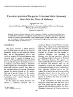
Fig. 1: ORTEP representation (JOHNSON, 1976) of the molecular structure and atom numbering scheme of the title
compound, the thermal ellipsoids are drawn at a probability of 50%.
A crystal with dimensions 0.46 x 0.41 x 0.11 mm was used for all X-ray investigations,
which were carried out on a STOE four circle diffractometer using Ni-filtered CuKα
radiation. Crystal data and details of the intensity data collection are summarized in Table 1.
The unit cell constants were determined by least-squares refinement of 43 reflections in a
range 30° < 2θ < 90°. The repeated measurement of three standard reflections (frequency: 90
minutes) revealed no significant decay (maximum intensity variation 3%) during the data
630
P. LUGER et al.: Crystal Structure
collection. The intensity data were corrected for Lorentz and polarization factors, but not for
absorption. The atomic scattering factors were taken from International Tables for X-ray
Crystallography, Vol. C.
The phase problem was solved by routine application of direct methods (program
SHELXS, SHELDRICK, 1986), least-squares refinements with anisotropic displacement
parameters for C, N and O and isotropic displacement parameters for H (calculated from
stereochemical considerations and allowed for free refinement), including an isotropic
extinction parameter, were executed using SHELXL97 (SHELDRICK, 1997). Details of the
refinement are included in Table 1, final atomic parameters are in Table 2, geometric
parameters are in Table 3.
Table 2: Fractional atomic coordinates and equivalent isotropic displacement parameters (Å2)
U eq = (1 3) ∑ i ∑ j U ij ai* a*j ai ⋅ a j
C1
C2
C3
C4
O4
C41
C5
C6
C7
C8
C9
O9
N10
C11
C12
C13
C14
x
1194(1)
1654(1)
2047(1)
1997(1)
2418(1)
2380(1)
1542(1)
1147(1)
768(1)
326(1)
-114(1)
-94(1)
-573(1)
-1035(1)
-1387(1)
-1148(1)
-655(1)
y
-2889(3)
-3114(3)
-4806(3)
-6347(2)
-7932(2)
-9616(3)
-6188(3)
-4472(3)
-1003(3)
-1914(3)
-91(2)
1963(2)
-861(2)
535(3)
-742(3)
-2997(4)
-3054(3)
z
9019(2)
8163(2)
8467(2
9662(1)
9889(1)
11062(2)
10534(2)
10196(2)
8671(2)
7620(2)
7328(2)
7772(2)
6511(1)
6244(2)
5406(2)
5127(2)
5813(2)
Ueq
54(1)
60(1)
60(1)
50(1)
67(1)
70(1)
56(1)
59(1)
65(1)
60(1)
53(1)
79(1)
53(1)
62(1)
68(1)
78(1)
69(1)
Table 3: Selected bond lengths (Å), angles (°) and torsion angles (°)
C1-C7
C4-O4
O4-C41
C7-C8
C8-C9
C9-O9
C9-N10
N10-C14
N10-C11
C11-C12
C12-C13
1.508(2)
1.370(2)
1.416(2)
1.514(2)
1.498(2)
1.207(2)
1.402(2)
1.381(2)
1.391(2)
1.340(2)
1.405(3)
Cryst. Res. Technol. 37 (2002) 6
C13-C14
O4-C4-C3
O4-C4-C5
C4-O4-C41
O9-C9-N10
O9-C9-C8
N10-C9-C8
C14-N10-C11
C14-N10-C9
C11-N10-C9
C12-C11-N10
C11-C12-C13
C14-C13-C12
C13-C14-N10
C3-C4-O4-C41
C6-C1-C7-C8
C2-C1-C7-C8
C1-C7-C8-C9
C7-C8-C9-O9
C7-C8-C9-N10
O9-C9-N10-C14
C8-C9-N10-C14
O9-C9-N10-C11
631
1.348(2)
115.6(1)
124.9(1)
118.2(1)
119.2(1)
123.5(1)
117.2(1)
107.3(1)
128.1(1)
124.6(1)
108.7(2)
107.6(2)
108.3(2)
108.2(2)
-177.7(1)
-89.8(2)
90.2(2)
177.1(1)
6.5(2)
-172.6(1)
174.5(2)
-6.4(2)
-4.6(2)
Results and Discussion
The molecular structure of the title compound is shown in Fig. 1 along with the atomic
numbering scheme. The molecule consists of two almost planar structural fragments, a
propanoyl pyrrole and a methoxyphenyl unit. The average deviations of contribution (non H)
atoms to least squares planes are 0.06 Å for the first plane and 0.009 Å for the latter one. The
interplanar angle is 93.8(3)°, hence the two fragments are almost perpendicular to each other.
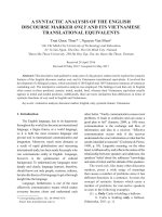
A very similar molecule, also from a Piper species (Piper brachystachyum), is reported in
the literature (KUMAR et al., 1998). 1-[3-(3,4-dimethoxyphenyl)propanoyl]pyrrole differs
from the title compound only by a further methoxy group in meta position of the phenyl ring.
The graphical superposition (Fig. 2) shows that both molecules differ by the dihedral angle
of the two major molecular planes which is 64.2(1)°, hence smaller for the dimethoxy
derivative. Otherwise the geometry of the two molecules is rather alike, e.g. the linear chain
is in an all trans arrangement, the methoxy group(s) is(are) in plane with the phenyl ring.
Bond lengths and angles are as expected and need no detailed discussion. Especially the
values in the amide group and the double bonds in the pyrrole ring are in line with the
dimethoxy derivative and a number of benzoyl and toluoyl substituted pyrrole derivatives
(BENNET, SOMAYAJI, BROWN & SANTARSIERO, 1991; BEACH, BATCHELOR, EINSTEIN &
BENNET, 1998).
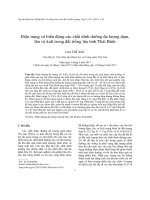
In the crystal lattice (see Fig. 3) the molecules are in head-to-tail arrangements forming
r
r
infinite chains in the a + c direction. Neighboured molecules in a chain are connected by
632
P. LUGER et al.: Crystal Structure
the shortest intermolecular contact in this structure being a C-H...O link from C12-H12 of the
pyrrole ring to the methoxy O4 of a glide plane related molecule, with H... O = 2.70(4)Å. A
second C-H... O contact is C8-H82 to the carbonyl O9 of a molecule related by a translation
r
in b -direction (H... O = 2.71(4)Å). Further close intermolecular contacts are not observed.
Fig. 2: Graphical superposition of the title molecule (solid lines) and 1-(3-(3,4-dimethoxyphenyl)propanoyl)pyrrole
(filled dashed lines), generated with SCHAKAL (KELLER, 1988).
r r
Fig. 3: Illustration of the crystal packing in a projection on the a — c -plane. H12...O4 (dashed line) is the shortest
intermolecular contact (SCHAKAL drawing, KELLER, 1988).
References
BEACH, L. J., BATCHELOR, R. J., EINSTEIN, F. W. B., BENNET, A. J.: Can. J. Chem. 76 (1998) 1410-1418.
BENNET, A. J., SOMAYAJI, V., BROWN, R. S., SANTARSIERO, B. D.: J. Am. Chem. Soc. 113 (1991) 75637571.
Cryst. Res. Technol. 37 (2002) 6
633
DUNG, N. X., THANH, L., KHOI, T. T., LECLERCQ, P. A.: J. Essent. Oil Res. 8 (1996) 649-652.
International Tables for X-Ray Crystallography Vol. C (1992): Kluwer Academic Publishers.
JOHNSON, C. K. (1976). ORTEP II, Report ORNL-5138, Oak Ridge National Laboratory, Tennessee,
USA.
KELLER, E: SCHAKAL88. A Fortran Program for the Graphical Representation of Molecular and
Crystallographic Models. Univ. Freiburg, Germany (1988).
KUMAR, R., PARMER, V. S., ERRINGTON, W., WENGEL, J., OLSEN, C. E.: Acta Cryst. C54 (1998) 233365.
LOI, D. T.: Cac cay thuoc va vi thuoc Viet Nam. Sci. and Tech. Pub. House (1995).
RANG, D. D., TUONG, D. T., LUU, V. T.: Chem. and Chem. Industry (in Vietnamese) 5 (2001) 25-29.
SHELDRICK, G. M. (1985): SHELXS-86. Crystallographic Computing 3, edited by G. M. Sheldrick, C.
Krüger, R. Goddard, pp. 175-189, Oxford University press.
SHELDRICK, G. M.: SHELXL-97 (1997), A FORTRAN-77 Program for Refinement of Crystal
Structures, Universität Göttingen, Germany.
TRUYEN, L. V., CHAU, N. G. (Editors): Selected Medicinal Plants in Vietnam, Science and Technology
Publishing House Hanoi 2 (1999) 182-184.