DSpace at VNU: Charge transfer at organic-inorganic interface of surface-activated PbS by DFT method
Bạn đang xem bản rút gọn của tài liệu. Xem và tải ngay bản đầy đủ của tài liệu tại đây (910.11 KB, 7 trang )
Surface Science 608 (2013) 67–73
Contents lists available at SciVerse ScienceDirect
Surface Science
journal homepage: www.elsevier.com/locate/susc
Charge transfer at organic–inorganic interface of surface-activated PbS by
DFT method
Nguyen Thuy Trang a,⁎, Luu Manh Quynh a, Tran Van Nam a, Hoang Nam Nhat b
a
b
Faculty of Physics, Hanoi University of Science, 334 Nguyen Trai, Thanh Xuan, Hanoi, Viet Nam
Faculty of Engineering Physics and Nanotechnology, University of Engineering and Technology, 144 Xuan Thuy, Cau Giay, Hanoi, Viet Nam
a r t i c l e
i n f o
Article history:
Received 10 July 2012
Accepted 24 September 2012
Available online 2 October 2012
Keywords:
Organic–inorganic interface
PbS
4-Aminothiophenol
Charge transfer
DFT
a b s t r a c t
Electronic structure of a surface-activated PbS by a bio-active molecule 4-aminothiophenol (4-ATP) was investigated using density functional theory (DFT). The obtained results demonstrated the importance of
charge transfer which accounted for the flipping of surface rumpling and the nature of the binding between
the activated surface and the capping agent. The influence of 4-ATP–PbS topology on bonding nature
between surface atoms was discussed. The capping-induced bonding nature shift was interpreted as surface
core level shifts (SCLSs) of PbS.
© 2012 Elsevier B.V. All rights reserved.
1. Introduction
Hybrid nanocomposites which are composed of nanoparticles (NPs)
with inorganic nanocrystalline cores and organic shells have been
strongly attracting researches because of their wide range of applications
from optoelectronics to biology [1–5]. One of the most brilliant candidates for fabricating such kind of composites is nanocrystalline lead sulfide PbS. The narrow bulk band gap Eg ~ 0.29 eV (at low temperature) [6]
and strong quantum confinement effect with Bohr radius ~18 nm [7]
allow to easily optimize the optical band gap as well as the absorption
and emission bands of the material by controlling the particle size [8].
As a result, optical properties of PbS nanocomposites have been intensively studied for photoemission elements in organic light emitting
diodes (OLEDs) and photovoltaic devices. The size-induced widening of
absorption and emission ranges from near infrared (NIR) to visible
(VIS) region of hybrid PbS NPs makes it easier to change the opticalactive region of the hybrid devices than the devices using organic molecules alone [9–11]. Hence PbS-based devices are more efficient than the
organic one. Moreover, recent electrochemical investigations have
suggested that PbS nanocomposites can act as electrochemical biosensor
[12–16]. Under applied voltages ~1.1 V, Pb2+ ions in PbS NCs can be
oxidized to neutral Pb atoms, which are recognizable with a high peak
⁎ Corresponding author.
E-mail address: (N.T. Trang).
0039-6028/$ – see front matter © 2012 Elsevier B.V. All rights reserved.
/>
in the cycle voltammogram [12]. This observation promised a high sensitivity of PbS-based bio-sensors.
The development of hybrid nanocomposite devices usually faces complicated interactions between different fragments including interparticle
interaction, NPs–solvent interaction, intraparticle interaction and interaction between NPs and organic electron-acceptor or donor agents in solutions. It has been shown via optical observations that those interactions
were in close relations with each other. The interparticle interaction
was shown to occur between NPs via long-range Forster resonant energy
transfer (FRET) which results in an enhancement of the low energy emission [17,18]. In solution, it was dominated by the interaction between NPs
and solvent [19]. Besides, the interactions between NPs and solvent dipoles tend to increase the intra-NP charge transfer (CT) rate via a
statistical mechanism which was attributed to the long time scales of CT
processes relative to the time scale of molecular motion [20]. Optical measurements on mixtures of PbS NPs and exposed the fact that due to the
energy level alignment, the charge exchanges of PbS NPs and electron
donating molecules only occur in excited state while that of PbS NPs
and electron accepting molecules can occur at ground state [19]. It was
shown that all of the CT processes strongly depend on NP size [20,21].
To clarify such complicated interactions, electronic structure aspects
should be involved. In this work, in the framework of density functional
theory (DFT), we investigated electronic structure of a PbS — organic
molecule junction at which a clear chemical bonding should occur so
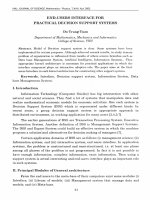
that the charge can be directly transferred. The selected organic molecule is 4-aminothiophenol (4-ATP) (Fig. 1a) because of two reasons.
Firstly, it has been frequently used for nanoparticle coating as efficient
surface stabilizer to prevent particle aggregations and especially as
bio-activator owing to its free amino group (\NH2) which is highly
68
N.T. Trang et al. / Surface Science 608 (2013) 67–73
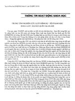
Fig. 1. (a) 4-ATP molecule, the green dash line represents the molecular axis; (b), (c) supercell model for (001) PbS surface, 4-ATP−H part is embedded into vacuum slab at different
geometries: S4-ATP conjugated with surface Sr atom (b) and Pbr atom (c).
bio-compatible [22,23]. Secondly, its thiol group (\SH) was known to
bind strongly with the Pb ion of NCs via chemical bonding [18,24].
2. Modeling details
Because (001) surface is the most dominating surface for “rock-salt”,
it has been chosen as calculated model. A supercell composed of a vacuum slab stacked on a PbS slab along (001) crystal direction was generated as quasi-2D simulation of such surface. We assumed only the
interaction of a most-top layer of PbS surface with 4-ATP and therefore
only 3 atomic layers were included in the PbS slab. The thickness of the
vacuum slab was chosen so that the interactions between different
atomic slabs vanish (about 30 Å).
According to experimental observations, the hydrogen atom in thiol
group (\SH) of 4-ATP molecule is able to be removed, leaving a free
bond on S atom which can form a combination with Pb atoms on PbS
surface. We embedded the 4-ATP molecule without H atom in thiol
group (\SH) into the vacuum slab (Fig. 1). The capping agent — solid
surface distance was changed from 2 Å to 5 Å. The vacuum slab thickness of about 30 Å is good enough for the interaction between neighboring 4-ATP fragment and PbS slab to vanish at the longest distance.
In order to confirm experimental observations that the remaining S
atom of (\SH) group prefers combining with Pb atoms to combining
with S atoms on PbS surface, potential curves of 4-ATP–(001) PbS surface
distance were produced for two topologies corresponding to S atom
from (\SH) group that directly binds with Pb and S atom on PbS surface
(Fig. 1b, c). For convenience, (001) PbS surface atoms which were directly conjugated with 4-ATP are called root atoms and indicated with “r”
index, i.e. Pbr and Sr. The others were called non-root surface atoms,
i.e. PbPbS-surface and SPbS-surface S atom from (\SH) group of 4-ATP was indicated by “4-ATP” index, i.e. S4-ATP, the remaining part of 4-ATP (4-ATP
without H atom) was 4-ATP−H and the one without (\SH) group was
4-ATP−(\SH). It was assumed that the axis of 4-ATP molecular (see
Fig. 1a) was perpendicular to the surface and the molecular was falling
straight forwards to Sr (S–S conjugation) (Fig. 1b) and Pbr (Pb–S conjugation) (Fig. 1c). The distance between S4-ATP and root sites was changed
from 2 Å to 5 Å.
All of our calculations were carried out using LDA functional with
the help of Dmol 3 code which provides atomic-like basis sets in numerical form of the size increasing from MIN to DNP type [25]. Basis functions of this basis set type are generated numerically as values on an
atomic-centered spherical-polar mesh. The angular portion is an appropriate spherical harmonic and the radial portion is obtained by solving
the atomic DFT equations numerically. In our calculations, we utilized
the DNP basis set which provides 2 numerical basis functions for each
occupied orbital in free atom. This basis set also complemented with
polarization functions, i.e. functions with angular momentum one
higher than that of the highest occupied orbital of free atom. In our
case, DNP basis set includes following basis functions for each atom:
H: two 1s, 2s and 2p functions; C, N, S: two 1s, 2s, 2p, 3s, 3p and 3d functions; Pb: two 1s, 2s, 2p, 3s, 3p, 3d, 4s, 4p, 4d, 4f, 5s, 5p, 5d, 6s, 6p and 6d
functions. Core electrons were treated at all electron relativistic level
(relativistic full-potential — Rel-FP).
3. Results and discussions
3.1. Ground state charge transfer at organic–inorganic interface
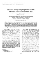
Removing hydrogen atom from thiol (\SH) group left one spin up
hole on 4-ATP−H fragment which was primarily located on S4-ATP atom
as demonstrated by the partial density of stats (DOSs) in Fig. 2a. This
suggested a strong ground state CT when the 4-ATP−H–PbS bond
formed. The ground state CT was examined along the potential curves
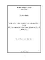
of 4-ATP−H–PbS surface distance. Fig. 3a shows the Pb–S and S–S conjugation potential curves drawn on the base of single point energy calculations. In both cases, the minimum of the potential well was at the
distance of 3.4 Å (see the inset). The Pb–S potential well which was
deeper than the S–S one suggested that Pb atoms were more preferable
than S atoms for the 4-ATP−H fragment to be attached to. This was in
agreement with experiment that thiol group (\SH) strongly binds
with Pb ions on NCs [18,24]. Fig. 3c shows electron deformation Δρ(r),
which is the difference between crystal electron density and the sum of
isolated atomic electron density, of 4-ATP−H–PbS interface at some distances. The electron-donating region between S4-ATP and Pbr, which was
clearly observable when a distance reduced below 3.9 Å, demonstrated
the ground state electron transfer between PbS surface and 4-ATP−H
fragment. According to the shape of this region, the electron transfer
was from Pbr to S4-ATP 2pz orbital.
Quantitative information of such charge transfer process was represented by the distance dependences of concentrated charges (Fig. 3b).
In the distance range from 3 to 4.5 Å, the positive charge of Pb r was increased while the total positive charge of the 4-ATP−H was reduced
with respect to the reduction of distance. This indicated two opposite
electron transfer processes on the intermediate atom S4-ATP: electron
transfer from Pbr to S4-ATP and from S4-ATP to 4-ATP−(\SH). A minimum
of S4-ATP negative charge was observed at the bottom of the potential
N.T. Trang et al. / Surface Science 608 (2013) 67–73
69
Fig. 2a. The bonding nature between non-root S and Pb atoms slightly
shifted towards covalence pole.
3.2. Atomic geometry and surface core level shifts of bared PbS surface
In order to address the reconstruction of geometry and electronic
structure due to the 4-ATP–PbS formation, it was beneficial to examine
the bared PbS surface first. To characterize the surface structural
reconstruction, surface relaxation δz and rumpling Δ12 were defined as
the following [28]:
z ¼ ðzS1 −zPb1 Þ=d0
ð1Þ
Δ12 ¼ 1=2 Ã ðzPb1 −zS2 þ zS1 −zPb2 Þ=d0 :
ð2Þ
Surface core level shifts (SCLSs) Δεs were also considered to evaluate effect of the bond formation on electronic structure of the PbS
surface, [28]:
Δs ¼ s −b :
ð3Þ
Another way to define SCLS was [29]:
Δs ¼ s −c :
Fig. 2. Partial DOS of PbS–4-ATP interface at distance of 3.4 Å (a) and 2.5 Å (b). The
Fermi level was normalized to zero and denoted by the dash vertical lines. Spin
up and spin down DOSs were denoted by positive and negative DOS channels,
respectively.
well, i.e. at Pb r–S4-ATP distance of 3.4 Å. On the right hand side of the
minimum, the Pbr–S4-ATP charge transfer was dominated by the
S4-ATP–4-ATP−(\SH) one then the S4-ATP charge became less negative
when the distance reduced. The domination of Pb r–S4-ATP electron
transfer on the left hand side of the minimum gave rise to the enhancement of the negative charge of S4-ATP when the distance reduced below
3.4 Å. In the distance range above 3 Å, the 4-ATP–PbS surface binding
should be supported by the ionic bond between opposite charge ions
S4-ATP and Pbr. Besides, the presence of 4-ATP gave insignificant effect
on the atomic charge of non-root surface atoms as well as the ionic
nature of bonding between them. When the distance was below ~3 Å
which is equal to total ionic radii of S2− RS2− = 1.84 Å [26] and Pb 2+
RPb2+ = 1.19 Å [27], the positive atomic charge of Pbr was reduced,
which indicated the enhancement of covalence nature. This scenario
was insured by the density of states (DOS) shown in Fig. 2b. For the distances below 3 Å, the spin up 2p hole on S4-ATP was filled by the
overlapping of S 2p orbital and Pb orbitals. Consequently, S 2p DOS
became symmetrical with both spin-up and -down S 2p bands that
are partially filled (Fig. 2b). At the same time, there was an increasing
of density of unoccupied Pb 6s states and low energy occupied 6p states.
So, the covalent bond was believed to originate from the overlapping
between partially filled S 2pz and Pb 6pz orbitals. The change in bond
nature increased the electron density at Pb r site on PbS surface.
Enhanced Coulomb field, which was induced by the increased electron
density, reduced electron density at nearest neighbor non-root surface S
site and thus reduced atomic charge of S on PbS surface as seen in
ð4Þ
Here, z specifies Cartesian coordination of atom on the direction perpendicular to surface; S1, Pb1 are S and Pb atoms in the most-top surface layer, S2, Pb2 are S and Pb atoms in the second-top layer which
was the center layer in our case; d0 is the calculated PbS bond length
of bulk model; εs, εb and εc are the eigenvalues of considered state
from atoms in surface layer, bulk material and center layer respectively.
Table 1, these parameters and SCLSs of the most-top surface layer of
bared PbS surface from our all electron full potential calculation were
compared with results of previous works. The absence of PbS surface reconstruction and SCLS have attracted both ab initio [28–32] and experimental investigations [33–35]. Despite of the divergence of surface
rumpling values and average surface relaxation by different theoretical
methods, all of them led to the same trends that the most-top atomic
layer processes the largest surface relaxation ranging from 4 to 9%.
Concerning the surface rumpling, full potential-linear augmented plan
wave (FP-LAPW) method [28] and core–shell model [36] were in
good agreement to predict that in the most-top atomic layer S atoms
considerably shift outwards in comparison with Pb atoms (S atom at
the top of the surface) but the use of pseudopotentials (PP-LAPW) predicted insignificant positive rumpling [31] or even flipped the rumpling
trend with Pb atom at the top of the surface [29,32]. Experiments were
involved to clarify the surface structural reconstruction trends. Unfortunately, X-ray standing wave (XSW) measurements on PbS at room temperature could not help to exactly estimate surface relaxation due to
phonon broadening effects [33]. Basing on these measurements, surface
relaxation was thought to be less than 1%. The better method to observe
surface structure, the low energy electron diffraction (LEED), was only
carried out on PbTe, an isoelectronic counterpart of PbS [37]. What
can be drawn from such experiment was that surface Pb atoms were
experimentally confirmed to shift inwards in comparison with nonmetallic atoms. Then it was believed that full potential calculations by
I.G. Batyrev et al. [28] and our group should be more reliable than the
one with pseudopotentials. It should be noted that those values from literatures were corresponding to 7-atomic-layer PbS slab model, meanwhile our calculation model was only 3-atomic-layers thick so average
surface relaxation and rumpling predicted by us were smaller than
that ones in Ref. [28].
To gain a deeper insight into the rumpling effect, we recalled the
simplest theory for cohesion in ideal ionic crystal which includes only
inter-ionic Coulomb interaction and the strong short-range core–core
repulsion due to Pauli's principle. According to this, the surface
70
N.T. Trang et al. / Surface Science 608 (2013) 67–73
Fig. 3. (a) Potential curves of 4-ATP–PbS surface distance. The insets zoom in the curves around minimum point at 3.4 Å. (b) Distance dependences of the concentrated charges of
different fragments and atoms at the organic–inorganic interface in case of Pb–S conjugation. (c) Electron deformation at 4-ATP–PbS interface on (100) slide at Pb–S distances of
2.5 Å (left) and 3.4 Å (right). The red color denotes electron-withdrawing area while the blue color denotes electron-donating area.
relaxation has purely corresponded to the reduction of Madelung's constant at the surface due to the reduction of the coordination number.
Then, the surface relaxations at every Pb and S site should be the
same. That means the surface rumpling should be absent for ideal
ionic crystals.
If one used the core–shell model additional effects were involved as
the core–shell model added more short-range interactions, i.e. intraionic core–shell interaction, shell–shell, core–shell interactions between
first and second neighbors [36]. These interactions correspond to electron polarization potential of each ionic shell and overlap potential of
wave functions at different sites, which usually occurs in covalence
bonding. The quantitative agreement between full-potential methods
and core–shell model rumpling suggested that the surface rumpling
may originate from the electronic polarization of surface ions due to
coordination imperfection and covalent bonding. On the other hand,
the softy of electron potential in pseudopotential methods seemed to underestimate the two factors.
Because the calculation reported in [29] failed to reproduce PbS surface rumpling, the obtained S 2p SCLS of 0.3 eV numerically coincided
with experimental value given in Ref. [34]. The S 2p SCLS from our calculation was in the opposite trend with the experiment if it was defined
in the same way as in [28]. It suggested that the 3-atomic layer slab in
use was not thick enough for electron density to converge with that
one of much thicker samples in the experiments. However, in this
study, we only concentrated on the effect of capping agent on the PbS
surface so the contribution of bared surface to structure deformation
and electron redistribution was given for calibration purpose only.
3.3. Structural and electronic structural deformation at 4-ATP–PbS
interface
The reconstructed structure and structure parameters of 4-ATP–PbS
interface were shown in the top panel of Fig. 4 and Table 1. During
optimization process, only 4-ATP−H part and the most-top surface
layer of PbS at the interface were allowed to relax, the center and surface layer on the other side were fixed at relaxed-bared-surface geometry. The energy gained after relaxation from the vertical absorption
geometry to the final geometry is ~0.345 eV. In this final geometry,
the molecular plane of 4-ATP was strongly inclined to make an angle
of 23.14° with PbS surface. The average surface relaxation in the presence of 4-ATP was strongly suppressed to −0.3% (only 13.6% of bared
surface relaxation remained) owing to the capping agent which compensated the surface coordination number imperfection. The flipping
of surface rumpling corresponded with the moving up of Pb atoms to
N.T. Trang et al. / Surface Science 608 (2013) 67–73
71
Table 1
A comparison of surface rumpling δr1, surface relaxation Δ12 of the most-top layer (in %), SCLSs of Pb 5d and S 2p states (in eV) between difference theoretical results
and experimental observations.
δr1
Δ12
Δεs of Pb 5d
Δεs of S 2p
εs − εb
εs − εc
εs − εb
–
–
Theoretical methods
Core–shell model (9-atomic layers) [36]
Madelung potential estimation [35]
2.1
–
−3.5
–
–
0.26
–
–
Ab initio calculations on 11-atomic layers
PP/Gaussian basis set/LDA [29]
−3.0
−4.1
–
–
Ab initio calculations on 7-atomic layers
PP/PW/GGA [31]
PP/PW/GGA [32]
FP/LAPW/GGA [28]
0.03
−1.3
2.9
−5.1
−8.4
−7.1
–
–
–
–
–
–
–
–
−0.41
0.91
−9.47
−2.20
−0.30
0.26
0.16
0.18
0.12
0.16
0.15
–
–
b1%
7
–
–
b1%
−4
–
0.0 ± 0.1
–
–
−0.30 ± 0.02
–
0.0
–
Ab initio calculations on 3-atomic layers
FP/DNP/LDA (our work for bared surface)
FP/DNP/LDA (our work for 4-ATP capped surface in
tilted -capping-fragment geometry)
Experimental measurements
XPS on PbS at low temperature T = 100 K [34]
XPS on PbS at room temperature [35]
XSW on PbS at room temperature [33]
LEED on PbTe [37]
the top of the surface δr1 = −9.47%. This seemed to be the response of
surface atoms against the change in their electronic dipole moments induced by charge transfer from Pb surface atoms to capping fragment. In
Fig. 5, we show the direction of electronic dipole moments of surface
atoms as inferred from the electron deformations of relaxed bared PbS
surfaces (Fig. 5a) and relaxed capped PbS surface (Fig. 5b) and schematized electron density in the form of positive charged nuclei and their
surrounding electron clouds (Fig. 5c). By this, the relaxation of surface
εs − εc
−0.30
−0.05
−0.01
with and without 4-ATP−H fragment could be explained in terms
of the relation between charge transfer and surface electronic dipole
moments.
The SCLS of the 4ATP-capped PbS surface corresponding to the tilted
geometry was also shown in Table 1. According to this, SCLSs of both
sulfur and lead was suppressed (reduced in magnitude). In order to interpret the change in SCLSs, it is worthwhile to remind that in the previous section we concluded that Pb–S conjugation is more preferable
Fig. 4. Optimized structure of 4-ATP–PbS interface (upper panel) together with corresponding electron deformation (lower panel) in (110) (a) and (−110) (b) slides.
72
N.T. Trang et al. / Surface Science 608 (2013) 67–73
Fig. 5. Schemes of electronic dipole moments of surface ions: (a) the electronic dipole moments of surface ions without capping agent and (b) with capping agent basing on analyzing electron deformation density; (c) the electronic dipole moments of surface ions are represented in terms of positive charged nuclei (circles with “+” inside) and their surrounding electron clouds (ellipses with “−” inside). 4-ATP−H fragment which is denoted by circles with “4-ATP−H” inside was put onto the surface (“relaxed, capped” panel) and
after relaxation, found its stable position (“relaxed, capped and relaxed again” panel).
than S–S one and Pbr–S 4-ATP bond should be ionic at distance above 3 Å
and polarized covalent when distance reduced below 3 Å. So at final
atomic geometry in which SPbS–S4-ATP distance was 3.4 Å and PbPbS–
S4-ATP distance was 2.976 Å, Pb–S 4-ATP ionic bonding with a weak covalence seemed to be more preferable than S–S4-ATP covalent bonding.
The direct binding of capping fragment to surface Pb atoms was
shown above to increase electron density on those atoms but reduce
electron density on surface S atoms. The increasing of electron density,
in turn, increased band–band Coulomb repulsion between Pb 5d core
levels and higher levels, increasing binding energy of Pb core state. As
a result, the positive shift of surface Pb 5d band was reduced. Whereas,
the electron density reduction on surface S atoms reduced the repulsion
on S core levels from the higher bands which, in turn, suppressed the
negative sulfur SCLS. Such change in SCLSs could also be interpreted
as the slight covalence shift of bonding between non-root S and Pb
atoms.
4. Conclusion
4-ATP capped PbS (001) surface was investigated by means of electronic structure methods in the frame work of density functional theory.
The capping compensated the surface imperfection of coordination
number and suppressed the average surface relaxation. However, the
charge transfer from PbS surface to 4-ATP−H fragment induced a
change in surface electronic dipole moments which in turn flipped surface rumpling of PbS. The direct bonding of capping fragment to surface
Pb atoms slightly shifted surface Pb–S bonding nature to covalence. This
shift can be interpreted as the reduction of SCLSs of both Pb and S.
Acknowledgments
This work is financially supported by Vietnam National University,
Hanoi (TRIG A project, no. QGTD 10.24). One of the authors, Nguyen
Thuy Trang, would like to thank TRIG A project of Hanoi University
of Science, Vietnam National University, Hanoi for supporting.
Appendix A. Supplementary data
Supplementary data to this article can be found online at http://
dx.doi.org/10.1016/j.susc.2012.09.014.
References
[1] W.U. Huynh, J.J. Dittmer, A.P. Alivisatos, Science 295 (2002) 2425.
[2] S. Coe, W.K. Woo, M. Bawendi, V. Bulovic, Nature 420 (2002) 800.
[3] G. Konstantatos, I. Howard, A. Fischer, S. Hoogland, J. Clifford, E. Klem, L. Levina,
E.H. Sargent, Nature 442 (2006) 180.
[4] J.A. Nozik, Phys. E. 14 (2002) 115.
[5] M. Bruchez, M. Moronne, P. Gin, S. Weiss, A.P. Alivisatos, Science 281 (1998) 2013.
[6] In: O. Madelung, U. Rossler, M. Schulz (Eds.), Semiconductors: Group IV Elements,
IV–IV and III–IV Compounds, Landolt–Bornstein, New Series, Group III, 41,
Springer-Verlag, Berlin, 2005, Pt. A.
[7] K.S. Babu, C. Vijayan, R. Devanathan, Mater. Lett. 58 (2004) 1223.
[8] M.A. Hines, G.D. Scholes, Adv. Mater. 15 (2003) 1844;
G. Konstabtatos, J. Clifford, L. Levina, E.H. Sargent, Nat. Photonics 1 (2007) 531.
[9] K.N. Bourdakos, D.M.N.M. Dissanayake, T. Lutz, S.R.P. Silva, R.J. Curry, Appl. Phys.
Lett. 92 (2008) 153311.
[10] S.A. McDonal, P.W. Cyr, L. Levina, E.H. Sargent, Appl. Phys. Lett. 84 (2004) 2089.
[11] A. Maria, P.W. Cyr, E.J.D. Klern, L. Levina, E.H. Sargent, Appl. Phys. Lett. 87 (2005)
213112.
[12] A.H. Cerdan, L.M. de la Rosa, C.A. Gonzalez, J. Genesca, J. Mater. Process. Technol.
143–144 (2003) 23.
[13] S. Guo, S. Dong, Trends Anal. Chem. 28 (2009) 96.
[14] S. Wang, X. Zhang, X. Mao, Q. Zeng, H. Xu, Y. Lin, W. Chen, G. Liu, Nanotechnology
19 (2008) 435501.
[15] N. Zhu, A. Zhang, Q. Wang, P. He, Y. Fang, Electroanalysis 16 (2004) 577.
[16] W. Sun, J. Zhong, P. Qin, K. Jiao, Anal. Biochem. 377 (2008) 115.
[17] C.R. Kagan, C.B. Murray, B.G. Bawendi, Phys. Rev. B: Condens. Matter 54 (1996) 8633.
[18] S.W. Clarl, J.M. Harbold, F.W. Wise, J. Phys. Chem. C 111 (2007) 7302.
[19] N.N. Muhammad, N.B. Konstatinos, J.C. Richard, Phys. Chem. Chem. Phys. 12 (2010)
7371.
[20] B.R. Hyun, A.C. Bartnik, J.K. Lee, H. Imoto, L. Sun, J.J. Choi, Y. Chujo, T. Hanrath, C.K.
Ober, F.W. Wise, Nano Lett. 10 (2010) 318.
[21] A. Gocalinska, M. Saba, F. Quochi, M. Marceddu, K. Szendreim, J. Caom, M.A. Loi,
M. Yarema, R. Seyrkammer, W. Heiss, A. Mura, G. Bongiovanni, J. Phys. Chem.
Lett. 1 (2010) 1149.
[22] N. Xiao, C. Yu, Anal. Chem. 82 (2010) 3659.
[23] M. Osawa, N. Matsuda, K. Yoshii, I. Uchida, J. Phys. Chem. 98 (1994) 12702.
[24] V.S. Gurin, K.N. Kasparov, E.A. Tyavlovskaya, Colloids Surf. A. 139 (1998) 1.
[25] B. Delley, J. Chem. Phys. 92 (1990) 508;
B. Delley, J. Chem. Phys. 113 (2000) 7756.
N.T. Trang et al. / Surface Science 608 (2013) 67–73
[26] L. Pauling, The Nature of the Chemical Bond, Cornell University Press, Ithaca, 1961.
[27] “Revised Effective Ionic Radii and Systematic Studies of Interatomic Distances in
Halides and Chalcogenides” by R. D. Shannon, Central Research and Development
Department, Experimental Station, E. I. Du Pont de Nemours and Company,
Wilmington, Delaware 19898, U.S.A.
[28] I.G. Batyrev, L. Kleinman, J. Leiro, Phys. Rev. B. 70 (2004) 073310.
[29] J. Muscat, J.D. Gale, Geochim. Cosmochim. Acta. 67 (2003) 799.
[30] G. Allan, Phys. Rev. B. 43 (1991) 9594.
[31]
[32]
[33]
[34]
[35]
[36]
[37]
73
A. Satta, S. de Gironcoli, Phys. Rev. B. 63 (2000) 033302.
A.B. Preobrajenski, T. Chasse, Appl. Surf. Sci. 166 (2000) 201.
T. Kendelewicz, P. Liu, G.E. Brown Jr., E.J. Nelson, Surf. Sci. 395 (1998) 229.
J.A. Leiro, K. Laajalehto, I. Kartio, M.H. Heinonen, Surf. Sci. Lett. 412/413 (1998) L918.
G. Paolucci, K.C. Prince, Phys. Rev. B. 41 (1990) 3851.
F.W. de Wette, W. Kress, U. Schroeder, Phys. Rev. B. 32 (1985) 4143.
A.A. Lazarides, C.B. Duke, A. Paton, A. Kahn, Phys. Rev. B. 52 (14) (1995) 895.