DSpace at VNU: Condylospora vietnamensis, a new Ingoldian hyphomycete isolated from fallen leaves in Vietnam
Bạn đang xem bản rút gọn của tài liệu. Xem và tải ngay bản đầy đủ của tài liệu tại đây (361.3 KB, 4 trang )
Mycoscience (2012) 53:326–329
DOI 10.1007/s10267-011-0166-8
SHORT COMMUNICATION
Condylospora vietnamensis, a new Ingoldian hyphomycete isolated
from fallen leaves in Vietnam
Le Thi Hoang Yen • Shigeki Inaba •
Yasuhisa Tsurumi • Sayaka Ban • Nguyen Lan Dung
Duong Van Hop • Katsuhiko Ando
•
Received: 6 December 2010 / Accepted: 30 November 2011 / Published online: 21 January 2012
Ó The Mycological Society of Japan and Springer 2012
Abstract A new Ingoldian hyphomycete was isolated
from fallen leaves in Bach Ma National Park, Vietnam, and
is described here as Condylospora vietnamensis. This
fungus is different from four known Condylospora species
in morphological characteristics, having U- or N-shaped
conidia.
Keywords
Anamorphic fungi Á Taxonomy
The genus Condylospora was established by Nawawi
(1976) based on C. spumigena Nawawi, isolated from a
foam sample collected in Malaysia. The hyaline and multiseptate conidia are unique in morphology; they are
wormlike and have a characteristic elbow-shaped bend
near the middle. Similar spores had been earlier reported as
unidentified fungi from Papua New Guinea (Tubaki 1965),
India (Ingold and Webster 1973), and Japan (Matsushima
1975). Condylospora spumigena has also been found in
India (Chandrashekar et al. 1990), Puerto Rico (SantosFlores et al. 1996), Thailand (Phongpaichit et al. 2002),
Poland (Czeczuga et al. 2003), South America (Schoenlein-Crusius and Piccolo 2003), and Venezuela (Cressa and
Smits 2007).
In 1985, two other Condylospora species were found
in the stream spora in Malaysia (Nawawi 1985). Later,
Nawawi and Kuthubutheen (1988) described these fungi on
L. T. H. Yen (&) Á N. L. Dung Á D. Van Hop
Institute of Microbiology and Biotechnology, Vietnam National
University, E2, 144 Xuan Thuy, Cau Giay, Hanoi, Vietnam
e-mail:
S. Inaba Á Y. Tsurumi Á S. Ban Á K. Ando
National Institute of Technology and Evaluation, 2-5-8,
Kazusakamatari, Kisarazu, Chiba 292-0818, Japan
123
submerged decaying twigs in Malaysia as the second and the
third species: C. gigantea Nawawi & Kuthub. and C. flexuosa Nawawi & Kuthub. Condylospora gigantea was recorded also from Puerto Rico (Santos-Flores et al. 1996) and
Poland (Czeczuga et al. 2003); and C. flexuosa in Puerto Rico
(Santos-Flores et al. 1996) and Venezuela (Smits et al. 2007).
A candidate for the fourth species producing mostly
N-shaped conidia was also found in Malaysia (Nawawi
1985; Nawawi and Kuthubutheen 1988), but it has not yet
been described formally; that is, it has not been previously
reported as a valid description.
During an investigation of microfungi in Vietnam, an
undescribed Condylospora-like fungus producing short and
small N-shaped conidia was found from the fallen leaves.
The purpose of this study is to describe this fungus as a
new species of Condylospora.
Fallen leaves were collected at Bach Ma National Park
in the central part of Vietnam in April 2005 by K. Ando.
The sample was kept in a moist chamber for 2–3 days in a
laboratory, then immersed into water in a 500-ml beaker
and stirred gently. A small amount of surface water was
collected using a glass slide (Bandoni and Koske 1974) and
spread on a low nutrient carbon agar medium (LCA; Miura
and Kudo 1970). After confirming the spores on the medium under a light microscope, single-spore cultures were
isolated by a Skerman’s micromanipulator to obtain the
pure culture.
These isolates were cultured at 25°C on LCA, cornmeal
agar (CMA; Nissui, Tokyo, Japan), and 2% malt agar (MA;
Becton–Dickinson, Sparks, MD, USA) for morphological
observation. Observation was made under a differential
interference contrast microscope (DIC; Axioplan 2, Zeiss,
Jena, Germany) and a scanning electron microscope (SEM;
JSM-6060, JEOL, Tokyo, Japan). For SEM, a small piece
(2 9 2 mm) of the colony was cut and fixed with 1% OsO4
Mycoscience (2012) 53:326–329
aqueous solution (aq. sol.) at room temperature for 2 h, then
dehydrated in an ethanol series and finally substituted
with isoamyl acetate. After critical point drying (HCP-2;
Hitachi, Tokyo, Japan) and coating with platinum–palladium
327
(JUC-5000; JEOL, Tokyo, Japan) the specimens were
observed under SEM at 15 kV.
Condylospora vietnamensis L.T.H. Yen & K. Ando, sp.
nov.
Figs. 1, 2, 3
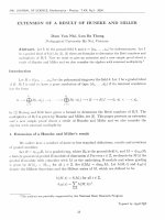
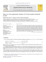
Fig. 1 Condylospora vietnamensis VTCCF-1208 on low nutrient carbon agar (LCA). a–c Conidial development: arrow in a shows the first
curving point of the conidial initial. d, e U-shaped mature conidia. f N-shaped mature conidium. Bars 5 lm
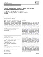
Fig. 2 Condylospora vietnamensis VTCCF-1208 on LCA. a U-shaped mature conidium. b Sympodially proliferating conidiogenous cell
(arrow 1) and a new conidium initial (arrow 2). Bars a 5 lm; b 2 lm
123
328
Mycoscience (2012) 53:326–329
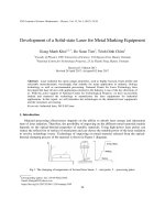
Fig. 3 Condylospora
vietnamensis VTCCF-1208 on
LCA. a Conidial development
on short conidiogenous cells
integrated in the hyphae.
b Sympodial development in a
conidiogenous cell (left) and
the detachment rachi (right).
c U-shaped (left) and N-shaped
(right) conidia. Bar 5 lm
Table 1 Comparison of conidium morphology in the Condylospora species
Species
Conidia
Number of
incurved bends
References
Shape
Number of septa
Length (lm)a
C. spumigena
1
L-shaped
10–15
72–102
Nawawi (1976)
C. flexuosa
3
S-shaped with straight tip
12–16
87–106
Nawawi and Kuthubutheen (1988)
C. gigantea
C. vietnamensis
1
2
L-shaped
U-shaped or N-shaped
25–36
(3–) 8–9 (–12)
131–200
38–99
Nawawi and Kuthubutheen (1988)
This study
Condylospora sp.
2
N-shaped or U-shapedb
27–42
150–180
Nawawi and Kuthubutheen (1988)
a
Total length of the proximal, middle, and distal portions in conidia
b
N-shaped and U-shaped conidia are shown in fig. 5 of Nawawi and Kuthubutheen (1988)
MycoBank no.: MB 519536
Colonies on MA and LCA growing slowly, somewhat white
to light cream, attaining 22–24 mm in diameter in 4 weeks at
25°C. Mycelium consisting of hyphae branched, septate,
thin-walled, hyaline, and 1–1.5 lm wide. Conidiogenous
cells integrated, intercalary in hyphae, undifferentiated,
short, simple, cylindrical (Figs. 1c, 3a), thin-walled; sometimes elongate, flexuous, sympodially proliferating
(Figs. 2b, 3b), 3–20 9 1.5–2 lm, provided with up to five
cylindrical denticles, 3–20 9 1.5–2 lm. Conidia holoblastic, hyaline, thin-walled, (3–)8–9(–12)-septate, incurved two
times, typically U-shaped (Figs. 1c–e, 2a, 3c), sometimes
123
N-shaped (Figs. 1f, 3c), consisting of the proximal, middle,
and distal portions that lie usually in a single plane; proximal
portion straight, 15–35 9 1–1.5 lm; middle portion forming
an angle of 60°–90° with the proximal part, 14–32 9
1–1.5 lm; distal portion lying parallel with the proximal
part, 9–32.5 9 1–1.5 lm.
Teleomorph: Unknown.
Type: VTCCF H-1008 (holotype: dried culture specimen,
from VTCCF-1208, on LCA) deposited in the Vietnam
Type Culture Collection, Hanoi (VTCC). NBRC H-12773
(isotype: dried culture specimen, from VTCCF-1208, on
Mycoscience (2012) 53:326–329
LCA) deposited in the NITE Biological Resource Center
(NBRC).
Ex-type culture: VTCCF-1208 (=NBRC 107639), isolated from fallen leaves of unidentified deciduous broadleaved tree, Bach Ma National Park, Hue Prov., Vietnam,
27 April 2005, collected by K. Ando.
According to the previous reports, the colonies in all the
species of Condylospora grow rather slowly on MA and
CMA, and they are dewy, white, or hyaline in color. No
sporulation was observed in most of the species even when
strips of agar culture were submerged in water and aerated,
although these Ingoldia fungi were reported from submerged substrates (Nawawi and Kuthubutheen 1988). The
new species, C. vietnamensis, readily produced conidia on
LCA and MA without being submerged in water.
Condylospora vietnamensis agrees well with the type
species in ontogeny, but it differs from other species of the
genus in the shape and size of the conidia (Table 1). Condylospora spumigena and C. gigantea have L-shaped conidia
with one incurved bend about the middle (Nawawi 1976;
Nawawi and Kuthubutheen 1988), and C. flexuosa has
S-shaped conidia with three bends around the middle
(Nawawi and Kuthubutheen 1988) that lie in one or more
plane. Condylospora vietnamensis has U-shaped or N-shaped
conidia with two bends. The undescribed Condylospora species from Malaysia has similar N-shaped or U-shaped conidia
(Nawawi 1985; Nawawi and Kuthubutheen 1988; cf. footnote
in Table 1), but these are larger than those in C. vietnamensis.
Acknowledgments This work was conducted under the Joint
Research Project on ‘‘Taxonomic and Ecological Studies of Microorganisms in Vietnam and the Utilization’’ between the Department
of Biotechnology, National Institute of Technology and Evaluation,
Japan, and Institute of Microbiology and Biotechnology, Vietnam
National University, and the project ‘‘Reservation of Microorganism
Genome’’ funded by the Ministry of Science and Technology, Vietnam. We sincerely thank Dr. Kaoru Yamaguchi, NBRC, for her
329
instruction in SEM observation, and also two anonymous reviewers
for their critical comments and helpful suggestions.
References
Bandoni RJ, Koske RE (1974) Monolayers and microbial dispersal.
Science 183:1079–1081
Chandrashekar KR, Sridhar KR, Kaveriappa KM (1990) Periodicity
of water-borne hyphomycetes in two streams of Western Ghat
forests (India). Acta Hydrochim Hydrobiol 18:187–204
Cressa C, Smits G (2007) Aquatic hyphomycetes in two blackwater
streams of Venezuela. Ecotropicos 20:82–85
Czeczuga B, Kiziewicz B, Mazalska B (2003) Further studies on
aquatic fungi in the River Biebrza within Biebrza National Park.
Pol J Environ Stud 12:531–543
Ingold CT, Webster J (1973) Some aquatic hyphomycetes from India.
Kavaka 1:5–9
Matsushima T (1975) Icones microfungorum a Matsushima lectorum.
Published by the author, Kobe
Miura K, Kudo MY (1970) An agar-medium for aquatic hyphomycetes. Trans Mycol Soc Jpn 11:116–118 (in Japanese)
Nawawi A (1976) Condylospora gen. nov., a hyphomycete from a
foam sample. Trans Br Mycol Soc 66:363–365
Nawawi A (1985) Aquatic hyphomycetes and other water-borne fungi
from Malaysia. Malay Nat J 39:75–134
Nawawi A, Kuthubutheen AJ (1988) Additions to Condylospora
(Hyphomycetes) from Malaysia. Mycotaxon 33:329–338
Phongpaichit S, Sakayaroj J, Hywel-Jones N, Jones G (2002)
Biodiversity of freshwater hyphomycetes at Ton-Nga-Chang
Wildlife Sanctuary, Southern Thailand. Res Rep Natl Inst
Environ Stud 171:165–170
Santos-Flores CJ, Nieves-Rivera AM, Betancourt-Lo´pez C (1996)
The genus Condylospora Nawawi (Hyphomycetes) in Puerto
Rico. Caribb J Sci 32:116–120
Schoenlein-Crusius IH, Piccolo RA (2003) The diversity of aquatic
hyphomycetes in South America. Braz J Microbiol 34:183–193
Smits G, Ferna´ndez R, Cressa C (2007) Preliminary study of aquatic
hyphomycetes from Venezuelan streams. Acta Bot Venez
30:345–355
Tubaki K (1965) Short notes on aquatic spora in East New Guinea.
Trans Mycol Soc Jpn 6:11–14
123