DSpace at VNU: An undescribed species of steinernema (Rhabditida: Steinernematidae) from chumomray national park (Vietnam)
Bạn đang xem bản rút gọn của tài liệu. Xem và tải ngay bản đầy đủ của tài liệu tại đây (3.57 MB, 8 trang )
VNU. JOURNAL OF SCIENCE, Nat.. Sci., & Tech., T.xx, N03. 2004
AN U N D E S C R IB E D SP E C IE S OF S T E IN E R N E M A (RHABDITIDA:
STEINERNEM ATIDAE) FROM CHUMOMRAY NATIONAL PARK
(VIETNAM)
P h a n Ke L o n g
In stitute o f Ecology and Biological Resources,
Vietnamese Academy o f Science and Technology
Abstract.
An
undescribed
species
of
Steinernema
(Rhabditida:
S t e i n e r n e m a t i d a e ) w a s i s o la te d f ro m f o r e s t soil o f t h e C h u m o m r a y N a t i o n a l p a r k
( K o n t u m p ro v .. S a T h a y d is t r ., S a S o n m u n i c i p a l i t y ) V i e t n a m . M o r p h o lo g i c a l a n d
morphometrical studies revealed that this species clearly differs from other
known Steinernema species. It has very large spicule as well as in s.
i n t e r m e d i u m b u t c a n b e s e p a r a t e d b y t h e l o n g e r I J t a i l l e n g t h , lo w e r r a t i o E%,
s h o r t e r s p ic u le , s h a p e of s p ic u le , t h e n u m b e r of g e n i t a l p a p i l l a e a t c a u d a l r e g io n
and the presence of mucro in male. Its lateral fields resemble the ones of s. sangi
b u t c a n b e s e p a r a t e d b y h i g h e r E% a n d D%, l a r g e r a n d s h o r t e r s p ic u le , t h e
m o r p h o l o g y o f s p ic u l e h e a d ( m a n u b r i u m ) a n d d o r s a l lobe o f sp ic u le .
Morphometries of IJs of this species are closed to s. monticolum but differ by the
p o s itio n o f e x c r e t o r y p o re , s h o r t e r a n d l a r g e r s p ic u le a n d t h e m o r p h o lo g y of
s p ic u le h e a d .
1. I n t r o d u c t i o n
Entomopathogenic nematodes (EPN) of the genus Steinernem a Travassos, 1927 and
Heterorhcibditis Poinar, 1976 have great potential for biological control of insect pests.
Currently, 33 species of the genus Steinernem a and 11 species of the genus Heterorhabditis
are described. Four species of the genus Steinenem a, s. tam i (Pham et a l., 2000), s. sangi
(Phan et al.y 2001a), s. loci and s. thanhi (Phan et al., 2001b), and one species of the genus
Heterorhabditis, H. baujardi (Phan et al.y 2003a) have been described from Vietnam.
Obviously, Vietnam has a high species diversity of entomopathogenic nematodes that may
provide good potential for biological control of insects. During a nematological survey
carried out in the Chum omray National P ark (Phan et al.j 2003b) an unknown
steinernem atid was detected. This isolate is distinguished from other Steinernem a species
by its morphology and morphometric characters.
2. Materials and methods
2.1. N em atodes
The entomopathogenic nematodes were isolated from soil samples taken in the forest
of Chumomray national park (Kontum prov., Sathay distr., Sason municipality) by the
Galleria mellonella L. baiting method and infective juveniles (IJs) were collected from
Galleria cadavers using White trap (Phan et al.y 2001a) and stored at 15° c in aerated
43
44
Phan K c Long
water. Co-ordinates and altitudes of the sampling sites were registered using GARMIN
GPS 12 ex.
2.2. Morphological observations
Nematodes were reared on G. mellonella. We used IJs collected during a week after
their first emergence from the insect cadavers; adults of the first generation were dissected
from the cadavers. Nematodes were killed and fixed in hot 4% formalin (50-60° C), and kept
in this solution for 48 h (Phan et aL, 2001a). Fixed nematodes were tran sferred to
anhydrous glycerine and mounted on slides. All m easurem ents were made using a drawing
tube attached to an Olympus BX50 light microscope (LM).
3. Description
3.1. M ale
Body curved ventrally, C-shaped when heat-killed (Figure 1A). Cuticle looks smooth
under LM. Head rounded, slightly offset from the body. Head with six pointed labial
papillae and four cephalic papillae. Amphids inconspicuous. Mouth opening funnel-shaped
or cup-shaped. Stoma shallow. Oesophagus muscular; procorpus cylindrical; metacorpus
slightly swollen non-valvate; isthm us distinct; basal bulb pyriform, valve distinct. Nerve
ring just above basal bulb. Cardia prominent and protruding into intestine lumen.
Excretory pore at middle of oesophagus. Excretory duct cuticularised; excretory gland
swollen and elongated. Monorchic gonad reflexed. Spicule paired, yellow-brownish in
colour, well curved and large (Figure 1G). Ratio SL/SPW about 4.5 (3.8-5.6). Spicule head
(manubrium) as long as wide. Blade arcuate with spicule tip straight. Three lobes on blade
well defined. Anterior, dorsal lobe enlarged dorsally and well curved, term inate posterior to
spicule tip. Lateral lobe prominent, usually enlarged anteriorly in width and term in ate at
spicule tip. Ventral lobe enlarged anteriorly at the ventral side, to form a prom inent
rostrum and term inate at spicule tip. Velum large, not covering spicule tip. Spicule tip
blunt. Gubernaculum about 70% of spicule length. In lateral view, gubernaculum boat
shaped, swollen at middle and proximal end with knob ventrally curved (Figure 1G). In
ventral view, cuneus long, bifurcate, not reaching to the end of corpus. Corpus separated
posteriorly. A single ventral precloacal papilla and eleven pairs of genital papillae present
and arranged as follows: five pairs subventrally preanal, one pair lateral a t the same level
of the single ventral precloacal papillae. One pair subventral ad-anal. Three pairs caudal,
subventral and one pair caudal, subdorsal. Tail conoid with mucron. Phasmids
inconspicuous.
3.2. Fem ale
Body robust, C-shaped when heat-killed. Cuticle looks smooth un der LM. Head
broadly rounded. Head with six pointed labial papillae and four cephalic papillae. Amphids
inconspicuous. Mouth opening funnel-shaped or cup-shaped. Stoma shallow. Oesophagus
with cylindrical procorpus; metacorpus slightly swollen and non-valvate; isthm us
indistinct; basal bulb pyriform, valve observed. Excretory pore at middle of oesophagus
(Figure 1C). Excretory duct cuticularised and excretory gland swollen. Cardia prominent
protruding into intestine lumen. Didelphic, amphidelphic gonad reflexed and tightly filled
V N U . Journal o f Science. N at., Sci.. & Tech.. T.xx, N t)3. 2004
45
A n undescribed species of.
with eggs. Vulva a transverse slit, protrunding from the body, without epiptygma (Figure
IF) and a t middle of body. Vagina short, oblique with m uscular walls. Post-anally slightly
swollen (Figure 1H). Tail dome shaped, shorter th a n anal body width with terminal peg.
A
D
YỈ f
ii i
100 ụm
D, E, I
40 ụm
A
20 ụm
c, F,
20 ụm
G
100 ụm
B
H
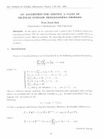
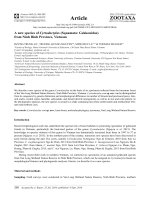
Figure 1. Drawing of the undescribed species of Steinernema from Chumomray National Park
(Vietnam). A & G: Male first generation. A. Entire view; G. Spicule & Gubernaculum. B, D. E & I:
Infective juveniles. B. Entire view; D & E. Bacterial vesicle; I. Tail in lateral view, c, F & H: Female
first generation, c. Oesophagus region; F. Vulva region; H. Tail in lateral view.
V N U . Journal o f Science. N at., Sci.. & Tecli.. 7 .XX. N lt3, 2004
46
Phan Ke Long
3.3. In fective ju v e n ile
When heat killed, body moderately C-shaped (Figure IB); often enclosed in cuticle of
second-stage; tapering regularly from base of oesophagus to anterior end and from anus to
terminus. Mouth and anus closed. In the head, labial papillae not observed; pore-like
amphids situated below labial disc just above cephalic papillae. Oesophagus long and
narrow, isthm us distinct and surrounded by nerve ring, basal bulb elongated with valve.
Cardia prominent. Excretory pore at middle of oesophagus. Hemizonid distinct and located
anteriorly to basal bulb. Bacterial vesicle elliptical or elongate (26-28 àm long and 7-10 àm
wide) (Figure ID, E). Lateral field with eight ridges (at mid-body), subm arginal and central
pair less distinct, sometimes the submarginal not observed. Tail long and constricted at
hyaline portion, especially on the dorsal side. Hyaline portion well pronounced about 54% of
tail length. Phasmids distinct and located in anterior half of tail (Figure II).
3.4. Differential diagnosis
The undescribed species is characterised by the body length about 712 (642-778) àm,
the distance from anterior end to excretory pore about 56 (50-68) am, the tail length about
75 (68-92) àm, the E% about 75 (67-87)%, and the lateral field at mid-body with eight ridges
(submarginal and central pair less distinct) of the IJs, as well as by the large spicules of the
males (SL/SPW about 4.5) (Table 1).
The morphometries of IJs of the undescribed species are close to those of s.
m onticolum (Stock et al., 1997) except for the position of the excretory pore (at 1/2 us at
anterior 1/3 of oesophagus). Moreover, the new species can be distinguished from s.
monticolum by male characters such as a shorter spicule length [58 (51-65) us 70 (61-80)
larger spicule [SL/SPW = 4.5 (3.8-5.6) vs 8.75 (8.0-8.71)] and the spicule head
(manubrium) elongated vs round (Table 1).
As Steinernem a interm edium (Poinar, 1985), this undescribed species has very large
spicules but can be separated from this species by the longer IJ tail length [75 (68-92) us 66
(53-74) am], the lower ratio E% [75 (67-87) us 96 (89-108)%]; shorter spicules [58 (51-65) vs
91 (84-100) am], the shape of the spicules (arcuate us well curved anteriorly, posterior
almost straight), the num ber of genital papillae at the caudal region (4 pairs vs 6 pairs),
and the presence of a mucron on the male tail (Table 1).
The undescribed species has a lateral field resembling to the one of s. sangi, also
found in Vietnam, but can be separated from this latter species by a higher E% [75 (67-87)
us 62 (56-70)], higher D% [46 (43-59) vs 40 (36-44)], larger spicule [ratio SL/SPW = 4.5 (3.8Õ.6) vs 5.25 (5.71-5.8)], shorter spicule length [58 (51-65) us 63 (58-80) am], the spicule head
(manubrium) (elongated and about 1/4 spicule length vs short, blunt and about 1/5 spicule
length), and the dorsal lobe of the spicule (not term inated at spicule tip vs term inated at
spicule tip) (Table 1).
V N U . Journal o f Science, N at., Sci., & Tech.,
T.xx.
N (i3. 2004
47
An undcscrihcd spccies of .
Table 1 Morphometric characters (in àm) of the undescribed species.
Measurement in form: mean ± SD (range)
Character*
1st generation male 1st generation female Infective juvenile
n
20
20
25
Body length (L)
1433 ± 106
(1320-1665)
127 ± 15
(105-150)
4± 1
(3-5)
6± 1
(5-8)
96 ± 5
(89-104)
120 ± 5
(110-129)
173 ± 7
(162-186)
264 ± 58
(165-360)
29 ± 5
(23-33)
-
3 2 0 6 ± 249
(2745-3765)
193 ± 18
(165-240)
6± 1
(5-8)
10 ± 1
(8-12)
108 ± 8
(90-117)
148 ± 9
(132-165)
226 ± 6
(216-239)
-
712 ± 4 3
(642-778)
28 ± 3
(26-35)
-
54 ± 5
(47-63)
73 ± 9
(59-87)
-
75 ± 5
(68-92)
54 ± 3
(49-62)
16 ± 1
(14-48)
-
-
-
-
-
-
-
-
-
55 ± 2
(50-58)
17 ± 1
(15-19)
14 ± 1
(13-16)
-
Body width (W)
Stoma length
Stoma width
EP
NR
ES
Testis flexure
Tail length
H%
Anal body width (ABW)
46 ± 4
(39-56)
Spicule length (SP)
58 ± 3
(51-65)
Spicule width (SPW)
13 ± 1
(11-15)
Gubernaculum length (GU) 41 ± 3
(36-44)
G ubernaculum width
6±1
(5-8)
SP/SPW
4.6 ±0.4
(4.2-5 6)
Vulva (%)
a (L/W)
b (L/ES)
1 -.................
11 ± 1
(9-13)
8±1
(7-9)
56 ± 4
(50-68)
84 ± 4
(80-100)
120 i: 7
(115-152)
-
25 ± 3
(18-29)
6± 1
(3.6-6.3)
EP = distance from anterior end to excretory po re ; N R = distance from anterior end to nerve ring;
ES = oesophagus length; H % = hyaline part/tail l e n g t h X 100.
V N U . Journal o f Science, N at.. Sci., & Tech.,
T.xx, N„3. 2004
Phan K c L o n e
4S
c (LIT)
D%=EP/ES Ì 100
E%=EP/Ti 100
SW=SP/ABW
GS=GU/SP
Mucron
50 ± 8
(40-67)
56 ± 5
(50-63)
-
60 ± 7
(49-70)
48 ± 3
(39-53)
-
1.29 ± 0.12
(1.11-1 50)
0.7 ±0.05
(0.64-0.79)
2.4 ±0.56
(1.5-3.0)
-
10 ± 1
(6-11)
46 ± 3
(43-59)
75 ± 5
(67-87)
-
-
-
-
-
4. D is c u ss io n
Precise identification of any organism is of outmost importance. Identification of
Steinernema and H eterorhabditis species by stan dard methods using morphology and
morphometries is rarely straightforward (Hominick et al.y 1997) because th a t kind of
investigation requires the examination of num erous characters, some being difficult to
observe. Moreover, morphometries of IJ vary within species and between populations
(Miduturi et al.y 1996). Some morphological characters are useful for distinguishing species
or groups of species of Steinernem a, e.g. lateral fields (Hominick et a l., 1997), amoeboid cells
(Spiridonov et al.y 1999), and morphology of spicula and gubernaculums (Nguyen & Smart,
1997). As a conclusion of their study of the morphometrical characters of several
populations of H cterorhabditis, Phan et al. (2003b) suggested th a t the morphometrical
characters, and the ratio e, ratio f and body diam eter of IJ as well as spicule length,
gubernaculum length and ratio s w of male along with the morphology of gubernaculums
should be considered when identifying and describing Heterorhabditis spp.
Hominick et al. (1996) argued th a t molecular techniques could be an addition to
traditional identification methods. Distinctions based on molecular characterisation may
elucidate species and groupings, which then can be studied for morphological characters
th at distinguish them from each other. Several modern techniques have been used for
identification of entomopathogenic nematodes. They include isozyme p attern s (Akhurst,
1987), total protein p atterns (Poinar & Kozodoi, 1988) or immunological techniques
(Jackson, I960). Initial research in molecular taxonomy and diagnostics of
entomopathogenic nematodes utilised cloned DNA probes and restriction fragment length
polymorphisms (RFLPs) as discriminatory methods (Roland & John, 1998). The internal
transcribed spacer region (ITS) is an ideal region for molecular taxonomic purposes. The
ribosomal genes flanking this region are highly conserved allowing the construction of
primers th a t enable PCR amplification of the highly variable ITS region between them
(Reid et a l 1997). Sequence variation in this region yields many RFLP, which can be used
for taxonomy. By comparison of the bands generated after restriction digests it was possible
to construct a provisional tree showing the relatedness of the Steinernem a species studied
(Reid et a l 1997). DNA sequences of ITS regions yield more detailed information about
variation within and among nematodes species th a n PCR-RFLP approaches. These spacer
sequences have been used successfully to diagnose species and populations of nematodes
V N U . Journal o f Science, N at.. Sci.. & Tech., I.X X , N itỉ , 2004
A n undescribcd species of.
49
(Phan et al.j 2003a). Analyses of ITS rDNA sequences also have been used to reconstruct
phylogenetic relationships of Steinernem a and H eterorhabditis species (Stock et a/., 2001;
Phan et al.y 2003a). The ongoing study in molecular characterisation of this undescribed
species may yield more interesting results for complete the description of this species.
The study of other characters of this interestin g species including the molecular ones
is going on in order to completely describe it in the n e a r future.
A c k n o w le d g e m e n ts . The fieldwork for this study was supported in part by grants
to Prof. Phan Ke Loc (Vietnam National University, Hanoi) and Dr Nguyen Tien Hiep
(Institute of Ecology and Biological Resources, Vietnamese Academy of Sciences and
Technology, Hanoi, Vietnam).
REFERENCE
1.
2.
Akhurst, R.J. Use of starch gel electrophoresis in the taxonomy of the genus
Heterorhabditis (Nematoda: Heterorhabditidae). Nematologica 33 (1987), pp 1-9.
Hominick, W.M., Reid, A.p., Bohan, D.A. & Briscoe, B.R. Entomopathogenic nematodes:
biodiversity, geographical distribution and the Convention on Biological Diversity.
Biocontrol Science and Technology 6 (1996), pp 317-331.
3.
Hominick, W.M., Briscoe, B.R., Del-Pino, F.G., Heng, J., Hunt, D.J., Kozodoy, E., Mracek,
z., Nguyen, K.B., Reid, A.p., Spiridonov, s., Stock, p., Sturhan, D., Waturu, c. & Yoshida,
M. Biosystematics of entomopathogenic nematodes: current status, protocols and
definitions. Journal o f Helminthology 71 (1997), pp 271-298.
4.Jackson, G.J. Differentiation of three species of Neoplectana
(Nematoda: Rhabditida)
grown axenically. Parasitology 55 (1965), pp 571-578.
5.
Miduturi, J.S., Matata,
Waeyenberge, L. & Moens, M. Naturally occurring
entomopathogenic nematodes in the province of East Flanders, Belgium. Nernatologia
Mediterranea 24 (1996), pp 287-293.
6. Nguyen, K.B. & Smart, G.c. Scanning electron microscope studies of spicules and
gubernacula of Steinernema spp. (Nemata: Steinernematidae). Nematologica 43 (1997), pp
465-480.
7. Pham, V.L., Nguyen, K.B., Reid, A.P., Spiridonov S.E. & Sturhan, D. Steinernema tami sp.
n. (Rhabditida: Steinernematidae) from Cat Tien forest, Vietnam. Russian Journal of
Nematology 8 (2000), pp 33-43.
8. Phan, K.L., Nguyen, N.c. & Moens, M. Steinernema sangi sp. n. (Rhabditida:
Steinernematidae) from Vietnam. Russian Journal of Nematology 9 (2001a), pp 1-7.
9.
Phan, K.L., Nguyen, N.c. & Moens, M. Steinernema loci sp. n. and Steinernema thanhi sp.
n. (Rhabditida: Steinernematidae) from Vietnam. Nematology 3 (2001b), pp 503-514.
10. Phan, K.L., Subbotin, s.A., Nguyen N.c. & Moens, M. Heterorhabditis baujardi sp. n.
(Rhabditida: Heterorhabditidae) from Vietnam with morphometric data for H. indica
populations. Nematology (2003a), pp 367-382.
11. Phan, K.L., Nguyen, N.c. & Moens, M. Natural distribution of entomopathogenic
nematodes (Rhabditida: Steinernema and Heterorhabditis) in Vietnam. Proceedings of the
2nd National conference in life science. Science & Technics Publishing House, Hanoi.
2003b, pp 670-673.
V N U . Journal o f Science, N a t.. Sci., & Tech.,
T.xx, Ay, 2004
Phan Ke L ong
50
Poinar, G.o. and Kozodoi, E. M. NeoapLectana glaseri and N. anom ali: sibling species or
parallelism. Revue de Nematologie 11 (1988), pp 13-19.
13. Poinar, G. o., Jr. Neoaplectana intermedia n. sp. (Steinernematidae: Nematoda) from South
Carolina. Revue de Nematologie 8 (1985), pp 321-327.
14. Reid, A.P., Hominick, W.M. & Briscoe, B.R. Molecular taxonomy and phylogeny of
entomopathogenic nematode species (Rhabditida: Steinernematidae) by RFLP analysis of
the ITS region of the ribosomal DNA repeat unit. Systematic Parasitology 37 (1997), pp
187-193.
15. Roland, N.p & John, T.J. The use of molecular biology techniques in Plant Nematology:
Past, present and future. Russian Journal o f Nematology 6 (1998), pp 47-56.
12.
16.
Spiridonov, S.E., Hominick, W.M. & Briscoe, B.R. Morphology of amoeboid cells in the
uterus of Steinernema species (Rhabditida: Steinernematidae). Russian Journal of
Nematology 7 (1999), pp 49-56.
Stock, S.P., Choo, H.Y. & Kaya, H.K. Steinernema monticolum sp. n. (Rhabditida:
Steinernematidae), an entomopathogenic nematode from Korea with a key to other species.
Nematologica 43 (1997), pp 15-29.
18. Stock, S.P., Campbell, J.F. & Nadler, S.A. Phylogeny of Steinernema Travassos, 1927
(Cephalobina: Steinernematidae) inferred from ribosomal DNA sequences and
morphological characters, Journal of Parasitology 87 (2001), pp 877-889.
17.
TAP CHI KHOA HỌC ĐHQGHN, KHTN & CN, T.xx, SỐ 3, 2004
VỂ MỘT LOÀI CHƯA ĐƯỢC MỒ TẢ THUỘC STEINERNEMA
(RHABDITIDA: STEINERNEMATIDAE) PHÂN LẬP Đ ư ợ c TỪ DAT RỪNG
CỦA VƯỜN QUỐC GIA CHƯ MOM RAY (VIỆT NAM)
P h a n K ế Long
Viện S in h thái và Tài nguyên sinh vật
Viện Khoa học và Công nghệ Việt N am
Loài chưa được mộ tả thuộc giông Steinernem a này (Rhabditida: Steinernematidae)
phân lập được từ đất rừng của Vườn Quốc gia Chư Mom Ray (tỉnh Kon Tum: huyện Sa
Thầy, xã Sa Sơn). Các nghiên cứu về hình th ái và sô' đo cho thấy nó khác biệt rõ ràng với tấ t
cả các loài đã biết của giông Steinernema. Gai giao cấu của nó rấ t lớn giông như ở
S.interm edium nhưng khác loài này vì có IJ đuôi dài hơn, tỷ lệ E% thấp hơn, gai giao cấu
ngắn hơn, ở kích thước của gai giao cấu, số’ lượng của nhú sinh dục ở vùng đuôi và ở sự có
mặt của mấu đuôi ỏ con đực. Loài chưa được mô tả này có các vùng bên giông như ở S.sangi
nhưng phân biệt với nó vì có E% và D% lớn hơn, gai giao cấu to hơn và ngắn hơn, ở hình
thái của đầu và thùy lưng của gai giao cấu. Các sô' đo IJ của loài chưa được mô tả này gần
như ở s.m onticolum nhưng khác bởi vị trí của lỗ bài tiết, gai giao cấu ngắn hơn và to hơn,
và ở hình thái đầu của gai giao cấu.
V N U . Journal o f Science, N at.. Sri.. & Tech.. T.XX, N J . 2004