Cathepsin L coexists with Cytotoxic T lymphocyte Antigen 2 alpha in distinct regions of the mouse brain
Bạn đang xem bản rút gọn của tài liệu. Xem và tải ngay bản đầy đủ của tài liệu tại đây (2.79 MB, 7 trang )
G Model
ACTHIS-51114; No. of Pages 7
ARTICLE IN PRESS
Acta Histochemica xxx (2016) xxx–xxx
Contents lists available at ScienceDirect
Acta Histochemica
journal homepage: www.elsevier.de/acthis
Cathepsin L coexists with Cytotoxic T-lymphocyte Antigen-2 alpha in
distinct regions of the mouse brain
Claudius Luziga a,∗ , Bui Thi To Nga b , Gabriel Mbassa a , Yoshimi Yamamoto c
a
b
c
Department of Veterinary Anatomy, Sokoine University of Agriculture, Morogoro, Tanzania
Department of Veterinary Pathology, Vietnam National University of Agriculture, Viet Nam
Laboratory of Biochemistry and Radiation Biology, Department of Veterinary Sciences, Yamaguchi University, Yamaguchi 753-8515, Japan
a r t i c l e
i n f o
Article history:
Received 23 May 2016
Received in revised form 12 August 2016
Accepted 17 August 2016
Available online xxx
Keywords:
Cathepsin L
CTLA-2␣
Immunofluorescence
Brain
Mouse
a b s t r a c t
Cathepsins B and L are two prominent members of cystein proteases with broad substrate specificity and
are known to be involved in the process of intra- and extra-cellular protein degradation and turnover. The
propeptide region of cathepsin L is identical to Cytotoxic T-lymphocyte antigen-2␣ (CTLA-2␣) discovered
in mouse activated T-cells and mast cells. CTLA-2␣ exhibits selective inhibitory activities against papain
and cathepsin L. We previously demonstrated the distribution pattern of the CTLA-2␣ protein in mouse
brain by immunohistochemistry, describing that it is preferentially localized within nerve fibre bundles
than neuronal cell bodies. In the present study we report colocalization of cathepsin L and CTLA-2␣ by
double labeling immunofluorescence analysis in the mouse brain. In the telencephalon, immunoreactivity was identified in cerebral cortex and subcortical structures, hippocampus and amygdala. Within
the diencephalon intense colocalization was detected in stria medullaris of thalamus, mammillothalamic
tract, medial habenular nucleus and choroid plexus. Colocalization signals in the mesencephalon were
strong in the hypothalamus within supramammillary nucleus and lateroanterior hypothalamic nucleus
while in the cerebellum was in the deep white matter, granule cell layer and Purkinje neurons but moderately in stellate, and basket cells of cerebellar cortex. The distribution pattern indicates that the fine
equilibrium between synthesis and secretion of cathespin L and CTLA-2␣ is part of the brain processes to
maintain normal growth and development. The functional implication of cathespin L coexistence with
CTLA-2␣ in relation to learning, memory and disease mechanisms is discussed.
© 2016 Published by Elsevier GmbH.
1. Introduction
Several kinds of proteolytic enzymes of mammalian proteases
have been identified including aspartic, cysteine, metallo, serine
and threonine (Rawlings et al., 2014). Cathepsins are cysteine proteases belonging to the papain subfamily. They are predominantly
endopeptidases located intracellularly in endolysosomal vesicles.
Various types of cathepsins have been discovered including cathepsin B, D, H, L, S and P (Barrett et al., 1981; Maubach et al., 1997).
Cathepsins B, L, and H are found in most cell types and body
tissues where they regulate diverse normal biological processes
such as cell death, proliferation, migration, invasion and protein
turnover (Barrett et al., 1981; Reddy et al., 1995; Maubach et al.,
1997; Deussing et al., 2002; Cowan et al., 2005). Cathepsin L in
secretory vesicle has been demonstrated for production of active
∗ Corresponding author.
E-mail address: (C. Luziga).
peptides required for cell to cell communication in the nervous and
endocrine systems (Funkelstein et al., 2010).
The expression of some cathepsins is high and regulated in specific cell types. Cathepsin B and L are expressed constitutively and
thought to participate in protein turnover and diseases. Studies in
mice deficient in cathepsin B or L have indicated a role for the
cathepsins in normal brain development. Mice deficient in both
cathepsin L and B show brain atrophy due to massive apoptosis
of cerebral and cerebellar neurons (Felbor et al., 2002). However,
prolonged activation of cathepsin B is associated with neuronal
degeneration in Alzheimer’s disease (Callahan et al., 1998; Nixon,
2000) while inhibition results in reduction of brain -amyloid peptides and significant improvement in memory in a mouse model of
Alzheimer’s disease (Hook et al., 2009). Similarly, gene-expression
profiling by cDNA microarrays also show that CTLA-2␣ is highly
expressed in mice brain tissues susceptible to cerebral malaria
(Delahaye et al., 2006). Regulation of cathepsin activity appears to
have a significant role in health and disease.
/>0065-1281/© 2016 Published by Elsevier GmbH.
Please cite this article in press as: Luziga, C., et al., Cathepsin L coexists with Cytotoxic T-lymphocyte Antigen-2 alpha in distinct regions
of the mouse brain. Acta Histochemica (2016), />
G Model
ACTHIS-51114; No. of Pages 7
ARTICLE IN PRESS
C. Luziga et al. / Acta Histochemica xxx (2016) xxx–xxx
2
Structural information indicates that cathepsins consist of a signal peptide, a propeptide, and a catalytic domain which is a mature
proteolytically active enzyme (Mach et al., 1994; Ménard et al.,
1998; Turk et al., 2012). The propeptides of some proteases are
reported to be potent inhibitors of the proteases from which they
were derived. The structure of Cytotoxic T-lymphocyte antigen2 alpha (CTLA-2␣) is homologous to the proregion of cathepsin L
(Denizot et al., 1989; Yamamoto et al., 2002) and that CTLA-2␣ is
a potent inhibitor of cathepsin L − like cystine proteases (Delaria
et al., 1994; Carmona et al., 1996; Deshapriya et al., 2010; Kurata
et al., 2003). Other propeptide-like cysteine proteinase inhibitor
proteins homologous to CTLA-2␣ have been identified in other
organisms including the Bombyx cysteine protease inhibitor (BCPI)
identified in Bombyx mori (Yamamoto et al., 1999a, 1999b; Kurata
et al., 2001) and the crammer peptide (CG10460 gene product)
found in Drosophila melanogaster (Comas et al., 2004).
The Drosophila crammer gene (CG10460) which is homologous
to mouse CTLA-2␣ gene, was found to be expressed in Drosophila
glial cells and mushroom bodies, the Drosophila olfactory memory centre, that form a prominent bilateral structure of the insect
brain. The concentration of expressed crammer is shown to be critical for the establishment of long-term memory, suggesting a role of
this inhibitor in memory formation through regulation of cathepsin
activity (Comas et al., 2004). In the hippocampus concurrent inhibition of multiple cysteine proteases induces a decrease in long-term
formation but not short-term spatial memory in mouse (Dash et al.,
2000).
In this context, information on the colocalization of cathepsin
L (a family of cysteine proteinases) with CTLA-2␣ in the central
nervous system is pertinent to several aspects of learning, memory establishment and diseases. This study was therefore aimed
at examining simultaneous localization of cathepsin L and CTLA2␣ in the mouse brain by double labeling immunofluorescence
microscopy.
tion and preparation of antiserum were performed as previously
described (Takahashi et al., 1993). The polyclonal anti-cathepsin
L antibody against rabbit cathepsin L protein and anti-CTLA-2␣
antibody against chicken CTLA-2␣ protein were obtained through
affinity chromatography column with recombinant cathepsin L and
CTLA-2␣ conjugated resins respectively. The specificity of the purified antibodies was characterized as previously described (Bui et al.,
2015)
2. Materials and methods
3.1. Colocalization pattern of cathepsin L with CTLA-2˛
protein in various structures of the mouse brain
2.3. Double immunofluolencence microscopy
Sections were deparaffinized and hydrated in a consecutive
series of xylene and ethanol to phosphate-buffered saline (0.01 M
PBS-pH 7.4). Endogenous peroxidase activity was blocked by
immersing the tissue sections in a solution of 0.3% v/v hydrogen
peroxide in distilled water for 30 min at room temperature (RT)
and then washed (3 × 5 min) in PBS. Afterwards, the sections were
blocked with 10% goat normal serum for 30 min at RT to avoid
nonspecific labeling. The sections were incubated with a mixture
of primary antibodies containing both cathepsin L and CTLA-2␣
(1:500) IgG and IgY in PBS, pH 7.4 for 24 h in a dark, humid chamber at 4 ◦ C. For negative control, 10% goat normal serum was applied
to some sections in place of primary antibody. Sections were then
washed (3 × 5 min) in PBS followed by incubation with a mixture
of Alexa Fluor® 488-conjugated donkey anti-rabbit IgG (FITC) and
Alexa Fluor® 594-conjugated goat anti-chicken IgY (TRITC) at a
dilution of 1:100 (Molecular Probes, Inc. Eugene, USA) for 1 h at RT.
At the end of incubation, the sections were washed (3 × 5 min) in
PBS and mounted. Immunoreactivity was examined using the BZ9000E HS all-in-one Fluorescence Microscope (KEYENCE, Japan).
Morphological structures refer to the neuron-anatomical atlas from
Paxinos and Franklin (2001) (Table 1).
3. Results
2.1. Animals and tissue preparation
A total of 10 mice were kept in a room at 19–21 ◦ C temperature,
with free access to food and water. All experimental procedures
were performed according to the guide for protection and control
of animal experimentation in Japan. Permission to use animals in
experiments was approved by the Animal Protection and Control
committee of Yamaguchi University. Ten adult male and female
mice five in each, aged 12 months were studied, sagittal and coronal
cutting planes were prepared. The mice were anesthetized with
sodium pentobarbital (70 mg/kg) by intraperitoneal injection and
transcardiac perfusion with 0.01 M phosphate buffered saline (PBS;
pH 7.4), followed by 4% paraformaldehyde (PFA; Sigma-Aldrich, St.
Louis, MO) in 0.1 phosphate buffer (PB; pH 7.4). Brain tissues were
dissected and postfixed in 4% PFA for 2 h at 4 ◦ C. The tissues were
then processed through graded ethanol series to paraffin wax and
sectioned at a thickness of 4 m using a microtome, then used for
immunofluorescence analysis.
2.2. Generation of antibodies
Recombinant cathepsin L and CTLA-2␣ were purified using
methods described previously with minor modifications. Affinitypurified rabbit anti-cathepsin L IgG and chicken anti-CTLA-2␣
IgY were generated. In brief, antiserum against cathepsin L was
obtained by immunizing rabbit against recombinant cathepsin L.
Antiserum against CTLA-2␣ was obtained by immunizing chicken
against recombinant CTLA-2␣ (Camenisch et al., 1999). Immuniza-
Immunofluorescence evaluation of cathespin L colocalization
with CTLA-2␣ was performed on sagittal and coronal sections of the
mouse brain. Cathepsin L with CTLA-2␣ displayed a region-specific
colocalization, being strongly present in some brain structures but
not detectable in others. Strong labeling was observed in the external capsule (ec); corpus callosum (cc); fimbria of hippocampus (fi);
interneurons in Cornu Ammonis 2, 3 fields of hippocampus; stria
medullaris (sm); fibres of mammillothalamic tract (mt) and anterior commissure (ac). Moderate labeling was detected in neocortex;
intermediate part of lateral septal nucleus (LSI) and in majority
of thalamic nuclei including anterodorsal thalamic nucleus (AD);
central part of mediodorsal thalamic nucleus (MDC) and medial
preoptic area (MPA) in sagittal section (Fig. 1).
3.2. Detailed analysis of cathepsin L colocalization with
CTLA-2˛ protein in coronal sections from various regions of
the mouse brain
3.2.1. Cerebral cortex and hippocampus
Consistent double labeling for cathepsin L and CTLA-2␣ proteins
was observed in the primary motor cortex, secondary motor cortex
and somatosensory cortex. High density of colocalization signals
was detected in the corpus callosum (cc) which is a structure that
connect the two hemispheres of the brain; the Cornu Ammonis
2, 3 fields of hippocampus; alveus of the hippocampus (Alv); in
neuron cell body of stratum pyramidale (Py) and fimbria of hippocampus(fi) (Fig. 2a,b).
Please cite this article in press as: Luziga, C., et al., Cathepsin L coexists with Cytotoxic T-lymphocyte Antigen-2 alpha in distinct regions
of the mouse brain. Acta Histochemica (2016), />
G Model
ACTHIS-51114; No. of Pages 7
ARTICLE IN PRESS
C. Luziga et al. / Acta Histochemica xxx (2016) xxx–xxx
3
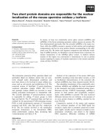
Fig. 1. Double labeling immunofluorescence images showing Cathepsin L and CTLA-2␣ immunoreactivity in sagittal sections from various regions of the mouse brain. (a)
Demonstrates labeling for cathepsin L (green; FITC), (b) CTLA-2␣ (red; TRITC) and (c) merged image. Very high immunoreactivity to indicate colocalization (yellow) in the
merged image is seen in external capsule (ec), corpus callosum (gcc), fimbria of hippocampus (fi), Cornu Ammonis 2, 3 fields of hippocampus (CA2 and CA3), stria medullaris
of thalamus (sm) and mammillothalamic tract (mt). Moderate colocalization in isocortex, Lateral septal nucleus, dorsal part (LSD), Lateral septal nucleus, intermediate part
(LSI), strial part of the preoptic area (StA) and anterodorsal thalamic nucleus (AD) but colocalization is not observed in molecular layer of hippocampus (Mol), dentate gyrus
(DG), nucleus of the horizontal limb of the diagonal band (HDB), medial preoptic area (MPA) and medial preoptic nucleus, medial part (MPOM). Scale bar: 150 m. (For
interpretation of the references to colour in this figure legend, the reader is referred to the web version of this article.)
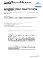
Fig. 2. (A) Double labeling immunofluorescence images showing Cathepsin L and CTLA-2␣ immunoreactivity in sagittal sections of the cortex and hippocampus. The first
column illustrates labeling for (a) cathepsin L (green; FITC), (b) CTLA-2␣ (red; TRITC) and (c) merged image in the subcortical structures and hippocampus. Colocalization of
Cathepsin L and CTLA-2␣ in the merged image is shown in corpus callosum (cc), Alveus of hippocampus (ALV) and Pyramidal cell layer of the hippocampus (Py). The second
column demonstrates labeling for (d) cathepsin L (green; FITC), (e) CTLA-2␣ (red; TRITC) and (f) merged image in secondary motor cortex (M2), corpus callosum (cc), Cornu
Ammonis 2 field of hippocampus (CA2), fimbria of the hippocampus (fi), stria medullaris of thalamus (sm) and anterodorsal thalamic nucleus (AD). CTLA-2␣ immunoreactivity
is observed in molecular layer of hippocampus (Mol) and dentate gyrus (DG), which does not colocalize with cathepsin L. Scale bar: 70 m. (B) Immunofluorescence images
demonstrating Cathepsin L and CTLA-2␣ double labeling immunoreactivity in coronal sections of the hippocampus. The first column shows labeling for (a) cathepsin L (green;
FITC), (b) CTLA-2␣ (red; TRITC) and (c) merged image in Cornu Ammonis 2, 3 fields of hippocampus (CA2 and CA3). Strong immunoreactivity for cathepsin L is observed in
CA2 and CA3 fields and colocalizes (yellow) with CTLA-2␣ in the merged image. The second column illustrates labeling for (d) cathepsin L (green; FITC), (e) CTLA-2␣ (red;
TRITC) and (f) merged image in the hippocampus. Labeling for cathepsin L is seen in Cornu Ammonis 3 field (CA3) and colocalizes with CTLA-2␣ (yellow) in the merged image
but colocalization is not observed in molecular layer of hippocampus (Mol), dentate gyrus (DG) and in the hilus (h). Scale bar: 50 m. (For interpretation of the references to
colour in this figure legend, the reader is referred to the web version of this article.)
3.2.2. Amygdala
The amygdala showed strong colocalization signals for cathesin
L and CTLA-2␣ proteins in the capsular part (CeC) and lateral division (CeL) of central amygdaloid nucleus while at low level in the
basolateral amygdaloid nucleus, anterior part (BLA) and absent in
the negative control sections incubated with 10% normal serum in
place of primary antibodies (Fig. 3).
3.2.3. Ventricular system and thalamus
Choroid plexus located in the ventricular system is important
in maintaining generation and flow of cerebrospinal fluid (CSF).
The plexus displayed high level of cathepsin L immunoreactivity colocalized with CTLA-2␣ within epindymal cells and medial
habenular nucleus, a chief relay nucleus of the descending dorsal diencephalic conduction system. The thalamic nuclei displayed
dense to moderate level of colocalization for cathepsin L and CTLA2␣ proteins. Intense labeling was detected in the stria medullaris
of thalamus (sm) and anteromedial thalamic nucleus (AM); moderately in paraventricular thalamic nucleus, posterior part (PVP),
paraventricular thalamic nucleus (PV) and interanteromedial thalamic nucleus (IAM) as well as in ventral reunions thalamic nucleus
(VRe), paraventricular hypothalamic nucleus, lateral magnocellular
Please cite this article in press as: Luziga, C., et al., Cathepsin L coexists with Cytotoxic T-lymphocyte Antigen-2 alpha in distinct regions
of the mouse brain. Acta Histochemica (2016), />
G Model
ACTHIS-51114; No. of Pages 7
ARTICLE IN PRESS
C. Luziga et al. / Acta Histochemica xxx (2016) xxx–xxx
4
Table 1
Morphological structures refer to the neuron-anatomical atlas from Paxinos and
Franklin (2001).
Brain Region
Density of
positive cells
Neocortex and subcortical regions
Primary somatosensory cortex
Secondary somatosensory cortex
Retrosplenial cortex
Secondary motor cortex
Corpus callosum
External capsule
++
++
++
++
++++
++++
Septum region
Fornix
Lateral septal nucleus, dorsal part
Lateral septal nucleus, intermediate part
Nucleus of the anterior commissure
++++
++
++
+++
Amygdala
BLA − Basolateral amygdaloid nucleus, anterior part
CeC − Central amygdaloid nucleus, capsular part
CeL − Central amygdaloid nucleus, lateral division
+
++++
+++
Hippocampus
Alveus of hippocampus
Fimbria of hippocampus
Dentate gyrus
Cornu Ammonis 1, fields of hippocampus
Cornu Ammonis 2, fields of hippocampus
Cornu Ammonis,3 fields of hippocampus
Hilus
Dentate gyrus
Molecular layer of hippocampus
Stratum oriens
Pyramidal cell layer of the hippocampus
++++
++++
−
−
+++
++++
−
−
−
−
++++
Thalamus
Stria medullaris of thalamus
Anterodorsal thalamic nucleus
Mediodorsal thalamic nucleus, central part
Mammillothalamic tract
External medullary lamina
Medial habenular bodies
Choroid plexus
Anteromedial thalamic nucleus
Paraventricular thalamic nucleus, posterior part
Paraventricular thalamic nucleus
Paraventricular thalamic nucleus, anterior part
Interanteromedial thalamic nucleus
Central medial thalamic nucleus
Ventral reunions thalamic nucleus
++++
+
+++
++++
+++
+++
++++
+++
+++
+++
−
+++
+
+
Hypothalamus
Strial part of the preoptic area
Medial preoptic area
Medial preoptic nucleus, medial part
Septohypothalamic nucleus
Paraventricular hypothalamic nucleus, posterior part
Paraventricular hypothalamic nucleus, lateral magnocellular part
Anterior hypothalamic area, anterior part
Anterior hypothalamic area, posterior part
Anterior hypothalamic area, central part
Lateroanterior hypothalamic nucleus
Posterior hypothalamic area
Medial mammillary nucleus, medial part
Medial mammillary nucleus, lateral part
Supramammillary nucleus
Interpeduncular nucleus, caudal subnucleus
+
+
+
+
+
+
–
+
+
+++
−
+++
++
+++
−
Raphe nuclei (Midbrain)
Paramedian raphe nucleus
Dorsal raphe nucleus
Median raphe nucleus
++
++
++
Cerebellum
Molecular layer
Purkinje cell layer
Granule cell layer
White matter
+
+++
+++
++++
The intensity of cathepsin L colocalization with CTLA-2␣ was classified as follows:
negative (−), moderate (++), high (+++), Very high (++++). The structures used to evaluate immunofluorescence colocalization were Dentate gyrus and Molecular layer
of hippocampus (−); Central medial thalamic nucleus and Anterior hypothalamic
area, posterior part (+); Lateral septal nucleus, dorsal part and Lateral septal nucleus,
intermediate part (++); Anteromedial thalamic nucleus and Paraventricular thalamic
nucleus (+++); Stria medullaris of thalamus and Mammillothalamic tract (++++).
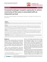
Fig. 3. Immunofluorescence images demonstrating Cathepsin L and CTLA-2␣ double labeling immunoreactivity in coronal sections of the amygdala. The first column
shows labeling for (a) cathepsin L (green; FITC), (b) CTLA-2␣ (red; TRITC) and (c)
merged image. Colocalization of Cathepsin L and CTLA-2␣ in the merged image is
strong in capsular part (CeC) and lateral division (CeL) of central amygdaloid nucleus
while at low level in the basolateral amygdaloid nucleus, anterior part (BLA). For control purposes, coronal sections from the amygdala were incubated with 10% normal
serum in place of primary antibodies. Labeling for cathepsin L and CTLA-2␣ is virtually absent in sections (d), (e) and (f) incubated with the 10% normal serum. Scale
bar: 50 m. (For interpretation of the references to colour in this figure legend, the
reader is referred to the web version of this article.)
part (PaLM), and paraventricular hypothalamic nucleus, posterior
part (PaPo) (Fig. 4A and B)
3.2.4. Hypothalamus
In the hypothalamus, strong immunoreactivity for cathepsin L
colocalized with CTLA-2␣ was confined to the anterior commissure
(ac), lateroanterior hypothalamic nucleus (LA); supramammillary
nucleus (SuM); lateral part of medial mammillary nucleus (ML);
moderately in anterior hypothalamic area, central part (AHC) and
medial mammillary nucleus, medial part (MM) (Figs. 4 B and 5 ).
3.2.5. Cerebellum
In the cerebellum, intense double immunolabeling for cayhepsin L and CTLA-2␣ proteins was strong in the internal white matter;
moderately in the granule layer in randomly distributed cells that
represent Golgi cells and/or granule cells and in cell bodies of Purkinje neurons and low in the molecular layer in stellate and basket
cells. (Fig. 6).
4. Discussion
Previous immunohistochemical studies show that CTLA-2␣ protein in the mouse brain is preferentially localized within dendrites
and axonal fibres (Luziga et al., 2007). CTLA-2␣ is also shown to
exhibit selective inhibition to mouse cathepsin L-like cysteine proteinases (Kurata et al., 2003). And that transient expression of
crammer (cysteine proteinases inhibitor) correlates well with the
establishment of long-term memory, suggesting a role of the crammer in memory formation through regulation of cathepsin activity
(Comas et al., 2004). Dash et al. (2000) also demonstrated that
concurrent inhibition of multiple caspases (a family of cysteine
proteases) in hippocampus blocks long-term but not short-term
spatial memory in mouse brain. In this context, we developed inter-
Please cite this article in press as: Luziga, C., et al., Cathepsin L coexists with Cytotoxic T-lymphocyte Antigen-2 alpha in distinct regions
of the mouse brain. Acta Histochemica (2016), />
G Model
ACTHIS-51114; No. of Pages 7
ARTICLE IN PRESS
C. Luziga et al. / Acta Histochemica xxx (2016) xxx–xxx
5
Fig. 4. (A) Double labeling Immunofluorescence images showing Cathepsin L and CTLA-2␣ immunoreactivity in coronal sections of the Ventricular system and Thalamus. The
first column illustrates labeling for (a) cathepsin L (green; FITC), (b) CTLA-2␣ (red; TRITC) and (c) merged image in medial habenular nucleus (MHb) and Choroid plexus (chp).
Very Strong labeling for both cathepsin L and CTLA-2␣ (yellow) is seen in medial habenular nucleus (MHb) and choroid plexus (chp). The second column shows labeling for
(d) cathepsin L (green; FITC), (e) CTLA-2␣ (red; TRITC) and (f) merged image in the thalamus. Very strong colocalization (yellow) is seen in stria medullaris of thalamus (sm)
and anteromedial thalamic nucleus (AM). Scale bar: 70 m. (B) Double labeling immunofluorescence images demonstrating Cathepsin L and CTLA-2␣ immunoreactivity in
sagittal sections of the thalamus. The first column illustrates labeling for (a) cathepsin L (green; FITC), (b) CTLA-2␣ (red; TRITC) and (c) merged image in thalamus. Moderate
double labeling for both Cathepsin L and CTLA-2␣ (yellow) in the merged image is seen in paraventricular thalamic nucleus, posterior part (PVP), paraventricular thalamic
nucleus (PV), interanteromedial thalamic nucleus (IAM) but is not observed in the anterior part of paraventricular thalamic nucleus, (PVA). The second column shows labeling
for (d) cathepsin L (green; FITC), (e) CTLA-2␣ (red; TRITC) and (f) merged image in the thalamus. Low intensity of colocalization (yellow) for both cathepsin L and CTLA-2␣ in
the merged is seen in ventral reunions thalamic nucleus (VRe), paraventricular hypothalamic nucleus, lateral magnocellular part (PaLM), and paraventricular hypothalamic
nucleus, posterior part (PaPo). Below the thalamus is the hypothalamus where moderate colocalization of cathepsin L and CTLA-2␣ is seen in the posterior part (AHP) but
not in the anterior part of the anterior hypothalamic area (AHA). Scale bar: 100 m. (For interpretation of the references to colour in this figure legend, the reader is referred
to the web version of this article.)
est to know the cellular colocalization of cathepsin L (a family of
cystein proteases) with CTLA-2␣ protein in the mouse brain. Understanding the cellular relationship of cathepsin inhibitory activity of
CTLA-2␣ in light of the emerging roles of cathepsins in memory
establishment, is essential in the development of treatments for
degenerative diseases associated with learning and memory loss.
In this study, immunoreactivity for cathepsin L and CTLA-2␣
was detected in nerve fibres bundles and in some nerve cell bodies. In the cerebral cortex and subcortical structures, colocalization
was very high in neocortex and corpus callosum. The neocortex
is involved in higher brain functions such as sensory perception, generation of motor commands, spatial reasoning, conscious
thought and language. It has also an influential role in sleep, memory and learning processes (Lui et al., 2011). The corpus callosum
plays a major role in most communications between different
regions of the brain. Studies show that cathepsin L in secretory
vesicles functions as a key protease for proteolytic processing of
proneuropeptides into active neuropeptides that are released to
mediate cell to cell communication in the nervous system for neurotransmission (Hook et al., 2012). Identification of cathepsin L and
CTLA-2␣ in neocortex and corpus callosum is suggestive of their
regulatory role in increasing or decreasing information transfer in
the brain and may also be involved in many other biological processes in the central nervous system that are yet to be identified.
The alveus, fimbria and the Pyramidal cell layer in Cornu Ammonis 2, 3 fields of the hippocampus also showed intense double
labeling for cathepsin L and CTLA-2␣. The alveus is composed of
the white myelinated fibres that arise from cell bodies of subculum
and hippocampus and eventually merges with the fimbria of the
hippocampus that goes on to become the fornix, which is also a
prominent white matter tracts passing above the thalamus. Fibres
of the fornix travel to the anterior commissure, a white matter
tract connecting both hemispheres and terminate in the mammillary bodies and continue upward through the mammilothalamic
tract towards the anterior nucleus of the thalamus (Maren, 1999;
Amunts et al., 2005; Marc and Sergio, 2014). All these structures are
part of the limbic system including amygdala, habenular and raphae
nucleus and were strongly labelled for cathepsin L and CTLA-2␣.
The presence of cathepsin L and CTLA-2␣ in these structures is
suggestive of their significant role in regulation of limbic system
functions that include motivation and reward, emotion, learning,
and memory establishment.
One of the most prominent labeling structures for both cathepsin L and CTLA-2␣ was the choroid plexus epithelium that produces
and secretes cerebrospinal fluid and controls movement of solutes
between the blood and the cerebrospinal fluid. The fluid provides
mechanical protection and a stable physiological environment for
the central nervous system and supplies the brain with certain
nutrients, hormones, and metal ions, while clearing cells that populate the anatomical compartment (Purves et al., 2001). Localization
of cathespin L and CTLA-2␣ in these structures correlated well with
our previous studies on the distribution of both cathespin L and
CTLA-2␣ in choroid plexus epithelium (Luziga et al., 2008, 2016).
Whether the colocalization of cathespin L and CTLA-2␣ to choroid
plexus epithelium is related to its role to produce and secrete cerebrospinal fluid; regulate and transfer of molecules across the bloodcerebrospinal fluid interface or to any other novel function is a
question that remains to be resolved.
The stria medullaris, mammillothalamic tract, anterodorsal thalamic nucleus and anteromedial thalamic nucleus in thalamus
displayed strong double immunofluorescence labeling for cathepsin L and CTLA-2␣. In functional perspective, the anterior nuclei
Please cite this article in press as: Luziga, C., et al., Cathepsin L coexists with Cytotoxic T-lymphocyte Antigen-2 alpha in distinct regions
of the mouse brain. Acta Histochemica (2016), />
G Model
ACTHIS-51114; No. of Pages 7
6
ARTICLE IN PRESS
C. Luziga et al. / Acta Histochemica xxx (2016) xxx–xxx
Fig. 5. Double labeling immunofluorescence images showing Cathepsin L and CTLA2␣ immunoreactivity in sagittal sections of the hypothalamus. The first column
shows labeling for (a) cathepsin L (green; FITC), (b) CTLA-2␣ (red; TRITC) and
(c) merged image in hypothalamus. Intense immunoreactivity of cathepsin L is
observed in lateroanterior hypothalamic nucleus (LA) and colocalizes with CTLA-2␣
in the merged imerge. Moderate colocalization is observed in anterior hypothalamic
area, central part (AHC). The second column illustrates labeling for (d) cathepsin L
(green; FITC), (e) CTLA-2␣ (red; TRITC) and (f) merged image in the hypothalamus.
Strong double labeling for cathepsin L and CTLA-2␣ is evident in supramammillary
nucleus (SuM) and lateral part of medial mammillary nucleus (ML); moderately in
medial mammillary nucleus, medial part (MM) but low in paramedian raphe nucleus
(PMnR) and absent in interpeduncular nucleus, caudal subnucleus (IPL). Scale bar:
100 m. (For interpretation of the references to colour in this figure legend, the
reader is referred to the web version of this article.)
and the mediodorsal thalamic nuclei act as nodal points of convergence where bilateral tissue damage can result in memory function
impairment similar to bilateral lesions of the mammillothalamic
tract, which connects the mammillary bodies to the anterior nuclei
(Berger, 2004). In rodents, bilateral disruption of this pathway has
also been shown to attenuate reinstatement of drug seeking behavior (Suzanne et al., 2001). Whether the colocalization of cathepsin
L and CTLA-2␣ to these structures is related to role in establishment of recall memory or to a novel function is a matter that needs
further studies.
In the cerebellum, abundant double labeling signals for cathepsin L and CTLA-2␣ were distributed in the white matter, granules
cell layer and Purkinje cells and moderately in the molecular layer.
The Purkinje cells are the only neurons responsible for sending
output from the cortex. On the other hand cathepsin L and CTLA2␣ were locacalized in deep cerebellar nuclei in the white matter
which receives input from the Purkinje cells and send output information to other brain regions. Studies show that Purkinje cells are
affected by the combined loss of cathepsin B and L while overexpression of the cathepsins contributes to neurodegeneration of
granule cells (Sevenich et al., 2006). Presence of cathepsin L and
CTLA-2␣ in the cerebellum is suggestive of their regulatory role in
prevention of Purkinje and granule cell degeneration
In conclusion, this study shows that cathepsin L coexists with
CTLA-2␣ in distinct regions of the mouse brain including cerebral
cortex, subcortical structures, hippocampus, amygdala, thalamic
nuclei, hypothalamus and the cerebellum. The colocalization indicate distinct structural or regional specific function of cathespin L
and CTLA-2␣ in various physiological and pathological processes of
the nervous system and that the fine equilibrium between the syn-
Fig. 6. Immunofluorescence images demonstrating Cathepsin L and CTLA-2␣
immunoreactivity double labeling in sagittal sections of the cerebellum. The first column illustrates labeling for (a) cathepsin L (green; FITC), (b) CTLA-2␣ (red; TRITC)
and (c) merged image in cerebellum. Colocalization (yellow); for cathepsin L and
CTLA-2␣ in the merged image is strong in the internal white matter; moderately
in the granule layer in randomly distributed cells that represent Golgi cells and/or
granule cells and in cell bodies of Purkinje neurons and low in the molecular layer
in stellate and basket cells. The second column shows larger magnification images
labelled for (d) cathepsin L (green; FITC), (e) CTLA-2␣ (red; TRITC) and (f) merged in
the cerebellum. Scale bar: a, b, c: 100 m; d, e, f: 50 m. (For interpretation of the
references to colour in this figure legend, the reader is referred to the web version
of this article.)
thesis and secretion of cathespin L and CTLA-2␣ is part of the brain
processes for establishment of long-term memory, storage of memory traces for spatial information, olfaction, emotion, behavior and
motivation and hence maintaining normal growth and development. This study also opens ways to new studies on the functional
implications of cathepsin L and CTLA-2␣ in the central nervous
system.
Acknowledgement
The authors gratefully acknowledge the Japanese Ministry of
Education, Culture, Sports, Science and Technology for financial
support.
References
Amunts, K., Kedo, O., Kindler, M., Pieperhoff, P., Mohlberg, H., Shah, N., Habel, U.,
Schneider, F., Zilles, K., 2005. Cytoarchitectonic mapping of the human
amygdala, hippocampal region and entorhinal cortex: intersubject variability
and probability maps. Anat. Embryol. 210, 343–352.
Barrett, A.J., Kirschke Cathepsin, H.B., Cathepsin, H., Cathepsin, L., 1981. In: Lorand,
L. (Ed.), Methods in Enzymology, 80. Academic Press, New York, pp. 535–561.
Berger, J.R., 2004. Memory and the mammillothalamic tract: editorial. Am. J.
Neuroradiol. 25, 906–907.
Bui, T.N., Luziga, C., Yamamoto, M., Takeshi, K., Yamamoto, Y., 2015. Identification
and characterization of the interactive proteins with cytotoxic T-lymphocyte
antigen-2␣ Bioscience. Biotechnol. Biochem. 79, 587–597.
Callahan, L.M., Chow, N., Cheetham, J.E., Cox, C., Coleman, P.D., 1998. Analysis of
message expression in single neurons of Alzheimer’s disease brain. Neurobiol.
Aging 19, 99–103.
Camenisch, G., Tini, M., Chilov, D., Kvietikove, I., Srinivas, V., Caro, J., Spielmann, P.,
Wenger, R.H., 1999. General applicability of chicken egg yolk antibodies: the
performance of IgY immunoglobulins raised against the hypoxia-inducible
factor 1␣. FASEB J. 13, 13–81.
Please cite this article in press as: Luziga, C., et al., Cathepsin L coexists with Cytotoxic T-lymphocyte Antigen-2 alpha in distinct regions
of the mouse brain. Acta Histochemica (2016), />
G Model
ACTHIS-51114; No. of Pages 7
ARTICLE IN PRESS
C. Luziga et al. / Acta Histochemica xxx (2016) xxx–xxx
Carmona, E., Dufour, E., Plouffe, C., Takebe, S., Mason, P., Mort, J.S., Ménard, R.,
1996. Potency and selectivity of the cathepsin L propeptide as an inhibitor of
cysteine proteases. Biochemistry 35, 8149–8157.
Comas, D., Petit, F., Preat, T., 2004. Drosophila long-term memory formation
involves regulation of cathepsin activity. Nature 430, 460–463.
Cowan, K.N., Leung, W.C., Mar, C., Bhattacharjee, R., Zhu, Y., Rabinovitch, M., 2005.
Caspases from apoptotic myocytes degrade extracellular matrix: a novel
remodeling paradigm. FASEB J. 19, 1848–1850.
Dash, P.K., Blum, S., Moore, A.N., 2000. Caspase activity plays an essential role in
long-term memory. Neuroreport 11, 2811–2816.
Delahaye, N.F., Coltel, N., Puthier, D., Flori, L., Houlgatte, R., Iraqi, F.A., Nguyen, C.,
Grau, G.E., Rihet1, P., 2006. Gene-expression profiling discriminates between
cerebral malaria (CM)/susceptible mice and CM-resistant mice. J. Infect. Dis.
193, 312–321.
Delaria, K., Florentino, L., Wallace, L., Tamburini, P., Brownell, E., Muller, D., 1994.
Inhibition of cathepsin L-like cysteine proteases by cytotoxic T-lymphocyte
antigen-2. J. Biol. Chem. 269, 25172–25177.
Denizot, F., Brunet, J.F., Roustan, P., Harper, K., Suzan, M., Luciani, M.F., Mattei, M.G.,
Golstein, P., 1989. Novel structures CTLA-2a and CTLA-2b expressed in mouse
activated T cells and mast cells and homologous to cysteine proteinase
proregions. Eur. J. Immunol. 19, 631–635.
Deshapriya, R.M.C., Yuhashi, S., Usui, M., Kageyama, T., Yamamoto, Y., 2010.
Identification of essential residues of CTLA-2␣ for inhibitory potency. J.
Biochem. 147, 393–404.
Deussing, J., Kouadio, M., Rehman, S., Weber, I., Schwinde, A., Peters, C., 2002.
Identification and characterization of a dense cluster of placenta-specific
cysteine peptidase genes and related genes on mouse chromosome 13.
Genomics 79, 225–240.
Felbor, U., Kessler, B., Mothes, W., Goebel, H.H., Ploegh, H.L., Bronson, R.T., Olsen,
B.R., 2002. Neuronal loss and brain atrophy in mice lacking cathepsins B and L.
Proc. Natl. Acad. Sci. 99, 7883–7888.
Funkelstein, L., Beinfeld, M., Minokadeh, A., Zadina, J., Hook, V., 2010. Unique
biological functions of cathepsin L in secretory vesicles for biosynthesis of
neuropeptides. Neuropeptides 44, 457–466.
Hook, V., Israel, S., Schechter, I., Demuth, H., Hook, G., 2009. Alternative pathways
for production of beta-amyloid peptides of alzheimer’s disease. Bio. Chem. 389,
993–1006.
Hook, V., Funkelstein, L., Wegrzyn, J., Bark, S., Kindy, M., Hook, G., 2012. Cysteine
cathepsins in the secretory vesicle produce activepeptides: cathepsin l
generates peptide neurotransmitters and cathepsin B produces beta-amyloid
of Alzheimer’s disease. Biochim. Biophys. Acta 1824, 89–104.
Kurata, M., Yamamoto, Y., Watabe, S., Makino, Y., Ogawa, K., Takahashi, S.Y., 2001.
Bombyx cysteine proteinase inhibitor (BCPI) homologous to propeptide
regions of cysteine proteinases is a strong, selective inhibitor of cathepsin
L-like cysteine proteinases. J. Biochem. 130, 857–863.
Kurata, M., Hirata, M., Watabe, S., Miyake, M., Takahashi, S.Y., Yamamoto, Y., 2003.
Expression, purification, and inhibitory activities of mouse cytotoxic
T-lymphocyte antigen-2. Protein Expression Purif. 32, 119–125.
Lui, J.H., Hansen, D.V., Kriegstein, A.R., 2011. Development and evolution of the
human neocortex. Cell 146, 18–36.
Luziga, C., Nakamura, O., Deshapriya, R.M.C., Usui, M., Miyaji, M., Wakimoto, M.,
Wada, N., Yamamoto, Y., 2007. Expression mapping of cytotoxic T-lymphocyte
antigen-2 alpha gene transcripts in mouse brain. Histochem. Cell Biol. 127,
569–579.
7
Luziga, C., Nakamura, O., Deshapriya, R.M.C., Usui, M., Miyaji, M., Wakimoto, M.,
Wada, N., Mbassa, G., Yamamoto, Y., 2008. Dendritic andaxonal localization of
cytotoxic T-lymphocyte antigen-2 alpha protein in mouse brain. Brain Res.
1204, 40–52.
Luziga, C., Bui, T.N., Kashoma, I., Katakweba, A., Yamamoto, Y., 2016. Localization
profile of cathepsin L in the brain of African giant rat (Cricestomys gambianus).
Anat. J. Africa 5, 618–630.
Ménard, R., Carmona, E., Takebe, S., Dufour, E., Plouffe, C., Mason, P., Mort, J.S.,
1998. Autocatalytic processing of recombinant human procathepsin L.
Contribution of both intermolecular and unimolecular events in the processing
of procathepsin L in vitro. J. Biol. Chem. 273, 4478–4484.
Mach, L., Mort, J.S., Glössl, J., 1994. Maturation of human procathepsin B.
Proenzyme activation and proteolytic processing of the precursor to the
mature proteinase, in vitro, are primarily unimolecular processes. J. Biol. Chem.
269, 13030–13035.
Maren, S., 1999. Long-term potentiation in the amygdala: a mechanism for
emotional learning and memory. Trends Neurosci. 22, 561–567.
Maubach, G., Schilling, K., Rommerskirch, W., Wenz, I., Schultz, J.E., Weber, E.,
Wiederanders, B., 1997. The inhibition of cathepsin S by its propeptide:
specificity and mechanism of action. Eur. J. Biochem. 250, 745–750.
Nixon, R.A., 2000. A protease activation cascade in the pathogenesis of Alzheimer’s
diasease. Ann. N.Y. Acad. Sci. 924, 117–131.
Paxinos, G., Franklin, K.B.J., 2001. The Mouse Brain in Stereotaxic Coordinates, 2nd
edition. Academic Press, Tokyo.
Purves, D., Augustine, G.J., Fitzpatrick, D., Katz, L.C., LaMantia, A.S., MacNamara,
J.O., Williams, S.M., 2001. Neuroscience, 2nd edition. Sinauer Associates Inc
2001, Sunderland, Mass.
Rawlings, N.D., Waller, M., Barrett, A.J., Bateman, A., 2014. MEROPS: the database
of proteolytic enzymes, their substrates and inhibitors. Nucleic Acids Res. 42,
D503–D509.
Reddy, V.Y., Zhang, Q.Y., Weiss, S.J., 1995. Pericellular mobilization of the
tissue-destructive cysteine proteinases, cathepsins B, L, and S, by human
monocyte-derived macrophages. Proc. Natl. Acad. Sci 92, 3849–3853.
Sevenich, L., Pennacchio, L., Peters, C., Reinheckel, T., 2006. Human cathepsin L
rescues the neurodegeneration and lethality in cathepsin B/L double-deficient
mice. Bio. Chem. 387, 885–891.
Takahashi, S.Y., Yamamoto, Y., Shionoya, Y., Kageyama, T., 1993. Cysteine
proteinase from the eggs of the silkmoth, Bombyx mori: identification of a
latent enzyme and characterization of activation and proteolytic processing
in vivo and in vitro. J. Biochem. 144, 267–272.
Turk, V., Stoka, V., Vasiljeva, O., Renko, M., Sun, T., Turk, B., Turk, D., 2012. Cysteine
cathepsins: from structure, function and regulation to new frontiers. Biochim.
Biophys. Acta 1824, 68–88.
Yamamoto, Y., Watabe, S., Kageyama, T., Takahashi, S.Y., 1999a. Purification and
characterization of Bombyx cysteine proteinase specific inhibitors from the
hemolymph of Bombyx mori. Arch. Insect Biochem. Physiol. 41, 119–129.
Yamamoto, Y., Watabe, S., Kageyama, T., Takahashi, S.Y., 1999b. A novel inhibitor
protein for Bombyx cysteine proteinase is homologous to propeptide regions
of cysteine proteinases. FEBS Lett. 448, 257–260.
Yamamoto, Y., Kurata, M., Watabe, S., Murakami, R., Takahashi, S.Y., 2002. Novel
cysteine proteinase inhibitors homologous to the proregions of cysteine
proteinases. Curr. Protein Pept. Sci. 3, 231–238.
Please cite this article in press as: Luziga, C., et al., Cathepsin L coexists with Cytotoxic T-lymphocyte Antigen-2 alpha in distinct regions
of the mouse brain. Acta Histochemica (2016), />