Báo cáo sinh học: "In vitro migration of cytotoxic T lymphocyte derived from a colon carcinoma patient is dependent on CCL2 and CCR2" pot
Bạn đang xem bản rút gọn của tài liệu. Xem và tải ngay bản đầy đủ của tài liệu tại đây (1.09 MB, 11 trang )
RESEARCH Open Access
In vitro migration of cytotoxic T lymphocyte
derived from a colon carcinoma patient is
dependent on CCL2 and CCR2
Klara Berencsi
1
, Pyapalli Rani
1
, Tianqian Zhang
1
, Laura Gross
1
, Michael Mastrangelo
2
, Neal J Meropol
3,4
,
Dorothee Herlyn
1
and Rajasekharan Somasundaram
1*
Abstract
Background: Infiltration of colorectal carcinomas (CRC) with T-cells has been associated with good prognosis.
There are some indications that chemokines could be involved in T-cell infiltration of tumors. Selective modulation
of chemokine activity at the tumor site could attract immune cells resulting in tumor growth inhibition. In mouse
tumor model systems, gene therapy with chemokines or administration of antibody (Ab)-chemokine fusion
proteins have provided potent immune mediated tumor rejection which was mediated by infiltrating T cells at the
tumor site. To develop such immunotherapeutic strategies for cancer patients, one must identify chemokines and
their receptors involved in T-cell migratio n toward tumor cells.
Methods: To identify chemokine and chemokine receptors involved in T-cell migration toward CRC cells, we have
used our previously published three-dimensional organotypic CRC culture system. Organotypic culture was initiated
with a layer of fetal fibroblast cells mixed with collagen matrix in a 24 well tissue culture plate. A layer of CRC cells
was placed on top of the fibroblast-collagen layer which was followed by a separating layer of fibroblasts in
collagen matrix. Anti-CRC specific cytotoxic T lymphocytes (CTLs) mixed with fibroblasts in collagen matrix were
placed on top of the separating layer. Excess chemokine ligand (CCL) or Abs to chemokine or chemokine receptor
(CCR) were used in migration inhibition assays to identify the chemokine and the receptor involved in CTL
migration.
Results: Inclusion of excess CCL2 in T-cell layer or Ab to CCL2 in separating layer of collagen fibroblasts blocked
the migration of CTLs toward tumor cells and in turn significantly inhibited tumor cell apoptosis. Also, Ab to CCR2
in the separating layer of collagen and fibroblasts blocked the migration of CTLs toward tumor cells and
subsequently inhibited tumor cell apoptosis. Expression of CCR2 in four additional CRC patients’ lymphocytes
isolated from infiltrating tumor tissues suggests their role in migration in other CRC patients.
Conclusions: Our data suggest that CCL2 secrete d by tumor cells and CCR2 recept ors on CTLs are involved in
migration of CTLs towards tumor. Gene therapy of tumor cells with CCL2 or CCL2/anti-tumor Ab fusion proteins
may attract CTLs that potentially could inhibit tumor growth.
Background
Chemokines play an important role in immune homeos-
tasis and immune surveillance (reviewed in [1-3]). Studies
have demonstrated that chemokines influence immune
rea ctions by regulating trafficking of dendritic cells (DC)
and lymphocytes [4]. In tumor bearing individuals, the
role of chemokines is paradoxical. Chemokines produced
by tumor cells are known to stimulate autocrine tumor
growth, progression and metastasis [4-10]. In contrast,
chemokines produced by tumor cells can also attract
chemokine receptor (CCR)-positive leukocytes into the
tumor area, potentially leading to tumor growth inhibi-
tion in vitro and in vivo [9,11-13]. In colorectal carci-
noma (CRC) patients, T-cell infiltration has been shown
to be associated with good prognosis (reviewed in [14]
* Correspondence:
1
The Wistar Institute, 3601 Spruce Street, Philadelphia, PA 19104, USA
Full list of author information is available at the end of the article
Berencsi et al . Journal of Translational Medicine 2011, 9:33
/>© 2011 Berencsi et al; licensee BioMed Central Ltd. This is an Open Access a rticle distributed under the terms of the Creative Commons
Attribution License ( 2.0), which permits unrestricted use, distribution, and reproduction in
any medium, provid ed the original work is properly cited.
and [15-19]). In these studies, favorable prognosis w as
correlated with the presence of tumor cells secreting che-
mokines such as CCL5, CXCL1 0 and CXCL1 that played
aroleinrecruitmentofCCR5
+
/CXCR3
+
T-helper (Th)1
cells or CX3CR1
+
,perforin
+
/granzyme B
+
T c ells [17],
[18]. Colorectal and pancreatic carcinoma cells are
known to secrete CCL2 which is associated with
increased tumor infiltration of macrophages [20-22].
However, there are mixed reports of good and bad prog-
nosis due to increased infiltration of tumor associated
macrophages in these studies [ 20-22]. In those s tudies,
the level of tumor derived-CCL2 and its influence on
T cell infiltration of tumor cells is unclear. In mouse
tumor model system, there are indications that mela-
noma cells secreting high amounts of CCL2 attract
macrophages resulting in tumor growth inhibition [23].
In another study, transfection of mouse CT-26 CRC cells
with CCL2 gene resulted in decreased metastasis and
increased susceptibility o f tumor cells to macrophage
lysis [24]. Thus, sele ctive modulation of chemokine activ-
ity at the tumor site could attract immune cells resulting
in tumor growth inhibition (reviewed in [25] and [1]).
In mo use systems, ex vivo transduction of chemokines
into tumor cells has provided potent tumor vaccines
inducing tumor rejection, which was mediated by infil-
trating T cells at the vaccine site. Infiltration of T cells
into the tumor area was followed by rejection of both
transduced and non-transduced tumor cells [11-13]. CD4
+
T-cell subsets have been implicated in tumor rejection
induced by vaccination of mice with CCL19-transduced
tumor cells [26], and CD8
+
CTL were instrumental in
tumor growth rejection in mice following intratumoral
delivery of CCL20 or CXCL12 via adenovirus vectors
[27] or injection of CCL16-expressing tumor cells. Both
CD4
+
and CD8
+
T cells were required for tumor growth
inhibition to occur in mice injected intratumorally with
CCL21 [28].
It has been suggested that immunological intervention
of cancer patients has been largely unsuccessful due to
limited ability of T cells to infiltrate tumors in vivo
([29,30]). Chemokines fused to anti-tumor Ab may be
utilized to attract adoptively transferred tumor antigen
(Ag) -specific T cells to the tumor site [31]. To develop
immunotherapeutic stra tegies for cancer patients based
on chemokines and their receptors, similar to the
approaches already successfully used in mice, one must
identify chemokines and their receptors involved in
T-cell migration toward tumor cells.
Recently, we have shown in an organotypic culture
system (reconstruct) that migration of CTL derived
from a CRC patient towards autologous tumor cells was
mediated by chemokine receptor CXCR3 expressed by
the T cells, and CXCL11 chemokine secreted by the
autologous tumor cells [32]. In the present study, we
show that migration of CTL derived from another CRC
patient is dependent on CCL2 and CCR2.
Materials and Methods
Cell lines
CTL 007, CTL020, CRC cell line (WC007) and fetal
colon fibroblast cell line (FCFB/1) were established and
maintained in culture as previously described [ 32,33].
Ten additional primary tumor tissues were obtained
from CRC patients of var ious diseas e stages whose
T cells were analyzed for chemokine receptor expression
(data from 4 patients whose T cells are positive for
CCR2 are shown in Table 1). Blood and tissue speci-
mens were obtained in co mpliance with Helsinki
Declaration with informed consent approved by Institu-
tional Review Board of Thomas Jefferson University
Hospital, Fo x Chase Cancer Center a nd The Wistar
Institute (Approval number 2109169).
Reagents
The following monoclonal antibodies (mAb) were used:
mAb Nok-1 to Fas ligand (BD- PharMingen, S an Diego,
CA); mAb CH-11 to CD95 and anti-CD11a mAb (Immu-
notech, Westbrook, ME); fluoresceinated (FITC) or phy-
coerythrin (PE) -conjugated anti-CD:4, 8, 25, 29, 40, 40L,
44, 49 a, 49b, 54 and 80; anti-CXCL-11 mAb; anti-human
CCR:1, 2, 3, 5, 6, 7 and 9 mAb; anti- CXCR:1, 2, 3, 4, 5
and6mAb(R&DSystems,Minneapolis,MN);anti-
human CCR:4, 8, and 10 mAb (Imgenex , S an Diego, CA);
anti-CCR11 and -CX3CR1 polyclonal Ab (Abcam, Cam-
bridge, MA); FITC conjugated goat anti-mouse IgG (Invi-
trogen, Carlsbad, CA). Recombinant human CXCL11 was
purchased from R&D Systems.
Chemokine determination by RT-PCR or ELISA
mRNA was extracted from CRC cells (5 × 10
6
)using
Fast Track 2.0 mRNA isolation kit (Invitrogen). The pri-
mers used were 5’ -G CC CGG TGT CAT C TT CCT
AAC CAA GC-3’ and 5’ -AGG GGA CAG GGG AAC
Table 1 CCR2 expression by T-cell lines established from
tumor infiltrating lymphocytes of CRC tissues
Patient
a
T cell
phenotype
Expression of CCR2
(% positive cells)
b
# Dukes’ Disease Stage
296674 A CD4 58.8
298884 B CD4/CD8 68
1003485 B CD4/CD8 48.2
05193 B CD4/CD8 27.4
a
Representative data from 4 patients whose T cells were positive for CCR2
expression.
b
CCR2 expression determined by FACS analysis. Data represented are from a
single experiment and the Results were confirmed in at least two
independent experiments.
Berencsi et al . Journal of Translational Medicine 2011, 9:33
/>Page 2 of 11
TCT CAG AGC AA-3’ for CCL3; 5’ -TGC TGC TTT
TCT TAC ACC GCG AGG AA-3’ and 5’-A GA AGG
GAC AGG AAC TGC GGA GAG GA-3’ for CCL4; 5’-
TCT GCA GCA CTT CTG TGT CTG-3’ and 5’-G GA
TCC TAG AAG GAG CTG GA-3’ for CCL7; 5’-CAG
TCC ATG AGA AGG AGT CCA-3’ and 5’-AGA TCC
TGC ACA GGA CTG TG-3’ for CCL8; 5’-AGG GCA
TGG GTT TTA TTA TAT ATA TAT-3’ and 5’ -TTT
AAA AAT AAC TGA TAT TCA TGG-3’ for CCL11;
5’-TCA TCT TTC CAC AAT AAC ATA TTT A-3’ and
5’-GTTTATTTGAGTATTGCTGATCTTT-3’ for
CCL13; 5’ -GGA CTT CCT GGA TCC TCC TC-3’ and
5’ -AGC AGT CAG CAG CAA AGT GA-3’ for CCL15;
5’-ATG GCC CTG CTA CTG GCC CTC AGC CTG-3’
and 5’ - TTA ACT GCT GCG GCG CTT CAT CTT
GGC-3’ for CCL19; 5’ -TGT AGG G CG ACG GTT
TTA-3’ and 5’-TCC ACC ACA ACA TGC AG-3’ for
CCL25; 5’-GGC CCT GCC CTT ATA GC-3’ and 5’ -
CTA ACT TGG GGT TGA CAT T-3’ for CXCL1-3; 5’-
TGT TGA GAG AGC TGC G-3’ and GGG TTC AGA
GAC CTC CA-3’ for CXCL5; 5’-GAA GTG GTA GCC
TC
C C-3’ and 5’-GCT TTC CCC CAC ACT C-3’ for
CXCL6; 5’ -TCC GCT GCA TGT GTA TAA AG-3’ and
5’-ATA GGT ATC CTG AAT AAA TGA GAA C-3’ for
CXCL7; 5’-CAT GCT GGT GAG CCA AGC AGT TTG
AA-3’ and 5’ -CAC TTC TGT GGG GTG TTG GGG
ACA AG-3 ’ for CXCL9; 5’- CGA TGC CTA AAT CCC
AAA TCG AAG CA-3’ and 5’ -AAT TGC TGG ACT
CCT TTG GGC AGT GG-3’ for CXCL11; 5’-ATG AAC
GCC AAG GTC GTG GTC-3’ and 5’ -TGG CTG TTG
TGC TTA CTT GTT T-3’ for CXCL12; 5’ -TCT CTC
CAG GCC ACG GTA TTC-3’ and 5’-ACC ATT TGG
CAC GAG GAT TCA C-3’ for CXCL13. CCL21 primer
was purchased from Biosource (Camarillo, CA) and
CCL2, CCL5, CXCL8 and CXCL10 primers were pur-
chased from R&D Systems. P CR reactions were per-
formed for 35 cycles (94°C, 45 sec; 60°C for CCL4,
CCL19, CCL21, CXC L1, CXCL5, CXCL6, CXCL7,
CXCL9, and CXCL11; 56°C for CXCL12, 55°C for
CCL2, CCL3, CCL5, CCL25, CXCL8 and CXCL10; 52°C
FOR CCL7 and CCL15; 48°C for CCL8, CCL11 and
CCL13, 45 sec; 72°C, 45 sec) using the SuperScript One-
Step RT-PCR kit (Invitrogen). All PCR involved an
initial denaturation step at 94°C for 45 sec to 1.5 min
and a final extension step for 7 min at 72°C. All PCR
products were analyzed using 10% novex-TBE gel (Invi-
trogen). Supernatants obtained from CRC cells on day 6
of culture were tested for the presence of CCL2, CCL3,
CCL15, CCL19, CCL21 and CXCL11 using ELISA kits
(R&D Systems).
Phenotyping
Phenotyping of tumor cells and T cells was performed
as described [32]. In brief, cultured cells were i ncubat ed
with saturating concentrations of FITC or PE-conju-
gated mAb (5 μ g/ml) detecting human lymphocyte and
tumor markers in FACS buffer for 1 h at 4°C, followed
by excess mAb removal by washing in FACS buffer.
Binding of the mAb was analyzed as described [32].
T-cell migration in organotypic CRC culture (reconstruct)
Organotypic CRC cul tures were initiated as described
[32].Inbrief,1.8×10
5
fetal fibroblast cells were mixed
with collagen matrix (450 μl) and plated in a 24-well tis-
sue culture treated plate (Corni ng, Corning, NY). After
24 h, WC007 CRC cells (1 × 10
5
) were seeded on top of
the collagen layer and after 24 h, a separating layer of
fibroblasts in collagen matrix (100 μl, 500 μm) was added
on top of the CRC cells. CTLs (1-10 × 10
5
)weremixed
with fibroblast s (1 × 10
5
) in 250 μlcollagenmatrixand
plated on top of the separating layer. In some cultures,
CRC cells were stained with CellTracker Blue CMAC
(15 μM, for 40 min at 37°C; Invitrogen) and CTL were
pre-stained with CFDA-Green (5 μM, Invitrogen). For
control reconstruct, autologous PHA blasts (PBMC sti-
mulated with PHA [1% v/v, Invitrogen] and propagated
in recombinant interleukin (IL)-2 [20 U/ml; a gift from
the Biological Resources Branch, National Cancer Insti-
tute-Frederick Cancer Research and Development
Center, Frederick, MD] for 3-4 weeks) were used. Recon-
structs were incubated in medium (50% DMEM, 50%
CRC medium supplement ed with 2% human AB serum) .
On various days after addition of T cells reconstructs
werefixedandprocessedforhistological evaluation as
described earlier [32]. The percentage of apoptoti c tumor
cells was determined by counting apoptotic nuclei and
intact tumor cells in sections stained with H&E.
Blocking of T-cell migration in reconstruct
T-cell migration in the reconstruct was performed in
the presence of anti-chemokine or chemokine receptor
Abs (10 μg/ml) or iso type-matched control Ab added
above the separat ing layer, followed by addition of CTL-
fibroblast collagen layer [32]. To evaluate whether
excess chemokine can block migration of T cells, the
chemokine (50 ng/ml) was added into the medium on
top of the T-cell layer. The percentage of apoptotic
tumor cells in the presence and absence of inhibitor was
determined and the percentage of inhibition of apoptosis
by Abs or chemokines was calculated [32].
Chemotaxis assay
CTL migration was evaluated using a 24-well, Transwell
plate (8. 0-μm pore size; Corning, Corning, NY) as
descri bed earlier [34]. In brief, T cells were washed once
with RPMI1640 medium, cell count re-adjusted (5 × 10
5
cells/mL) in T cell medium [33] and an aliquot (100 μL)
of T-cell suspension was placed in the top chamber of
Berencsi et al . Journal of Translational Medicine 2011, 9:33
/>Page 3 of 11
the Transwell. Bottom chamber of Transwell plate
received chemokine (500 μLinTcellmedium)atthe
indicated concentration prior to the addition of T cells in
the top chamber. After 90 min incubation at 37
o
Cina
5% CO2 atmosphere, the top chamber was rem oved, and
the number of T cells that had migrated into the bottom
chamber was counted under the microscope.
Immunohistochemistry
Formalin-fixed paraffin-embedded sections (5 μm) were
deparaffinized by sequential application of Sub-xylene
substitute (3 × 10 min, Surgipath Medical Industries,
Richmond, IL) and re-hydrated through a graded series
of ethanol after which they were rinsed shortly in phos-
phate buffered saline (PBS). In situ end labeling (ISEL)
was performed as described earlier [35]. Briefly: sections
were air-dried and digested with proteinase K (1.25 μg/
ml in 50 mM Tris-HCl, 1 mM EDTA, pH 8.0) for
30 min at 40°C in a humidified chamber. After incuba-
tion, sections were rinsed in distilled water followed by
sequential changes of ethanol (70-95%), and air dried.
Sections were end-labeled by incubating with bioti ny-
lated dCTP, dATP, and no n biotinylated dTTP, dGTP
(0.01 mM each, Invitrogen) in the presence o f DNA
polymerase Klenow I fragment (Pr omega Corporation,
Madison, WI) for 60 min at 40° C. Sections were
blocked later for endogenous peroxidase activity with
H
2
O
2
(0.3% in methanol, for 20 min at RT, serially
rinsed with distilled water and PBS) and the bi otin-
labeled DNA sequences were detected by horseradish
peroxidase (HRP)-conjugated streptavidin for 40 min at
37° C. Slides were washed and incubated with 2’ ,5’ -
diaminobenzidine (DAB, Vector Laboratories, Burlin-
game, CA) followed by c ounte rstaining with hematoxy-
lin. Caspase staining was performed using an Ab specific
for the active form of caspase 3 (pre sent only in cells
undergoing apoptosis). In brief, sections were blocked for
endogeneous peroxidase as above, followed by addition
of avidin/biotin and protein blocking using respective
blocking kits (Vector Laboratories and Immunotech).
After blocking, slides were incubated with rabbit anti-
human active caspase 3 polyclonal Ab (1:1500 dilution, R
& D Systems), at 4°C overnight, followed by incubation
with biotinylated anti-rabbit Ab a nd HRP-conjugated
streptavidin (both from Vector Laboratories). Signals
were visualized with 2’ ,5’ DA B as the subs trate. The
slides were counterstained with hematoxylin. Normal
rabbit gamma globulin was used as a negative control
(MP Biomedical Services, Santa Ana, CA).
Statistical analyses
Differences between experimental and control values were
analyzed for significance by 2-sample Student’s t-test.
Results
Functional characteristics of CTL007 in reconstruct
We have shown that CTL007 specifically lyses autolo-
gous WC007 colon carcinoma target cells in an HLA-
class I (A1) restricted manner and does not have any
inhibitory T cell function [33]. Studies of CTL007 in the
reconstructshowedthattheseCTLinducetumorcell
apoptosis. Apoptosis of WC007 cells was determined
microscopically in H&E-stainedcultures,andbyhisto-
chemistry (Figure 1B [a-f]). Reconstruct containing auto-
logous PHA blasts has large numbers of healthy WC007
tumor cells (Figure 1B [a]). In contrast, culture estab-
lished with CTL007 shows greater proportion of dead
tumor cells (Figure 1B [b]) which was further confirmed
by apoptosis assays [caspase staining] (Figure 1B [c and
d]) and in situ end labeling (ISEL; Figure 1B [e and f]).
Tumor cell apoptosis was quantified microscopically by
enumerating apoptotic tumor cells in H&E-stained cul-
tures (Table 2). The CTL induced significant apoptosis
in the autologous CRC cells, as compared to recon-
structs with tumor cells alone or tumor cells plus PHA
blasts. The percentage of apoptotic WC007 cells
depended on E: T ratios in the reconstruct (Table 2 ).
CTL007 showed significantly (p = 0.005) higher lytic
capacity on day 5 of lympho cyte culture compared to
day 3, irrespective of the presence or absence of a separ-
ating collagen/fibroblast layer (Figure 2). Thus, longer
exposure of tumo r cells to CTL007 result s in increased
apoptosis of tumor cells.
Migration of CTL was visualized in reconstructs using
T cells labeled with CFDA-Green and tumor cells
labeled with CMAC-Blue. Lymphocytes migrated from
the top layer of collagen and fibroblasts through a separ-
ating layer of collagen and fibroblasts toward WC007
tumor cells (Figure 1B [h]). Although a small proportion
of PHA blasts also migrated to the tumor cell layer, the
PHA blasts did not induce significant apoptosis in the
tumor cells (Figure 1B [a, c, e, g; Table 2).
Phenotypic characteristics of CTL007 and WC007 CRC
cells
CTL007 and WC007 CRC cells were phenotyped with
special emphasis on molecules that might be involved in
the interactions of these cells with each other and com-
ponents of the reconstruct (Table 3). CTL007 is a CD4+
(> 96%) T cell line that expresses several adhesion and
co-stimu latory molecules as described in Table 3. It also
expresses a2 (CD49b) and b1(CD29) integrins (Table 3)
that are important for T-cell interaction with collagen in
the rec onstruct, which might result in T-cell activation
[36,37]. In addition, T cells also express LFA-1a
(CD11a), ICAM-1 (CD54), and CD44 (Table 3) and
these molecules facilitate interaction of the lymphocytes
Berencsi et al . Journal of Translational Medicine 2011, 9:33
/>Page 4 of 11
with fibroblasts in the reconstruct [38,39]. This interac-
tion results in the activation of both lymphocytes and
fibroblasts through secretion of growth and survival fac-
tors, cytokines, and fibronectin [38-41].
WC007 CRC cells e xpress both HLA class I and II
molecules, FAS, ICAM-1, and various integrins.
Expression of a2 and b1 integrins by the CRC cells may
facilitate their binding to collagen [42]. CRC cells also
express FAS ligand (< 19%) and are positive for B7-1
and ICAM-1. B7-1 and ICAM- 1 on the CRC cells
potentially interact with CD28 and LFA-1a on the CTL,
respectively, which may result in T-cell stimulation [43].
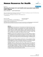
Figure 1 Migration of CTL007 toward WC007 CRC cells in the reconstruct. A. Reconstruct schema. B. [a-f]: The bottom layer of reconstructs
contained 1.8 × 10
5
fibroblasts in 450 μl type I collagen gel which was followed by addition of CRC cells (1 × 10
5
) on top after 24 hr. After
further 24 hr, a separating layer of fibroblasts in collagen gel (100 μl, 500 μm) was added on top of cancer cells, followed by the top layer
containing CTL (1 × 10
5
[32]), or autologous PHA blasts (control lymphocytes [a, c, e]) mixed with 1 × 10
5
fibroblasts and collagen. Reconstructs
were harvested on day 6 (3 days after adding T cells), fixed in buffered formalin and embedded in paraffin. a, b: Staining with H&E. c, d: Specific
brown staining of apoptotic cells by anti-caspase 3 Ab. e, f: In situ end-labeling (ISEL); black staining of nuclei of cells undergoing apoptosis.
Apoptosis was significantly higher in presence of CTL (d and f) than in presence of autologous PHA blasts (c and e; p < 0.0005). g-h:
Reconstructs were prepared as in a-f, but tumor cells were stained with CellTracker Blue CMAC; autologous PHA blasts (g) and CTL007 (h) were
pre-stained with CFDA-Green. Reconstructs were harvested on day 4 (2 days after adding T cells), and sections were photographed in the Nikon
fluorescence microscope using appropriate filters. Arrows indicate binding of CTL007 to the tumor cells.
Berencsi et al . Journal of Translational Medicine 2011, 9:33
/>Page 5 of 11
Thus, several phenotypic markers are expressed by
CTL007 and WC007, which are known to facilitate
interactions between these cells and between the lym-
phocytes or tumor cells and collagen or fibroblasts in
the r econstruct, leading to activation of T cells as well
as T-cell migration toward tumor cells.
Chemokine and chemokine receptor involved in CTL007
migration toward WC007 cells
CTL007 express the chemokine receptors CCR1, CCR2,
CCR3, CCR5, CCR7, CCR9, CXCR1, CXCR2, CXCR3,
CXCR4, and CXCR5 (Table 4). For each chemokine
Table 2 Apoptosis induction by CTL007 in reconstruct with autologous WC007 CRC cells*
Lymphocytes E:T Total number of tumor cells mean
± SD/field (10 fields)
Number of apoptotic tumor cells mean
± SD/field (10 fields)
Percentage of apoptotic cells, mean
± SD/field (10 fields)
Experiment I
CTL007 1:1 80.5 ± 38.8 23.5 ± 8.7 31.7 ± 11
c, d
PHA blast 1:1 136.2 ± 54.8 15.9 ± 6.8 12.1 ± 3.9
c
No
lymphocytes
NA 90.1 ± 3.5 7.1 ± 2.8 8.1 ± 3.5
d
Experiment II
CTL007 10:1 116.7 ± 19.4 94.8 ± 19 79.5 ± 6.7
a, b
PHA blast 10:1 118.3 ± 23.6 34.7 ± 11.8 27.5 ± 4.7
a
No
lymphocytes
NA 121.1 ± 22.3 25.8 ± 10.7 21.3 ± 10.1
b
a-d
Values with the same symbol differ significantly from each other (Student’s 2-sided t-test; p < 0.0001.
* Reconstructs were established with a separating collagen-fibroblast layer, CTL007 or autologous PHA blasts were added onto the top layer. Reconstructs were
harvested either on day 3 (experiment I) or day 4 (experiment II) after adding T cells. Day of harvesting of reconstruct cultures was based on optimum
constriction of the collagen gel of reconstruct cultures. The ratio of apoptotic tumor cells was determined by counting apoptotic nuclei and intact tumor cells in
sections stained with H&E. Data represented are from two independent experiments.
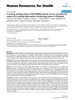
Figure 2 T ime course of WC007 CRC apoptosis induction by
CTL007 in reconstruct. Reconstructs were prepared as in Fig.1. An
E: T ratio of 2:1 was used in this assay, reconstructs harvested on
day 6 or 8 (3 or 5 days after adding T cells), fixed, processed and
enumerated as described in Fig.1. Values represent mean
percentage of apoptosis/field (total of 10 fields), of cultures with
lymphocytes corrected by the value obtained without lymphocytes,
± SD (bars). Percent apoptotic tumor cells of reconstruct cultures
with CTL (▲) were significantly higher than cultures with PHA blast
(■) on both days, for cultures with and without separating layer (at
p < 0.05 level). Percent apoptotic tumor cells of reconstruct cultures
was significantly (p = 0.005) higher on day 5 than on day 3 for
cultures with and without separating layer. The separating layer had
no significant effect on percent apoptotic cells.
Table 3 Phenotypic and functional markers of anti-CRC
CTL007 and autologous WC007 tumor cell line
Parameter investigated
a
Cell lines (% cells positive)
CRC WC007 CTL007
HLA Class I 99.8 95.6
HLA Class II < 1 60
CD4 NA 96
CD8 NA < 1
CD25 < 1 30
CD40L 2.1 13
CD44 48 79.7
CD80 (B7-1) 85 < 1
CD49a (a1 integrin) 48.2 26.1
CD49b (a2 integrin) 63.2 32.5
CD29 (b1 integrin) 56.9 79.8
CD95 (FAS) 65.1 95.1
CD95L (FASL) 19 < 1
CD54 (ICAM-1) 84.5 89.3
CD11a (LFA-1a) < 1 84.2
NA = not applicable.
a
All markers were determined by flow cytometry and data represented are
from a single experiment and the Results wer e confirmed in at least two
independent experiments.
Berencsi et al . Journal of Translational Medicine 2011, 9:33
/>Page 6 of 11
receptor, with the exception of CCR3, CXCR1, CXCR2,
CXCR4, and CXCR5 the corresponding chemokine(s)
was expressed by WC007 CRC cells, as determined by
RT-PCR and protein expression confirmed by ELISA
(Table 4).
We evaluated possible roles of chemokine receptors
CCR1, CCR2, CCR3, CCR5, CCR7 and CXCR3 expressed
by the T cells in the migration of the CTL cells toward
CRC cells in the reconstruct. T-cell migration was mea-
sured as a function of tumor cell apoptosis and not abso-
lute number of T cells at the tumor cell layer, since
T cells may themselves undergo apoptosis after inducing
tumor cell apoptosis and one T cell may induce apoptosis
in more than one tumor cell. Onl y apoptotic tumor cells,
and not T cells, were counted. Apoptotic tumor cells
could be distinguished from apoptotic T cells based on
Table 4 Chemokine receptors expressed by CTL007, and chemokines produced by WC007 CRC cells
Chemokine receptors expressed by CTL007
a
Chemokines
Chemokine receptors % positive
cells
Known to bind
to receptor
Expressed by WC007
b
RT-PCR ELISA (pg/ml)
CCR1 5.7 CCL3 + < 30
c
CCL5 - ND
d
CCL7 - ND
CCL13 - ND
CCL15 + 35
CCR2 65 CCL2 + 51.6
CCL7 - ND
CCL8 - ND
CCL13 - ND
CCR3 19.4 CCL7 - ND
CCL8 - ND
CCL11 - ND
CCL13 - ND
CCL24 - ND
CCR5 10.1 CCL3 + < 30
c
CCL4 + 16
CCL5 - ND
CCR7 9.5 CCL19 + < 20
c
CCL21 + < 30
c
CCR9 16.4 CCL25 - ND
CXCR1 16.7 CXCL6 - ND
CXCL8 - ND
CXCR2 15.4 CXCL1 - ND
CXCL5 - ND
CXCL6 - ND
CXCL7 - ND
CXCL8 - ND
CXCR3 16.9 CXCL9 - ND
CXCL10 - ND
CXCL11 + 35.6
CXCL13 - ND
CXCR4 17.6 CXCL12 - ND
CXCR5 32.4 CXCL13 - ND
a
Chemokine receptor expression as determined by FACS and the following chemokine receptors were not expressed by CTL007:CCR4, CCR6, CCR8, CCR10,
CX3CR1.
b
Chemokine expression was detected by RT-PCR and if they were positive in RT-PCR, then protein expression was confirmed by ELISA. CCL3, CCL19 and CCL21
mRNA were detected in WC007 cells by RT-PCR, but the protein expression was below detection limit of.
ELISA. Data represented are from a single experiment and the Results were confirmed in at least two independent experiments.
c
below detection limit.
d
ND-Not determined.
Berencsi et al . Journal of Translational Medicine 2011, 9:33
/>Page 7 of 11
size difference. However, evaluation of apoptotic tumor
cells does not allow us to distinguish between T cells
with high migratory a nd l ow lytic activity and T cells
with low migratory and high lytic activity. Nevertheless
the ratio of apoptotic tumor cells correlates with CTL
migration. Blocking of chemokine receptor CCR2 but not
CCR1, CCR3, CCR5, CCR7 or CXCR3 on CTL007 with
Abs significantly inhibited tumor cell apoptosis (Table 5).
To determine the involvement of CCR2 ligand (CCL2) in
T cell migration and apoptosis, excess recombinant
CCL2 o r anti-CCL2 A b was ad ded to the top of the
T-cell layer or to the separating layer. Both excess CCL2
and anti-CCL2 Ab were able to inhibit T cell migration
and tu mor cell apoptosis significantly (p < 0.05, Ta ble 5).
InvolvementofCCR2andCCL2ininductionofmigra-
tion of CTL007 was further confirmed in chemotaxis
assay u sing Transwell plates (Figure 3). Treatment of
T cells wi th anti-CCR2 Ab significantly inhibited (p <
0.001) the migration of CTL007 toward recombinant
CCL2, whereas control IgG treatment of T cells had no
effect(Figure3).Thesedatafurther confirm of our find-
ing that CCR2 receptor on T cells and CCL2 secreted by
tumor cells are involved in migration of T c ells t owards
tumor cells. Predominant e xpression of CCR2 (65%)
when compared to other chemokine receptors (most
<20% except CXC5; Table 4 Figure 3B) further supports
the role of CCR2 in T cell migration. Relative high
expressions of CCR2 (65%) on CTL007 and CXCR3
(48.3%) on CT020 (Figure 3B) respectively suggests
dominant expression of certain chemokine receptors
(CCR2 for CTL007 and CXCR3 for CTL020) may be a
decisive factor in migration of lymphocytes.
CCR2 and CCL2 expression by T cells and tumor cell lines,
respectively, derived from additional CRC patients
We inv estigated the distribution of CCR2 and CCL2 in
T cells and tumor cell lines, respectively, established
from specimens of additional CRC patients. Expression
of CCR2 receptor and its ligand by the cells of addi-
tional CRC patients would suggest that involvement of
the receptor/ligand in T-cell migration toward tumor
cells may not be a unique observation made in a single
CRC patient, but may be found in other p atients. Ten
tumor reactive T-cell lines derived from TIL of addi-
tional CRC patients were analyzed for CCR2 expression
and four of these showed CCR2 expression (Table 1).
Furthermore, two of the established CRC cell lines pro-
duced CCL2 (data not shown).
Discussion
We have demonstrated here that CTL007 migrate
through a 500 μm collagen/fibroblast separating layer
toward tumor cells, resulting in tumor cell apoptosis.
We have also shown that migration is dependent on
CCR2 expressed by T cells and CCL2 secreted by tumor
cells.
Our recently developed novel three-dimensional cul-
ture system offers a unique way of studying migration of
leukocytes toward tumor cells and the factors that influ-
ence leukocyte migration under physiological conditions
[32,34]. As described in our previous studies, human
CRC is grown in vitro under three-dimensional condi-
tions using a mixture of collagen and fibroblasts [32,44].
Interaction of a2andb1 integrins on CRC-specific
T cells with collagen and the presence of activated fibro-
blasts help to maint ain Ag-specific T cells in a state of
activation in absence of exogenous addition of IL-2
[36,37]. In addition, T cells cou ld interact with fibro-
blasts via adhesion molecules like LFA-1a, ICAM-1 and
CD44 which could in turn stimulate fibroblasts to
secrete inflammatory cytokines such as IL-1, IL-6, IL-7
[38,39], and fibronectin [41]. IL-1 could stimulate
T cells to express IL-2 receptor and induce secretion of
IL-2 [45]. IL-6 and IL-7 are T-cell survival factors [46],
and fibronectin stimulates predominantly resting lym-
phocytes [41]. Other investigators have used collagen
matrices to study interaction of leukocytes with tumor
cells, but they have not d emonstrated CTL migration
resulting in tumor cell apoptosis in a culture system
similar to the reconstruct cultures shown here [47-49].
In the present study, CCL2 produced by CRC cells
attracts CTL through binding of CCL2 to its corre-
sponding chemokine receptor CCR2 on the T cells. This
was demonstrated by blocking of T-cell migration in
presence of addition of excess chemokine CCL2 in the
T-cell layer of th e reconstruct or the addition of Ab s to
CCL2 or CCR2, each applied on top of the separating
collagen/fibroblast layer. Involvement of CCR2 in T-cell
migration was further confir med in chemo taxis assay in
Transwell plate experiment by treating T cells with anti-
CCR2 Ab. Although CTL007 has several chemokine
receptors matching chemokine produced by CRC cells,
predominant expression of CCR2 (65%) by the T cells
and t he relatively higher amount of CCL2 secretion b y
tumor cells may have been a decisive factor in T-cell
migration.
Many carcinomas, including breast, colorectal, pancrea-
tic and renal c arcinomas, and neuro-ectodermal tumors
such as melanomas, medullo blastomas, neuroblastomas
and glioblastomas are known to produce CCL2 (reviewed
in [1]; [20-22,50]. CCL2 secretion by tumor cells can aid
in tumor progression, angiogenesis and metastasis [1,5,10].
Also, the secretion of CCL2 by tumor cells results in infil-
tration of tumor cells by leukocytes including T cells,
NKT cells and macrophages (reviewed in [1]; [20-22]). To
our knowledge, the role of CCL2-dependent T-cell migra-
tion in CRC is l argely unknown. In mouse tumor model
systems, melanoma cells secreting high amounts of CCL2
Berencsi et al . Journal of Translational Medicine 2011, 9:33
/>Page 8 of 11
attract macrophages resulting in inhibition of tumor
growth. However, tumor cells secreting low amounts
of CCL2 promote tumor growth by stimulating angio-
genesis [23]. In another study, tumor cells transfected
with CCL2 showed decreased metastasis due to
increased infiltration of macrophages and susceptibility
of tumor cells to lysis by infiltrating macrophages [24].
Thus, patients may be vaccinated with chemokine-
transduced tumor cells [51] or tumor-associated Ags
fused to chemokines [52]; alternatively, patients may
be treated with anti-tumor Ab/chemokine fusion pro-
tein, which may attract adoptively transferred lympho-
cytes to the tumor area [12,52,53]. In light of the
mouse study by Nesbit et.al. , [23], one needs to care-
fully modulate the expression of CCL2 to attract
immune cells toward tumor cells. Thus, chemokines
may be useful for immunotherapy of cancer patients.
In addition to therapeutic implications, the results of
our study have prognostic potential. Infiltration of
CRC with T lymphocytes is correlated with a favorable
prognosis [54], and chemokine r eceptor expression by
T lymphocytes a s well as chemokine producti on by
tumor cells should be explored for their possible asso-
ciation with prognosis. Identification of tumor cells
secreting CCL2 or T-cells expressing CCR2 in the
tumor microenvironment couldbeausefulprognostic
marker in CRC patients [17,18,22,55-58]. In our earlier
study, we have shown that migration of CTL derived
from a CRC patient towards autologous tumor cells
was mediated by CXCR3 expresse d by the T cells, and
CXCL11 chemokine secreted by the autologous tumor
cells [32]. In the present study, we show that migration
of CTL is dependent on CCL2 and CCR2. Presence of
CCR2 in T cells obtained from four of the ten addi-
tional patients suggests that CCR2 expression may not
be that uncommon. It is likely that in each individual
CRC patient a unique chemokine/ch emokine receptor
pair might be involved in attraction of T cells towards
tumorcells.Hence,itisessentialtoidentifytheche-
mokine/chemokine receptor pair responsible for
Table 5 Induction of tumor apoptosis by CTL007 is
inhibited by anti-CCR2 and CCL2 Abs and excess CCL2a
Blocking
agent
Number
of
tumor
cells/
field
(10
fields)
Number of
apoptotic cells/
field (10 fields)
%of
apoptotic
cells
% of tumor
cell apoptosis
inhibition
None 21.8 ± 9.7 8.2 ± 9 40.5 ± 9.7
b
-
Control
IgG
21 ± 2.2 8.4 ± 1.1 40.1 ± 4.9
c
-
Anti-CCR1
Ab
22 ± 3.4 10.0 ± 1.6 45.6 ± 5.0 -13.7
Anti-CCR2
Ab
19.6 ± 1.1 2.6 ± 0.5 13.2 ± 2.3
c
67.1
Anti-CCR3
Ab
22.4 ± 2.8 9.2 ± 1.9 40.7 ± 4.2 1.5
Anti-CCR5
Ab
21 ± 2.6 9.6 ± 1.8 45.5 ± 3.8 -13.4
Anti-CCR7
Ab
18.8 ± 2.4 7.2 ± 1.6 38.0 ± 4.1 5.2
Anti-
CXCR3
Ab
21.4 ± 1.5 9.2 ± 0.8 43.1 ± 1.8 -7.5
CCL2 21.4 ± 4.4 3.6 ± 1.1 17.2 ± 6.1
b
57.5
Anti-CCL2
Ab
17.8 ± 2.6 2.2 ± 0.4 12.7 ± 3.8
c
68.6
a
Reconstructs consisted of a bottom layer of collagen and fibroblasts, followed
by a tumor cell layer and a separating layer of collagen and fibroblasts. Anti-
chemokine receptor or control Abs were added, followed by a top layer
containing CTL mixed with fibroblasts and collagen (E: T = 3:1). Excess CCL2
(50 ng/ml) was added to the T-cell layer. Percentage of apoptotic tumor cells
in 6 day cultures was determined.
(b, c)
Values with same letter differ
significantly from each other (p < 0.05). Data represented are from a single
experiment and the Results were confirmed in at least two independent
experiments.
Figure 3 A. CTL007 migrat es toward CCL2 in Transwell. T cells were placed in the top chamber of the Transwell. CCL2 (10 ng/ml) in T cell
medium, or T cell medium alone were added to the bottom of the Transwell (duplicate wells). After 90 min of culture, the number of migrated
cells in the bottom chamber was counted under the microscope. Some T cell cultures were pre-incubated and treated with saturating
concentration (10 μg/ml) of mouse anti-CCR2 Ab or mouse IgG as control. B. Expression of CCR2. T cells grown in log phase were incubated
with either anti-CCR2 or anti-CXCR3 Ab (black or grey solid line) or mouse IgG control (dotted line) in RPMI 1640 with 5% human AB serum for
1 h at 4°C. After washing, FITC-labeled anti-mouse IgG was added. Expression of chemokine receptors was detected by flow cytometry.
Berencsi et al . Journal of Translational Medicine 2011, 9:33
/>Page 9 of 11
attraction of T cells towards tumor in each CRC
patient for individualized therapy.
Conclusions
Our study demonstrates the role of CCR2 and CCL2 in
migration o f CTLs towards tumor. Data obtained from
additional CRC patients further strengthen the role of
CCR2 and CCL2 in lymphocyte migration. Identification
of chemokine/chemokine receptor p air responsible for
attraction of T cells towards tumor in each patient will
aid individualized therapy approaches using gene modi-
fication of tumor cells with chemokine or chemokine/
anti-tumor Ab fusion proteins to attract CTLs that
potentially could inhibit tumor growth.
Acknowledgements
We thank James Hayden and Frederick Keeney for technical help on
microscopy imaging and Jeffrey S. Faust, David Ambrose and Daniel Hussey
for assistance in flow cytometry analyses. We thank Elsa Aglow and Russell
Delgiacco for providing assistance in histotechnology. We also thank Drs.
Yingtao Bi and Ramana Davuluri for statistical data analyses. This work is
supported by National Institute of Health grants CA74294, and CA10815, by
a grant from Corixa Corporation, by Intramural National Cancer Institute
Funds, and by the Commonwealth Universal Research Enhancement
program, Pennsylvania Department of Health.
Author details
1
The Wistar Institute, 3601 Spruce Street, Philadelphia, PA 19104, USA.
2
Department of Medical Oncology, Thomas Jefferson University, 1015 Walnut
Street, Philadelphia, PA 19107, USA.
3
Department of Medical Oncology, Fox
Chase Cancer Center, 333 Cottman Avenue, Philadelphia, PA 19111, USA.
4
Division of Hematology and Oncology, University Hospitals Seidman Cancer
Center and Case Western Reserve University, 11100 Euclid Avenue, Lakeside
1200, Cleveland, OH 44106-5065, USA.
Authors’ contributions
KB carried out reconstruct studies and all the data analyses; PR carried out
histological staining, RT-PcR of chemokines; TZ performed ELISA assays for
chemokines; LG cultured and expanded CTLs for the assay, and
characterized chemokine receptor expression on T cells; MM and NM
recruited patients for the study, DH and RS designed the study and in
coordination with all others drafted the manuscript. All authors read and
approved the final version of the manuscript.
Competing interests
The authors declare that they have no competing interests.
Received: 15 September 2010 Accepted: 30 March 2011
Published: 30 March 2011
References
1. Somasundaram R, Herlyn D: Chemokines and the microenvironment in
neuroectodermal tumor-host interaction. Semin Cancer Biol 2009,
19:92-96.
2. Luster AD: Chemokines–chemotactic cytokines that mediate
inflammation. N Engl J Med 1998, 338:436-445.
3. Baggiolini M: Chemokines in pathology and medicine. J Intern Med 2001,
250:91-104.
4. Mackay CR: Chemokines: immunology’s high impact factors. Nat Immunol
2001, 2:95-101.
5. Fridlender ZG, Buchlis G, Kapoor V, Cheng G, Sun J, Singhal S, Crisanti MC,
Wang LC, Heitjan D, Snyder LA, Albelda SM: CCL2 blockade augments
cancer immunotherapy. Cancer Res 70:109-118.
6. Payne AS, Cornelius LA: The role of chemokines in melanoma tumor
growth and metastasis. J Invest Dermatol 2002, 118:915-922.
7. Raman D, Baugher PJ, Thu YM, Richmond A: Role of chemokines in tumor
growth. Cancer Lett 2007, 256:137-165.
8. Ghadjar P, Rubie C, Aebersold DM, Keilholz U: The chemokine CCL20 and
its receptor CCR6 in human malignancy with focus on colorectal cancer.
Int J Cancer 2009, 125:741-745.
9. Akishima-Fukasawa Y, Nakanishi Y, Ino Y, Moriya Y, Kanai Y, Hirohashi S:
Prognostic significance of CXCL12 expression in patients with colorectal
carcinoma. Am J Clin Pathol 2009, 132:202-210, quiz 307.
10. Loberg RD, Ying C, Craig M, Day LL, Sargent E, Neeley C, Wojno K,
Snyder LA, Yan L, Pienta KJ: Targeting CCL2 with systemic delivery of
neutralizing antibodies induces prostate cancer tumor regression in
vivo. Cancer Res 2007, 67:9417-9424.
11. Frederick MJ, Clayman GL: Chemokines in cancer. Expert Rev Mol Med 2001,
3:1-18.
12. Homey B, Muller A, Zlotnik A: Chemokines: agents for the
immunotherapy of cancer? Nat Rev Immunol 2002, 2:175-184.
13. Vicari AP, Caux C: Chemokines in cancer. Cytokine Growth Factor Rev 2002,
13:143-154.
14. Erreni M, Bianchi P, Laghi L, Mirolo M, Fabbri M, Locati M, Mantovani A,
Allavena P: Expression of chemokines and chemokine receptors in
human colon cancer. Methods Enzymol 2009, 460:105-121.
15. Galon J, Costes A, Sanchez-Cabo F, Kirilovsky A, Mlecnik B, Lagorce-Pages C,
Tosolini M, Camus M, Berger A, Wind P, Zinzindohoué F, Bruneval P,
Cugnenc PH, Trajanoski Z, Fridman WH, Pagès F: Type,
density, and
location of immune cells within human colorectal tumors predict clinical
outcome. Science 2006, 313:1960-1964.
16. Pages F, Berger A, Camus M, Sanchez-Cabo F, Costes A, Molidor R,
Mlecnik B, Kirilovsky A, Nilsson M, Damotte D, Meatchi T, Bruneval P,
Cugnenc PH, Trajanoski Z, Fridman WH, Galon J: Effector memory T cells,
early metastasis, and survival in colorectal cancer. N Engl J Med 2005,
353:2654-2666.
17. Musha H, Ohtani H, Mizoi T, Kinouchi M, Nakayama T, Shiiba K, Miyagawa K,
Nagura H, Yoshie O, Sasaki I: Selective infiltration of CCR5(+)CXCR3(+) T
lymphocytes in human colorectal carcinoma. Int J Cancer 2005,
116:949-956.
18. Ohta M, Tanaka F, Yamaguchi H, Sadanaga N, Inoue H, Mori M: The high
expression of Fractalkine results in a better prognosis for colorectal
cancer patients. Int J Oncol 2005, 26:41-47.
19. Tosolini M, Kirilovsky A, Mlecnik B, Fredriksen T, Mauger S, Bindea G,
Berger A, Bruneval P, Fridman WH, Pages F, Galon J: Clinical impact of
different classes of infiltrating T cytotoxic and helper cells (Th1, th2,
treg, th17) in patients with colorectal cancer. Cancer Res 71:1263-1271.
20. Baier PK, Eggstein S, Wolff-Vorbeck G, Baumgartner U, Hopt UT:
Chemokines in human colorectal carcinoma. Anticancer Res 2005,
25:3581-3584.
21. Bailey C, Negus R, Morris A, Ziprin P, Goldin R, Allavena P, Peck D, Darzi A:
Chemokine expression is associated with the accumulation of tumour
associated macrophages (TAMs) and progression in human colorectal
cancer. Clin Exp Metastasis 2007, 24:121-130.
22. Monti P, Leone BE, Marchesi F, Balzano G, Zerbi A, Scaltrini F, Pasquali C,
Calori G, Pessi F, Sperti C, Di Carlo V, Allavena P, Piemonti L: The CC
chemokine MCP-1/CCL2 in pancreatic cancer progression: regulation of
expression and potential mechanisms of antimalignant activity. Cancer
Res 2003, 63:7451-7461.
23. Nesbit M, Schaider H, Miller TH, Herlyn M: Low-level monocyte
chemoattractant protein-1 stimulation of monocytes leads to tumor
formation in nontumorigenic melanoma cells. J Immunol 2001,
166:6483-6490.
24. Huang S, Singh RK, Xie K, Gutman M, Berry KK, Bucana CD, Fidler IJ, Bar-Eli M:
Expression of the JE/MCP-1 gene suppresses metastatic potential in
murine colon carcinoma cells. Cancer Immunol Immunother 1994, 39:231-238.
25. Dranoff G: Cytokines in cancer pathogenesis and cancer therapy. Nat Rev
Cancer 2004, 4:11-22.
26. Braun SE, Chen K, Foster RG, Kim CH, Hromas R, Kaplan MH, Broxmeyer HE,
Cornetta K: The CC chemokine CK beta-11/MIP-3 beta/ELC/Exodus 3
mediates tumor rejection of murine breast cancer cells through NK cells.
J Immunol 2000, 164:4025-4031.
27. Fushimi T, O’Connor TP, Crystal RG: Adenoviral gene transfer of stromal
cell-derived factor-1 to murine tumors induces the accumulation of
dendritic cells and suppresses tumor growth. Cancer Res 2006,
66:3513-3522.
Berencsi et al . Journal of Translational Medicine 2011, 9:33
/>Page 10 of 11
28. Sharma S, Stolina M, Luo J, Strieter RM, Burdick M, Zhu LX, Batra RK,
Dubinett SM: Secondary lymphoid tissue chemokine mediates T cell-
dependent antitumor responses in vivo. J Immunol 2000, 164:4558-4563.
29. Mitchell MS, Darrah D, Yeung D, Halpern S, Wallace A, Voland J, Jones V,
Kan-Mitchell J: Phase I trial of adoptive immunotherapy with cytolytic T
lymphocytes immunized against a tyrosinase epitope. J Clin Oncol 2002,
20:1075-1086.
30. Buckanovich RJ, Facciabene A, Kim S, Benencia F, Sasaroli D, Balint K,
Katsaros D, O’Brien-Jenkins A, Gimotty PA, Coukos G: Endothelin B receptor
mediates the endothelial barrier to T cell homing to tumors and
disables immune therapy. Nat Med 2008, 14:28-36.
31. Penichet ML, Morrison SL: Antibody-cytokine fusion proteins for the
therapy of cancer. J Immunol Methods 2001, 248:91-101.
32. Berencsi K, Meropol NJ, Hoffman JP, Sigurdson E, Giles L, Rani P,
Somasundaram R, Zhang T, Kalabis J, Caputo L, Furth E, Swoboda R,
Marincola F, Herlyn D: Colon carcinoma cells induce CXCL11-dependent
migration of CXCR3-expressing cytotoxic T lymphocytes in organotypic
culture. Cancer Immunol Immunother 2007, 56:359-370.
33. Somasundaram R, Jacob L, Swoboda R, Caputo L, Song H, Basak S,
Monos D, Peritt D, Marincola F, Cai D, Birebent B, Bloome E, Kim J,
Berencsi K, Mastrangelo M, Herlyn D: Inhibition of cytolytic T lymphocyte
proliferation by autologous CD4+/CD25+ regulatory T cells in a
colorectal carcinoma patient is mediated by transforming growth factor-
beta. Cancer Res 2002, 62:5267-5272.
34. Zhang T, Somasundaram R, Berencsi K, Caputo L, Gimotty P, Rani P,
Guerry D, Swoboda R, Herlyn D: Migration of cytotoxic T lymphocytes
toward melanoma cells in three-dimensional organotypic culture is
dependent on CCL2 and CCR4. Eur J Immunol 2006, 36:457-467.
35. Felgar RE, Furth EE, Wasik MA, Gluckman SJ, Salhany KE: Histiocytic
necrotizing lymphadenitis (Kikuchi’s disease): in situ end-labeling,
immunohistochemical, and serologic evidence supporting cytotoxic
lymphocyte-mediated apoptotic cell death. Mod Pathol 1997, 10:231-241.
36. Friedl P, Entschladen F, Conrad C, Niggemann B, Zanker KS: CD4+ T
lymphocytes migrating in three-dimensional collagen lattices lack focal
adhesions and utilize beta1 integrin-independent strategies for
polarization, interaction with collagen fibers and locomotion. Eur J
Immunol 1998, 28:2331-2343.
37. Rao WH, Hales JM, Camp RD: Potent costimulation of effector T
lymphocytes by human collagen type I. J Immunol 2000, 165:4935-4940.
38. Aiba S, Nakagawa S, Hara M, Tomioka Y, Deguchi M, Tagami H: Cultured
murine dermal cells can function like thymic nurse cells. J Invest
Dermatol 1994, 103:162-167.
39. Murakami S, Okada H: Lymphocyte-fibroblast interactions. Crit Rev Oral
Biol Med 1997, 8:40-50.
40. Crowston JG, Salmon M, Khaw PT, Akbar AN: T-lymphocyte-fibroblast
interactions. Biochem Soc Trans 1997, 25:529-531.
41. Maas-Szabowski N, Stark HJ, Fusenig NE: Keratinocyte growth regulation in
defined organotypic cultures through IL-1-induced keratinocyte growth
factor expression in resting fibroblasts. J Invest Dermatol 2000,
114:1075-1084.
42. Maaser K, Wolf K, Klein CE, Niggemann B, Zanker KS, Brocker EB, Friedl P:
Functional hierarchy of simultaneously expressed adhesion receptors:
integrin alpha2beta1 but not CD44 mediates MV3 melanoma cell
migration and matrix reorganization within three-dimensional
hyaluronan-containing collagen matrices. Mol Biol Cell 1999, 10:3067-3079.
43. Binnerts ME, van Kooyk Y, Simmons DL, Figdor CG: Distinct binding of T
lymphocytes to ICAM-1, -2 or -3 upon activation of LFA-1. Eur J Immunol
1994, 24:2155-2160.
44. Zhang T, Somasundaram R, Berencsi K, Caputo L, Rani P, Guerry D, Furth E,
Rollins BJ, Putt M, Gimotty P, Swoboda R, Herlyn M, Herlyn D: CXC
chemokine ligand 12 (stromal cell-derived factor 1 alpha) and CXCR4-
dependent migration of CTLs toward melanoma cells in organotypic
culture. J Immunol 2005, 174:5856-5863.
45. Starnes HF Jr: Biological effects and possible clinical applications of
interleukin 1. Semin Hematol 1991, 28:34-41.
46. Rathmell JC, Farkash EA, Gao W, Thompson CB: IL-7 enhances the survival
and maintains the size of naive T cells. J Immunol 2001, 167:6869-6876.
47. Wei WZ, Miller B, Gutierrez RM: Inhibition of tumor growth by peptide
specific cytotoxic T lymphocytes in a three-dimensional collagen matrix.
J Immunol Methods 1997, 200:47-54.
48. Jacobs N, Moutschen MP, Franzen-Detrooz E, Boniver V, Boniver J,
Delvenne P: Organotypic culture of HPV-transformed keratinocytes: a
model for testing lymphocyte infiltration of (pre)neoplastic lesions of
the uterine cervix. Virchows Arch 1998, 432:323-330.
49. Tasaki A, Yamanaka N, Kubo M, Matsumoto K, Kuroki H, Nakamura K,
Nakahara C, Onishi H, Kuga H, Baba E, Tanaka M, Morisaki T, Katano M:
Three-dimensional two-layer collagen matrix gel culture model for
evaluating complex biological functions of monocyte-derived dendritic
cells. J Immunol Methods 2004, 287:79-90.
50. Brown CE, Vishwanath RP, Aguilar B, Starr R, Najbauer J, Aboody KS,
Jensen MC: Tumor-derived chemokine MCP-1/CCL2 is sufficient for
mediating tumor tropism of adoptively transferred T cells. J Immunol
2007, 179:3332-3341.
51. Zibert A, Balzer S, Souquet M, Quang TH, Paris-Scholz C, Roskrow M,
Dilloo D: CCL3/MIP-1alpha is a potent immunostimulator when
coexpressed with interleukin-2 or granulocyte-macrophage colony-
stimulating factor in a leukemia/lymphoma vaccine. Hum Gene Ther 2004,
15:21-34.
52. Biragyn A, Tani K, Grimm MC, Weeks S, Kwak LW: Genetic fusion of
chemokines to a self tumor antigen induces protective, T-cell
dependent antitumor immunity. Nat Biotechnol 1999, 17:253-258.
53. Biragyn A, Kwak LW: Designer cancer vaccines are still in fashion. Nat
Med 2000, 6:966-968.
54. Guidoboni M, Gafa R, Viel A, Doglioni C, Russo A, Santini A, Del Tin L,
Macri E, Lanza G, Boiocchi M, Dolcetti R:
Microsatellite instability and high
content of activated cytotoxic lymphocytes identify colon cancer
patients with a favorable prognosis. Am J Pathol 2001, 159:297-304.
55. Longo-Imedio MI, Longo N, Trevino I, Lazaro P, Sanchez-Mateos P: Clinical
significance of CXCR3 and CXCR4 expression in primary melanoma. Int J
Cancer 2005, 117:861-865.
56. Kim J, Mori T, Chen SL, Amersi FF, Martinez SR, Kuo C, Turner RR, Ye X,
Bilchik AJ, Morton DL, Hoon DS: Chemokine receptor CXCR4 expression in
patients with melanoma and colorectal cancer liver metastases and the
association with disease outcome. Ann Surg 2006, 244:113-120.
57. Monteagudo C, Martin JM, Jorda E, Llombart-Bosch A: CXCR3 chemokine
receptor immunoreactivity in primary cutaneous malignant melanoma:
correlation with clinicopathological prognostic factors. J Clin Pathol 2007,
60:596-599.
58. Varney ML, Johansson SL, Singh RK: Distinct expression of CXCL8 and its
receptors CXCR1 and CXCR2 and their association with vessel density
and aggressiveness in malignant melanoma. Am J Clin Pathol 2006,
125:209-216.
doi:10.1186/1479-5876-9-33
Cite this article as: Berencsi et al.: In vitro migration of cytotoxic T
lymphocyte derived from a colon carcinoma patient is dependent on
CCL2 and CCR2. Journal of Translational Medicine 2011 9:33.
Submit your next manuscript to BioMed Central
and take full advantage of:
• Convenient online submission
• Thorough peer review
• No space constraints or color figure charges
• Immediate publication on acceptance
• Inclusion in PubMed, CAS, Scopus and Google Scholar
• Research which is freely available for redistribution
Submit your manuscript at
www.biomedcentral.com/submit
Berencsi et al . Journal of Translational Medicine 2011, 9:33
/>Page 11 of 11