DSpace at VNU: An in silico study on antidiabetic activity of bioactive compounds in Euphorbia thymifolia Linn.
Bạn đang xem bản rút gọn của tài liệu. Xem và tải ngay bản đầy đủ của tài liệu tại đây (3.73 MB, 13 trang )
Nguyen Vo et al. SpringerPlus (2016) 5:1359
DOI 10.1186/s40064-016-2631-5
Open Access
RESEARCH
An in silico study on antidiabetic activity
of bioactive compounds in Euphorbia thymifolia
Linn.
T. Hoang Nguyen Vo1†, Ngan Tran1†, Dat Nguyen1 and Ly Le1,2*
Abstract: Herbal medicines have become strongly preferred treatment to reduce the negative impacts of diabetes
mellitus (DM) and its severe complications due to lesser side effects and low cost. Recently, strong anti-hyperglycemic
effect of Euphorbia thymifolia Linn. (E. thymifolia) on mice models has reported but the action mechanism of its
bioactive compounds has remained unknown. This study aimed to evaluate molecular interactions existing between
various bioactive compounds in E. thymifolia and targeted proteins related to Type 2 DM. This process involved the
molecular docking of 3D structures of those substances into 4 targeted proteins: 11-β hydroxysteroid dehydrogenase
type 1, glutamine: fructose-6-phosphate amidotransferase, protein-tyrosine phosphatase 1B and mono-ADP-ribosyltransferase sirtuin-6. In the next step, LigandScout was applied to evaluate the bonds formed between 20 ligands
and the binding sites of each targeted proteins. The results identified seven bioactive compounds with high binding
affinity (<−8.0 kcal/mol) to all 4 targeted proteins including β-amyrine, taraxerol, 1-O-galloyl-β-d-glucose, corilagin,
cosmosiin, quercetin-3-galactoside and quercitrin. The pharmacophore features were also explained in 2D figures
which indicated hydrophobic interactions, hydrogen bond acceptors and hydrogen bond donors forming between
carbonyl oxygen molecules of those ligands and active site residues of 4 targeted protein.
Keywords: E. thymifolia, Diabetes mellitus, Molecular docking, Pharmacophore, LigandScout
Background
Euphorbia thymifolia is a small prostrate herbaceous
annual weed which is abundant in waste places and open
grasslands and distributes in most Asian countries. This
medicinal plant has been studied for many bioactivities and therapeutic effects such as anti-microbial effect
(Killedar et al. 2011), bronchial asthma (Sharma and Tripathi 1984) and the anti-hyperglycemic effect of Euphorbiaceae family has been fully reviewed (Bnouham et al.
2006). Besides that, E. thymifolia has also been traditionally used for treatment of gastrointestinal disorder,
inflammatory and respiratory diseases (Loi 2015).
Diabetes mellitus (DM) and its complications are main
causes of deaths in most countries. Type 2 DM has also
been known as another terms “Non-insulin dependent
*Correspondence:
†
T. Hoang Nguyen Vo and Ngan Tran contributed equally to this work
1
International University – Vietnam National University - HCMC, Quarter
6, Linh Trung Ward, Thu Duc District, Ho Chi Minh City, Vietnam
Full list of author information is available at the end of the article
diabetes mellitus (NIDDM)” which accounted for more
than 90 % of diagnosed cases of DM in adults (International Diabetes Federation (IDF) 2015). In accordance
with Ford et al. (2002), the statistics of patients suffering
Type 2 DM and metabolic syndromes were estimated
about 50 million in the US and 314 million around the
world and this number was predicted to increase dramatically in the next decades. The feature of Type 2 DM
is the partial or complete failure in using insulin (insulin
resistance) even though the functional insulin is available
and then causes hyperglycemia. To overcome this resistance, the pancreatic β cells produce extra mount of insulin to maintain glucose in the normal range. However,
this process is only effective in the short term as burnout
β cell occurs. At this time, the patients have suffered Type
2 DM.
Many efforts to figure out the effective treatments for
Type 2 DM have been increased. For many years, scientists have endeavored to apply not only pharmacological
methods but also non-pharmacological approaches but
© 2016 The Author(s). This article is distributed under the terms of the Creative Commons Attribution 4.0 International License
( which permits unrestricted use, distribution, and reproduction in any medium,
provided you give appropriate credit to the original author(s) and the source, provide a link to the Creative Commons license,
and indicate if changes were made.
Nguyen Vo et al. SpringerPlus (2016) 5:1359
none of them met all safety requirements in medication.
Losing weight and doing exercise have been highly recommended as two major non-drug therapies to increase
insulin sensitivity. In aspect of pharmaceutical science,
although metformin and thiazolidinedione both have
good effect in insulin resistance, they cannot be widely
used because of their undesirable side effects. Currently,
research on relationships between antioxidant compounds and Type 2 DM has been well published and documented. People revealed that an intake of antioxidant in
diet has contributed to reduce the development of Type
2 DM (Montonen, et al. 2004; Evans 2007). Besides, in
2005, Fraga investigated that the intake of dark chocolate
which was a rich source of flavonols could decrease blood
pressure and improved insulin sensitivity in healthy persons (Fraga 2005).
In the light of these evidences, the objective of this
research is to test the anti-hyperglycemic activity of antioxidant compounds in the ethanolic extracts of E. thymifolia by using them as ligands for four targeted proteins to
determine which compound is effective binder. The chemical composition analyzed by GC–MS from areal part of
E. thymifolia suggested three main families: tannin, flavonoid and terpenoid (Sandeep et al. 2009; Prasad and
Bisht 2011) which are strong anti-oxidant compounds.
Possessing polyphenol structure involving high number
of hydroxyl group inside, tannin and flavonoid were, thus,
predicted to be able to form hydrogen bonds with various
reactive oxygen species, such as singlet oxygen, peroxynitrite and hydrogen peroxide which are major causes of cell
damages. Due to this mechanism, tannin and flavonoid
were considered to play potential roles in reducing the
oxidative stress related to Type 2 DM (Evans 2007; Maiese
et al. 2007). Terpenoid is an enormous class of organic
compound in plant whose potential antioxidant activity
has already studied (Gonzalez-Burgos and Gomez-Serranillos 2012). However, there are no research indicating
their affinity for Type 2 DM. Four targeted proteins used
in this study was previously investigated to serve as potential drug target for Type 2 DM (Nguyen and Le 2012; Shi
2009; Vogel 2002). 11β-HSD1 (11β-hydroxysteroid dehydrogenase type I) or “cortisone reductase” is an NADPHdependent enzyme highly expressed in main metabolic
tissues including liver, adipose tissue, and the central
nervous system. In these tissues, HSD11B1 reduces cortisone to the active hormone cortisol that activates glucocorticoid receptors. 11βHSD1 inhibition is a tempting
target for the treatment of glucortinoid-associated diseases, especially of Type 2 DM (Davani, et al. 2004;
Andrews and Walker 1999). Glutamine-fructose-6-phosphate amidotransferase (GFAT or GFPT) is the first and
Page 2 of 13
rate-limiting enzyme of the hexosamine pathway. GFAT
controls the flux of glucose into the hexosamine pathway
and catalyzes the formation of glucosamine 6-phosphate.
The majority of glucose will enter the glycolysis pathway,
with a small percentage entering the hexosamine pathway.
GFPT or GFAT regulate the hexosamine pathway products. Therefore, this enzyme involved in a therapeutic
target against Type 2 DM (Chou 2004). Protein-tyrosine
phosphatase 1B (PTP1B) is a negative regulator of the
insulin signaling pathway and is considered a promising
potential therapeutic target, in particular for treatment
of Type 2 DM. It has also been implicated in the development of breast cancer and has been explored as a potential therapeutic target in that avenue as well. Sirtuin-6 or
Mono-ADP ribosyltransferase-sirtuin-6 (SIRT6) is a stress
responsive protein deacetylase and mono-ADP ribosyltransferase enzyme encoded by the SIRT6 gene. SIRT6
functions in multiple molecular pathways related to aging,
including DNA repair, telomere maintenance, glycolysis
and inflammation. Promisingly, the absence of enzyme
SIRT6 may lead to dramatically induced of blood sugar
level (Hasan et al. 2002). The objective of this study was
to display a range of bioactive compounds from all three
families and determine if and how they interact with proteins that is important to Type 2 DM (Muthumani et al.
2016, Prasad and Bisht 2011, PROTA 2008) (Table 2).
Methods
Molecular docking
Receptor preparation
3D structure of 11-β HSD1, GFAT, PTP1B, SIRT6 were
taken from Protein Data Bank as following 11β-HSD1
(PDB code 1XU7), GFAT (PDB code 2ZJ3), PTP1B
(PDB code 4Y14) and SIRT6 (PDB code 3K35). To verify the capacity of the model in reproducing experimental observation with new ligand, all these structures
were tested again at the binding site. Following this
way, 11β-HSD1 (PDB code 1XU7) was tested again with
molecule:
NADPH
dihydro-nicotinamide-adeninedinucleotide phosphate (NDP), GFAT (PDB code 2ZJ4)
was tested with 2-deoxy-2-amino glucitol-6-phosphate
(AGP), SIRT6 (PDB code 3K35) with adenosine-5-diphosphoribose (APR) and PTP1B (PDB code 4Y14) with
3-bromo-4-[difluoro(phosphono)methyl]-N-methyl-Nalpha-(methylsulfonyl)-l-phenylalaninamide. This work
was done by Autodock Vina (Trott and Olson 2009) and
VMD was used for visualization (Humphrey et al. 1996).
Bioactive compound preparation
Most of the 3D structures of drug molecules in
E.thymifolia were downloaded from PubChem
Nguyen Vo et al. SpringerPlus (2016) 5:1359
Compound section of National Center for Biotechnology
Information (NCBI) and the others were drawn by GaussView 5 (Dennington et al. 2009). Ligands during this
process also being checked for Torsion count to detect
currently active bonds with default settings. Importantly,
amide bonds were checked and treated as non-rotatable.
Ligands were also utilized to merge non-polar hydrogens.
The 2D structures of 20 ligands are illustrated in Table 1.
Docking simulations
Autodock Vina (Trott and Olson 2009) was employed for
binding affinity measurement. The content of configure
file was determined as position of receptor file, ligand
file, data of Grid-box’s three coordinates X, Y, Z were
18.125, −27.72, −0.34 respectively in case of 11β-HSD1,
8.82, 5.31, −7.903 for GFAT, −11.21, −22.77, −6.75 in
PTP1B, 14.5, −18.02 and 17.04 in SIRT6, the size of Grid
box which was set up in 30 × 30 × 30 points, number of
modes which were 10 and the energy range which was set
up at 9 kcal/mol. Docking process in AutoDock Vina has
been performed with 1000 of exhaustiveness for enhancing accuracy.
Pharmacophore analysis
This part of process was carried out by using the pharmacophore tool included in LigandScout (Wolber and
Langer 2005). The program showed us the 2D and 3D
structure with the position and interaction of ligand in
the binding pocket of the receptor. From these 2D figures, some types of bond were identified by color and
symbol. Four features namely hydrogen bond acceptor
(HBA), hydrogen bond donor (HBD), negative ionizable
area (NIA), hydrophobic interaction were labeled as red
arrow, green arrow, red star and orange bubble (supporting information) respectively.
Results and discussion
Free energy binding of bioactive compound to targeted
proteins
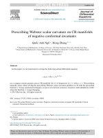
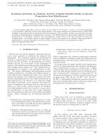
The line chart (Fig. 1) showed the binding capacity of
all three family bioactive compounds: tannin, flavonoid
and terpenoid in E. thymifolia on 4 proteins related to
Type 2 DM in humans. In this chart, tannin and flavonoid families included first seven compounds. Among
those docking result, the absolute value of binding energy
ranged from 7.2 to 10.4 (kcal/mol; Fig. 1). In this range,
the greatest result was in five compounds of both families
which were higher 8 kcal/mol in term of absolute value.
Those are cosmosiin, quercetin-3-galactoside, quercitrin, corilagin, 1-O-galloyl-β-d-glucose and which were
selected for pharmacophore analysis step. Besides that,
Page 3 of 13
kaempferol and quercetin of flavonoid family also had
good results but this was different in each protein, perhaps the amino acid construction of each protein. For
example, the binding affinity of quercetin was 9.7 and
8.3 kcal/mol in 11β HSD1 (1XU7) and SIRT6 (3K35)
respectively, compared to 7.8 and 7.6 in PTP1B (4Y14)
and GFAT1 (2ZJ3). Although there have been fluctuations in this range, the result of tannin and flavonoid
were still high. This reflected the fact that the polyphenol structure with high number of hydroxyl group which
serve to facilitate ligands in forming hydrogen bonds
with free residue of receptor.
In addition, Fig. 1 also indicated the best receptor for
these bioactive compounds in E. thymifolia. Following
this chart, the line for 11β-HSD1 (1XU7) stayed at the
upper level, followed by GFAT1 and SIRT6 at middle,
and then the line of protein PTP1B (4Y14) located at bottom of chart. This proves that the 11β-HSD1 was the best
receptor for binding of tannin and flavonoid family. In
term of terpenoid family, 12 compounds have 3D structure on NCBI website, and their absolute value of binding
energy was illustrated in Fig. 1. The good binding energy
(>|−8| kcal/mol) belonged to line of 11β-HSD1 (1XU7).
This line has half of result which was larger 10 kcal/mol
in term of absolute value. For this reason, the 11β-HSD1
line located at top of chart. Followed by SIRT6 protein
line which had 6 molecules in range of 9 and 11.5 kcal/
mol, the next position is GFAT1 line and then in the bottom of chart, the PTP1B owned 10 compounds which
had low results (<|−8| kcal/mol). Terpenoid family had
a highest in number of ligands in this study, but there
were only two compounds β-amyrine and taraxerol
were chosen for pharmacophore analysis step. Half of
them, 6 compounds were rejected because of low result.
Those were 2-(4-methyl-3-cyclohexene-1-yl)-2-propanol,
limonene, phytol, piperiterone, safranal, caryophyllene
oxide. Their absolute value of binding energy to all four
proteins ranged from 4.7 to 6.5 kcal/mol. They all shared
a simple structure with only one ring and few hydroxyl
groups outside which may explain their low binding affinity. Thus, these molecules appear to have a low capacity
to form a complex with the four target proteins.
Overall, the result of this part indicated 7 compounds which had high binding capacity (|binding
energy| > 8 kcal/mol) to all four receptors 11β-HSD1,
PTP1B, GFAT1, SIRT6. Both tannin and terpenoid family had 2 representers, β-amyrine and taraxerol for terpenoid group, corilagin and 1-O-galloyl-β-d-glucose for
tannin family. Three last compounds belong to flavonoid
family, cosmosiin, quercetin-3-galactoside and quercitrin. Besides that, in three families, the line of 11β-HSD1
Nguyen Vo et al. SpringerPlus (2016) 5:1359
Table 1 2D structures of 20 drug candidates suggested from PubChem—NCBI
Page 4 of 13
Nguyen Vo et al. SpringerPlus (2016) 5:1359
Page 5 of 13
Table 1 continued
always stayed in highest level. It means that there is
stronger interaction of ligand on this protein, compared
to other three receptors. In addition, in the active site
of PTP1B, GFAT1 and SIRT6, many compounds of E.
thymifolia had stronger binding capacity than the controls and 70 % of compounds in E. thymifolia can interact with 11β-HSD1 by absolute value of binding energy
higher 8.5 kcal/mol (Table 2). All these statistical number
proved that, E. thymifolia is potential drug for some proteins related to Type 2 DM.
Pharmacophore analysis
11β‑HSD1 and GFAT1
Pharmacophore analysis is an explanation step for
docking result: low or high binding affinity of ligand to
receptors. Five molecules of tannin and flavonoid group
Binding energy (kcal/mol, abosulute values)
Nguyen Vo et al. SpringerPlus (2016) 5:1359
Page 6 of 13
13
11β-HSD1 (1XU7)
12
PTP1B (4Y14)
11
GFAT (2ZJ3)
SIRT6 (3K35)
10
9
8
7
6
5
4
Fig. 1 Absolute values of binding energy of 20 ligands to 4 receptors. The abbreviation of these ligands were listed as COS cosmosiin, KAE kaempferol, QUE Que, QUG quercetin-3-galactoside, QUT quercitrin, COR corilagin, GAL 1-O-galloyl-β-d-glucose1-O-galloyl-β-d-glucose, EUP euphorbol,
2-4MET 2-(4 methyl-3-cyclohexene-1-yl)-2-propanol, 24METOL 24 methylencycloartenol, BAMY Β-amyrine, BSTI Β-sitosterol, CAM campesterol, CAR
caryophyllene oxide, LIM limonene, PHY phytol, PIP piperiterone, SAF safranal, STI stigmasterol, TAX taraxerol. Besides that, blue line represented for
11β-HSD1 protein, followed by the purple, green and red were labeled for PTP1B, GFAT1, SIRT6, respectively
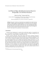
(1-O-galloyl-β-d-glucose, corilagin, cosmosiin, quercetin-3-galactoside, quercitrin) were frequently within
hydrogen contact with residues Ile 46, Tyr 183, Ile 121,
Ser 170 (Fig. 2). From this observation, four residues
seemed to be an important substrate recognition site
of 11β-HSD1. This conclusion is strongly supported by
studies on crystal structures and biochemical of 11βHSD1 (Hosfield et al. 2005; Hult et al. 2006). Especially,
Ile 46 and Ile 121, both of them were dual role leading to close contact with five compounds by hydrogen
bonds and also establish more hydrophobic interactions
with benzene ring on ligand [Fig. 2(1, 2, 4)]. In addition,
1-O-galloyl-β-d-glucoseand cosmosiin could link to the
receptor with a high number of hydrogen bonds compared to corilagin, quercetin-3-galactoside and quercitrin. This is proper explanation for high binding affinity of
cosmosiin. This action can be explained by the affinity of
each steroidal hydroxyl group for the receptor. For example, the functional group in cosmosiin could donate two
or three hydrogen bonds with different residue such as
Ser 43, Ser67, Arg 66, Lys 44, Gly 41, Asn 119. In tannin
family, although 1-O-galloyl-β-d-glucose showed much
stronger interaction than corilagin in term of hydrogen
bond, its binding capacity was lower. To fully understand
this phenomenon, molecular dynamic (MD) simulation
on the complexes is suggested.
Along with hydrogen bond, hydrophobic interactions
were also displayed. Β-amyrine and taraxerol seemed
to be rich on hydrophobic contact at position of the
methyl group which was non-polar [Fig. 2(6, 7)]. These
two compounds were also in contact with this receptor
because of the presence of the benzene ring. The residue
Thr 124, Thr 220 and Thr 222 were three residues which
could form not only hydrophobic interaction with terpenoid family but also hydrogen bond with 1-O-galloyl-βd-glucose, quercetin-3-galactoside, quercitrin, members
of tannin, and flavonoid group. Furthermore, in Fig. 2(2),
the residues Thr 220, Thr 222, Ala 223, Ile 121, Leu 217
were frequently observed in ligand-receptor interactions between, so they could be a critical part in binding
pocket. One important thing that Ser 261 and Arg 269
was shown as largely hydrophobic residues in previous
Nguyen Vo et al. SpringerPlus (2016) 5:1359
Page 7 of 13
Table 2 Binding energy (kcal/mol) of bio-molecules in E. thymifolia to 11β-HSD1, PTP1B, GFAT and SIRT6
Family
Ligand
Control
Flavonoid
Kaempferol
Quercetin
Quercetin-3-galactoside
Quercitrin
Corilagin
1-O-Galloyl-beta-d-glucose
Terpenoid
11β-HSD1 (1XU7)
PTP1B (4Y14)
GFAT (2ZJ3)
SIRT6 (3K35)
NDP: −12.5
C0A: −8.2
AGP: −6.5
APR: −11.0
−10.0
−7.7
−9.9
−9.0
−9.7
−7.8
−7.6
−8.3
Sample
Cosmosiin
Tannin
Binding energy (kcal/mol)
Euphorbol
2-(4 methyl-3-cyclohexene-1-yl)-2-propanol
24 methylen cycloartenol
β-Amyrine
β-Sitosterol
Campesterol
Caryophyllene
Limonene
Phytol
Piperiterone
Safranal
Stigmasterol
Taraxerol
−9.1
−8.9
−9.4
−8.9
−7.4
−7.8
−7.9
−8.4
−7.7
−9.1
−9.0
−8.9
−8.7
−6.4
−8.0
−6.0
−6.1
−5.4
−10.2
−11.1
−11.6
−10.3
−10.1
−7.8
−5.5
−6.0
−5.7
−5.6
−11.0
−12.1
study involving crystal structure analysis (Hult et al.
2006) but in the figures from our study, these hydrophobic interactions were not present.
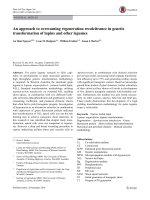
In term of GFAT1, this protein also had good binding
energy and in some cases it had higher or equal to result
of 11β-HSD1. Quercetin-3-galactoside, corilagin and cosmosiin were good illustration. Figure 3(1, 2, 3) supported
this statement with high number of hydrogen bonds and
hydrophobic interaction with receptor. The hydrogen
bonds were established between GFAT and members of
tannin and flavonoid family at position of Ser 420, Lys
675, Gln 421, Thr 375, Ser 422 in binding pocket. This was
also the conclusion in case of E.hirta and previous article
of Kuo-Chen and his partners (Chou 2004). In Fig. 3, Thr
375 and Thr 425 were especial case due to the bond they
linked to receptor. This residue closed to not only methyl
group but also to hydroxyl group of taraxerol and benzene
ring of cosmosiin and quercetin-3-galactoside, quercitrin.
−7.4
−7.9
−8.2
−7.0
−6.8
−6.0
−5.6
−5.2
−6.2
−5.4
−7.7
−8.4
−8.3
−7.9
−8.1
−8.8
−9.3
−9.0
−7.9
−10.4
−6.9
−9.0
−9.0
−10.9
−8.2
−9.4
−7.8
−7.1
−4.8
−5.2
−5.4
−5.5
−8.5
−8.9
−9.4
−7.3
−6.3
−6.5
−5.8
−5.7
−9.7
−11.5
Therefore, it could bind to the receptor by hydrogen and
hydrophobic interaction. Besides that, hydrophobic was
also displayed between Val 677, Ala 674, Thr 375 and two
members of terpenoid family: β-amyrine and taraxerol.
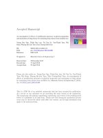
SIRT6 and PTP1B
1-O-Galloyl-β-d-glucose, corilagin, cosmosiin, quercetin3-galactoside, quercitrin interacted with SIRT6 with the
result of binding energy 8, 9, 9, 8.8, 9.3, 10.9, 11.5 in term
of absolute value (Table 2). These results were smaller
than 11β-HSD1. But there was a similarity with interaction of 11β-HSD1 and ligands. All these compounds can
form either hydrogen bond or hydrophobic interaction
with free residue in active site of SIRT6. Tannin and flavonoid family can build up hydrogen bond with Gln 111,
Thr 213, Ser 214 [Fig. 4(1, 2, 3, 4, 5)]. Three residues that
seem to have critical role in active site of SIRT6, but this
output was totally difference in the studying of structure
(See figure on next page.)
Fig. 2 Binding modes of selective compounds with 11β-HSD1. 1 Cosmosiin, 2 quercetin-3-galactoside, 3 quercitrin, 4 corilagin, 5 1-O-galloyl- β-dglucose, 6 β-amyrine, 7 taraxerol (The red and blue arrows were hydrogen donor and receptor bonds and the black round dot line was hydrophobic
interaction. Yellow dot was hydrophobic region of ligand.)
Nguyen Vo et al. SpringerPlus (2016) 5:1359
Page 8 of 13
Nguyen Vo et al. SpringerPlus (2016) 5:1359
Page 9 of 13
Nguyen Vo et al. SpringerPlus (2016) 5:1359
Page 10 of 13
(See figure on previous page.)
Fig. 3 Binding modes of selective compounds with GFAT. 1 Cosmosiin, 2 quercetin-3-galactoside, 3 quercitrin, 4 corilagin, 5 1-O-galloyl- β-dglucose, 6 β-amyrine, 7 taraxerol. The red and blue arrows were hydrogen donor and receptor bonds and the black round dot line was hydrophobic
interaction. Yellow dot was hydrophobic region of ligand
and biochemical function of SIRT6 of Patricia and coworker (Pan et al. 2011). This can be explained by the different tested site in our research.
In addition, the hydrophobic interactions also played
an important role in docking result. The good illustration
was the difference in one methyl group at carbon number
6 of rhamnoside ring (IUPAC name) of quercitrin compared to quercetin-3-galactoside structure [Fig. 4(2, 3)].
This conduct to 9.3 kcal/mol binding affinity of quercitrin compared to 8.8 kcal/mol of quercetin-3-galactoside.
For this reason, this kind of bond between five of seven
ligands and SIRT6 was also considerable point; these
compounds form hydrophobic interaction with Ile 217,
Trp186, Phe 62 at two hydrophore groups: benzene ring
in flavonoid family and methyl group in terpenoid family
[Fig. 4(1, 3, 6, 7)].
The docking result of PTP1B was lower compared
to three other receptors. This can be explained by the
number of hydrogen bond and hydrophobic interaction in the link of ligands and SIRT6. For example,
the number of hydrophobic interaction and hydrogen
bond between taraxerol and four 11β-HSD1, SIRT6,
GFAT1 and PTP1B were 32 [Fig. 2(7)], 23 [Fig. 3(7)], 11
[Fig. 4(7)], 8 [Fig. 5(7)] respectively, and docking results
were 12.1, 11.5, 8.9, 8.4 kcal/mol respectively in term of
absolute value (Table 2). In case of corilagin, the number of hydrogen bond in PTP1B was 8 [Fig. 5(4)] compared to 2 hydrogen bonds of SIRT6 [Fig. 4(4)] but the
docking result was smaller. This action can be explained
by the maintain time of interaction between ligand and
receptors. The same with hydrogen bond, the number of
hydrophobic interaction was also significantly reduced
in arrangement from 11β-HSD1 to PTP1B. There were
only 4 bonds between β-amyrine and PTP1B, whereas
24 bonds in case of 11β-HSD1. The duration time of
the interaction between ligand and receptor is high frequency of residues Tyr 29, Phe 52, Ile 219 (Fig. 5) seem to
be the significant region in active site of PTP1B.
Conclusion
In summary, from the list of 20 compounds, seven compounds were chosen due to high absolute value of binding energy to all four receptors (>8 kcal/mol). They are
β-amyrine, taraxerol, 1-O-galloyl-β-d-glucose, corilagin,
cosmosiin, quercetin-3-galactoside and quercitrin. Polyphenol, the frame of tannin and flavonoid family had
high binding affinity to all four receptors. Besides that,
the binding affinity of two of the terpenoid compounds
also suggested that this family is also a good prospect for
the treatment of Type 2 DM.
Although the basic concepts of interaction between
20 ligands of E. thymifolia and 4 receptors had been
already defined, many questions still remained unclear
for relationship between docking result in autodock step
and number of bonds in 2D structure of pharmacophore analysis step. Therefore, further research is required
using, the molecular dynamic (MD) and hydrogen bond
analysis to clearly determined the stability of the hydrogen bonds and hydrophobic interactions between ligands
and receptors.
(See figure on next page.)
Fig. 4 Binding modes of selective compounds with SIRT6. 1 Cosmosiin, 2 quercetin-3-galactoside, 3 quercitrin, 4 corilagin, 5 1-O-galloyl-β-dglucose, 6 β-amyrine, 7 taraxerol. The red and blue arrows were hydrogen donor and receptor bonds and the black round dot line was hydrophobic
interaction. Yellow dot was hydrophobic region of ligand
Nguyen Vo et al. SpringerPlus (2016) 5:1359
Page 11 of 13
Nguyen Vo et al. SpringerPlus (2016) 5:1359
Page 12 of 13
Nguyen Vo et al. SpringerPlus (2016) 5:1359
Page 13 of 13
(See figure on previous page.)
Fig. 5 Binding modes of selective compounds with PTP1B. 1 Cosmosiin, 2 quercetin-3-galactoside, 3 quercitrin, 4 corilagin, 5 1-O-galloyl-β-dglucose, 6 β-amyrine, 7 taraxerol. The red and blue arrows were hydrogen donor and receptor bonds and the black round dot line was hydrophobic
interaction. Yellow dot was hydrophobic region of ligand
Authors’ contributions
THNV, NT and DN have been responsible for the all technical matters, scientific
issues/values and the manuscript preparation. LL has been responsible for
data analysis, reading and approving the final manuscript. All authors read and
approved the final manuscript.
Author details
1
International University – Vietnam National University - HCMC, Quarter
6, Linh Trung Ward, Thu Duc District, Ho Chi Minh City, Vietnam. 2 Institute
of Computational Science and Technology - HCMC, Ho Chi Minh City, Vietnam.
Acknowledgements
This project is funded by Vietnam National University at Ho Chi Minh City
under grant number C2016-28-01. The authors would like to appreciate
passionate support from Computational Biology Center at IU and Institute of
Computational Science and Technology for supporting us to complete this
project.
Competing interests
The authors declare that they have no competing interest.
Received: 26 January 2016 Accepted: 20 June 2016
References
Andrews RC, Walker BR (1999) Glucocorticoids and insulin resistance: old
hormones, new targets. Clin Sci 96(5):513–523
Bnouham M, Ziyyat A, Mekhfi H, Tahri A, Legssyer A (2006) Medicinal plants
with potential antidiabetic activity—a review of ten years of herbal medicine research (1990–2000). Int J Diabetes Metab 14(1):1–25
Chou K-C (2004) Molecular therapeutic target for type-2 diabetes. J Proteome
Res 3(6):1284–1288
Davani B et al (2004) Aged transgenic mice with increased glucocorticoid
sensitivity in pancreatic β-cells develop diabetes. Diabetes (American
Diabetes Association) 53(suppl 1):S51–S59
Dennington R, Keith T, Millam J (2009) GaussView, version 5. Prod. Shawnee
Mission Semichem Inc., Shawnee
Evans JL (2007) Antioxidants: do they have a role in the treatment of insulin
resistance? Indian J Med Res 125(3):355
Ford ES, Giles WH, Dietz WH (2002) Prevalence of the metabolic syndrome
among US adults: findings from the third National Health and Nutrition
Examination Survey. JAMA 287(3):356–359
Fraga CG (2005) Cocoa, diabetes, and hypertension: should we eat more
chocolate? Am J Clin Nutr 81(3):541–542
Gonzalez-Burgos E, Gomez-Serranillos MP (2012) Terpene compounds in
nature: a review of their potential antioxidant activity. Curr Med Chem
19(31):5319–5341
Hasan S et al (2002) Acetylation regulates the DNA end-trimming activity of
DNA polymerase β. Mol Cell 10(5):1213–1222
Hosfield DJ et al (2005) Conformational flexibility in crystal structures of
human 11β-hydroxysteroid dehydrogenase type I provide insights into
glucocorticoid interconversion and enzyme regulation. J Biol Chem
280(6):4639–4648
Hult M et al (2006) Active site variability of type 1 11β-hydroxysteroid dehydrogenase revealed by selective inhibitors and cross-species comparisons.
Mol Cell Endocrinol 248(1–2):26–33
Humphrey W, Dalke A, Schulten K (1996) VMD: visual molecular dynamics. J
Mol Graph 14(1):33–38
International Diabetes Federation (IDF) (2015) What is Diabetes. In: Cavan D, da
Rocha Fernandes J, Makaroff L, Ogurtsova K, Webber S (eds) IDF diabetes
atlas, 7th edn. International Diabetes Federation, Brussels
Killedar SG, Desai RG, Kashid UT, Bhore NV, Mahamuni SS (2011) Antimicrobial
activity and phytochemical screening of fresh latex of Euphorbia thymifolia Linn. Int J Res Ayurveda Pharm 2(5)
Loi DT (2015) Cac cay thuoc va vi thuoc chua ly, Part B. Chua ly truc trung. In:
Loi DT (ed) Cay Thuoc va Vi Thuoc Vietnam (Vietnamese medicinal plants
and herbal formulations), chap IV, Part B. Hong Duc, Ha Noi, pp 199–200
Maiese K, Daniela Morhan S, Zhong Chong Z (2007) Oxidative stress biology
and cell injury during type 1 and type 2 diabetes mellitus. Curr Neurovasc
Res 4(1):63–71
Montonen J, Knekt P, Järvinen R, Reunanen A (2004) Dietary antioxidant intake
and risk of type 2 diabetes. Diabetes Care (American Diabetes Association) 27(2):362–366
Muthumani D, Hedina A, Kausar J, Anand V, Pushpa (2016) Phytopharmacological activities of Euphorbia thymifolia Linn. Syst Rev Pharmacy 7(1):30–34
Nguyen NDT, Le LT (2012) Targeted proteins for diabetes drug design. Adv Nat
Sci Nanosci Nanotechnol 3:013001
Pan PW, Feldman JL, Devries MK, Dong A, Edwards AM, Denu JM
(2011) Structure and biochemical functions of SIRT6. J Biol Chem
286(16):14575–14587
Prasad K, Bisht G (2011) Evaluation of nutritive minerals and antioxidants
values of Euphorbia thymifolia Linn. Curr Res Chem 3:98–105
PROTA (2008) Plant resources of tropical Africa. In: Schmelzer GH, Gurib-Fakim
A (eds) Medicinal plants, vol 11(1). PROTA Foundation - Backhuys - CTA,
Wageningen, pp 294–296
Sandeep K, Rahul A, Vishvesh A, Chandrakant M (2009) Laxative and antihelmintic activity of aqueous extract of Euphorbia thymifolia Linn. Res J
Pharmacogn Phytochem 1(3):182–184
Sharma GD, Tripathi SN (1984) Experimental evaluation of Dugdhika
(Euphorbia prostrata W. Ait) for the treatment of ‘Tamaka Svasa’ (bronchial
asthma). Anc Sci Life 3(3):143
Shi Yigong (2009) Serine/threonine phosphatases: mechanism through structure. Cell 139(3):468–484
Trott O, Olson AJ (2009) AutoDock Vina: improving the speed and accuracy of
docking with a new scoring function, efficient optimization, and multithreading. J Comput Chem 31(2):455–461
Vogel GH (ed) (2002) Drug discovery and evaluation: pharmacological assays.
Springer, Berlin, Heidelberg
Wolber G, Langer T (2005) LigandScout: 3-D pharmacophores derived from
protein-bound ligands and their use as virtual screening filters. J Chem
Inf Model 45(1):160–169