DSpace at VNU: Application of ultrasound to microencapsulation of coconut milk fat by spray drying method
Bạn đang xem bản rút gọn của tài liệu. Xem và tải ngay bản đầy đủ của tài liệu tại đây (368.24 KB, 5 trang )
J Food Sci Technol
DOI 10.1007/s13197-014-1285-y
ORIGINAL ARTICLE
Application of ultrasound to microencapsulation of coconut milk
fat by spray drying method
Hoang Du Le & Van Viet Man Le
Revised: 23 January 2014 / Accepted: 7 February 2014
# Association of Food Scientists & Technologists (India) 2014
Abstract Mixtures of coconut milk and gelatin solution were
treated by ultrasound, mixed with maltodextrin and subsequently spray-dried to yield powder. The effects of ultrasonic
power and sonication time on the microencapsulation efficiency (ME) and microencapsulation yield (MY) of coconut fat
were investigated. The results indicated that increase in ultrasonic power from 0 to 5.68 W/g and in sonication time from 0
to 2.5 min augmented ME and MY of coconut fat. However,
treatment with sonication power higher than 5.68 W/g led to a
drop in fat ME and MY, mainly due to aggregation of fat
particles and that blocked the adsorption of gelatin molecules
on the particle surface.
Keywords Coconut milk . Coconut fat . Coconut milk
powder . Ultrasound . Encapsulation efficiency .
Encapsulation yield
Introduction
Coconut milk is an oil-in-water emulsion extracted from grated coconut meat with or without added water. This natural
product is highly susceptible to microbiological, chemical and
biochemical deterioration (Waisundara et al. 2007). Therefore,
over the years, many attempts have been done to extend shelflife of coconut milk. One of the best methods for the preservation of coconut milk is spray-drying to yield powder (Seow
and Gwee 1997).
According to Gharsallaoui et al. (2007), spray-drying, a
process of microencapsulation, has been used for decades to
microencapsulate food ingredients such as flavours, lipids,
H. D. Le : V. V. M. Le (*)
Department of Food Technology, Ho Chi Minh City University of
Technology, Ho Chi Minh City, Vietnam
e-mail:
and carotenoids (core materials). Microencapsulation by spray
drying consists of three basic steps: preparation of the emulsion; homogenization of the emulsion; and atomization of
food material into the drying chamber. These authors reported
that the first two steps significantly affected the microencapsulation of the core materials.
The first step is the formation of a fine and stable emulsion
of the core material in the wall solution. The mixture is
prepared by dispersing the core material into a solution of
the coating agents (Gharsallaoui et al. 2007). The first coating
agents used in the production of coconut milk powder were
decaglycerol monostearate, sodium caseinate, and dextrin
(Seow and Gwee 1997). Other coating agents such as maltodextrin, casein or skim milk, and/or corn syrup were also used
for fat microencapsulation during the spray-drying (Seow and
Gwee 1997). Recently, the use of gelatin as wall material for
phospholipid microencapsulation by spray drying has
attracted considerable interest (Bruschi et al. 2003;
Gharsallaoui et al. 2007; Vinetsky and Magdassi 1997;
Yoshii et al. 2001). Gelatin, a water-soluble material, has all
the properties of an effective entrapping agent: high emulsifying activity, high stabilizing activity, and high tendency to
form a fine dense network upon drying (Gharsallaoui et al.
2007). Furthermore, gelatin has the ability to stimulate the
early formation of the surface crust, which prevents the loss of
core material during spray drying (Gharsallaoui et al. 2007;
Yoshii et al. 2001). However, gelatin has not been used for the
spray drying of coconut milk yet.
In the second step, the emulsion is homogenized by high
pressure or ultrasound to obtain the uniform and small fat
droplets. The particle size distribution (PSD) of fat droplets
plays an important role in the stability of emulsion (Jena and
Das 2006) and in the microencapsulation efficiency by spray
drying (Gharsallaoui et al. 2007). High pressure homogenization is a method of choice commonly used in many researches
for enhancing the stability of coconut milk emulsion
J Food Sci Technol
(Chiewchan et al. 2006; Tangsuphoom and Coupland 2008,
2009) and for preparing stable emulsion before spray-drying
(Hogan et al. 2001; Liu et al. 2001; Yoshii et al. 2001).
Recently, the use of ultrasound for homogenization of oil in
water emulsion has attracted considerable attention (Kentish
et al. 2008; Leong et al. 2011). In addition, ultrasound had a
very good homogenization effect at high power levels compared with high pressure homogenization (Wu et al. 2000). In
case of coconut milk emulsion, the modeling of particle size
distribution of sonicated coconut milk emulsion was investigated by Jena and Das (2006) and the results showed that
suitable sonication time reduced the fat droplet size.
Until now, application of ultrasound to fat microencapsulation in the production of instant coconut milk powder has
not been clearly considered. Therefore, this work was aimed
to investigate the effects of sonication variables on the ME and
MY of coconut fat using gelatin and maltodextrin as emulsifiers. In addition, the information obtained from this work
would give a clearer understanding of phenomena that happen
during the ultrasonic process of coconut milk as well as other
oil-in-water emulsions.
Materials and methods
Materials
Grated coconut meat was purchased from a local market in
Ben Tre, Vietnam. Wall materials used in this study included
gelatin and maltodextrin. Gelatin (Bloom: 150) was originated
from Gelita Australia Pty Ltd (Australia) and Maltodextrin
(Dextrose equivalent: 18) was purchased from Qinhuangdao
Lihua Starch Co., Ltd (China). Solvents and chemicals were
obtained from Guangzhou Jinhuada Chemical Reagent Co.,
Ltd (China).
Experimentation
Grated coconut meat was mixed with water at a weight ratio of
1:1. Subsequently, the mixture was heated to 50 °C for 10 min,
filtered through a cheesecloth and pressed to extract coconut
milk. Samples of the coconut milk were collected for further
analysis. In this experiment, the solid content (% w/w) and
total fat content (% w/w) of coconut milk were 19.54±0.19
and 14.66±0.25 respectively.
Emulsifier solutions were prepared as follows:
&
&
10 % (w/w) gelatin solution was obtained by dissolving
gelatin in distilled water, stirring at 750 rpm, and heating
at 50 °C for 6 h.
10 % (w/w) maltodextrin solution was obtained by dissolving maltodextrin in distilled water, stirring at 750 rpm,
and room temperature for 1 h.
The emulsifier solutions were then filtered through a
cheesecloth to ensure that all undissolved particles were
eliminated.
Effect of ultrasonic power on ME and MY of coconut fat
Mixture of coconut milk and gelatin solution was homogenized by a Model VC 750 ultrasonic probe (Sonics &
Materials Inc., USA) at different power levels (2.27–6.82 W/
g) for 2.5 min. Samples were taken from the obtained emulsions for further analysis. The sonicated emulsions were then
mixed with maltodextrin solution and stirred by a magnetic
stirrer at 750 rpm for 30 min. Our preliminary investigations
(unpublished data) showed that a core (gelatin) to wall ratio
(w/w) of 7.5/10 and a gelatin to maltodextrin ratio (w/w) of
4:1 were the appropriate conditions for the microencapsulation of coconut fat. These ratios were therefore chosen to carry
out all experiments in this study. The solid concentration of
the resultant emulsion was adjusted to 15 % (w/w) by adding
distilled water. The final emulsion was spray dried by a
Mobile Minor—Model E spray-drier (Niro A/S, Denmark).
The spray-drier was equipped with a chamber with dimensions of 0.8 m diameter and 0.6 m height, a centrifugal
atomizer, a cyclone separator and an exhaust blower. The
emulsion was fed into the chamber at the rate of 22.9 mL/
min by a 505S peristaltic pump (Matson-Marlow, England).
The drying took place with an air inlet temperature of 160 °C,
outlet temperature of 42 °C and an air pressure of 0.35 MPa at
the atomizer. Control samples without ultrasonic treatment
were also carried out. The powder of each run was collected
for further analysis.
Effect of sonication time on ME and MY of coconut fat
In this experiment, the mixture of coconut milk and gelatin
solution was homogenized by ultrasound at a suitable value of
sonication power obtained from the experiment in previous
section. Sonication time was varied between 0.5 and 3 min.
The following steps were similar to those in section “Effect of
ultrasonic power on ME and MY of coconut fat”. Control
samples without ultrasonic treatments were also carried out.
The sonicated emulsion and powder of each run were
sampled for further analysis.
Determination of solid content and total fat content of coconut
milk emulsion
Solid content of coconut milk emulsion was determined by
drying at 130±3 °C until constant weight (Lakshanasomya
et al. 2011).
The total fat content of coconut milk emulsion was determined by using a method proposed by Lakshanasomya et al.
(2011). Ten milliliters of coconut milk emulsion was taken
J Food Sci Technol
into the fat extraction flask for analysis. Firstly, 1.5 ml of
ammonium hydroxide was added and mixed followed by
10 ml of alcohol (95 %) and the contents were again well
mixed. Secondly, 25 ml diethyl ether was added to the flask,
then it was shook vigorously for 1 min. Finally, 25 ml of light
petroleum ether (b.p. 40–60 °C) was added and the flask was
shook vigorously for 1 min. After separation was complete,
the fat solution was transferred into a petri dish and the petri
dish was dried at 102±2 °C for 1 h and weighed. The total fat
content was calculated as the difference between weight of
petri dish with fat and weight of initial petri dish.
Determination of surface fat content, encapsulated fat content,
and total fat content of coconut milk powder
The fat on the surface of coconut milk powder particles was
determined by a method described by Jafari et al. (2008). One
gram of coconut milk powder was accurately weighed into the
extraction flask. Subsequently, 25 ml of petroleum ether (b.p.
40–60 °C) was added and the mixture was shook vigorously
for 10 min. The mixture was then filtered through a cloth. The
filtrate was transferred into the petri dish, dried at 102±2 °C
for 1 h and weighed. The surface fat content was calculated as
the difference between weight of petri dish with fat and weight
of initial petri dish.
The total fat content of coconut milk powder was determined by using a method described by Lakshanasomya et al.
(2011). One gram of coconut milk powder was accurately
weighed into the fat extraction flask. Water was added to
complete the volume to 10 ml and mixed. The total fat content
in the emulsion was then determined similarly to the process
in section “Determination of solid content and total fat content
of coconut milk emulsion”.
The encapsulated fat content was calculated as a difference
of the total fat content and the surface fat content of the
powder obtained.
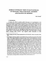
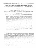
Fig. 1 Effect of sonication power on ME (black diamond) and MY
(black square)
coater E-102 (Hitachi- Japan). The S-4800 model scanning
electron microscope (Hitachi, Japan) was used to study the
outer surface of the coconut milk powder. The examination
was operated at an accelerating voltage of 2 kV. The S-4800
software (Hitachi, Japan) was used to present the micrographs
of the powder microstructure.
PSD of coconut milk emulsion
Particle size distributions of raw and homogenized coconut milk
emulsion were assessed by using a Model LA 920 laser diffraction particle analyzer (Horiba, Japan). Samples were diluted to
approximately 0.005 wt% in an effort to avoid multiple scattering effect. The average particle size was calculated as a volume
mean diameter, or d43 (d43 =∑ni.D4i /∑ni.D4i ) (Tangsuphoom and
Coupland 2008) where ni is the number of the droplets of
diameter Di.
Statistical analysis
All experiments were performed in triplicate. Mean values
were considered significantly different when P<0.05. OneWay analysis of variance was performed using the software
Statgraphics Centurion XV.
Microencapsulation efficiency (ME) and microencapsulation
yield (MY) of coconut fat
ME and MY were calculated by formulas reported by Shu
et al. (2006).
ME was defined as a ratio between the mass of the encapsulated fat and the mass of the total fat in the coconut milk
powder.
MY was defined as a ratio between the mass of the total fat
of the coconut milk powder and the mass of the total fat of the
emulsion before spray drying.
0 W/g
6.84 W/g
3.41 W/g
Scanning electron microscopy (SEM) of coconut milk powder
The powder sample was placed on one surface of a doublefaced adhesive tape and coated with gold by using an ion-
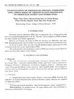
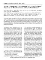
Fig. 2 Effect of sonication power on fat particle size distribution of
sonicated coconut milk emulsions: 3.41 W/g (black square) and
6.82 W/g (black triangle) and control (0 W/g, black diamond)
J Food Sci Technol
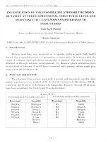
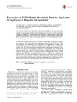
Fig. 3 SEM pictures of coconut
milk powder produced from
sonicated coconut milk at
different ultrasonic powers:
3.41 W/g (a), 6.82 W/g (b) and
0 W/g (c, control sample)
a
Results and discussion
Effect of ultrasonic power on ME and MY of coconut fat
The results in Fig. 1 demonstrate that the ME and MY reached
the highest level when the sonication power was between 3.41
and 5.68 W/g, then decreased when the ultrasonic power was
higher than 5.68 W/g. The reasons for these changes can be
explained through the effects of ultrasonic power on PSD of
coconut milk emulsion. PSD of the sonicated coconut milk
(treated at 3.41 W/g and 6.82 W/g) and non-sonicated coconut
milk was evaluated. Emulsion without sonication exhibited a
trimodal distribution as shown in Fig. 2. By contrast, emulsion
sonicated at 3.41 W/g had a monomodal distribution. An
interesting result from Fig. 2 was that emulsion homogenized
at 6.82 W/g (the highest applied power) had a bimodal
distribution.
The application of low frequency ultrasound is known to
cause acoustic cavitation which plays an important role in the
reduction of primary droplets of oil-in-water emulsion
(Kentish et al. 2008). Therefore, it would be expected that
increase in ultrasonic power would improve the amount of
shear force that breaks up primary droplets into the smaller
ones. However, the high mean particle size (d43 ≈9.7 μm) was
obtained in the emulsion homogenized at the highest power
level (6.82 W/g). By contrast, emulsion homogenized at
3.41 W/g had the smallest particle size (d 43 ≈ 6 μm).
Decrease in particle size lead to increase in surface area of
fat particle where gelatin molecules adsorb on. As a consequence, ME and MY of coconut fat increased when fat droplet
size decreased.
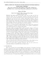
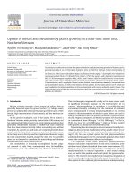
Fig. 4 Effect of sonication time on ME (black diamond) and MY (black
square) of coconut fat (The ultrasonic power was 3.41 W/g)
b
c
Based on the results obtained, we proposed a hypothesis
about the effects of sonication power on ME and MY of
coconut fat. Small droplet size was reported to prevent flocculation of the droplets (Tadros 2005). Consequently, decrease in
particle size will facilitate the adsorption of gelatin on the
droplet surface. As a result, ME and MY increased. However,
when the sonication power reached the “over-processing”
levels, the coalescence of droplets enhanced the formation of
bigger droplets or “aggregates” with higher diameter.
Consequently, the adsorption of gelatin on the newly formed
droplet interface was obstructed. In this case, we supposed that
gelatin molecules just partially adsorbed on the droplet surface.
As a consequence, microencapsulation efficiency of gelatin
declined. In all experiments, sonicated coconut milk with gelatin was added with maltodextrin before spray-dried to yield
powder. Suitable concentration of maltodextrin was believed to
decrease the number of cracks on the surface of powder particles (Sheu and Rosenberg 1998). ME and MY of coconut fat
were therefore enhanced. A convincing result that strongly
supported our hypothesis was that powder particles in SEM
pictures from 6.82 W/g sonicated and non-sonicated emulsions
appeared agglomerated (Fig. 3). This phenomenon was probably due to the formation of “aggregates”. The aggregates of fat
droplets in the emulsion could result in the formation of aggregates of particles in the powder obtained. By contrast, few
agglomerated particles were observed in SEM picture of the
powder from 3.41 W/g ultrasonic emulsion. According to
Hogan et al. (2001) the powder particles that appeared highly
agglomerated had the high level of surface fat content.
Fig. 5 Effect of sonication time on fat particle size distribution of
coconut milk with gelatin emulsions: 0 min (control sample) (black
diamond), 1 min (black square), 2 min (black triangle), 2.5 min (x) and
3 min (+) (The ultrasonic power was 3.41 W/g)
J Food Sci Technol
Therefore, coconut milk powder that exhibited aggregated
particles had low ME and MY of coconut fat.
A similar trend between droplet size and applied sonication
power has been observed by Jafari et al. (2008) and Kentish
et al. (2008) who homogenized the internal phase of fish oil
and flax seed oil with the emulsifiers of proteins and Tween
40, respectively. These studies showed that when the ultrasonic power reached an “over-processing” level, droplet aggregation was enhanced. Generally, ultrasound increases ME
and MYof fat when the ultrasonic power lower than the “overprocessing” level. Very high ultrasonic power decreases ME
and MY of fat.
Effect of sonication time on ME and MY of coconut fat
Figure 4 shows that prolongation of sonication time resulted in
the increase in ME and MY of coconut fat. The sonication
time of 2.5 min was suitable for coconut fat microencapsulation, in which ME and MY of the treated samples increased
22.6 % and 24.0 % respectively as compared to ME and MY
of the non-treated samples. Treatment with longer time did not
significantly increase ME and MY of coconut fat.
In order to clearly understand the effects of sonication time
on ME and MY of coconut fat, we determined the PSD of
coconut milk emulsion treated at 3.41 W/g with different
sonication times: 0 min (control sample), 1 min, 2 min,
2.5 min and 3 min. As shown in Fig. 5, coconut milk treated
at 2, 2.5 and 3 min had a monomodal distribution. By contrast,
non-treated and 1 min treated coconut milk with gelatin emulsion had a trimodal and bimodal distribution, respectively. The
average particle size (D43) of the non-treated, 1 min, 2 min,
2.5 min, 3 min treated coconut milk were 10.3, 7.6, 7.4, 6.2
and 6.0 μm, respectively. Similar trend between sonication
time and droplet size reduction was observed by Jena and Das
(2006) for coconut fat microencapsulated by gum acacia and
maltodextrin. As a result of droplet size reduction, ME and
MY increased when sonication time increased.
Conclusion
Ultrasonic treatment increased ME and MYof coconut fat due
to its ability to break fat droplets into smaller size droplets. ME
and MY reached the highest levels when the sonication power
increased from 3.41 to 5.68 W/g. However, when the sonication power increased from 5.68 to 6.82 w/g, the formation of
“aggregates” with high diameter blocked the adsorption of
gelatin on particle surface and that led to a drop in MY and
MY of coconut milk fat. Suitable sonication time for coconut
fat microencapsulation was 2.5 min.
References
Bruschi ML, Cardoso MLC, Lucchesi MB, Gremião MPD (2003)
Gelatin microparticles containing propolis obtained by spraydrying technique: preparation and characterization. Int J Pharm
264:45–55
Chiewchan N, Phungamngoen C, Siriwattanayothin S (2006) Effect of
homogenizing pressure and sterilizing condition on quality of
canned high fat coconut milk. J Food Eng 73:38–44
Gharsallaoui A, Roudaut G, Chambin O, Voilley A, Saurel R (2007)
Applications of spray-drying in microencapsulation of food ingredients: an overview. Food Res Int 40:1107–1121
Hogan SA, McNamee BF, O’Riordan ED, O’Sullivan M (2001)
Emulsification and microencapsulation properties of sodium
caseinate/carbohydrate blends. Int Dairy J 11:137–144
Jafari SM, Assadpoor E, Bhandari B, He Y (2008) Nano-particle encapsulation of fish oil by spray drying. Food Res Int 41:172–183
Jena S, Das H (2006) Modeling of particle size distribution of sonicated
coconut milk emulsion: effect of emulsifiers and sonication time.
Food Res Int 39:606–611
Kentish S, Wooster TJ, Ashokkumar M, Balachandran S, Mawson R,
Simons L (2008) The use of ultrasonics for nanoemulsion preparation. Innov Food Sci Emerg Technol 9:170–175
Lakshanasomya N, Danudol A, Ningnoi T (2011) Method performance
study for total solids and total fat in coconut milk and products. J
Food Compos Anal 24:650–655
Leong TSH, Wooster TJ, Kentish SE, Ashokkumar M (2011) Minimising
oil droplet size using ultrasonic emulsification. Ultrason Sonochem
16:721–727
Liu X-D, Atarashi T, Furuta T, Yoshii H, Aishima S, Ohkawara M, Linko
P (2001) Microencapsulation of emulsified hydrophobic flavors by
spray drying. Dry Technol 19:1361–1374
Seow CC, Gwee CN (1997) Coconut milk: chemistry and technology. Int
J Food Sci Technol 32:189–201
Sheu T-Y, Rosenberg M (1998) Microstructure of microcapsules
consisting of whey proteins and carbohydrates. J Food Sci 63:
491–494
Shu B, Yu W, Zhao Y, Liu X (2006) Study on microencapsulation of
lycopene by spray-drying. J Food Eng 76:664–669
Tadros T (2005) Applied surfactants: principles and applications. Wiley VCH
Tangsuphoom N, Coupland JN (2008) Effect of surface-active stabilizers
on the microstructure and stability of coconut milk emulsions. Food
Hydrocoll 22:1233–1242
Tangsuphoom N, Coupland JN (2009) Effect of surface-active stabilizers
on the surface properties of coconut milk emulsions. Food
Hydrocoll 23:1801–1809
Vinetsky V, Magdassi S (1997) Microencapsulation by Surfactant–
Gelatin insoluble complex: effect of pH and surfactant concentration. J Colloid Interface Sci 189:83–91
Waisundara VY, Perera CO, Barlow PJ (2007) Effect of different
pre-treatments of fresh coconut kernels on some of the quality attributes of the coconut milk extracted. Food Chem 101:
771–777
Wu H, Hulbert GJ, Mount JR (2000) Effects of ultrasound on milk
homogenization and fermentation with yogurt starter. Innov Food
Sci Emerg Technol 1:211–218
Yoshii H, Soottitantawat A, Liu X-D, Atarashi T, Furuta T,
Aishima S, Ohgawara M, Linko P (2001) Flavor release from
spray-dried maltodex-trin/gum Arabic or soy matrices as a
function of storage relative humidity. Innov Food Sci Emerg
Technol 2:55–61