DSpace at VNU: CHEMICAL CONSTITUENTS FROM THE LEAVES OF XYLOPIA POILANEI AND THEIR BIOACTIVITY
Bạn đang xem bản rút gọn của tài liệu. Xem và tải ngay bản đầy đủ của tài liệu tại đây (124.01 KB, 8 trang )
HETEROCYCLES, Vol. 78, No. 3, 2009
763
HETEROCYCLES, Vol. 78, No. 3, 2009, pp. 763 - 770. © The Japan Institute of Heterocyclic Chemistry
Received, 3rd September, 2008, Accepted, 10th November, 2008, Published online, 11th November, 2008
DOI: 10.3987/COM-08-11540
CHEMICAL CONSTITUENTS FROM THE LEAVES OF XYLOPIA
POILANEI AND THEIR BIOACTIVITY
Tran Dinh Thang,a Ping-Chung Kuo,b Ngo Xuan Luong,*,a Nguyen Xuan
Dung,c Le Van Hac,a Yao-Haur Kuo,d Mei-Lin Yang,e and Tian-Shung
Wu*,e
a
Department of Chemistry, Vinh University, Vinh City, Nghean Province,
Vietnam, b Department of Biotechnology, National Formosa University, Yunlin
632, Taiwan,
c
Department of Chemistry, College of Natural Sciences, Hanoi
National University, Hanoi 10000, Vietnam,
Chinese Medicine, Taipei 105, Taiwan,
e
d
National Research Institute of
Department of Chemistry, National
Cheng Kung University, Tainan 701, Taiwan
*Tel: 886-6-2747538, Fax: 886-6-2740552, E-mail:
Abstract – A new flavonoid glycoside, xylopoillin A (1), and nine known
compounds were isolated from the methanolic extracts of the leaves of Xylopia
poilanei. The structure of this new compound was completely elucidated using a
combination of 2D NMR techniques (COSY, NOESY, HMQC and HMBC) and
HR-ESI-MS analyses. The other chemical structures of known compounds were
identified by comparison of their spectroscopic and physical data with those
reported in the literature.
The Xylopia genus (Annonaceae) comprises about 160 species with occurrence in South and Central
America, Africa and Asia.1 Approximately four Xylopia species were identified in Vietnam.2 These species
produce a wide variety of metabolites including alkaloids,3-8 lignans,9 acetogenins,10-11 diterpenoids,12-16
and sesquiterpenoids.17-19 Termite antifeedant, cardiovascular and diuretic activities were reported for this
genus.20-21 Xylopia poilanei is utilized traditionally in the regions of India, Malaya, Philippine islands, and
Indo-China for the medicinal uses as febrifuge, anthelmintic, anti-emetic, anti-inflammatory, and in
urinary and uterine discharges, piles, and lumbago.22 In our investigations of phytochemical diversity of
this plant, the structure of a new flavonoid, xylopoillin A (1), was established by spectroscopic methods
764
HETEROCYCLES, Vol. 78, No. 3, 2009
including 1D and 2D NMR and MS techniques. In addition, nine known compounds, including four
alkaloids, three flavonoids, and two carbohydrates, were also characterized by comparison of their
spectral and physical data with those reported in the literatures. The present study wishes to report the
structural assignments of this new compound, and their cytotoxic activity of the isolates.
The air-dried and powdered leaves of X. poilanei were extracted with hot methanol and concentrated to
give a dark brown syrup. The methanol extract was suspended in H2O and partitioned with ethyl acetate
and n-butanol. The successive purification of n-butanol soluble extracts by a combination of conventional
chromatographic techniques afforded a new compound (1). In addition, nine known compounds including
of dicentrine (2),23 dicentrinone (3),24 nordicentrine (4),25 oxoanolobine (5),26 rutin (6),27 quercitrin (7),28
naringenin 7-neohesperidoside (8),29 myo-inositol (9),30 and sucrose (10)31 were also characterized from
this plant. The structure of this new flavonoid glycoside, xylopoillin A (1), was established on the basis of
1D and 2D NMR and mass spectroscopic analyses.
The HR-ESI-MS of the new compound (1) displayed a sodium adduct ion peak at m/z 603.1328 [M+Na]+,
corresponding to the pseudomolecular formula of C26H28O15Na (calcd. 603.1326). The UV absorption
maxima at 360, 269, and 257 nm were characteristic of a flavone skeleton.32 The IR absorption bands at
3405 and 1656 cm-1 displayed the presence of a hydroxyl group and a hydrogen-bonded carbonyl group.
In the 1H NMR spectrum, a typical set of ABX signals at δ 7.74 (1H, d, J = 2.0 Hz), 7.60 (1H, dd, J = 8.5,
2.0 Hz), and 6.90 (1H, d, J = 8.5 Hz) were attributed to the trisubstituted B-ring. Two mutually coupled
doublet at δ 6.42 (1H, d, J = 2.0 Hz) and 6.23 (1H, d, J = 2.0 Hz) was assumed to be H-6 and H-8 since
they displayed 2J ,3J-HMBC correlations with the carbon signals at δ 165.9 (C-7), 158.4 (C-5), 105.7
(C-10), 99.9 (C-8); and δ 165.9 (C-7), 163.0 (C-9), 105.7 (C-10), 94.7 (C-6), respectively. Two anomeric
proton signals at δ 5.19 (d, J = 6.5 Hz) and 5.13 (d, J = 1.5 Hz) suggested the presence of two sugar units.
In addition, there are oxygenated methine and methylene protons at δ 3.44 (1H, t, J = 9.5 Hz), 3.47 (1H,
dd, J = 12.5, 1.5 Hz), 3.74 (1H, dd, J = 8.5, 3.0 Hz), 3.82 (1H, dd, J = 12.5, 4.0 Hz), 3.86 (2H, m), 3.93
(1H, m), 4.05 (1H, dd, J = 3.5, 1.5 Hz), and 4.08 (1H, dd, J = 8.5, 6.5 Hz), which were identified as the
proton signals of the sugar moieties. Moreover, the upfield methyl doublet at δ 1.28 (3H, d, J = 6.5 Hz)
was the characteristic absorption for the rhamnose fragment. In the 13C NMR and DEPT spectral analyses,
the sugar portion of 1 displayed one methyl (δ 18.1), one oxymethylene (δ 67.1), seven oxymethine (δ
68.9, 70.2, 71.8, 72.1, 72.2, 74.1, 80.0), and two anomeric signals (δ 103.0 and 104.7), which were
identified as L-arabinose (δ 104.7, 80.0, 71.8, 68.9, and 67.1)33 and D-rhamnose (δ 103.0, 74.1, 72.2, 72.1,
70.2, and 18.1)34 by comparison with the literature values and the assistance of comprehensive 2D NMR
spectroscopic analyses, including the COSY, NOESY, HMQC, and HMBC experiments. The sugar
fraction of acid hydrolysate of compound 1 was further analyzed with HPLC by comparison of their
retention times and optical rotations with those of authentic samples as described in the reference.35 In the
HETEROCYCLES, Vol. 78, No. 3, 2009
765
HMBC spectrum, long range correlations from H-1" (δ 5.19) to C-3 (δ 135.8) and H-1"' (δ 5.13) to C-3"
(δ 67.1) confirmed the presence of 3-O-α-D-rhamnosyl-(1→3)-β-L-arabinose moiety and the
configurations of arabinose and rhamnose were determined as β and α, respectively, according to their
coupling constants of anomeric protons. In addition, the proton signals at δ 7.74 (H-2') and 7.60 (H-6')
displayed 3J-HMBC correlations with the carbon signal at δ 158.6 (C-2), respectively, confirmed the
substitution pattern of B-ring to be 1,3,4-trisubstituted and the groups attached at C-3' and -4' were
hydroxyl functions with the assistance of molecular formula and
13
C NMR spectroscopic analysis. The
successive NOESY and HMBC experiments furnished the complete assignments of proton and carbon
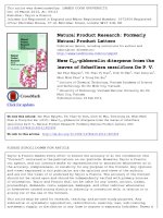
signals of 1 and thus its chemical structure was established as shown in Figure 1 and named trivially as
xylopoillin A.
OH
3'
8
HO
O
2
5'
1'
9
7
OH
4'
2'
OH
6'
O
6
10
4
3
O
5
OH
HO
O
1'''
4"
2"
1"
O
5"
OH
6'''
5'''
2'''
3'''
4'''
OH
OH
O
3"
1
Figure 1. Structure and significant HMBC correlations of compound 1
O
O
N
O
Me
N
O
H
O
MeO
MeO
OMe
OMe
2
3
O
O
NH
O
N
O
H
O
MeO
OMe
OH
4
5
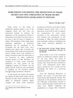
Figure 2. Structures of alkaloids 2-5
766
HETEROCYCLES, Vol. 78, No. 3, 2009
Some of the isolated compounds were assayed for cytotoxic activity against tumor cell lines Daoy (human
medulloblastoma), Hep-2 (human laryngeal carcinoma), MCF-7 (human breast adenocarcinoma), and
Hela (human cervical epitheloid carcinoma), as described previously,36 and the EC50 values are
summarized in Table 1. Only compound 4 displayed moderate cytotoxicity with the EC50 values of 3.84,
2.57, 2.16, 1.28 µg/mL against the Daoy, Hep-2, MCF-7, and Hela tumor cell lines, respectively.
Tested compounds
ED50 (µg/mL)
Daoy
Hep2
MCF-7
Hela
1
(-)
(-)
(-)
(-)
2
(-)
(-)
(-)
(-)
3
(-)
(-)
(-)
(-)
4
3.84
2.57
2.16
1.28
5
(-)
(-)
(-)
(-)
6
(-)
(-)
(-)
(-)
Mitomycin C
0.13
0.15
0.14
0.15
(-): ED50 40
µg/mL
Table 1. EC50 values of tested compounds against human tumor cell line panel
EXPERIMENTAL
General Melting points were determined using an Electrothermal IA-9200 melting points measuring
apparatus. The UV spectra were determined on an Agilent UV-VIS recording spectrophotometer. The IR
spectra were obtained, as KBr discs, on a Hitachi 270-30 type spectrometer. Optical rotations were
determined on a Jasco DIP-1000 KUY polarimeter. The electrospray ionization (ESI) mass spectra were
obtained using an AGILENT 1100 LC-MSD Trap spectrometer. The HR-ESI-MS spectra were measured
on a Bruker APEX II mass spectrometer. 1H- and 13C-NMR, COSY, NOESY, HMQC, and HMBC spectra
were recorded on the Bruker Avance-500 NMR spectrometer, using tetramethylsilane (TMS) as the
internal standard. Standard pulse sequences and parameters were used for the NMR experiments and all
chemical shifts were reported in parts per million (ppm, δ). Column chromatography (CC) was performed
on silica gel (Kieselgel 60, 70-230 mesh and 230-400 mesh, E. Merck).
Plant Materials The leaves of Xylopia poilanei (Annonaceae) was collected from Nghe An, Vietnam,
during May 2007 and the plant materials were identified and authenticated by Assoc. Prof. Dr. Vu Xuan
Phuong, Institute of Ecology and Biological Resources, Vietnamese Academy of Science and Technology.
HETEROCYCLES, Vol. 78, No. 3, 2009
767
A voucher specimen (20070505) was deposited in the herbarium of the Institute of Ecology and
Biological Resources, Vietnamese Academy of Science and Technology, Hanoi, Vietnam.
Extraction and Isolation
The leaves of Xylopia poilanei (4.4 Kg) were powdered and soaked with
MeOH (5 L × 3) at rt, and the combined extracts were concentrated under reduced pressure to give a deep
brown syrup (260 g). This was partitioned between H2O and EtOAc followed by n-BuOH. The n-BuOH
soluble residue (105 g) was chromatographed over a silica gel column, which was developed by gradient
elution with CHCl3 and increasing concentrations of MeOH to afford seven fractions. Chromatography of
fraction 3 on silica gel column by eluting with mixture of CHCl3-MeOH (19:1) and step gradient with
MeOH led to the isolation of pure compound 2 (35 mg), 3 (42 mg), 4 (23 mg), 5 (21 mg). Separation of
fraction 4 by silica gel column chromatography with CHCl3-MeOH (9:1) afforded compound 1 (55 mg),
6 (26 mg), 7 (20 mg), and 8 (27 mg), successively. Fraction 6 was subjected to a series of silica gel
column chromatographic separation using CHCl3-MeOH-H2O (9:1:0.05) yielded compounds 9 (43 mg)
and 10 (38 mg).
Xylopoillin A (1): yellow powder, mp 265−268 °C; [α]D30 −76.7° (c 1.0, MeOH); UV λ max (MeOH) (log
ε): 360 (4.20), 270 (4.25, sh), 257 (4.35) nm; IR (KBr) ν max: 3405, 2924, 1656, 1607, 1563, 1500, 1358,
1292, 1211 cm-1; 1H-NMR (500 MHz, CD3OD) δ : 1.28 (3H, d, J = 6.5 Hz, Me-6"'), 3.44 (1H, t, J = 9.5
Hz, H-4"'), 3.47 (1H, dd, J = 12.5, 1.5 Hz, H-5"), 3.74 (1H, dd, J = 8.5, 3.0 Hz, H-3"), 3.82 (1H, dd, J =
12.5, 4.0 Hz, H-5"), 3.86 (2H, m, H-3"' & -5"'), 3.93 (1H, m, H-4"), 4.05 (1H, dd, J = 3.5, 1.5 Hz, H-2"'),
4.08 (1H, dd, J = 8.5, 6.5 Hz, H-2"), 5.13 (1H, d, J = 1.5 Hz, H-1"'), 5.19 (1H, d, J = 6.5 Hz, H-1"), 6.23
(1H, d, J = 2.0 Hz, H-8), 6.42 (1H, d, J = 2.0 Hz, H-6), 6.90 (1H, d, J = 8.5 Hz, H-5'), 7.60 (1H, dd, J =
8.5, 2.0 Hz, H-6'), 7.74 (1H, d, J = 2.0 Hz, H-2');13C-NMR (125 MHz, CD3OD) δ : 18.1 (C-6'''), 67.1
(C-5''), 68.9 (C-4''), 70.2 (C-5'''), 71.8 (C-2''), 72.1 (C-3'''), 72.2 (C-2'''), 74.1 (C-4'''), 80.0 (C-3''), 94.7
(C-6), 99.9 (C-8), 103.0 (C-1'''), 104.7 (C-1''), 105.7 (C-10), 116.2 (C-5'), 117.4 (C-2'), 122.9 (C-1'),
123.2 (C-6'), 135.8 (C-3), 146.0 (C-3'), 149.9 (C-4'), 158.4 (C-5), 158.6 (C-2), 163.0 (C-9), 165.9 (C-7),
179.4 (C-4); ESI-MS m/z: 581 ([M+H]+, 5). HR-ESI-MS m/z: 603.1328 [M+Na]+ (calcd for C26H28O15Na,
603.1326).
Acid hydrolysis of Compound 1
Compound 1 (12.0 mg) was refluxed at 100 ºC for 1 h with 2N HCl
(10 mL). The acid hydrolysate was extracted with EtOAc and evaporated to dryness to yield a colorless
amorphous solid which on recrystallization from MeOH afforded 2.0 mg of quercetin,37 identified by mp,
UV, IR, 1H-, 13C-NMR and mass spectral analysis, while the sugars in the aqueous layer were identified
as L-arabinose and D-rhamnose, respectively, with HPLC analysis by comparison of their retention times
and optical rotations with those of authentic samples as described in the reference.35
In Vitro Cytotoxicity Assay. All stock cultures were grown in T-25 flasks. Freshly trypsinized cell
suspensions were seeded in 96-well microtiter plates at densities of 1500-7500 cells per well with
768
HETEROCYCLES, Vol. 78, No. 3, 2009
compounds added from DMSO-diluted stock. After 3 days in culture, attached cells were fixed with cold
50 % trichloroacetic acid and then stained with 0.4 % sulforhodamine B (SRB). The absorbency at 562
nm was measured using a microplate reader after solubilizing the bound dye. The mean EC50 is the
concentration of agent that reduces cell growth by 50 % under the experimental conditions and is the
average from at least three independent determinations that were reproducible and statistically significant.
Four human cancer cell lines, Daoy, Hep2, MCF-7, and Hela were used in the assay. Mitomycin C (5 µM,
final concentration) and DMSO (0.3 %, final concentration) were used as positive and vehicle controls.
Results were expressed as percent of DMSO control.
ACKNOWLEDGEMENTS
The authors would like to thank Assoc. Prof. Dr. Vu Xuan Phuong, Institute of Ecology and Biological
Resources, Vietnamese Academy of Science and Technology, for the plant identification. Authors also
want to thank the National Science Council, Taiwan (NSC 97-2323-B-006-002) for partial financial
support of this research. We are also grateful to the National Center for High-performance Computing,
Taiwan, for computer time and facilities.
REFERENCES
1.
A. Takhtajan, In ‘Floristic Regions of the World,’ Univ. California Press: Los Angeles, 1986.
2.
P. H. Ho, In ‘Flora in Vietnam,’ Montreal Pub, 1992.
3.
R. Hocquemiller and A. Cave, J. Nat. Prod., 1981, 44, 551.
4.
A. Jossang, M. Lebœuf, A. Cavé, and J. Pusset, J. Nat. Prod., 1991, 54, 466.
5.
G. G. Harrigan, A. A. L. Gunatilaka, D. G. I. Kingston, G. W. Chan, and R. K. Johnson, J. Nat.
Prod., 1994, 57, 68.
6.
E. M. K. Wijeratne, Y. Hatanaka, T. Kikuchi, Y. Tezuka, and A. A. L. Gunatilaka, Phytochemistry,
1996, 42, 1703.
7.
Y. Nishiyama, M. Moriyasu, M. Ichimaru, K. Iwasa, A. Kato, S. M. Mathenge, P. B. C. Mutiso, and
F. D. Juma, Phytochemistry, 2004,65 , 939.
8.
Y. Nishiyama, M. Moriyasu, M. Ichimaru, K. Iwasa, A. Kato, S. M. Mathenge, P. B. C. Mutiso, and
F. D. Juma, Phytochemistry, 2006, 67, 2671.
9.
A. Wahl, F. Roblot, and A. Cavé, J. Nat. Prod., 1995, 58, 786.
10. T. C. Saizarbitoria, Z. M. Gu, G. X. Zhao, L. Zeng, J. F. Kozlowski, and J. L. McLaughlin, J. Nat.
Prod., 1995, 58, 532.
11. D. Alfonso, T. C. Saizarbitoria, G. X. Zhao, G. Shi, Q. Ye, J. T. Schwedler, and J. L. McLaughlin,
Tetrahedron, 1996, 52, 4215.
HETEROCYCLES, Vol. 78, No. 3, 2009
769
12. G. G. Harrigan, D. S. Bolzani Vanderlan, A. A. L. Gunatilaka, and D. G. I. Kingston,
Phytochemistry,1994, 36 , 109.
13. J. A. Takahashi, M. A. D. Boaventura, J. D. C. Bayma, and A. B. Oliveira, Phytochemistry, 1995, 40,
607.
14. S. Ngouela, B. Nyasse, E. Tsamo, M. C. Brochier, and C. Morin, J. Nat. Prod., 1998, 61, 264.
15. J. F. Tavares, K. F. Queiroga, M. V. B. Silva, M. F. F. M. Diniz, J. M. B. Filho, E. V. L. da-Cunha,
C. A. Simone, J. X. A. Junior, P. S. Melo, M. Haun, and M. S. Silva, J. Nat. Prod., 2006, 69, 960.
16. I. C. Moreira, N. F. Roque, and J. H. G. Lago, Biochem. Syst. Ecol., 2006, 34, 833.
17. D. Martins, E. Osshiro, N. F. Roque, V. Marks, and H. E. Gottlieb, Phytochemistry, 1998, 48, 677.
18. C. Kamperdick, N. M. Phuong, T. V. Sung, and G. Adam, Phytochemistry, 2001, 56, 335.
19. C. Kamperdick, N. M. Phuong, G. Adam, and T. V. Sung, Phytochemistry, 2003, 64, 811.
20. L. Lajide, P. Escoubas, and J. Mizutani, Phytochemistry, 1995, 40, 1105.
21. L. I. Somova, F. O. Shode, K. Moodley, and Y. Govender, J. Ethnopharmacol., 2001,77, 165.
22. N. T. Ban. In ‘Checklist of plant species of Vietnam,’ Agricultural Publishing House, Hanoi, 2003.
23. S. Ayers, D. L. Zink, K. Mohn, J. S. Powell, C. M. Brown, T. Murphy, R. Brand, S. Pretorius, D.
Stevenson, D. Thompson, and S. B. Singh, Planta Med., 2007, 73, 296.
24. E. M. K. Wijeratne, Y. Hatanaka, T. Kikuchi, Y. Tezuka, and A. A. L. Gunatilaka, Phytochemistry,
1996, 42, 1703.
25. K. Likhitwitayawuid, C. K. Angerhofer, H. Chai, J. M. Pezzuto, G. A. Cordell, and N. Ruangrungsi,
J. Nat. Prod., 1993, 56, 1468.
26. C. Y. Chen, F. F. Chang, and Y. C. Wu, J. Chin. Chem. Soc., 1997, 44, 313.
27. J. H. Lee, C. H. Ku, N. I. Baek, S. H. Kim, H. W. Park, and D. K. Kim, Arch. Pharm. Res., 2004, 27,
40.
28. C. H. Jeong and K. H. Shim, Biosci. Biotecnol. Biochem., 2004, 68, 1984.
29. E. J. Chang, W. J. Lee, S. H. Cho, and S. W. Choi, Arch. Pharm. Res., 2003, 26, 620.
30. E. Breitmaier and W. Voelter, In ‘Carbon-13 NMR Spectroscopy,’ VCH: Weinheim, 1986, p. 379.
31. K. Nakano, Y. Murakami, Y. Takaishi, and T. Tomimatsu, Chem. Pharm. Bull., 1986, 34, 5005.
32. T. J. Mabry, K. R. Markham, and M. B. Thomas, In ‘The Systematic Identification of Flavonoids,’
Springer-Verlag, New York, 1970, p. 45.
33. T. A. Giudici and J. S. Griffin, Carbohydr. Res., 1974, 33, 287.
34. I. Burger, B. V. Burger, C. F. Albrecht, H. S. C. Spies, and P. Sánder, Phytochemistry, 1998, 49,
2087.
35. M. H. Shin, W. Wang, K. I. Nam, Y. Jo, J. H. Jung, and K. S. Im, J. Nat. Prod., 2003, 66, 1351.
36. D. A. Scudiero, R. H. Shoemaker, K. D. Paull, A. Monks, S. Tierney, T. H. Nofziger, M. J. Currens,
770
HETEROCYCLES, Vol. 78, No. 3, 2009
D. Seniff, and M. R. Boyd, Cancer Res., 1988, 48, 4827.
37. C. C. Shen, Y. S. Chang, and L. K. Ho, Phytochemistry, 1993, 34, 843.