DSpace at VNU: Flavonoids from leaves of Tetracera scandens L. 9. Bui Thanh Tung
Bạn đang xem bản rút gọn của tài liệu. Xem và tải ngay bản đầy đủ của tài liệu tại đây (326.25 KB, 4 trang )
Available online www.jocpr.com
Journal of Chemical and Pharmaceutical Research, 2015, 7(3):2123-2126
Research Article
ISSN : 0975-7384
CODEN(USA) : JCPRC5
Flavonoids from leaves of Tetracera scandens L.
Tung Bui Thanh1*, Hai Nguyen Thanh2, Huong Duong Thi Ly1, Huong Le-Thi-Thu3, Loi
Vu Duc3 and Tung Nguyen Huu4
1
Department of Pharmacology and Clinical Pharmacy, School of Medicine and Pharmacy, Vietnam National
University, Hanoi, 144 Xuan Thuy, Cau Giay, Ha Noi, Vietnam
2
Department of Pharmaceutics and Pharmaceutical Technology, School of Medicine and Pharmacy, Vietnam
National University, Hanoi, 144 Xuan Thuy, Cau Giay, Ha Noi, Vietnam
3
Department of Pharmacognosy and Traditional Medicines, School of Medicine and Pharmacy, Vietnam National
University, Hanoi, 144 Xuan Thuy, Cau Giay, Ha Noi, Vietnam
4
Department of Pharmaceutical Chemistry and Drug Quality Control, School of Medicine and Pharmacy, Vietnam
National University, Hanoi, 144 Xuan Thuy, Cau Giay, Ha Noi, Vietnam
_____________________________________________________________________________________________
ABSTRACT
Tetracera scandens L. (Dilleniaceae) is a genus of flowering plants of the Dilleniaceae family. It has been reported
that Tetracera scandens L. content hight amout of flavonoids. From the ethanol extract of leaves of Tetracera
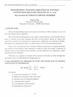
scandens L, we have been isolated six known flavonoids compounds. Their structures were identified as quercetin-3O-L-rhamnoside (1), genistein (2), quercetin (3), quercetin 3-O-β-D-glucuronide (4), kaempferol (5) and
quercetin-3-O-α-L-arabinofuranoside (6) on the basis of spectroscopic data and by comparing their
physicochemical and spectral data with those published in literatures.
Keywords: Flavonoids, Quercetin, Tetracera scandens L.
_____________________________________________________________________________________________
INTRODUCTION
Tetracera scandens L. (Dilleniaceae) is a genus of flowering plants of the Dilleniaceae family. It is widely used in
Vietnamese traditional medicine to treat the diseases of hepatitis, gout and inflammation. Therefore, we conducted
the investigation about the chemical composition of leaves of Tetracera scandens L, which were collected in Nha
Trang, Vietnam. From the ethanol extract of leaves of Tetracera scandens L, we have been isolated together with six
known compounds, which were first reported in this material. Their structures were elucidated on the basis of
spectroscopic data and comparing with the reported 1H-NMR and 13C-NMR data in literature.
GENERAL AND EXPERIMENTAL PROCEDURES
UV spectra were recorded in MeOH on a JASCO V-550 UV/vis spectrometer with a 0.5 nm resolution. NMR
spectra were obtained on a Varian Unity Inova 500 MHz spectrometer with tetramethylsilane (TMS) as the internal
standard. Silica gel (Merck, 63-200 µm particle size), RP- 18 (Merck, 40-63 µm particle size), and Sephadex LH-20
were used for column chromatography. TLC was carried out with silica gel 60 F254 and RP-18 F254 plates. HPLC
was carried out using a Gilson system with a UV detector and Optima Pak C18 column (10 × 250 mm, 10 µm
particle size, RS Tech, Korea). All solvents used for extraction and isolation were of analytical grade.
PLANT MATERIAL
The leaves of Tetrtacera scandens L. were collected in October 2013 from Nha Trang province, Vietnam and
authenticated by Prof. Nguyen Thanh Hai (School of Medicine and Pharmacy, Vietnam National University, Hanoi).
A voucher specimen (No. SMP-2013-0012) was deposited at the Herbarium of SMP, VNU.
2123
Tung Bui Thanh et al
J. Chem. Pharm. Res., 2015, 7(3):2123-2126
______________________________________________________________________________
EXTRACTION AND ISOLATION
The leaves of Tetracera scandens L. (2.5 kg) were extracted with ethanol (10 L × 3 times) at room temperature for a
week. The combined ethanol extract was then concentrated to yield a dry residue (251 g). This crude extract was
suspended in water (2.5 L) and partitioned successively with n-hexane (3 × 2 L), EtOAC (3 × 2 L), and BuOH (3 × 2
L). The EtOAC and BuOH soluble fractions were combined (117 g) and chromatographed over a silica gel column
(10 × 30 cm; 63–200 µm particle size) eluting with gradient solvent CHCl3/acetone (19:1, 18:2…1:19, each 2.5 L),
to yield six fractions (F1: 13.2 g; F2: 22.6 g; F3: 28.4 g; F4: 14.5 g; F5: 23.11 g; F6: 10.87 g) based on the TLC
profile. Fraction F3 was further applied to an RP-18 column (7 × 30 cm; 40-63 µm particle size) eluting with a
stepwise gradient of MeOH/H2O (1:2 to 10:1) to afford four sub-fractions (F3.1–F3.4). Fraction F3.1 (90 mg) was
separated by HPLC [Optima Pak C18 column (10 × 250 mm, 10 µm particle size, RS Tech, Korea); mobile phase
MeOH in H2O containing 0.1% formic acid (0–50 min: 63% MeOH, 50–55 min: 100% MeOH, 55–60 min: 100%
MeOH); flow rate 2 mL/min; UV detection at 205 and 254 nm] to give compound 1 (tR = 36.0 min, 16.0 mg)
(quercetin-3O-L-rhamnoside), compound 2 (tR = 39.0 min, 10.0 mg) (genistein). Fraction F3.2 (80 mg) was
purified by HPLC (0–50 min: 75% MeOH, 50–55 min: 100% MeOH) to yield compound 3 (tR = 40.0 min, 5.8 mg)
(quercetin). Fraction F3.4 (48 mg) was purified by HPLC (0–50 min: 75% MeOH, 55 min: 100% MeOH) to yield
compounds 4 (tR = 42.0 min, 3.5 mg) (quercetin 3-O-β-D-glucuronide). Fraction F4 was chromatographed over a
Sephadex LH-20 column (7 × 40 cm) using MeOH as the eluting solvent to afford three subfractions (F4.1–F4.3).
Subfraction F4.2 (3.1 g) was further chromatographed over a silica gel column (5 × 40 cm; 40–63 µm particle size)
with a gradient solvent of CHCl3/ MeCN (9:1, 8:2…1:9, each 2.5 L) to yield five fractions (F4.2.1–F4.5.5).
Subfraction F4.2.2 (150 mg) was purified by HPLC (0–40 min: 70% MeOH, 40–45 min: 100% MeOH) to yield and
compound 5 (tR = 19.0 min, 3.5 mg) (kaempferol) and compound 6 (tR = 24.0 min, 4.5 mg) (quercetin-3-O-α-Larabinofuranoside) (Figure 1).
Quercetin-3-O-L-rhamnoside (1): yellow powder, UV spectrum (MeOH, λmax, nm) 257, 359; PMR spectrum
(500 MHz, acetone-d6, δ, ppm, J/Hz): 6.73 (1H, br, s H-6), 6.94 (1H, br, s, H-8), 7.97 (1H, d, J = 1.5, H-2’), 7.45
(1H, d, J = 8.4, H-5’), 7.85 (1H, d, J = 8.4, H-6’), 5.99 (1H, m, H-1”), 4.16 (1H, m, H-2”), 3.80 (1H, m, H-3”), 3.61
(1H, m, H-4”), 4.65 (1H, m, H-5”), 1.37 (3H, d, J = 6.0, CH3-6”); 13C-NMR spectrum (125 MHz, acetone-d6, δ,
ppm): 157.9 (C-2), 135.8 (C-3), 179.2 (C-4), 163.8 (C-5), 99.4 (C-6), 164.7 (C-7), 94.5 (C-8), 158.3 (C-9), 105.7 (C10), 122.8 (C-1’), 116.1 (C-2’), 145.7 (C-3’), 148.9 (C-4’), 116.7 (C-5’), 122.6 (C-6’), 102.7 (C-1”), 72.0 (C-2”),
72.9 (C-3”), 78.7 (C-4”): 71.3 (C-5”), 17.7 (C-6”). These data are in agreement with those originally reported for
quercetin-3 -O-L- rhamnoside [1].
Genistein (2): yellow powder, UV spectrum (MeOH, λmax, nm): 260, 366; PMR spectrum (500 MHz, acetone-d6,
δ, ppm, J/Hz): 7.87(1H, s, H-2), 6.32(1H, d, J=2.17, H-6), 6.58(1H, d, J=2.17, H-8), 7.74, (2H, d, J= 8.73, H-2’, H6’, 7.04 (2H, d, J= 8.72, H-3’, H-5’). 13C-NMR spectrum (125 MHz, acetone-d6, δ, ppm): 148.3 (C-2), 146.7 (C-3),
176.5 (C-4), 162.1 (C-5), 99.1 (C-6), 165.0 (C-7), 94.5 (C-8), 157.8 (C-9), 104.1 (C-10), 121.8 (C-1’), 121.5 (C-2’),
115.9 (C-3’), 146.9 (C-4’), 115.9 (C-5’), 121.5 (C-6’). These data are in agreement with those originally reported for
genistein [2].
Quercetin (3): yellow powder, PMR spectrum (500 MHz, acetone-d6, δ, ppm, J/Hz): 6.27 (1H, d, J=2.2, H-6),
6.53 (1H, d, J=2.2, H-8), 7.83 (1H, d, J=2.2, H-2’), 6.99 (1H, d, J=9.1, H-5’), 7.69 (1H, dd, J=9.0 Hz, J= 2.2, H-6’);
13
C-NMR spectrum (125 MHz, acetone-d6, δ, ppm): 124.9 (C-2), 136.7 (C-3), 176.5 (C-4); 162.1 (C-5), 99.1 (C-6),
164.9 (C-7), 94.5 (C-8), 157.8 (C-9), 104.1 (C-10), 123.8 (C-1’), 115.8 (C-2’); 145.8 (C-3’), 148.3 (C-4’), 116.2 (C5’), 121.5 (C-6’). These data are in agreement with those originally reported for quercetin [3].
Quercetin 3-O-β-D-glucuronide (4): yellow powder, UV spectrum (MeOH, λmax, nm) 256, 358; PMR spectrum
(500 MHz, acetone-d6, δ, ppm, J/Hz): 6.23 (1H, d, J=2.0, H-6), 6.47 (1H, d, J=2.0, H-8), 7.82 (1H, d, J=2.4, H-2’),
6.90 (1H, d, J=8.6, H-5’), 7.69 (1H, dd, J=2.4, J=8.6, H- 6’), 5.44 (1H, m, H-1”), 3.52 (1H, m, H-2”), 3.57 (1H, m,
H-3”), 3.84 (1H, m, H-4”), 3.87 (1H, m, H-5”); 13C-NMR spectrum (125 MHz, acetone-d6, δ, ppm): 157.8 (C-2),
135.1 (C-3), 178.8 (C-4), 162.8 (C-5); 99.6 (C-6), 165.1 (C-7), 94.6 (C-8), 158.1 (C-9), 105.4 (C-10), 123.1 (C-1’),
115.8 (C-2’), 145.3 (C-3’), 149.2 (C-4’), 117.2 (C-5’), 122.5 (C-6’), 104.1 (C-1”), 75.1 (C-2”), 76.2 (C-3”), 72.3 (C4”), 77.4 (C-5”), 170.1 (C-6”). These data are in agreement with those originally reported for quercetin 3-O-β-Dglucuronide [4].
Kaempferol (5): yellow powder, PMR spectrum (500 MHz, acetone-d6, δ, ppm, J/Hz): 6.27 (1H, d, J=2.0 H-6),
6.54 (1H, d, J=2.0, H-8), 8.15 (2H, d, J=8.8, H-2’, H-6’), 7.01 (2H, d, J= 8.8, H-3’, H-5’); 13C-NMR spectrum (125
MHz, acetone-d6, δ, ppm): 147.0 (C-2), 136.7 (C-3), 176.6 (C-4), 162.1 (C-5), 99.1 (C-6), 165.0 (C-7), 94.6 (C-8),
157.9 (C-9), 104.1 (C-10), 123.4 (C-1’), 130.5 (C-2’), 116.4 (C-3’), 160.1 (C-4’); 116.4 (C-5’), 130.5 (C-6’). These
data are in agreement with those originally reported for kaempferol [3].
2124
Tung Bui Thanh et al
J. Chem. Pharm. Res., 2015, 7(3):2123-2126
______________________________________________________________________________
Quercetin-3-O-α-L-arabinofuranoside (6): yellow powder, UV spectrum (MeOH, λmax, nm) 257, 357; PMR
spectrum (500 MHz, acetone-d6, δ, ppm, J/Hz): 6.28 (1H, d, J=2.1, H-6); 6.51 (1H, d, J= 2.1, H-8), 7.73 (1H, d,
J=2.1, H-2’); 6.99 (1H, d, J=8.5, H-5’), 7.58 (1H, dd, J=8.5, J= 2.4, H-6’), 5.48 (1H, d, J=0.6, H-1”); 4.10(1H, m, H2”), 3.61(1H, m, H-3”), 3.97 (1H, m, H-4”), 4.28 (2H, m, CH2-5”); 13C-NMR spectrum (125 MHz, acetone-d6, δ,
ppm): 158.1 (C-2), 135.4 (C-3), 179.9 (C-4), 162.8 (C-5), 99.6 (C-6), 165.2 (C-7), 94.7 (C-8), 158.0 (C-9), 105.5 (C10), 122.9 (C-1’), 116.6 (C-2’), 145.8 (C-3’), 149.2 (C-4’), 116.4 (C-5’), 122.5 (C-6’), 109.2 (C-1”), 82.3 (C-2”),
78.9 (C-3”), 89.5 (C-4”), 62.7 (C-5”). These data are in agreement with those originally reported for quercetin-3-Oα-L-arabinofuranoside [5].
1
2
OH
OH
HO
O
HO
OH
O
O
O
OH
OH
O
OH
3
HO
OH
O
OH
OH
OH
O
OH
OH
4
OH
OH
HO
O
O
O
OH
O
OH
O
5
HO
OH
HOOC
OH
O
OH
6
OH
OH
OH
HO
OH
O
O
1.
Quercetin-3O-rhamnoside
2.
Genistein
3.
Quercetin
4.
Quercetin 3-O-β-D-glucuronide
5.
Kaempferol
6.
Quercetin-3-O-α-L-arabinofuranoside
O
OH
O
OH
O
HOH2C
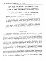
Figure 1. Structure of flavonoids isolatedfrom leaves of Tetracera scandens L.
2125
OH
Tung Bui Thanh et al
J. Chem. Pharm. Res., 2015, 7(3):2123-2126
______________________________________________________________________________
Competing interests
We declare that we have no conflict of interest.
Acknowledgements
The research was supported by has been financed by the “Program Tay Bac” with grants number: KHCNTB05C/13-18. Also we would like to thank VNU-VSL (Vietnam National University, HaNoi-VNU- Scientist Links)
for support to submit the manuscript.
REFERENCES
[1] X Ma, W Tian, L Wu, X Cao, Y Ito, J Chromatogr A., 2005, 1070, 211-214.
[2] H. A. Almahy, N. I. Alhassan, J of Science and Tech., 2011, 3,118-124.
[3] Z. P. Xiao, H. K. Wu, T. Wu, H. Shi, B. Hang, H. A. Aisa, Chem. Nat. Cmpd.,2006, 42, 736-737.
[4] JH. Moon, T. Tsushida, K. Nakahara, J. Terao,Free Radic Biol Med.,2001, 30, 1274-1285.
[5] X. Zhang, PT. Thuong, W. Jin, ND. Su, DE. Sok, K Bae, SS Kang, Arch Pharm Res., 2005, 28, 22-27.
2126