DSpace at VNU: Flavonoids and other compound isolated from leaves of Aconitum carmichaeli Debx. growing in Viet Nam 10. Bui Thanh Tung
Bạn đang xem bản rút gọn của tài liệu. Xem và tải ngay bản đầy đủ của tài liệu tại đây (149.74 KB, 7 trang )
Available online www.jocpr.com
Journal of Chemical and Pharmaceutical Research, 2015, 7(6):228-234
Research Article
ISSN : 0975-7384
CODEN(USA) : JCPRC5
Flavonoids and other compound isolated from leaves of Aconitum carmichaeli
Debx. growing in Viet Nam
Loi Vu Duc1*, Tung Bui Thanh1, Hai Nguyen Thanh1 and Vung Nguyen Tien2
1
School of Medicine and Pharmacy, Viet Nam National University, Hanoi
2
National Institute of Forensic Medicine, Hanoi
_____________________________________________________________________________________________
ABSTRACT
Two flavonol glycosides: 5,7,3'-trimethoxyquercetin -3-O-β-D-fructofuranoside (1), 7,4′-O-dimethylluteolin 5-O-[αL-arabinofuranosyl-(1→6)-β-D-glucopyranoside] (2) and one fatty acid ester (Z)-3-hydroxypentan-2-yl-10aminooctacos-9-enoate (3) were isolated from the leaves of Aconitum carmichaeli Debx growing in Ha Giang
province, Vietnam. Their structures were elucidated by spectroscopic methods, including IR, MS and NMR. This is
first time these compounds were isolated from leaves of A. carmichaeli Debx.
Keywords: Aconitum carmichaeli, flavonoids, trimethoxyquercetin, dimethylluteolin
_____________________________________________________________________________________________
INTRODUCTION
The Aconitum L. genus which is mainly characterized by the presence of highly toxic diterpene alkaloids has been
traditionally used in China as source of arrow poison for 2000 years. The alkaloid composition of different European
Aconitum species has been analyzed in some reports. In Vietnam, Bui Hong Cuong has been reported to chemical
compositions of A. carmichaeli Dexb. var. carmichaeli collected in Sa Pa, Lao Cai province including alkaloids,
acid amin, free sugars, organic acids. Its rhizomes contain fatty acid, sterol and its stems contain carotenoids, its
leaves and flowers present carotenoids, sterols, flavonoids. Several alkaloids named karacolin, benzoylmesaconin
also reported in this study [1]. In the last years the study of Aconitum genus lead to the other secondary metabolites
such as flavonoids. The flavonol composition of the aerial parts of A. jaluense, A. pseudolaeve and A. chiisanense
have been reported [2]. Regarding the European species, the flowers of A. paniculatum have shown the presence of
three flavonol glycosides [3]. However, there have been no reports of flavonoid compositions from A. carmichaeli
collected in Ha Giang province, Viet Nam. In this study we describe the isolation and structure elucidation of
compounds isolated from the leaves of Aconitum carmichaeli Dexb.
EXPERIMENTAL SECTION
Plant material
The leaves of Aconitum Carmichaeli Debx.were collected in Ha Giang province, Viet Nam during 2012 and
authenticated by the National Institute of Medicinal Materials (NIMM). A voucher specimen has been deposited in
the NIMM.
228
Loi Vu Duc et al
J. Chem. Pharm. Res., 2015, 7(6):228-234
______________________________________________________________________________
General experimental procedures
Melting points were measured on Mikroskopheiztisch PHMK-50 (VEB Waegetechnik Rapido, Germany). The FTIR spectra were recorded on an IMPACT-410FT-IR spectrometer (CARL ZEISS JENA). The NMR [1H (500 MHz),
13
C (125 MHz), and DEPT-90 and 135 MHz)] spectrum were recorded on an AVANCE spectrometer AV 500
(Brucker, Germany) in the Institute of Chemistry, Viet Nam Academy of Science and Technology (VAST).
Chemical shifts were reported in ppm downfield from TMS with J in Hz. Electrospray Ionization Mass Spectrum
(ESI-MS) were recorded on a Varian Agilent 1100 LC-MSD mass spectrometer. Analytical TLC was performed on
Kieselgel 60 F254 (Merck) plates (silica gel, 0.25 mm layer thickness) and RP-18 F254 (Merck) plates (0.25 mm layer
thickness). Spots were visualized using ultraviolet radiation (at 254 and 365 nm) and by spraying with 10% H2SO4,
followed by heating with a heat gun. Column chromatography was performed on silica gel (70–230 and 230–400
mesh, Merck). Organic solvents were of analytical grade.
Extraction and isolation
The dried and powdered leaves of A. carmichaeli (1.5 kg) were extracted with 96% ethanol (5L x 4 times) at room
temperature. The ethanol extract were combined, filtrated, and evaporated to dryness in vacuum at 40°C. The
residue (80 g) was suspended in water and then partitioned with EtOAc. The EtOAc extract (15 g) was subjected to
column chromatography (CC) over silica gel with a gradient solvent system [n-hexane : EtOAc (EtOAc 0%-100%)
and EtOAc : MeOH (0%-80%)] to give 5 fractions: A, B, C, D, E. The fraction B (0.8 g) was subjected to repeated
silica gel CC and eluted with CHCl3 - EtOAc (90:10) to give compound 3 (16 mg). The fraction E (2.1 g) were
subjected to repeat silica gel CC and eluted with MeOH - EtOAc (90:10) to yield compound 1 (12 mg) and
compound 2 (22 mg).
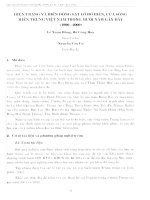
Compound 1: 5,7,3'-trimethoxyquercetin -3-O-β-D-fructofuranoside
M= 506, Rf = 0.4 (CHCl3- MeOH, 10:90). IR (KBr, vmax cm-1): 3060 (OH), 2924 (CH), 1687 (C=O), 1073 (C–O–C).
The 1H-NMR (500 MHz, CDCl3-d6, δ, ppm, J/Hz), 13C-NMR (125 MHz, CDCl3-d6, δ, ppm, J/Hz) and DEPT data
for compound 1 are presented in table 1.
OCH 3
3'
OH
4'
H 3CO
O
7
1'
3
6''
O
5
OCH 3
O
2'' HO
O
HO
4'' H
1''
H
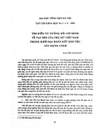
Figure 1. Structure of compound 1
229
H
OH
OH
Loi Vu Duc et al
J. Chem. Pharm. Res., 2015, 7(6):228-234
______________________________________________________________________________
Table 1. 1H-NMR, 13C-NMR and DEPT Data for compound 1
HMBC
(C→H)
C
C
C
C
CH
C
CH
C
C
CH3
CH3
C
CH
C
C
CH
CH
CH3
δC
ppm
156,2
130,0
177,47
161,9
110,6
162,2
110,9
148,4
111,3
55,8
53,7
129,1
128,8
145,1
147,5
124,5
127,9
55,9
δH
ppm
6,19 s
6,79 s
3,42 s
3,61 s
8,25 s
8,12 d (7,0)
8,13 d (7,0)
3,86 s
CH2
C
CH
CH
CH
CH2
60,3
111,7
73,1
67,9
75,8
60,2
3,83 dd (2,5; 13,0); 3,43 dd (3,0; 12,5)
3,17 dd (4,0; 15,0)
4,46 dd (4,5; 14,5)
4,93 dd (4,0; 14)
3,09 m
Position C
DEPT
2
3
4
5
6
7
8
9
10
5-OCH3
7-OCH3
1’
2’
3’
4’
5’
6’
’
3 -OCH3
4’-OCH3
1’’
2’’
3’’
4’’
5’’
6’’
5; 7; 8; 10
6; 7; 9; 10
5
7
2; 3’; 4’; 6’
1’
2; 2’; 4’
3’
1; 2’; 3’
4’; 5’
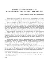
Compound 2: 7,4′-O-dimethylluteolin 5-O-[α-L-arabinofuranosyl-(1→6)-β-D-glucopyranoside]
M = 608, Rf = 0,3 (CHCl3- MeOH, 15:85), ESI-MS: m/z 609,2 [M+H]+, IR (KBr, vmax cm-1): 3414 (OH), 2936 (CH),
1725 (C=O), 1100 (C–O–C). The 1H-NMR (500 MHz, CDCl3-d6, δ, ppm, J/Hz), 13C-NMR (125 MHz, CDCl3-d6,
δ, ppm, J/Hz) and DEPT data for compound 2 are presented in table 2.
OH
3'
2'
4''' HO
5'''
HO
O
O
3'''
H
H
2'''
6
6''
9
10
C
2
5''
O
1''
3''
2''
6'
3
4
O
O
OH
OH
Figure 2. Structure of compound 2
230
5'
1'
5
4''
1'''
HO
H HO
O
7
A
H
B
8
H 3 CO
OCH 3
4'
Loi Vu Duc et al
J. Chem. Pharm. Res., 2015, 7(6):228-234
______________________________________________________________________________
Table 2. 1H-NMR, 13C-NMR and DEPT Data for compound 2
Position C
DEPT
2
3
4
5
6
7
8
9
10
7-OCH3
1'
2'
3'
4'
5'
6'
4’–OCH3
1''
2''
3''
4''
5''
6''
1'''
2'''
3'''
4'''
C
CH
C
C
CH
C
CH
C
C
CH3
C
CH
C
C
CH
CH
CH3
CH
CH
CH
CH
CH
CH2
CH
CH
CH
CH
δC
ppm
165,0
106,3
180,2
159,5
104,3
165,9
97,5
160,6
110,4
56,8
121,3
110,1
150,4
155,0
117,6
122,0
56,7
104,8
74,8
77,3
71,8
77,3
68,4
110,5
83,3
78,8
85,7
5'''
CH2
63,0
δH
ppm
6,61 s
6,90 d (2,0)
6,95 d (2,0)
3,7 s
7,45 d (2,0)
6,89 d (7,5)
7,51 dd (2,0, 8,0)
3,96 s
4,86
3,62
3,70
3,42
3,53 t (7,5)
3,65
4,97 s
4,05
3,87 dd (3,5; 6,0)
4,01 m
3,61
3,75 dd (3,5; 12,0)
HMBC
(C→H)
2; 4; 1'; 2'
5; 7; 8; 10
6; 7; 9; 10
7
2; 3'; 4'; 6'
1'
2; 2'; 4'
4'
5
6''
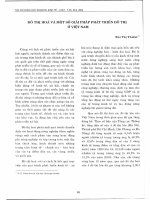
Compound 3: (Z)-3-hydroxypentan-2-yl-10-aminooctacos-9-enoate
M=524, Rf = 0,3 (CH2Cl2 - EtOAc, 90:10), ESI-MS: m/z 547 [M+Na]+, IR: (KBr, ν cm-1): 3426, 3334 (NH2); 2923,
2853 (CH); 1658 (C=O); 1625 (C=C); 1548 (C-N); 1089 (C-O-C). The 1H-NMR (500 MHz, CDCl3-d6, δ, ppm,
J/Hz), 13C-NMR (125 MHz, CDCl3-d6, δ, ppm, J/Hz) and DEPT data for compound 3 are presented in table 3.
NH2
OH
28
1
9
O
Figure 3. Structure of compound 3
231
O
4'
1'
2'
Loi Vu Duc et al
J. Chem. Pharm. Res., 2015, 7(6):228-234
______________________________________________________________________________
Table 3.1H-NMR, 13C-NMR and DEPT Data for compound 3
Position C
DEPT
1
C=O
δC
ppm
173,1
2
CH2
36,9
3
4
5
6
7
8
9
10
11
12
13
14
15
16
CH2
CH2
CH2
CH2
CH2
CH2
CH
C
CH2
CH2
CH2
CH2
CH2
CH2
CH2
CH2
CH2
CH2
CH2
CH2
CH2
CH2
CH2
CH2
CH2
CH3
CH
CH
CH2
CH3
26,0
29,5
29,5
29,5
29,5
22,6
118,2
162,1
49,4
29,5
29,5
29,5
29,5
29,5
29,5
29,5
29,5
29,5
29,5
29,5
29,5
29,5
29,5
33,6
25,8
14,2
76,7
74,4
31,9
14,1
17
18
19
20
21
22
23
24
25
26
27
28
1’
2’
3’
4’
δH
ppm
H-2a :2,28 d, (6,0)
H-2b: 1,62 d, (7,0)
1,44-1,45 m
1,25 m
1,25 m
1,25 m
1,25 m
2,15-2,19 m
5,69 d, (7,5)
1,50-1,60 m
1,25 m
1,25 m
1,25 m
1,25 m
1,25 m
1,25 m
1,25 m
1,25 m
1,25 m
1,25 m
1,25 m
1,25 m
1,25 m
1,25 m
1,25 m
1,25 m
1,09 d, (7,0)
4,00-4,03 m
3,62 d, (5,0)
1,37-1,40 m
0,88 t, (14,0)
RESULTS AND DISCUSSION
Compound 1: ESI-MS spectrum gave the molecular ion peak at m/z 507,3 [M+H]+, corresponded to molecular
mass M= 506. The IR spectrum revealed the presence of hydroxyl (O-H) and conjugated carbonyl groups (C=O) at
3060 cm-1 and 1687 cm-1, respectively. It also present the absorption bands at 2944 cm-1 (C-H) and 1073 cm -1 (C-OC).
13
C-NMR, DEPT 90 and DEPT 135 spectrum of compound 1 showed signals for all 24 carbons. Among them, one
signal belonged to carbonyl group at δ 177.4 ppm; fours signals belonged to aromatic carbons bearing an oxygen
atom at δ 162,2; 148,9; 145,1; 147,5 ppm and three signals belonged to methoxy group at δ 55,9; 55,8; 53,7 ppm.
Furthermore, 13C-NMR spectrum showed six conjugated signals from δ 60,2 to δ 111,7 ppm indicating the presence
of one sugar moiety. The J values (7.51 Hz) of the anomeric proton indicated β-orientations of the glycosidic
linkages. Two signals at δ 60,3 and δ 60,2 ppm data permitted the identification of the sugar moiety as fructose.
1
H-NMR spectrum of compound 1 displayed signals for three aromatic protons at δ 8,25 (1H, s, H-2”,), δ 8,13 (1H,
d, J =7,0 Hz, H-5”) and δ 8,12 (1H, d, J = 7,0 Hz, H-6”), indicating the presence of a 1,3,4-trisubstituted benzene
ring. The remaining aromatic protons at δ 6.79 (1H, d, J=2.0 Hz, H-6) and δ6.19 (1H, d, J=2.0 Hz, H-8) showed the
presence of a 1,2,3,5-tetrasubstituted benzene ring.1H-NMR spectrum displayed the signal of one anomeric protons
at δ 4,93 ppm (1H, dd, J=4,0; 14 Hz, H-5”) suggested the presence of beta-fructose sugar moiety. Therefore, based
on the above data and compared with previous published [4], the structure of compound 1 was established as 5,7,3'trimethoxyquercetin-3-O-β-D-fructofuranosid (Fig. 1).
Compound 2 was obtained as a yellow powder. ESI-MS spectrum gave the molecular ion peak at m/z 609,2
[M+H]+, corresponded to molecular mass M = 608. The IR spectrum revealed the presence of hydroxyl (O-H) and
232
Loi Vu Duc et al
J. Chem. Pharm. Res., 2015, 7(6):228-234
______________________________________________________________________________
conjugated carbonyl groups (C=O) at 3414 cm-1 and 1725 cm-1, respectively. It also present the absorption bands at
2936 cm-1 (C-H) and 1100 cm -1(C-O-C).
1
H-NMR spectrum displayed the signal corresponding to two aromatic rings of flavonol skeleton were observed at δ
6,62 (1H, s, H-3), and δ 6,90 (1H, d, J= 2,0; H-6) và δ 6,95 (1H, d, J= 2,0; H-8). The 1H-NMR spectrum of 2 showed
three signals assignable to ABX coupled aromatic protons at 1,3,4 positions at δ 7,45 (1H, d, J=2,0, H-2'); δ 6,89
(1H, d, J= 8,0; H-5'); δ 7,51 (1H, dd, J=2,0, 8,0; H-6'). Moreover, two methoxyl group δ 3,96 (3H, s, OCH3) và δ
3,97 (3H, s, OCH3) at were also presented. The J values (7.53 Hz) of the two anomeric protons indicated βorientations of the glycosidic linkages. Furthermore, 1H-NMR spectrum displayed the signal of two anomeric
protons at δ 4,86 (1H, H-1'') and δ 4,97 (H, s, H-1''') indicating the presence of two sugar moieties. The positions of
the H-1'' and H-1''' were confirmed by HSQC correlations between H-1'' with δ 104,8 (C-1'') and H-1''' with δ 110,5
(C-1'''). Four methylene protons of two groups oxymethylene in the two sugar moieties were revealed at δ 3,65 (2H,
H-6'', overlap) and δ 3,61 (1H, Ha-5'''), δ 3,75 (1H, dd, J=3,5; 12,0; Hb-5'''). The oxymethine proton signals were
revealed at δ 3,62 (1H, H-2''); δ 3,70 (1H, H-3''); δ 3,42 (1H, H-4''); δ 3,53 (1H, H-5''); δ 4,05 (1H, H-2'''); δ 3,87
(1H, dd, J=3,5; 6,0; H-3'''); δ 4,01 (1H, m, H-4'''). The sugar moieties were confirmed through correlations in HSQC
spectrum. 13C-NMR spectrum showed six conjugated signals at δ 104,8 (C-1''); δ 74,8 (C-2''); δ 77,3 (C-3''); δ 71,8
(C-4''); δ 77,3 (C-5''); δ 68,4 (C-6'') indicating the molecular of glucose. In the 13C NMR spectrum, the downfield
signal at δ 68,4 (C-6'') indicated a ether group. Beside the presence of 17 carbon atoms of aglycon moiety (including
two signals of methoxyl groups) there were 5 signals at δ 110,5 (CH, C-1'''); δ 83,3 (CH, C-2'''); δ 78,8 (CH, C-3''');
δ 85,7 (CH, C-4'''); δ 63,0 (CH2, C-5'''). From this data, associated with the 13C-NMR data, it was obvious that the
sugar unit was O-[-L-arabinofuranosyl-(16)-β-D-glucopyranosid]. The positions of substituted groups were
confirmed by 1H-13C correlation in HMBC. The correlation of proton H-1” and C-5; group methoxyl and C-7
indicated position of sugar moiety at C-5 and group methoxyl at C-7 in ring A. Also the correlations of proton H-1”’
and C-6” was further confirmed the positions linked of two molecular sugar.
In the ring B, position of methoxyl group at C-4’ was confirmed through the correlation between protons in
methoxyl group and C-4’; H-6’ and C-4’. This indicated that C-6’ and carbon (-hydroxy) are in para positions.
Based upon these evidences and compared with previous published [5], compound 2 was established as 7,4’-Odimethylluteolin 5-O-[α-L-arabinofuranosyl-(16)-β-D-glucopyranosid] (Fig. 2).
Compound 3: ESI-MS spectrum gave the molecular ion peak at m/z 547547 [M+Na]+, corresponded to molecular
mass M= 524 and molecular formula C33H65NO3. The compound has absorption bands at 3426, 3334 cm-1 (NH2);
2923, 2853 cm-1 (C-H); 1658 cm-1 (C=O); 1625 (C=C, cis); 1548 cm-1 (C-N); 1089 cm-1 (C-O-C) in its IR spectrum.
The 1H-NMR spectrum of 3 showed signal doublet with J = 7,5 Hz at δ 5,69 indicated a cis double bond.
The 1H-NMR spectrum of 3 displayed signals corresponding to methyl group bonds to methine at δ 1,09 ppm (J
=7,0 Hz), two methyl groups bond to methylen groups (J = 14 Hz), two signals of methinegroups at δ 4,00-4,03 and
δ 3,62 (d, J = 5 Hz) and 25 signals of methylen groups. The 13C-NMR, DEPT 90 and DEPT 135 spectrum of
compound 3 showed signals for all 33 carbons. Among them, one signal belonged to carbonyl group at δ 173,1 ppm;
one quaternary carbon signal belonged to the olefinic carbon bond to N at δ 162.1 ppm; two signals of methine
group bearing an oxygen atom at δ 76,7 ppm and δ 74,4 ppm; three signals of methyl group and 25 signals of
methylene group. Based upon these evidences and compared with previous published [6, 7], compound 3 was
established as (Z)-3-hydroxypentan-2-yl 10-aminooctacos-9-enoate (Fig. 3).
CONCLUSION
Our phytochemical study of the 96% ethanol extracts of the leaves of A. carmichaeli Dexb. collected in Ha Giang
province, Vietnam has resulted in the isolation of three compounds: 5,7,3'-trimethoxyquercetin-3-O-β-Dfructofuranoside (1), 7,4′-O-dimethylluteolin 5-O-[α-L-arabinofuranosyl-(1→6)-β-D-glucopyranoside] (2) and (Z)3-hydroxypentan-2-yl-10-aminooctacos-9-enoate (3). These were isolated for the first timefrom leaves of A.
carmichaeli Dexb. Our results could be beneficial for the search of new chemical agents from Vietnamese plants.
Acknowledgments
The research was supported by has been financed by the “Program Tay Bac” with grants number: KHCNTB.05C/13-18.
233
Loi Vu Duc et al
J. Chem. Pharm. Res., 2015, 7(6):228-234
______________________________________________________________________________
REFERENCES
[1] Bui H. C., Phung H. B., Vu C. N., Journal of medicinal materials-Ha Noi, 2005,10 (2), 55-59.
[2] Jeong H. J., Whang W. K., Kim I. H., Planta Med., 1997, 63 (4): 329-334.
[3] Gelsomina F., Alessandra B., Anna Ri. B., Franca T., Ivano M., J Nat Prod , 2000, 63(11), 1563-1565.
[4] Zahir A., Jossang A., Bodo B., Provost J., Cosson J. P., Sévenet T., Journal of Natural Products, 1999, 62(2),
241-243.
[5] Hai L., Karl W. K. T., Gui-Xin C., Jun-Ming W., et al., Chemistry & Biodiversity, 2011, 8 (11), 2110–2116,
[6] Volodymyr M., Volodymyr S., Chemistry and chemical technology, 2010, 4(3), 171-178.
[7] Zakia K., Rosina K., Medicinal chemistry research, 2011, 11(3), 658-665.
234