Microbiology a systems approach 3rd ed cowan BBS part 2
Bạn đang xem bản rút gọn của tài liệu. Xem và tải ngay bản đầy đủ của tài liệu tại đây (35.58 MB, 481 trang )
13.2 The Progress of an Infection
367
Microbe X
Virulence
Percentage of optimal
infectious dose
Correct portal
of entry
High
>100
Yes
Low
0
No
Microbe passes
through unnoticed.
Possible
outcomes
Host
Genetic profile
that resists
Microbe X
(Nonspecific
defenses)
Previous exposure
to Microbe X
(Specific immunity)
General level
of health; production
of stress hormones
Microbe becomes
established
without disease
(colonization or
infection).
Microbe causes
disease.
Yes
No
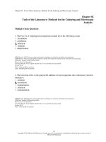
Figure 13.2 Will disease result from an encounter between a (human) host and a microorganism? In most cases, all of the
slider bars must be in the correct ranges and the microbe’s toggle switch must be in the “yes” position while the host’s toggle switch must be in
the “no” position in order for disease to occur.
developed virulence properties. Examples of opportunistic
pathogens include Pseudomonas species and Candida albicans.
Factors that greatly predispose a person to infections, both primary and opportunistic, are shown in table 13.4.
The relative severity of the disease caused by a particular microorganism depends on the virulence of the microbe.
Although the terms pathogenicity and virulence are often used
interchangeably, virulence is the accurate term for describing the degree of pathogenicity. The virulence of a microbe is
determined by its ability to
1. establish itself in the host, and
2. cause damage.
There is much involved in both of these steps. To establish
themselves in a host, microbes must enter the host, attach
firmly to host tissues, and survive the host defenses. To cause
damage, microbes produce toxins or induce a host response
that is actually injurious to the host. Any characteristic or
structure of the microbe that contributes to the preceding
activities is called a virulence factor. Virulence can be due
to single or multiple factors. In some microbes, the causes of
virulence are clearly established, but in others they are not. In
the following section, we examine the effects of virulence fac-
tors while simultaneously outlining the stages in the progress
of an infection.
Note that different healthy individuals have widely
varying responses to the same microorganism. This is determined in part by genetic variation in the specific components
of their defense systems. That is why the same infectious
agent can cause severe disease in one individual and mild or
no disease in another.
Table 13.4 Factors That Weaken Host Defenses and
Increase Susceptibility to Infection*
• Old age and extreme youth (infancy, prematurity)
• Genetic defects in immunity, and acquired defects in
immunity (AIDS)
• Surgery and organ transplants
• Underlying disease: cancer, liver malfunction, diabetes
• Chemotherapy/immunosuppressive drugs
• Physical and mental stress
• Other infections
*These conditions compromise defense barriers or immune responses.
368
Chapter 13
INSIGHT 13.1
Microbe-Human Interactions
Life Without Microbiota
For years, questions lingered about how essential the microbiota
is to normal life and what functions various members of the biota
might serve. The need for animal models to further investigate
these questions led eventually to development of laboratory
strains of germ-free, or axenic, mammals and birds. The techniques and facilities required for producing and maintaining a
germ-free colony are exceptionally rigorous. After the young
mammals are taken from the mother aseptically by cesarean
section, they are immediately transferred to a sterile isolator or
incubator. The newborns must be fed by hand through gloved
ports in the isolator until they can eat on their own, and all materials entering their chamber must be sterile. Rats, mice, rabbits,
guinea pigs, monkeys, dogs, hamsters, and cats are some of the
mammals raised in the germ-free state.
A dramatic characteristic of germ-free animals is that they
live longer and have fewer diseases than normal controls, as
long as they remain in a sterile environment. From this standpoint, it is clear that the biota is not needed for survival in this
rarefied environment. At the same time, it is also clear that axenic
life is highly impractical. Studies have revealed important facts
about the effect of the biota on various organs and systems. For
example, the biota contributes significantly to the development
of the immune system. When germ-free animals are placed in
contact with normal control animals, they gradually develop a
biota similar to that of the controls. However, germ-free subjects
are less tolerant of microorganisms and can die from infections
Sterile enclosure for rearing and handling germ-free laboratory animals.
Why is there variation? In chapter 7, we described coevolution as changes in genetic composition by one species in
response to changes in another. Infectious agents evolve in
response to their interaction with a host (as in the case of
antibiotic resistance). Hosts evolve, too. And although their
pace of change is much slower than that of a microbe, eventually changes show up in human populations due to their
past experiences with pathogens. One striking example is
sickle cell disease. Persons who are carriers of a mutation
Table 13.A Effects of the Germ-Free State
Germ-Free Animals Display
Suggesting That:
Enlargement of the cecum;
other degenerative diseases
of the intestinal tract of rats,
rabbits, chickens
Microbes are needed
for normal intestinal
development.
Vitamin deficiency in rats
Microbes are a significant
nutritional source of
vitamins.
Underdevelopment of
immune system in most
animals
Microbes are needed to
stimulate development of
certain host defenses.
Higher rates of
autoimmune disease
Microbes are needed to
“occupy” the immune
system.
by relatively harmless species. This susceptibility is due to the
immature character of the immune system of germ-free animals.
Germ-free animals also have stunted intestinal tracts. Table 13.A
summarizes some major conclusions arising from studies with
germ-free animals.
In 2008 researchers found that Bacteroides fragilis in the gut
produce a molecule that fends off colonization by Helicobacter
pylori. When scientists isolated this molecule and fed it to mice,
it protected them from colitis. And normal biota in the mouth
apparently are important contributors to taste, according to a
recent study. It seems that the thiols released from fruits and vegetables (and wines!), which give them their flavor, are released
due to the action of oral bacteria. Germ-free experiments have
clarified the dynamics of several infectious diseases. Perhaps the
most striking discoveries were made in the case of oral diseases.
For years, the precise involvement of microbes in dental caries
had been ambiguous. Studies with germ-free rats, hamsters,
and beagles confirmed that caries development is influenced by
heredity, a diet high in sugars, and poor oral hygiene. Even when
all these predisposing factors are present, however, germ-free
animals still remain free of caries unless they have been inoculated with specific bacteria. Further discussion on dental diseases
is found in chapter 22.
in their hemoglobin gene (i.e., who inherited one mutated
hemoglobin gene and one normal) have few or no sickle cell
disease symptoms but are more resistant to malaria than people who have no mutations in their hemoglobin genes. When
a person inherits two alleles for the mutation (from both parents), that person enjoys some protection from malaria but
will suffer from sickle cell disease.
People of West African descent are much more likely to
have one or two sickle cell alleles. Malaria is endemic in West
13.2 The Progress of an Infection
Africa. It seems the hemoglobin mutation is an adaptation
of the human host to its long-standing relationship with the
malaria protozoan.
In another example, AIDS researchers have found that
people with a particular gene are less likely to be infected
by HIV, and are slower to develop symptoms from it. Conversely, possessing a different gene gives you weaker cellmediated immunity and that predisposes you to infections
that others might not experience. These are examples of the
variability represented by the slider bar in the lower lefthand corner of figure 13.2. Scientists have also found that
bacteria in the human body respond to human stress hormones, such as norepinephrine. For example, when nerve
cells in the gut produce this hormone, E. coli were found
to increase their numbers up to ten-thousandfold. Other
studies have found that some stress hormones can induce
bacteria to adhere to hard surfaces, raising the possibility of
biofilm formation, and that bacteria increase the expression
of pathogenic genes in these hormones. These phenomena
(depicted with the lower right slider bar in figure 13.2),
suggest an intriguing new area of research into the prevention of microbial disease. Even though human factors are
important, the Centers for Disease Control and Prevention
has adopted a system of biosafety categories for pathogens
based on their general degree of pathogenicity and the relative danger in handling them. This system is explained in
more detail in Insight 13.2.
Becoming Established:
Step One—Portals of Entry
To initiate an infection, a microbe enters the tissues of the
body by a characteristic route, the portal of entry, usually a
cutaneous or membranous boundary. The source of the infectious agent can be exogenous, originating from a source outside the body (the environment or another person or animal),
or endogenous, already existing on or in the body (normal
biota or a previously silent infection).
For the most part, the portals of entry are the same
anatomical regions that also support normal biota: the skin,
gastrointestinal tract, respiratory tract, and urogenital tract.
The majority of pathogens have adapted to a specific portal
of entry, one that provides a habitat for further growth and
spread. This adaptation can be so restrictive that if certain
pathogens enter the “wrong” portal, they will not be infectious. For instance, inoculation of the nasal mucosa with the
influenza virus invariably gives rise to the flu, but if this virus
contacts only the skin, no infection will result. Likewise, contact with athlete’s foot fungi in small cracks in the toe webs
can induce an infection, but inhaling the fungus spores will
not infect a healthy individual. Occasionally, an infective
agent can enter by more than one portal. For instance, Mycobacterium tuberculosis enters through both the respiratory and
gastrointestinal tracts, and pathogens in the genera Streptococcus and Staphylococcus have adapted to invasion through
several portals of entry such as the skin, urogenital tract, and
respiratory tract.
369
Infectious Agents That Enter the Skin
The skin is a very common portal of entry. The actual sites of
entry are usually nicks, abrasions, and punctures (many of
which are tiny and inapparent) rather than unbroken skin.
Intact skin is a very tough barrier that few microbes can penetrate. Staphylococcus aureus (the cause of boils), Streptococcus
pyogenes (an agent of impetigo), the fungal dermatophytes, and
agents of gangrene and tetanus gain access through damaged
skin. The viral agent of cold sores (herpes simplex, type 1) enters
through the mucous membranes near the lips.
Some infectious agents create their own passageways
into the skin using digestive enzymes. For example, certain
helminth worms burrow through the skin directly to gain
access to the tissues. Other infectious agents enter through
bites. The bites of insects, ticks, and other animals offer an
avenue to a variety of viruses, rickettsias, and protozoa. An
artificial means for breaching the skin barrier is contaminated
hypodermic needles by intravenous drug abusers. Users who
inject drugs are predisposed to a disturbing list of well-known
diseases: hepatitis, AIDS, tetanus, tuberculosis, osteomyelitis,
and malaria. Contaminated needles often contain bacteria from
the skin or environment that induce heart disease (endocarditis), lung abscesses, and chronic infections at the injection site.
Although the conjunctiva, the outer protective covering
of the eye, is ordinarily a relatively good barrier to infection,
bacteria such as Haemophilus aegyptius (pinkeye), Chlamydia
trachomatis (trachoma), and Neisseria gonorrhoeae have a special affinity for this membrane.
The Gastrointestinal Tract as Portal
The gastrointestinal tract is the portal of entry for pathogens contained in food, drink, and other ingested substances. They are adapted to survive digestive enzymes
and abrupt pH changes. The best-known enteric agents
of disease are gram-negative rods in the genera Salmonella, Shigella, Vibrio, and certain strains of Escherichia coli.
Viruses that enter through the gut are poliovirus, hepatitis
A virus, echovirus, and rotavirus. Important enteric protozoans are Entamoeba histolytica (amoebiasis) and Giardia
lamblia (giardiasis). Recent research has also shown that the
intestines contain a wide variety of plant bacteria (which
enter on food). It is not known whether these organisms
cause disease, but scientists speculate they may be responsible for complaints that doctors can’t diagnose. The anus
is a portal of entry in people who practice anal sex. See
chapter 22 for details of these diseases.
The Respiratory Portal of Entry
The oral and nasal cavities are also the gateways to the respiratory tract, the portal of entry for the greatest number
of pathogens. Because there is a continuous mucous
membrane surface covering the upper respiratory tract,
the sinuses, and the auditory tubes, microbes are often
transferred from one site to another. The extent to which an
agent is carried into the respiratory tree is based primarily
370
Chapter 13
Microbe-Human Interactions
INSIGHT 13.2
Laboratory Biosafety Levels and Classes of Pathogens
Personnel handling infectious agents in the laboratory must be protected from possible
infection through special risk management or containment procedures. These involve:
1. carefully observing standard laboratory aseptic and sterile procedures while handling cultures and infectious samples;
2. using large-scale sterilization and disinfection procedures;
3. refraining from eating, drinking, and smoking; and
4. wearing personal protective items such as gloves, masks, safety glasses, laboratory
coats, boots, and headgear.
Some circumstances also require additional protective equipment such as biological safety
cabinets for inoculations and specially engineered facilities to control materials entering and leaving the laboratory in the air and on personnel. Table 13.B summarizes the
primary biosafety levels and agents of disease as characterized by the Centers for Disease
Control and Prevention.
TABLE 13.B Primary Biosafety Levels and Agents of Disease
Biosafety Level
Facilities and Practices
Risk of Infection and Class of Pathogens
1
Standard, open bench, no special facilities needed;
typical of most microbiology teaching labs;
access may be restricted.
Low infection hazard; microbes not generally considered
pathogens and will not colonize the bodies of healthy
persons; Micrococcus luteus, Bacillus megaterium,
Lactobacillus, Saccharomyces.
2
At least level 1 facilities and practices; plus
personnel must be trained in handling
pathogens; lab coats and gloves required; safety
cabinets may be needed; biohazard signs posted;
access restricted.
Agents with moderate potential to infect; class 2
pathogens can cause disease in healthy people but can
be contained with proper facilities; most pathogens
belong to class 2; includes Staphylococcus aureus,
Escherichia coli, Salmonella spp., Corynebacterium
diphtheriae; pathogenic helminths; hepatitis A, B, and
rabies viruses; Cryptococcus and Blastomyces.
3
Minimum of level 2 facilities and practices; plus
all manipulation performed in safety cabinets;
lab designed with special containment features;
only personnel with special clothing can enter;
no unsterilized materials can leave the lab;
personnel warned, monitored, and vaccinated
against infection dangers.
Agents can cause severe or lethal disease especially
when inhaled; class 3 microbes include Mycobacterium
tuberculosis, Francisella tularensis, Yersinia pestis, Brucella
spp., Coxiella burnetii, Coccidioides immitis, and yellow
fever, WEE, and HIV.
4
Minimum of level 3 facilities and practices; plus
facilities must be isolated with very controlled
access; clothing changes and showers required
for all people entering and leaving; materials
must be autoclaved or fumigated prior to
entering and leaving lab.
Agents are highly virulent microbes that pose extreme
risk for morbidity and mortality when inhaled in
droplet or aerosol form; most are exotic flaviviruses;
arenaviruses, including Lassa fever virus; or
filoviruses, including Ebola and Marburg viruses.
on its size. In general, small cells and particles are inhaled
more deeply than larger ones. Infectious agents with this
portal of entry include the bacteria of streptococcal sore
throat, meningitis, diphtheria, and whooping cough and
the viruses of influenza, measles, mumps, rubella, chickenpox, and the common cold. Pathogens that are inhaled into
the lower regions of the respiratory tract (bronchioles and
lungs) can cause pneumonia, an inflammatory condition
of the lung. Bacteria (Streptococcus pneumoniae, Klebsiella,
Mycoplasma) and fungi (Cryptococcus and Pneumocystis)
are a few of the agents involved in pneumonias. Other
agents causing unique recognizable lung diseases are
13.2 The Progress of an Infection
Mycobacterium tuberculosis and fungal pathogens such as
Histoplasma. Chapter 21 describes infections of the respiratory system.
and gonorrhea have been supplanted by a large and growing list of STDs led by genital warts, chlamydia, and herpes.
Evolving sexual practices have increased the incidence of
STDs that were once uncommon, and diseases that were
not originally considered STDs are now so classified.2 Other
common sexually transmitted agents are HIV (AIDS virus),
Trichomonas (a protozoan), Candida albicans (a yeast), and
hepatitis B virus. STDs are described in detail in chapter 23,
with the exception of HIV (see chapter 20) and hepatitis B
(see chapter 22).
Not all urogenital infections are STDs. Some of these
infections are caused by displaced organisms (as when normal biota from the gastrointestinal tract cause urinary tract
infections) or by opportunistic overgrowth of normal biota
(“yeast infections”).
Urogenital Portals of Entry
The urogenital tract is the portal of entry for many pathogens that are contracted by sexual means (intercourse or
intimate direct contact). Sexually transmitted diseases
(STDs) account for an estimated 4% of infections worldwide, with approximately 13 million new cases occurring
in the United States each year. The most recent available
statistics for the estimated incidence of common STDs are
provided in table 13.5.
The microbes of STDs enter the skin or mucosa of the
penis, external genitalia, vagina, cervix, and urethra. Some
can penetrate an unbroken surface; others require a cut or
abrasion. The once predominant sexual diseases syphilis
Pathogens That Infect During
Pregnancy and Birth
The placenta is an exchange organ—formed by maternal and
fetal tissues—that separates the blood of the developing fetus
from that of the mother yet permits diffusion of dissolved
nutrients and gases to the fetus. The placenta is ordinarily an
effective barrier against microorganisms in the maternal circulation. However, a few microbes such as the syphilis spirochete
can cross the placenta, enter the umbilical vein, and spread by
the fetal circulation into the fetal tissues (figure 13.3).
Other infections, such as herpes simplex, can occur perinatally when the child is contaminated by the birth canal.
Table 13.5 Incidence of Common Sexually
Transmitted Diseases
STD
Estimated Number of New
Cases per Year in United States
Human papillomavirus
Trichomoniasis
Chlamydiosis
Herpes simplex
Gonorrhea
Hepatitis B
AIDS
Syphilis
6,000,000
5,000,000
3,000,000
1,600,000
356,000
77,000
41,000
41,000
371
2. Amoebic dysentery, scabies, salmonellosis, and Strongyloides worms are
examples.
Placenta
Maternal blood pools
within intervillous space
Bacterial
cells
Umbilical cord
Umbilical
vein
Placenta
Umbilical
arteries
Maternal
blood vessel
(a)
(b)
Umbilical
cord
Figure 13.3 Transplacental infection of the fetus. (a) Fetus in the womb. (b) In a closer view, microbes are shown penetrating the
maternal blood vessels and entering the blood pool of the placenta. They then invade the fetal circulation by way of the umbilical vein.
372
Chapter 13
Microbe-Human Interactions
The common infections of fetus and neonate are grouped
together in a unified cluster, known by the acronym TORCH,
that medical personnel must monitor. TORCH stands for
toxoplasmosis, other diseases (hepatitis B, AIDS, and chlamydia), rubella, cytomegalovirus, and herpes simplex virus. The
most serious complications of TORCH infections are spontaneous abortion, congenital abnormalities, brain damage,
prematurity, and stillbirths.
The Size of the Inoculum
Another factor crucial to the course of an infection is the
quantity of microbes in the inoculating dose. For most agents,
infection will proceed only if a minimum number, called the
infectious dose (ID), is present. This number has been determined experimentally for many microbes. In general, microorganisms with smaller infectious doses have greater
virulence. On the low end of the scale, the ID for rickettsia,
the causative agent of Q fever, is only a single cell, and it is
only about 10 infectious cells in tuberculosis, giardiasis, and
coccidioidomycosis. The ID is 1,000 bacteria for gonorrhea
and 10,000 bacteria for typhoid fever, in contrast to
1,000,000,000 bacteria in cholera. Numbers below an infecF
Bacteria
Host cell
(a) Fimbriae
Bacterial cell
C
Host cell
(b) Capsules
S
tious dose will generally not result in an infection. But if the
quantity is far in excess of the ID, the onset of disease can be
extremely rapid.
Becoming Established:
Step Two—Attaching to the Host
How Pathogens Attach
Adhesion is a process by which microbes gain a more stable foothold on host tissues. Because adhesion is dependent
on binding between specific molecules on both the host and
pathogen, a particular pathogen is limited to only those
cells (and organisms) to which it can bind. Once attached,
the pathogen is poised advantageously to invade the body
compartments. Bacterial, fungal, and protozoal pathogens
attach most often by mechanisms such as fimbriae (pili),
surface proteins, and adhesive slimes or capsules; viruses
attach by means of specialized receptors (figure 13.4). In
addition, parasitic worms are mechanically fastened to
the portal of entry by suckers, hooks, and barbs. Adhesion
methods of various microbes and the diseases they lead to
are shown in table 13.6. Firm attachment to host tissues is
Table 13.6 Adhesive Properties of Microbes
Microbe
Disease
Adhesion Mechanism
Neisseria gonorrhoeae
Gonorrhea
Fimbriae attach to genital
epithelium.
Escherichia coli
Diarrhea
Fimbrial adhesin
Shigella
Dysentery
Fimbriae attach to
intestinal epithelium.
Mycoplasma
Pneumonia
Specialized tip at ends
of bacteria fuse tightly to
lung epithelium.
Pseudomonas
aeruginosa
Burn, lung
infections
Fimbriae and slime layer
Streptococcus
pyogenes
Pharyngitis,
impetigo
Lipoteichoic acid and
capsule anchor cocci to
epithelium.
Streptococcus
mutans, S. sobrinus
Dental caries
Dextran slime layer glues
cocci to tooth surface
after initial attachment.
Influenza virus
Influenza
Viral spikes attach to
receptor on cell surface.
Poliovirus
Polio
Capsid proteins attach to
receptors on susceptible
cells.
HIV
AIDS
Viral spikes adhere
to white blood cell
receptors.
Giardia lamblia
(protozoan)
Giardiasis
Small suction disc on
underside attaches to
intestinal surface.
Virus
Host cell
(c) Spikes
Figure 13.4 Mechanisms of adhesion by pathogens.
(a) Fimbriae (F), minute bristlelike appendages. (b) Adherent
extracellular capsules (C) made of slime or other sticky substances.
(c) Viral envelope spikes (S). See table 13.6 for specific examples.
373
13.2 The Progress of an Infection
almost always a prerequisite for causing disease since the
body has so many mechanisms for flushing microbes and
foreign materials from its tissues.
Cell
cement
Epithelial cell
Bacteria
Becoming Established:
Step Three—Surviving Host Defenses
Microbes that are not established in a normal biota relationship in a particular body site in a host are likely to encounter
resistance from host defenses when first entering, especially
from certain white blood cells called phagocytes. These
cells ordinarily engulf and destroy pathogens by means of
enzymes and antimicrobial chemicals (see chapter 14).
Antiphagocytic factors are a type of virulence factor used
by some pathogens to avoid phagocytes. The antiphagocytic
factors of resistant microorganisms help them to circumvent
some part of the phagocytic process (see figure 13.5c). The
most aggressive strategy involves bacteria that kill phagocytes outright. Species of both Streptococcus and Staphylococcus
produce leukocidins, substances that are toxic to white blood
cells. Some microorganisms secrete an extracellular surface
layer (slime or capsule) that makes it physically difficult for
the phagocyte to engulf them. Streptococcus pneumoniae, Salmonella typhi, Neisseria meningitidis, and Cryptococcus neoformans are notable examples. Some bacteria are well adapted
to survival inside phagocytes after ingestion. For instance,
pathogenic species of Legionella, Mycobacterium, and many
rickettsias are readily engulfed but are capable of avoiding
further destruction. The ability to survive intracellularly in
phagocytes has special significance because it provides a
place for the microbes to hide, grow, and be spread throughout the body.
Causing Disease
How Virulence Factors Contribute
to Tissue Damage
Virulence factors from a microbe’s perspective are simply
adaptations it uses to invade and establish itself in the host.
(You will remember from chapter 9 that many virulence factors can be found on pathogenicity islands, genetic regions
that have been passed horizontally from other microbes.)
These same factors determine the degree of tissue damage
that occurs. The effects of a pathogen’s virulence factors on
tissues vary greatly. Cold viruses, for example, invade and
multiply but cause relatively little damage to their host. At
the other end of the spectrum, pathogens such as Clostridium
tetani or HIV severely damage or kill their host. Microorganisms either inflict direct damage on hosts through the use of
exoenzymes or toxins (figure 13.5a,b), or they cause damage
indirectly when their presence causes an excessive or inappropriate host response (figure 13.5c). For convenience, we
divide the “directly damaging” virulence factors into exoenzymes and toxins. Although this distinction is useful, there
is often a very fine line between enzymes and toxins because
many substances called toxins actually function as enzymes.
Microbial virulence factors are often responsible for
inducing the host to cause damage, as well. The capsule
(a) Exoenzymes
Bacteria
Exotoxins
Epithelial cells
Nucleus
(b) Toxins
Capsule
Bacteria cannot
be engulfed
Blocked
Phagocyte
Continued presence
of microbes damages
host tissue
(c) Blocked phagocytic response
Figure 13.5 Three ways microbes damage the host.
(a) Exoenzymes. Bacteria produce extracellular enzymes that dissolve
intracellular connections and penetrate through or between cells to
underlying tissues. (b) Toxins (primarily exotoxins) secreted by bacteria
diffuse to target cells, which are poisoned and disrupted. (c) Bacterium
has a property that enables it to escape phagocytosis and remain as an
“irritant” to host defenses, which are deployed excessively.
of Streptococcus pneumoniae is a good example. Its presence
prevents the bacterium from being cleared from the lungs by
phagocytic cells, leading to a continuous influx of fluids into
the lung spaces, and the condition we know as pneumonia.
Extracellular Enzymes Many pathogenic bacteria, fungi,
protozoa, and worms secrete exoenzymes that break down
and inflict damage on tissues. Other enzymes dissolve the
host’s defense barriers and promote the spread of microbes
to deeper tissues.
Examples of enzymes are:
1. mucinase, which digests the protective coating on
mucous membranes and is a factor in amoebic dysentery;
374
Chapter 13
Microbe-Human Interactions
2. keratinase, which digests the principal component of skin
and hair, and is secreted by fungi that cause ringworm;
3. collagenase, which digests the principal fiber of connective tissue and is an invasive factor of Clostridium species
and certain worms; and
4. hyaluronidase, which digests hyaluronic acid, the ground
substance that cements animal cells together. This enzyme
is an important virulence factor in staphylococci, clostridia,
streptococci, and pneumococci.
Some enzymes react with components of the blood. Coagulase, an enzyme produced by pathogenic staphylococci, causes
clotting of blood or plasma. By contrast, the bacterial kinases
(streptokinase, staphylokinase) do just the opposite, dissolving
fibrin clots and expediting the invasion of damaged tissues.
In fact, one form of streptokinase (Streptase) is marketed as a
therapy to dissolve blood clots in patients with problems with
thrombi and emboli.3
Bacterial Toxins: A Potent Source of Cellular Damage A
toxin is a specific chemical product of microbes, plants,
and some animals that is poisonous to other organisms.
Toxigenicity, the power to produce toxins, is a genetically
controlled characteristic of many species and is responsible for the adverse effects of a variety of diseases generally
called toxinoses. Toxinoses in which the toxin is spread by
the blood from the site of infection are called toxemias (teta3. These conditions are intravascular blood clots that can cause circulatory
obstructions.
nus and diphtheria, for example), whereas those caused by
ingestion of toxins are intoxications (botulism). A toxin is
named according to its specific target of action: Neurotoxins
act on the nervous system; enterotoxins act on the intestine;
hemotoxins lyse red blood cells; and nephrotoxins damage
the kidneys.
Another useful scheme classifies toxins according to their
origins (figure 13.6). A toxin molecule secreted by a living
bacterial cell into the infected tissues is an exotoxin. A toxin
that is not actively secreted but is shed from the outer membrane is an endotoxin. Other important differences between
the two groups are summarized in table 13.7.
Exotoxins are proteins with a strong specificity for a target
cell and extremely powerful, sometimes deadly, effects. They
generally affect cells by damaging the cell membrane and
initiating lysis or by disrupting intracellular function. Hemolysins (hee-mahl′-uh-sinz) are a class of bacterial exotoxin
that disrupts the cell membrane of red blood cells (and some
other cells, too). This damage causes the red blood cells to
hemolyze—to burst and release hemoglobin pigment. Hemolysins that increase pathogenicity include the streptolysins of
Streptococcus pyogenes and the alpha (α) and beta (β) toxins of
Staphylococcus aureus. When colonies of bacteria growing on
blood agar produce hemolysin, distinct zones appear around
the colony. The pattern of hemolysis is often used to identify
bacteria and determine their degree of pathogenicity.
The exotoxins of diphtheria, tetanus, and botulism, among
others, attach to a particular target cell, become internalized,
and interrupt an essential cell pathway. The consequences of
Exotoxins
Cell wall
Endotoxin
(a)
Target organs are damaged;
heart, muscles, blood
cells, intestinal tract show
dysfunctions.
(b)
General physiological effects—
fever, malaise, aches, shock
Figure 13.6 The origins and effects of circulating exotoxins and endotoxin. (a) Exotoxins, given off by live cells, have
highly specific targets and physiological effects. (b) Endotoxin, given off when the cell wall of gram-negative bacteria disintegrates, has more
generalized physiological effects.
13.2 The Progress of an Infection
Table 13.7 Differential Characteristics of Bacterial
Exotoxins and Endotoxin
Characteristic
Exotoxins
Endotoxin
Toxicity
Toxic in minute
amounts
Toxic in high doses
Effects on the body
Specific to a cell
type (blood, liver,
nerve)
Systemic: fever,
inflammation
Chemical
composition
Small proteins
Lipopolysaccharide
of cell wall
Heat denaturation
at 60°C
Unstable
Stable
Toxoid formation
Can be converted to
toxoid*
Cannot be converted
to toxoid
Immune response
Stimulate
antitoxins**
Does not stimulate
antitoxins
Fever stimulation
Usually not
Yes
Manner of release
Secreted from live
cell
Released by cell via
shedding or during
lysis
Typical sources
A few gram-
All gram-negative
positive and gramnegative
bacteria
*A toxoid is an inactivated toxin used in vaccines.
**An antitoxin is an antibody that reacts specifically with a toxin.
is not a trait inherent in microorganisms, but is really a consequence of the interplay between microbe and host.
Of course, it is easier to study and characterize the
microbes that cause direct damage through toxins or enzymes.
For this reason, these true pathogens were the first to be fully
understood as the science of microbiology progressed. But in
the last 15 to 20 years, microbiologists have come to appreciate exactly how important the relationship between microbe
and host is, and this has greatly expanded our understanding
of infectious diseases.
The Process of Infection and Disease
Establishment, Spread, and Pathologic Effects
Aided by virulence factors, microbes eventually settle in a
particular target organ and cause damage at the site. The type
A Note About Terminology
Words in medicine have great power and economy. A single
technical term can often replace a whole phrase or sentence,
thereby saving time and space in patient charting. The beginning
student may feel overwhelmed by what seems like a mountain of
new words. However, having a grasp of a few root words and a
fair amount of anatomy can help you learn many of these words
and even deduce the meaning of unfamiliar ones. Some examples of medical shorthand follow.
•
cell disruption depend upon the target. One toxin of Clostridium tetani blocks the action of certain spinal neurons; the toxin
of Clostridium botulinum prevents the transmission of nervemuscle stimuli; pertussis toxin inactivates the respiratory cilia;
and cholera toxin provokes profuse salt and water loss from
intestinal cells. More details of the pathology of exotoxins are
found in later chapters on specific diseases.
In contrast to the category exotoxin, which contains
many specific examples, the word endotoxin refers to a single
substance. Endotoxin is actually a chemical called lipopolysaccharide (LPS), which is part of the outer membrane of gramnegative cell walls. Gram-negative bacteria shed these LPS
molecules into tissues or into the circulation. Endotoxin differs
from exotoxins in having a variety of systemic effects on tissues
and organs. Depending upon the amounts present, endotoxin
can cause fever, inflammation, hemorrhage, and diarrhea.
Blood infection by gram-negative bacteria such as Salmonella,
Shigella, Neisseria meningitidis, and Escherichia coli are particularly dangerous, in that it can lead to fatal endotoxic shock.
Inducing an Injurious Host Response Despite the extensive discussion on direct virulence factors, such as enzymes
and toxins, it is probably the case that more microbial diseases are the result of indirect damage, or the host’s excessive or inappropriate response to a microorganism. This is an
extremely important point because it means that pathogenicity
375
•
•
•
The suffix -itis means an inflammation and, when affixed
to the end of an anatomical term, indicates an inflammatory condition in that location. Thus, meningitis is an
inflammation of the meninges surrounding the brain;
encephalitis is an inflammation of the brain itself; hepatitis involves the liver; vaginitis, the vagina; gastroenteritis,
the intestine; and otitis media, the middle ear. Although
not all inflammatory conditions are caused by infections,
many infectious diseases inflame their target organs.
The suffix -emia is derived from the Greek word haeima,
meaning blood. When added to a word, it means “associated with the blood.” Thus, septicemia means sepsis
(infection) of the blood; bacteremia, bacteria in the
blood; viremia, viruses in the blood; and fungemia, fungi
in the blood. It is also applicable to specific conditions
such as toxemia, gonococcemia, and spirochetemia.
The suffix -osis means “a disease or morbid process.”
It is frequently added to the names of pathogens to
indicate the disease they cause: for example, listeriosis,
histoplasmosis, toxoplasmosis, shigellosis, salmonellosis,
and borreliosis. A variation of this suffix is -iasis, as in
trichomoniasis and candidiasis.
The suffix -oma comes from the Greek word onkomas
(swelling) and means tumor. Although it is often used to
describe cancers (sarcoma, melanoma), it is also applied
in some infectious diseases that cause masses or swellings (tuberculoma, leproma).
The Classic Stages of Clinical Infections
and scope of injuries inflicted during this process account for
the typical stages of an infection (Insight 13.3), the patterns
of the infectious disease, and its manifestations in the body.
In addition to the adverse effects of enzymes, toxins, and
other factors, multiplication by a pathogen frequently weakens host tissues. Pathogens can obstruct tubular structures
such as blood vessels, lymphatic channels, fallopian tubes,
and bile ducts. Accumulated damage can lead to cell and tissue death, a condition called necrosis. Although viruses do
not produce toxins or destructive enzymes, they destroy cells
by multiplying in and lysing them. Many of the cytopathic
effects of viral infection arise from the impaired metabolism
and death of cells (see chapter 6).
Patterns of Infection Patterns of infection are many and
varied. In the simplest situation, a localized infection, the
microbe enters the body and remains confined to a specific
tissue (figure 13.7a). Examples of localized infections are
boils, fungal skin infections, and warts.
Many infectious agents do not remain localized but
spread from the initial site of entry to other tissues. In fact,
spreading is necessary for pathogens such as rabies and
Height of
infection
Convalescent period
healing nature of the immune response. During this period many
patients stop taking their antibiotics, even though there are still
pathogens in their system. And think about it—the ones still alive
at this stage of treatment are the ones in the population with the
most resistance to the antibiotic. In most cases, continuing the
antibiotic dosing will take care of them. But stop taking the drug
now and the bugs that are left to repopulate are the ones with the
higher resistance.
The transmissibility of the microbe during these four stages
must be considered on an individual basis. A few agents are
released mostly during incubation (measles, for example); many
are released during the invasive period (Shigella); and others can
be transmitted during all of these periods (hepatitis B).
Period of invasion
There are four distinct phases of infection and disease: the incubation period, the prodrome, the period of invasion, and the
convalescent period.
The incubation period is the time from initial contact with
the infectious agent (at the portal of entry) to the appearance of
the first symptoms. During the incubation period, the agent is
multiplying at the portal of entry but has not yet caused enough
damage to elicit symptoms. Although this period is relatively
well defined and predictable for each microorganism, it does
vary according to host resistance, degree of virulence, and distance between the target organ and the portal of entry (the farther
apart, the longer the incubation period). Overall, an incubation
period can range from several hours in pneumonic plague to
several years in leprosy. The majority of infections, however,
have incubation periods ranging between 2 and 30 days.
The earliest notable symptoms of infection appear as a
vague feeling of discomfort, such as head and muscle aches,
fatigue, upset stomach, and general malaise. This short period
(1–2 days) is known as the prodromal stage. The infectious
agent next enters a period of invasion, during which it multiplies at high levels, exhibits its greatest toxicity, and becomes
well established in its target tissue. This period is often marked
by fever and other prominent and more specific signs and
symptoms, which can include cough, rashes, diarrhea, loss of
muscle control, swelling, jaundice, discharge of exudates, or
severe pain, depending on the particular infection. The length
of this period is extremely variable.
As the patient begins to respond to the infection, the
symptoms decline—sometimes dramatically, other times slowly.
During the recovery that follows, called the convalescent period,
the patient’s strength and health gradually return owing to the
Prodromal stage
INSIGHT 13.3
Microbe-Human Interactions
Incubation period
Chapter 13
Intensity of Symptoms
376
Initial
exposure
to microbe
Time
Stages in the course of infection and disease. Dashed lines represent
periods with a variable length.
hepatitis A virus, whose target tissue is some distance from
the site of entry. The rabies virus travels from a bite wound
along nerve tracts to its target in the brain, and the hepatitis A
virus moves from the intestine to the liver via the circulatory
system. When an infection spreads to several sites and tissue fluids, usually in the bloodstream, it is called a systemic
infection (figure 13.7b). Examples of systemic infections are
viral diseases (measles, rubella, chickenpox, and AIDS); bacterial diseases (brucellosis, anthrax, typhoid fever, and syphilis); and fungal diseases (histoplasmosis and cryptococcosis).
Infectious agents can also travel to their targets by means of
nerves (as in rabies) or cerebrospinal fluid (as in meningitis).
A focal infection is said to exist when the infectious agent
breaks loose from a local infection and is carried into other tissues (figure 13.7c). This pattern is exhibited by tuberculosis or
by streptococcal pharyngitis, which gives rise to scarlet fever.
In the condition called toxemia,4 the infection itself remains
localized at the portal of entry, but the toxins produced by the
pathogens are carried by the blood to the actual target tissue.
4. Not to be confused with toxemia of pregnancy, which is a metabolic
disturbance and not an infection.
13.2 The Progress of an Infection
377
Primary
(urinary)
infection
Localized
infection (boil)
Systemic
infection
(e)
Focal infection
(c)
Various
microbes
(a)
Secondary
(vaginal)
infection
Mixed infection
(d)
(b)
Figure 13.7 The occurrence of infections with regard to location, type of microbe, and order of infection. (a) A localized
infection, in which the pathogen is restricted to one specific site. (b) Systemic infection, in which the pathogen spreads through circulation to
many sites. (c) A focal infection occurs initially as a local infection, but circumstances cause the microbe to be carried to other sites systemically.
(d) A mixed infection, in which the same site is infected with several microbes at the same time. (e) In a primary-secondary infection, an initial
infection is complicated by a second one in the same or a different location and caused by a different microbe.
In this way, the target of the bacterial cells can be different from
the target of their toxin.
An infection is not always caused by a single microbe. In
a mixed infection, several agents establish themselves simultaneously at the infection site (figure 13.7d). In some mixed or
synergistic infections, the microbes cooperate in breaking down
a tissue. In other mixed infections, one microbe creates an environment that enables another microbe to invade. Gas gangrene,
wound infections, dental caries, and human bite infections tend
to be mixed. These are sometimes called polymicrobial diseases.
Some diseases are described according to a sequence of
infection. When an initial, or primary, infection is complicated
by another infection caused by a different microbe, the second
infection is termed a secondary infection (figure 13.7e). This
pattern often occurs in a child with chickenpox (primary
infection) who may scratch his pox and infect them with
Staphylococcus aureus (secondary infection). The secondary
infection need not be in the same site as the primary infection,
and it usually indicates altered host defenses.
Infections that come on rapidly, with severe but short-lived
effects, are called acute infections. Infections that progress and
persist over a long period of time are chronic infections.
Figure 13.8 is a summary of the pathway a microbe follows when it causes disease.
Finding a Portal
of Entry
Attaching Firmly
Surviving Host
Defenses
Causing Damage
(Disease)
Skin
GI tract
Respiratory tract
Urogenital tract
Fimbriae
Capsules
Surface proteins
Viral spikes
Avoiding
phagocytosis
Avoiding death
inside phagocyte
Absence of specific
immunity
Direct damage
Toxins and/or
enzymes
Indirect damage
Endogenous biota
Figure 13.8 The steps involved when a microbe causes disease in a host.
Inducing
inappropriate,
excessive host
response
Exiting Host
Portals of exit
Respiratory tract,
salivary glands
Skin cells
Fecal matter
Urogenital tract
Blood
378
Chapter 13
Microbe-Human Interactions
Signs and Symptoms: Warning
Signals of Disease
When an infection causes pathologic changes leading to disease, it is often accompanied by a variety of signs and symptoms. A sign is any objective evidence of disease as noted by
an observer; a symptom is the subjective evidence of disease
as sensed by the patient. In general, signs are more precise
than symptoms, though both can have the same underlying
cause. For example, an infection of the brain might present
with the sign of bacteria in the spinal fluid and symptom
of headache. Or a streptococcal infection might produce a
sore throat (symptom) and inflamed pharynx (sign). Disease
indicators that can be sensed and observed can qualify as
either a sign or a symptom. When a disease can be identified
or defined by a certain complex of signs and symptoms, it is
termed a syndrome. Signs and symptoms with considerable
importance in diagnosing infectious diseases are shown in
table 13.8. Specific signs and symptoms for particular infectious diseases are covered in chapters 18 through 23.
Signs and Symptoms of Inflammation
The earliest symptoms of disease result from the activation of
the body defense process called inflammation. The inflammatory response includes cells and chemicals that respond
nonspecifically to disruptions in the tissue. This subject is
discussed in greater detail in chapter 14, but as noted earlier, many signs and symptoms of infection are caused by
the mobilization of this system. Some common symptoms
of inflammation include fever, pain, soreness, and swelling.
Signs of inflammation include edema, the accumulation
of fluid in an afflicted tissue; granulomas and abscesses,
walled-off collections of inflammatory cells and microbes in
the tissues; and lymphadenitis, swollen lymph nodes.
Rashes and other skin eruptions are common symptoms
and signs in many diseases, and because they tend to mimic
each other, it can be difficult to differentiate among diseases on
this basis alone. The general term for the site of infection or dis-
Table 13.8 Common Signs and Symptoms
of Infectious Diseases
Signs
Symptoms
Fever
Septicemia
Microbes in tissue fluids
Chest sounds
Skin eruptions
Leukocytosis
Leukopenia
Swollen lymph nodes
Abscesses
Tachycardia (increased
heart rate)
Antibodies in serum
Chills
Pain, ache, soreness, irritation
Malaise
Fatigue
Chest tightness
Itching
Headache
Nausea
Abdominal cramps
Anorexia (lack of
appetite)
Sore throat
ease is lesion. Skin lesions can be restricted to the epidermis and
its glands and follicles, or they can extend into the dermis and
subcutaneous regions. The lesions of some infections undergo
characteristic changes in appearance during the course of disease and thus fit more than one category (see Insight 18.3).
Signs of Infection in the Blood
Changes in the number of circulating white blood cells, as
determined by special counts, are considered to be signs of
possible infection. Leukocytosis (loo″-koh′-sy-toh′-sis) is an
increase in the level of white blood cells, whereas leukopenia
(loo″-koh-pee′-nee-uh) is a decrease. Other signs of infection
revolve around the occurrence of a microbe or its products in
the blood. The clinical term for blood infection, septicemia,
refers to a general state in which microorganisms are multiplying in the blood and are present in large numbers. When
small numbers of bacteria or viruses are found in the blood,
the correct terminology is bacteremia or viremia, which
means that these microbes are present in the blood but are not
necessarily multiplying.
During infection, a normal host will invariably show
signs of an immune response in the form of antibodies in the
serum or some type of sensitivity to the microbe. This fact
is the basis for several serological tests used in diagnosing
infectious diseases such as AIDS or syphilis. Such specific
immune reactions indicate the body’s attempt to develop
specific immunities against pathogens. We concentrate on
this role of the host defenses in chapters 14 and 15.
Infections That Go Unnoticed
It is rather common for an infection to produce no noticeable symptoms, even though the microbe is active in the
host tissue. In other words, although infected, the host does
not manifest the disease. Infections of this nature are known
as asymptomatic, subclinical, or inapparent because the
patient experiences no symptoms or disease and does not
seek medical attention. However, it is important to note that
most infections are attended by some sort of sign. In the section on epidemiology, we further address the significance of
subclinical infections in the transmission of infectious agents.
The Portal of Exit: Vacating the Host
Earlier, we introduced the idea that a parasite is considered
unsuccessful if it does not have a provision for leaving its host
and moving to other susceptible hosts. With few exceptions,
pathogens depart by a specific avenue called the portal of exit
(figure 13.8 and figure 13.9). In most cases, the pathogen is
shed or released from the body through secretion, excretion,
discharge, or sloughed tissue. The usually very high number
of infectious agents in these materials increases the likelihood that the pathogen will reach other hosts. In many cases,
the portal of exit is the same as the portal of entry, but some
pathogens use a different route. As we see in the next section,
the portal of exit concerns epidemiologists because it greatly
influences the dissemination of infection in a population.
13.2 The Progress of an Infection
379
motility speeds up peristalsis, resulting in diarrhea, and the
more fluid stool provides a rapid exit for the pathogen. A
number of helminth worms release cysts and eggs through
the feces (see chapter 22). Feces containing pathogens are a
public health problem when allowed to contaminate drinking water or when used to fertilize crops.
Coughing,
sneezing
Urogenital Tract
Insect bite
A number of agents involved in sexually transmitted infections leave the host in vaginal discharge or semen. This is
also the source of neonatal infections such as herpes simplex,
Chlamydia, and Candida albicans, which infect the infant as
it passes through the birth canal. Less commonly, certain
pathogens that infect the kidney are discharged in the urine:
for instance, the agents of leptospirosis, typhoid fever, tuberculosis, and schistosomiasis.
Skin cells
(open lesion)
Removal
of blood
Urine
Feces
Figure 13.9 Major portals of exit of infectious diseases.
Removal of Blood or Bleeding
Although the blood does not have a direct route to the outside, it can serve as a portal of exit when it is removed or
released through a vascular puncture made by natural or
artificial means. Blood-feeding animals such as ticks and fleas
are common transmitters of pathogens (see Insight 20.2). The
AIDS and hepatitis viruses are transmitted by shared needles
or through small gashes in a mucous membrane caused by
sexual intercourse. Blood donation is also a means for certain
microbes to leave the host, though this means of exit is now
unusual because of close monitoring of the donor population
and blood used for transfusions.
Respiratory and Salivary Portals
Mucus, sputum, nasal drainage, and other moist secretions are
the media of escape for the pathogens that infect the lower or
upper respiratory tract. The most effective means of releasing
these secretions are coughing and sneezing (see figure 13.13),
although they can also be released during talking and laughing. Tiny particles of liquid released into the air form aerosols
or droplets that can spread the infectious agent to other people.
The agents of tuberculosis, influenza, measles, and chickenpox
most often leave the host through airborne droplets. Droplets
of saliva are the exit route for several viruses, including those
of mumps, rabies, and infectious mononucleosis.
Skin Scales
The outer layer of the skin and scalp is constantly being shed
into the environment. A large proportion of household dust
is actually composed of skin cells. A single person can shed
several billion skin cells a day. Skin lesions and their exudates
can serve as portals of exit in warts, fungal infections, boils,
herpes simplex, smallpox, and syphilis.
Fecal Exit
Feces are a very common portal of exit. Some intestinal pathogens grow in the intestinal mucosa and create an inflammation that increases the motility of the bowel. This increased
The Persistence of Microbes
and Pathologic Conditions
The apparent recovery of the host does not always mean that
the microbe has been completely removed or destroyed by the
host defenses. After the initial symptoms in certain chronic
infectious diseases, the infectious agent retreats into a dormant
state called latency. Throughout this latent state, the microbe
can periodically become active and produce a recurrent disease. The viral agents of herpes simplex, herpes zoster, hepatitis B, AIDS, and Epstein-Barr can persist in the host for long
periods. The agents of syphilis, typhoid fever, tuberculosis,
and malaria also enter into latent stages. The person harboring
a persistent infectious agent may or may not shed it during
the latent stage. If it is shed, such persons are chronic carriers
who serve as sources of infection for the rest of the population.
Some diseases leave sequelae in the form of long-term or
permanent damage to tissues or organs. For example, meningitis can result in deafness, strep throat can lead to rheumatic
heart disease, Lyme disease can cause arthritis, and polio can
produce paralysis.
Reservoirs: Where Pathogens Persist
In order for an infectious agent to continue to exist and
be spread, it must have a permanent place to reside. The
380
Chapter 13
Microbe-Human Interactions
reservoir is the primary habitat in the natural world from
which a pathogen originates. Often it is a human or animal
carrier, although soil, water, and plants are also reservoirs.
The reservoir can be distinguished from the infection source,
which is the individual or object from which an infection is
actually acquired. In diseases such as syphilis, the reservoir
and the source are the same (the human body). In the case of
hepatitis A, the reservoir (a human carrier) is usually different from the source of infection (contaminated food).
Living Reservoirs
Persons or animals with frank symptomatic infection are
obvious sources of infection, but a carrier is, by definition,
an individual who inconspicuously shelters a pathogen and
spreads it to others without any notice. Although human
carriers are occasionally detected through routine screening
(blood tests, cultures) and other epidemiological devices,
they are unfortunately very difficult to discover and control.
Asymptomatic
Incubation
As long as a pathogenic reservoir is maintained by the carrier state, the disease will continue to exist in that population,
and the potential for epidemics will be a constant threat. The
duration of the carrier state can be short or long term, and it is
important to remember that the carrier may or may not have
experienced disease due to the microbe.
Several situations can produce the carrier state.
Asymptomatic (apparently healthy) carriers are infected
but they show no symptoms (figure 13.10a). A few asymptomatic infections (gonorrhea and genital warts, for
instance) can carry out their entire course without overt
manifestations. Figure 13.10b demonstrates three types of
carriers who have had or will have the disease but do not at
the time they transmit the organism. Incubating carriers
spread the infectious agent during the incubation period.
For example, AIDS patients can harbor and spread the virus
for months and years before their first symptoms appear.
Recuperating patients without symptoms are considered
Convalescent
Chronic
Stages of release during infection
(a)
(b) Time
Passive
(c)
Figure 13.10
Transfer of infectious agent through contact
Infectious agent
(a) An asymptomatic carrier is infected without symptoms. (b) Incubation, convalescent, and chronic carriers can transmit the
infection either before or after the period of symptoms. (c) A passive carrier is contaminated but not infected.
13.2 The Progress of an Infection
convalescent carriers when they continue to shed viable
microbes and convey the infection to others. Diphtheria
patients, for example, spread the microbe for up to 30 days
after the disease has subsided.
An individual who shelters the infectious agent for a
long period after recovery because of the latency of the infectious agent is a chronic carrier. Patients who have recovered
from tuberculosis or hepatitis infections frequently carry the
agent chronically. About one in 20 victims of typhoid fever
continues to harbor Salmonella typhi in the gallbladder for
several years, and sometimes for life. The most infamous of
these was “Typhoid Mary,” a cook who spread the infection
to hundreds of victims in the early 1900s. (Salmonella infection is described in chapter 22.)
The passive carrier state is of great concern during patient
care (see a later section on nosocomial infections). Medical
and dental personnel who must constantly handle materials
that are heavily contaminated with patient secretions and
blood risk picking up pathogens mechanically and accidently
transferring them to other patients (figure 13.10c). Proper
handwashing, handling of contaminated materials, and aseptic techniques greatly reduce this likelihood.
Animals as Reservoirs and Sources Up to now, we have
lumped animals with humans in discussing living reservoirs
or carriers, but animals deserve special consideration as
vectors of infections. The word vector is used by epidemiologists to indicate a live animal that transmits an infectious
agent from one host to another. (The term is sometimes misused to include any object that spreads disease.) The majority
of vectors are arthropods such as fleas, mosquitoes, flies, and
ticks, although larger animals can also spread infection—for
example, mammals (rabies), birds (psittacosis), or lizards
(salmonellosis).
By tradition, vectors are placed into one of two categories, depending on the animal’s relationship with the
microbe (figure 13.11). A biological vector actively participates in a pathogen’s life cycle, serving as a site in which it
can multiply or complete its life cycle. A biological vector
(a) Biological vectors are infected. Example: The
Anopheles mosquito carries the malaria
protozoan in its gut and salivary glands and
transmits it to humans when it bites.
381
communicates the infectious agent to the human host by biting, aerosol formation, or touch. In the case of biting vectors,
the animal can
1. inject infected saliva into the blood (the mosquito)
(figure 13.11a),
2. defecate around the bite wound (the flea), or
3. regurgitate blood into the wound (the tsetse fly).
A detailed discussion of arthropod vectors is found in
Insight 20.2.
Mechanical vectors are not necessary to the life cycle of
an infectious agent and merely transport it without being
infected. The external body parts of these animals become
contaminated when they come into physical contact with a
source of pathogens. The agent is subsequently transferred
to humans indirectly by an intermediate such as food or,
occasionally, by direct contact (as in certain eye infections).
Houseflies (figure 13.11b) are noxious mechanical vectors.
They feed on decaying garbage and feces, and while they
are feeding, their feet and mouthparts easily become contaminated. They also regurgitate juices onto food to soften
and digest it. Flies spread more than 20 bacterial, viral,
protozoan, and worm infections. Various flies transmit tropical ulcers, yaws, and trachoma. Cockroaches, which have
similar unsavory habits, play a role in the mechanical transmission of fecal pathogens as well as contributing to allergy
attacks in asthmatic children.
Many vectors and animal reservoirs spread their own
infections to humans. An infection indigenous to animals
but naturally transmissible to humans is a zoonosis (zoh″uh-noh′-sis). In these types of infections, the human is
essentially a dead-end host and does not contribute to the
natural persistence of the microbe. Some zoonotic infections
(rabies, for instance) can have multihost involvement, and
others can have very complex cycles in the wild (see plague in
chapter 20). Zoonotic spread of disease is promoted by close
associations of humans with animals, and people in animaloriented or outdoor professions are at greatest risk. At least
150 zoonoses exist worldwide; the most common ones are
(b) Mechanical vectors are not infected. Example:
Flies can transmit cholera by landing on feces
then landing on food or a drinking glass.
Figure 13.11 Two types of vectors. (a) Biological vectors serve as hosts during pathogen development. One example is the mosquito, a
carrier of malaria. (b) Mechanical vectors such as the housefly transport pathogens on their feet and mouthparts.
382
Chapter 13
Microbe-Human Interactions
listed in table 13.9. Zoonoses make up a full 70% of all new
emerging diseases worldwide. It is worth noting that zoonotic
infections are impossible to completely eradicate without also
eradicating the animal reservoirs. Attempts have been made
to eradicate mosquitoes and certain rodents, and in 2004
China slaughtered tens of thousands of civet cats who were
thought to be a source of the respiratory disease SARS.
A 2005 United Nations study warned that one of the most
troublesome trends is the increase in infectious diseases due
to environmental destruction. Deforestation and urban sprawl
cause animals to find new habitats, often leading to new patterns of disease transmission. For example, the fatal Nipahvirus
seems to have begun to infect humans although it previously
only infected Asian fruit bats. The bats were pushed out of
their forest habitats by the creation of palm plantations. They
encountered domesticated pigs, passing the virus to them, and
the pigs in turn transmitted it to their human handlers.
Nonliving Reservoirs
Clearly, microorganisms have adapted to nearly every habitat in the biosphere. They thrive in soil and water and often
find their way into the air. Although most of these microbes
are saprobic and cause little harm and considerable benefit to
humans, some are opportunists and a few are regular pathogens. Because human hosts are in regular contact with these
environmental sources, acquisition of pathogens from natural habitats is of diagnostic and epidemiological importance.
Table 13.9 Common Zoonotic Infections
Disease
Primary Animal Reservoirs
Viruses
Rabies
Yellow fever
Viral fevers
Hantavirus
Influenza
West Nile virus
All mammals
Wild birds, mammals, mosquitoes
Wild mammals
Rodents
Chickens, birds, swine
Wild birds, mosquitoes
Bacteria
Rocky Mountain spotted fever
Psittacosis
Leptospirosis
Anthrax
Brucellosis
Plague
Salmonellosis
Tularemia
Dogs, ticks
Birds
Domestic animals
Domestic animals
Cattle, sheep, pigs
Rodents, fleas
Variety of mammals, birds, and
rodents
Rodents, birds, arthropods
Miscellaneous
Ringworm
Toxoplasmosis
Trypanosomiasis
Trichinosis
Tapeworm
Domestic mammals
Cats, rodents, birds
Domestic and wild mammals
Swine, bears
Cattle, swine, fish
Soil harbors the vegetative forms of bacteria, protozoa,
helminths, and fungi, as well as their resistant or developmental stages such as spores, cysts, ova, and larvae. Bacterial pathogens include the anthrax bacillus and species of
Clostridium that are responsible for gas gangrene, botulism,
and tetanus. Pathogenic fungi in the genera Coccidioides and
Blastomyces are spread by spores in the soil and dust. The
invasive stages of the hookworm Necator occur in the soil.
Natural bodies of water carry fewer nutrients than soil does
but still support pathogenic species such as Legionella, Cryptosporidium, and Giardia.
The Acquisition and Transmission
of Infectious Agents
Infectious diseases can be categorized on the basis of how
they are acquired. A disease is communicable when an
infected host can transmit the infectious agent to another host
and establish infection in that host. (Although this terminology is standard, one must realize that it is not the disease
that is communicated but the microbe. Also be aware that the
word infectious is sometimes used interchangeably with
the word communicable, but this is not precise usage.) The
transmission of the agent can be direct or indirect, and the
ease with which the disease is transmitted varies considerably from one agent to another. If the agent is highly communicable, especially through direct contact, the disease is
contagious. Influenza and measles move readily from host
to host and thus are contagious, whereas Hansen’s disease
(leprosy) is only weakly communicable. Because they can be
spread through the population, communicable diseases are
our main focus in the following sections.
In contrast, a noncommunicable infectious disease does
not arise through transmission of the infectious agent from
host to host. The infection and disease are acquired through
some other special circumstance. Noncommunicable infections occur primarily when a compromised person is invaded
by his or her own microbiota (as with certain pneumonias,
for example) or when an individual has accidental contact
with a microbe that exists in a nonliving reservoir such as soil.
Some examples are certain mycoses, acquired through inhalation of fungal spores, and tetanus, in which Clostridium tetani
spores from a soiled object enter a cut or wound. Persons thus
infected do not become a source of disease to others.
Patterns of Transmission
in Communicable Diseases
The routes or patterns of disease transmission are many
and varied. The spread of diseases is by direct or indirect
contact with animate or inanimate objects and can be horizontal or vertical. The term horizontal means the disease is
spread through a population from one infected individual
to another; vertical signifies transmission from parent to offspring via the ovum, sperm, placenta, or milk. The extreme
complexity of transmission patterns among microorganisms
makes it very difficult to generalize. However, for easier
organization, we will divide microorganisms into two major
13.2 The Progress of an Infection
groups, as shown in figure 13.12: transmission by some form
of direct contact or transmission by indirect routes, in which
some vehicle is involved.
Modes of Contact Transmission In order for microbes
to be directly transferred, some type of contact must occur
between the skin or mucous membranes of the infected person and that of the new infectee. It may help to think of this
route as the portal of exit meeting the portal of entry without
the involvement of an intermediate object, substance, or
space. Most sexually transmitted diseases are spread directly.
In addition, infections that result from kissing or bites by
biological vectors are direct. Most obligate parasites are far
too sensitive to survive for long outside the host and can be
transmitted only through direct contact. Diseases transmitted vertically from mother to baby fit in this contact category
also. The trickiest type of “contact” transmission is droplet
contact, in which fine droplets are sprayed directly upon
a person during sneezing or coughing (as distinguished
from droplet nuclei that are transmitted some distance by
air). While there is some space between the infecter and the
383
infectee, it is still considered a form of contact because the
two people have to be in each other’s presence, as opposed
to indirect forms of contact.
Routes of Indirect Transmission For microbes to be indirectly transmitted, the infectious agent must pass from an
infected host to an intermediate conveyor and from there
to another host. This form of communication is especially
pronounced when the infected individuals contaminate
inanimate objects, food, or air through their activities. The
transmitter of the infectious agent can be either openly
infected or a carrier.
Indirect Spread by Vehicles: Contaminated Materials
The term vehicle specifies any inanimate material commonly used by humans that can transmit infectious agents.
A common vehicle is a single material that serves as the source
of infection for many individuals. Some specific types of
vehicles are food, water, various biological products (such
as blood, serum, and tissue), and fomites. A fomite is an
inanimate object that harbors and transmits pathogens. The
list of possible fomites is as long as your imagination allows.
Communicable
Infectious Diseases
Contact: Kissing,
sex (Epstein-Barr
virus, gonorrhea)
Fomites
(Staphylococcus
aureus)
Fecal-oral contamination
can also lead to both of
these types of transmission
Droplets
(colds,
chickenpox)
Food, water,
biological products
(Salmonella, E. coli)
Direct
Vertical
(HIV, syphilis)
Droplet
nuclei
Biological
vector (West
Nile virus,
malaria)
Aerosols
Figure 13.12 Summary of how communicable infectious diseases are transmitted.
Air (tuberculosis,
hantavirus)
Indirect
(vehicles)
384
Chapter 13
Microbe-Human Interactions
Probably highest on the list would be objects commonly
in contact with the public such as doorknobs, telephones,
handheld remote controls, and faucet handles that are readily
contaminated by touching. Shared bed linens, handkerchiefs,
toilet seats, toys, eating utensils, clothing, personal articles,
and syringes are other examples. Although paper money is
impregnated with a disinfectant to inhibit microbes, pathogens are still isolated from bills as well as coins.
Outbreaks of food poisoning often result from the role
of food as a common vehicle. The source of the agent can
be soil, the handler, or a mechanical vector. Because milk
provides a rich growth medium for microbes, it is a significant means of transmitting pathogens from diseased
animals, infected milk handlers, and environmental
sources of contamination. The agents of brucellosis, tuberculosis, Q fever, salmonellosis, and listeriosis are transmitted by contaminated milk. Water that has been
contaminated by feces or urine can carry Salmonella, Vibrio
(cholera) viruses (hepatitis A, polio), and pathogenic protozoans (Giardia, Cryptosporidium).
In the type of transmission termed the oral-fecal route,
a fecal carrier with inadequate personal hygiene contaminates food during handling, and an unsuspecting person
ingests it. Hepatitis A, amoebic dysentery, shigellosis, and
typhoid fever are often transmitted this way. Oral-fecal
transmission can also involve contaminated materials
such as toys and diapers. It is really a special category
of indirect transmission, which specifies that the way
in which the vehicle became contaminated was through
contact with fecal material and that it found its way to
someone’s mouth.
Indirect Spread by Vehicles: Air as a Vehicle Unlike soil
and water, outdoor air cannot provide nutritional support for
microbial growth and seldom transmits airborne pathogens.
On the other hand, indoor air (especially in a closed space)
can serve as an important medium for the suspension and
dispersal of certain respiratory pathogens via droplet nuclei
and aerosols. Droplet nuclei are dried microscopic residues
created when microscopic pellets of mucus and saliva are
ejected from the mouth and nose. They are generated forcefully in an unstifled sneeze or cough (figure 13.13) or mildly
during vocalizations. The larger beads of moisture settle rapidly. If these settle in or on another person, it is considered
droplet contact, as described earlier; but the smaller particles
evaporate and remain suspended for longer periods. They
can be encountered by a new host who is geographically or
chronologically distant; thus, they are considered indirect
contact. Droplet nuclei are implicated in the spread of hardier
pathogens such as the tubercle bacillus and the influenza
virus. Aerosols are suspensions of fine dust or moisture particles in the air that contain live pathogens. Q fever is spread
by dust from animal quarters, and psittacosis is spread by
aerosols from infected birds. An unusual outbreak of coccidioidomycosis (a lung infection) occurred during the 1994
Figure 13.13 The explosiveness of a sneeze. Special
photography dramatically captures droplet formation in an unstifled
sneeze. When such droplets dry and remain suspended in air, they are
droplet nuclei.
Southern California earthquake. Epidemiologists speculate
that disturbed hillsides and soil gave off clouds of dust containing the spores of Coccidioides.
In the disease chapters of this book (see chapters 18–23),
the modes of transmission appearing in the pink boxes in
figure 13.12 will be used to describe the diseases.
Nosocomial Infections: The Hospital
as a Source of Disease
Infectious diseases that are acquired or develop during a hospital stay are known as nosocomial (nohz″-oh-koh′-mee-al)
infections. This concept seems incongruous at first thought,
because a hospital is regarded as a place to get treatment for a
disease, not a place to acquire a disease. Yet it is not uncommon
for a surgical patient’s incision to become infected or a burn
patient to develop a case of pneumonia in the clinical setting.
The rate of nosocomial infections can be as low as 0.1% or as
high as 20% of all admitted patients depending on the clinical
setting, with an average of about 5%. In light of the number of
admissions, this adds up to 2 to 4 million cases a year, which
result in nearly 90,000 deaths. Nosocomial infections cost time
and money as well as suffering. By one estimate, they amount
to 8 million additional days of hospitalization a year and an
increased cost of $5 to $10 billion.
So many factors unique to the hospital environment are
tied to nosocomial infections that a certain number of infections are virtually unavoidable. After all, the hospital both
attracts and creates compromised patients, and it serves
as a collection point for pathogens. Some patients become
infected when surgical procedures or lowered defenses
permit resident biota to invade their bodies. Other patients
acquire infections directly or indirectly from fomites, medical
equipment, other patients, medical personnel, visitors, air,
and water.
13.2 The Progress of an Infection
The health care process itself increases the likelihood
that infectious agents will be transferred from one patient
to another. Treatments using reusable instruments such as
respirators and thermometers constitute a possible source of
infectious agents. Indwelling devices such as catheters, prosthetic heart valves, grafts, drainage tubes, and tracheostomy
tubes form ready portals of entry and habitats for infectious
agents. Because such a high proportion of the hospital population receives antimicrobial drugs during their stay, drugresistant microbes are selected for at a much greater rate than
is the case outside the hospital.
The most common nosocomial infections involve the
urinary tract, the respiratory tract, and surgical incisions
(figure 13.14). Gram-negative intestinal biota (Escherichia
coli, Klebsiella, Pseudomonas) are cultured in more than half of
patients with nosocomial infections. Gram-positive bacteria
(staphylococci and streptococci) and yeasts make up most of
the remainder. True pathogens such as Mycobacterium tuberculosis, Salmonella, hepatitis B, and influenza virus can be
transmitted in the clinical setting as well.
The federal government has taken steps to incentivize
hospitals to control nosocomial transmission. In the fall of
2008 the Medicare and Medicaid programs announced they
would not reimburse hospitals for nosocomial catheter-associated urinary tract infections, vascular catheter-associated
bloodstream infections, and surgical site infections. It will
be interesting to see whether this regulation has any effect
on the rate of nosocomial infections.
Medical asepsis includes practices that lower the microbial
load in patients, caregivers, and the hospital environment.
Septicemia
6%
Skin
8%
Urinary tract
40%
Other (meningitis,
gastroenteritis)
12%
Respiratory
15%
Surgical sites
19%
Figure 13.14 Most common nosocomial infections.
Relative frequency by target area.
385
These practices include proper hand washing, disinfection,
and sanitization, as well as patient isolation. The goal of these
procedures is to limit the spread of infectious agents from
person to person. An even higher level of stringency is seen
with surgical asepsis, which involves all of the strategies listed
previously plus ensuring that all surgical procedures are conducted under sterile conditions. This includes sterilization of
surgical instruments, dressings, sponges, and the like, as well
as clothing personnel in sterile garments and scrupulously
disinfecting the room surfaces and air.
Hospitals generally employ an infection control officer
who not only implements proper practices and procedures
throughout the hospital but is also charged with tracking
potential outbreaks, identifying breaches in asepsis, and
training other health care workers in aseptic technique.
Among those most in need of this training are nurses and
other caregivers whose work, by its very nature, exposes
them to needlesticks, infectious secretions, blood, and physical contact with the patient. The same practices that interrupt
the routes of infection in the patient can also protect the health
care worker. It is for this reason that most hospitals have
adopted universal precautions that recognize that all secretions from all persons in the clinical setting are potentially
infectious and that transmission can occur in either direction.
Universal Blood and Body
Fluid Precautions
Medical and dental settings require stringent measures to
prevent the spread of nosocomial infections from patient to
patient, from patient to worker, and from worker to patient.
But even with precautions, the rate of such infections is rather
high. Recent evidence indicates that more than one-third of
nosocomial infections could be prevented by consistent and
rigorous infection control methods.
Previously, control guidelines were disease-specific, and
clearly identified infections were managed with particular
restrictions and techniques. With this arrangement, personnel tended to handle materials labeled infectious with much
greater care than those that were not so labeled. The AIDS
epidemic spurred a reexamination of that policy. Because of
the potential for increased numbers of undiagnosed HIVinfected patients, the Centers for Disease Control and Prevention laid down more stringent guidelines for handling
patients and body substances. These guidelines have been
termed universal precautions (UPs), because they are based
on the assumption that all patient specimens could harbor
infectious agents and so must be treated with the same
degree of care. They also include body substance isolation
(BSI) techniques to be used in known cases of infection.
It is worth mentioning that these precautions are designed
to protect all individuals in the clinical setting—patients, workers, and the public alike. In general, they include techniques
designed to prevent contact with pathogens and contamination and, if prevention is not possible, to take purposeful measures to decontaminate potentially infectious materials.
386
Chapter 13
Microbe-Human Interactions
The universal precautions recommended for all health
care settings are:
1. Barrier precautions, including masks and gloves, should
be taken to prevent contact of skin and mucous membranes with patients’ blood or other body fluids. Because
gloves can develop small invisible tears, double gloving
decreases the risk further. For protection during surgery,
venipuncture, or emergency procedures, gowns, aprons,
and other body coverings should be worn. Dental workers should wear eyewear and face shields to protect
against splattered blood and saliva.
2. More than 10% of health care personnel are pierced each
year by sharp (and usually contaminated) instruments.
These accidents carry risks not only for AIDS but also
for hepatitis B, hepatitis C, and other diseases. Preventing inoculation infection requires vigilant observance of
proper techniques. All disposable needles, scalpels, or
sharp devices from invasive procedures must immediately
be placed in puncture-proof containers for sterilization and
final discard. Under no circumstances should a worker
attempt to recap a syringe, remove a needle from a syringe,
or leave unprotected used syringes where they pose a risk
to others. Reusable needles or other sharp devices must be
heat-sterilized in a puncture-proof holder before they are
handled. If a needlestick or other injury occurs, immediate
attention to the wound, such as thorough degermation and
application of strong antiseptics, can prevent infection.
3. Dental handpieces should be sterilized between patients,
but if this is not possible, they should be thoroughly
disinfected with a high-level disinfectant (peroxide,
hypochlorite). Blood and saliva should be removed completely from all contaminated dental instruments and
intraoral devices prior to sterilization.
4. Hands and other skin surfaces that have been accidently
contaminated with blood or other fluids should be
scrubbed immediately with a germicidal soap. Hands
should likewise be washed after removing rubber gloves,
masks, or other barrier devices.
5. Because saliva can be a source of some types of infections, barriers should be used in all mouth-to-mouth
resuscitations.
6. Health care workers with active, draining skin or mucous
membrane lesions must refrain from handling patients or
equipment that will come into contact with other patients.
Pregnant health care workers risk infecting their fetuses
and must pay special attention to these guidelines. Personnel should be protected by vaccination whenever possible.
infection is caused by a certain microbe, but such has not
always been the case. More than a century ago, Robert Koch
realized that in order to prove the germ theory of disease he
would have to develop a standard for determining causation that would stand the test of scientific scrutiny. Out of his
1
Specimen from patient ill with
infection of unknown etiology
is carried through an isolation
procedure.
2 A pure culture of the
suspected agent is
made.
Full microscopic and
biological characterization
Inoculation of test subject
3
Observe animal for
disease characteristics
Specimen taken
4
Isolation procedures for known or suspected infections
should still be instituted on a case-by-case basis.
Which Agent Is the Cause? Using Koch’s
Postulates to Determine Etiology
An essential aim in the study of infection and disease is
determining the precise etiologic, or causative, agent. In our
modern technological age, we take for granted that a certain
Pure culture and
identification procedures
Process Figure 13.15 Koch’s postulates: Is this the
etiologic agent? The microbe in the initial and second isolations
and the disease in the patient and experimental animal must be
identical for the postulates to be satisfied.
13.2 The Progress of an Infection
experimental observations on the transmission of anthrax
in cows came a series of proofs, called Koch’s postulates,
that established the principal criteria for etiologic studies
(figure 13.15). These postulates direct an investigator to
1. find evidence of a particular microbe in every case of a
disease,
2. isolate that microbe from an infected subject and cultivate it in pure culture in the laboratory,
3. inoculate a susceptible healthy subject with the laboratory isolate and observe the same resultant disease, and
4. reisolate the agent from this subject.
Valid application of Koch’s postulates requires attention
to several critical details. Each isolated culture must be pure,
observed microscopically, and identified by means of characteristic tests; the first and second isolates must be identical; and
the pathologic effects, signs, and symptoms of the disease in the
first and second subjects must be the same. Once established,
these postulates were rapidly put to the test, and within a short
time, they had helped determine the causative agents of tuberculosis, diphtheria, and plague. Today, most known infectious
diseases have been directly linked to a known infectious agent.
Koch’s postulates continue to play an essential role in
modern epidemiology. Every decade, new diseases challenge the scientific community and require application of the
postulates.
Koch’s postulates are reliable for many infectious diseases, but they cannot be completely fulfilled in certain
situations. For example, some infectious agents are not
readily isolated or grown in the laboratory. If one cannot
elicit a similar infection by inoculating it into an animal, it
is very difficult to prove the etiology. It is difficult to satisfy
Koch’s postulates for viral diseases because viruses usually
have a very narrow host range. Human viruses may only
cause disease in humans, or perhaps in primates, though the
disease symptoms in apes will often be different. To address
this, T. M. Rivers proposed modified postulates for viral
infections. These were used in 2003 to definitively determine
the coronavirus cause of SARS (Insight 13.4).
It is also usually not possible to use Koch’s postulates to
determine causation in polymicrobial diseases. Diseases such
as periodontitis and soft tissue abscesses are caused by complex mixtures of microbes. While it is theoretically possible to
isolate each member and to re-create the exact proportions of
individual cultures for step 3, it is not attempted in practice.
INSIGHT 13.4
387
Koch’s Postulates Still
Critical
SARS (severe acute respiratory syndrome) hit the news in the
winter of 2002, and though it was deadly, ultimately killing
hundreds of people of the 8,000 or so it infected, it was contained in a period of 7 months, even though it was new to
the medical community. The epidemic was brought to a halt
quickly because the response by the scientific and medical
personnel was lightning fast. By April of 2003, scientists had
sequenced the entire genome of the suspected agent, a coronavirus. In May of 2003, Dutch scientists published a paper in
the journal Nature with the title “Aetiology: Koch’s Postulates
Fulfilled for SARS Virus.”
The set of Koch’s postulates used in this study was that
modified by Rivers in 1937 for viral diseases. There are six
postulates in the modified version, not four as in the original
Koch’s postulates. In their SARS paper, the scientists noted
that the first three postulates had been met by other researchers. The final three were examined in the work described in
the article. The scientists inoculated two macaque monkeys
with the SARS virus that had been isolated from a fatal human
case and cultivated in cell culture. The two macaques became
lethargic. One of them suffered respiratory distress, and
both of them excreted virus from their noses and throats. At
autopsy, the macaques were found to have histological signs
of pneumonia that were indistinguishable from human cases
(postulate 4). The virus that was recovered from the monkeys
was shown by PCR and electron microscopy to be identical
to the one used for inoculation (postulate 5). Finally, 2 weeks
after infection, the macaques’ blood tested positive for antibody to the SARS virus. This fulfilled the last postulate and
gave scientists the proof they needed to rapidly design interventions targeted at this particular coronavirus.
Koch’s Postulates as Modified by Rivers
1. Virus must be isolated from each diseased host.
2. Virus must be cultivated in cell culture.
3. Virus must be filterable, that is, must pass through
pores small enough to impede bacteria and other
microorganisms.
4. Virus must produce comparable disease when inoculated
into the original host species or a related one.
5. The same virus must be reisolated from the new host.
6. There must be a specific immune response to the original
virus in the new host.
13.2 Learning Outcomes—Can You . . .
5. . . . differentiate between pathogenicity and virulence?
6. . . . define opportunism?
7. . . . list the steps a microbe has to take to get to the point where
it can cause disease?
8. . . . list several portals of entry?
9. . . . define infectious dose?
10. . . . describe three ways microbes cause tissue damage?
11. . . . differentiate between endotoxins and exotoxins?
12. . . . provide a definition of virulence factors?
13. . . . draw and label a curve representing the course of clinical
infection?
14. . . . discuss the topic of reservoirs thoroughly?
15. . . . list seven different modes of transmission of infectious
agents?
16. . . . define nosocomial infection and list the three most common
types?
17. . . . list Koch’s postulates, and when they might not be appropriate in establishing causation?
388
Chapter 13
Microbe-Human Interactions
13.3 Epidemiology: The Study
of Disease in Populations
So far, our discussion has revolved primarily around the
impact of an infectious disease in a single individual. Let us
now turn our attention to the effects of diseases on the community—the realm of epidemiology. By definition, this term
involves the study of the frequency and distribution of disease and other health-related factors in defined populations.
It involves many disciplines—not only microbiology but also
anatomy, physiology, immunology, medicine, psychology,
sociology, ecology, and statistics—and it considers all forms
of disease, including heart disease, cancer, drug addiction,
and mental illness.
A groundbreaking British nurse named Florence Nightingale helped to lay the foundations of modern epidemiology. She arrived in the Crimean war zone in Turkey in the
mid-1850s, where the British were fighting and dying at an
astonishing rate. Estimates suggest that 20% of the soldiers
there died (by contrast, 2.6% of U.S. soldiers in the Vietnam
war died). Even though this was some years before the discovery of the germ theory, Nightingale understood that filth
contributed to disease and instituted methods that had never
been seen in military field hospitals. She insisted that separate linens and towels be used for each patient, and that the
floors be cleaned and the pipes of sewage unclogged. She
kept meticulous notes of what was killing the patients and
was able to demonstrate that many more men died of disease
than of their traumatic injuries. This was indeed one of the
earliest forays into epidemiology—trying to understand how
diseases were being transmitted and using statistics to do so.
The techniques of epidemiology are also used to track
behaviors, such as exercise or smoking. The epidemiologist
is a medical sleuth who collects clues on the causative agent,
pathology, sources, and modes of transmission and tracks
the numbers and distribution of cases of disease in the community. In fulfilling these demands, the epidemiologist asks
who, when, where, how, why, and what about diseases. The
outcome of these studies helps public health departments
develop prevention and treatment programs and establish a
basis for predictions.
Who, When, and Where? Tracking
Disease in the Population
Epidemiologists are concerned with all of the factors covered earlier in this chapter: virulence, portals of entry and
exit, and the course of disease. But they are also interested
in surveillance—that is, collecting, analyzing, and reporting
data on the rates of occurrence, mortality, morbidity, and
transmission of infections. Surveillance involves keeping
data for a large number of diseases seen by the medical community and reported to public health authorities. By law,
certain reportable, or notifiable, diseases must be reported
to authorities; others are reported on a voluntary basis.
A well-developed network of individuals and agencies
at the local, district, state, national, and international levels
Case File 13
Continuing the Case
Initially, Google was only compiling information about flu trends in the United
States and Canada. But after the H1N1
virus appeared in Mexico in 2009, the
CDC asked Google to go back and look at
Internet searches conducted by people in Mexico
M i during
d i that
th t
time. The search data showed an uptick (peak in graph below)
about a week before the CDC data recorded it.
◾ Based on the Google graph, what do you think was
happening in January and February 2009?
Mexico flu activity:
Low
2008-2009
Jun
Jul
Minimal Low
Aug
Sep
Oct
Nov
Dec
Jan
Feb
Moderate High
Mar
Apr
Intense
May
keeps track of infectious diseases. Physicians and hospitals
report all notifiable diseases that are brought to their attention. These reports are either made about individuals or in the
aggregate, depending on the disease.
Traditionally, local public health agencies first receive the
case data and determine how they will be handled. In most
cases, health officers investigate the history and movements
of patients to trace their prior contacts and to control the further spread of the infection as soon as possible through drug
therapy, immunization, and education. In notifiable sexually
transmitted diseases, patients are asked to name their partners so that these persons can be notified, examined, and
treated. It is very important to maintain the confidentiality of
the persons in these reports. The principal government
agency responsible for keeping track of infectious diseases
nationwide is the Centers for Disease Control and Prevention
(CDC) in Atlanta, Georgia; the CDC is a part of the U.S. Public Health Service. The CDC publishes a weekly notice of
diseases (the Morbidity and Mortality Report) that provides
weekly and cumulative summaries of the case rates and
deaths for about 50 notifiable diseases, highlights important
and unusual diseases, and presents data concerning disease
occurrence in the major regions of the United States. It is
available to anyone at Ultimately, the CDC shares its statistics on disease with the
World Health Organization (WHO) for worldwide tabulation
and control.
389
13.3 Epidemiology: The Study of Disease in Populations
Epidemiological Statistics: Frequency of Cases
The prevalence of a disease is the total number of existing cases with respect to the entire population. It is often
thought of as a snapshot and is usually reported as the percentage of the population having a particular disease at any
given time. Disease incidence measures the number of new
cases over a certain time period. This statistic, also called the
case, or morbidity, rate, indicates both the rate and the risk
of infection. The equations used to figure these rates are:
Total number of
cases in population × 100 = %
Prevalence = ______________________
Total number of
persons in population
Number of
new cases
Incidence = _____________________
(Usually reported
Total number of
per 100,000 persons)
susceptible persons
The changes in incidence and prevalence are usually
followed over a seasonal, yearly, and long-term basis and
are helpful in predicting trends (figure 13.16). Statistics
of concern to the epidemiologist are the rates of disease
with regard to sex, race, or geographic region. Also of
importance is the mortality rate, which measures the total
number of deaths in a population due to a certain disease.
Over the past century, the overall death rate from infectious
Age
20–24 years
25–29 years
Race & Hispanic Origin
White, not Hispanic
Black, not Hispanic
Mexican
Cases per 100,000 population
30
Acute hepatitis A
Education
No HS diploma or GED
HS diploma or GED
Some college or more
25
20
Percent of Poverty Level
Below 100%
100%–<200%
200% or more
15
10
Number of Sexual Partners
in Past 12 Months
One
Two
Three or more
Acute hepatitis B
5
Acute hepatitis C
45
27
33
47
30
38
42
34
46
38
31
32
42
62
0
1966 1970 1975 1980 1985 1990 1995 2000 2006
(a) Hepatitis incidence: cases per year, United States, 1966–2006.
0
20
40
(b) HPV infection among young adults age 20–29 years.
Prevalence in United States 2003–2004.
60
Malaria everywhere
Malaria presence varies
No known malaria
(c) Malaria activity, 2009.
Figure 13.16 Graphical representation of epidemiological data. The Centers for Disease Control and Prevention collects
epidemiological data that are analyzed with regard to (a) time frame, (b) age and other characteristics, and (c) geographic region.
80
Microbe-Human Interactions
60
Number of cases
diseases in the developed world has dropped, although the
number of persons afflicted with infectious diseases (the
morbidity rate) has remained relatively high.
When there is an increase in disease in a particular geographical area, it can be helpful to examine the epidemic
curve (incidence over time) to determine if the infection is a
point-source, common-source, or propagated epidemic. A
point-source epidemic, illustrated in figure 13.17a, is one in
which the infectious agent came from a single source, and all
of its “victims” were exposed to it from that source. The classic example of this is food illnesses brought on by exposure
to a contaminated food item at a potluck dinner or restaurant. Common-source epidemics or outbreaks result from
common exposure to a single source of infection that can
occur over a period of time (figure 13.17b). Think of a contaminated water plant that infects multiple people over the
course of a week, or even of a single restaurant worker who
is a carrier of hepatitis A and does not practice good hygiene.
Lastly, a propagated epidemic (figure 13.17c) results from an
infectious agent that is communicable from person to person
and therefore is sustained—propagated—over time in a
population. Influenza is the classic example of this. The
point is that each of these types of spread become apparent
from the shape of the outbreak or epidemic curves.
An additional term, the index case, refers to the first
patient found in an epidemiological investigation. How the
cases unfurl from this case helps explain the type of epidemic it is. The index case may not turn out to be the first
case—as the investigation continues earlier cases may be
found—but the index case is the case that brought the epidemic to the attention of officials. Monitoring statistics also
makes it possible to define the frequency of a disease in the
population. An infectious disease that exhibits a relatively
steady frequency over a long time period in a particular
geographic locale is endemic (figure 13.18a). For example,
Lyme disease is endemic to certain areas of the United
States where the tick vector is found. A certain number of
new cases are expected in these areas every year. When a
disease is sporadic, occasional cases are reported at irregular intervals in random locales (figure 13.18b). Tetanus and
diphtheria are reported sporadically in the United States
(fewer than 50 cases a year).
When statistics indicate that the prevalence of an endemic
or sporadic disease is increasing beyond what is expected for
that population, the pattern is described as an epidemic
(figure 13.18c). The time period is not defined—it can range
from hours in food poisoning to years in syphilis—nor is
an exact percentage of increase needed before an outbreak
can qualify as an epidemic. Several epidemics occur every
year in the United States, most recently among STDs such as
chlamydia and gonorrhea. The spread of an epidemic across
continents is a pandemic, as exemplified by AIDS and influenza (figure 13.18d).
One important epidemiological truism might be called
the “iceberg effect,” which refers to the fact that only a small
50
40
30
20
10
January 15
16
17
18
19
20
21
16
17
18
19
20
21
16
17
18
19
20
21
(a)
60
Number of cases
Chapter 13
50
40
30
20
10
January 15
(b)
90
80
70
Number of cases
390
60
50
40
30
20
10
January 15
(c)
Figure 13.17 Different outbreak or epidemic curves with
different shapes. (a) Point-source epidemic, (b) common-source
epidemic, (c) propagated epidemic.
portion of an iceberg is visible above the surface of the ocean,
with a much more massive part lingering unseen below
the surface. Regardless of case reporting and public health
screening, a large number of cases of infection in the community go undiagnosed and unreported. (For a list of reportable
diseases in the United States, see table 13.10.) In the instance
of salmonellosis, approximately 40,000 cases are reported
391
Cases
(a) Endemic Occurrence
(c) Epidemic Occurrence
(b) Sporadic Occurrence
(d) Pandemic Occurrence
Figure 13.18 Patterns of infectious disease occurrence. (a) In endemic occurrence, cases are concentrated in one area at a relatively
stable rate. (b) In sporadic occurrence, a few cases occur randomly over a wide area. (c) An epidemic is an increased number of cases that often
appear in geographic clusters. The clusters may be local, as in the case of a restaurant-related food-borne epidemic, or nationwide, as is the case
with Chlamydia. (d) Pandemic occurrence means that an epidemic ranges over more than one continent.
Table 13.10 Reportable Diseases in the United States*
•
•
•
•
•
•
•
•
•
•
•
•
Anthrax
Botulism
Brucellosis
Chancroid
Chlamydia trachomatis
genital infections
Cholera
Cryptosporidiosis
Cyclosporiasis
Dengue fever
Diphtheria
Ehrlichiosis
Encephalitis/meningitis,
arboviral
• Encephalitis/meningitis,
California serogroup
viral
• Encephalitis/meningitis,
eastern equine
• Encephalitis/meningitis,
Powassan
• Encephalitis/meningitis,
St. Louis
• Encephalitis/meningitis,
western equine
• Encephalitis/meningitis,
West Nile
• Giardiasis
• Gonorrhea
• Haemophilus influenzae
invasive disease
• Hansen’s disease (leprosy)
• Hantavirus pulmonary
syndrome
• Hemolytic uremic
syndrome
• Hepatitis, viral, acute
• Hepatitis A, acute
• Hepatitis B, acute
• Hepatitis B virus,
perinatal infection
• Hepatitis C, acute
• Hepatitis, viral, chronic
• Chronic hepatitis B
• Hepatitis C virus
infection (past or present)
• HIV infection
• Influenza-associated
pediatric mortality
• Legionellosis
• Listeriosis
• Lyme disease
• Malaria
• Measles
•
•
•
•
•
•
•
•
•
•
•
•
•
•
•
•
•
•
•
*Reportable to the CDC; other diseases may be reportable to state departments of health.
Source: Centers for Disease Control and Prevention, 2010.
Meningococcal disease
Mumps
Novel influenza A infections
Pertussis
Plague
Poliomyelitis, paralytic
Poliovirus infection
Psittacosis
Q fever
Rabies
• Rabies, animal
• Rabies, human
Rubella
Rubella, congenital
syndrome
Salmonellosis
Severe acute respiratory
syndrome–associated
coronavirus (SARS-CoV)
disease
Shiga toxin–producing
Escherichia coli (STEC)
Shigellosis
Smallpox
Spotted fever rickettsiosis
Streptococcal disease,
invasive, group A
• Streptococcal toxic shock
syndrome
• Streptococcus pneumoniae,
invasive disease
• Syphilis
• Syphilis, congenital
• Tetanus
• Toxic shock syndrome
• Trichinellosis
• Tuberculosis
• Tularemia
• Typhoid fever
• Vancomycin-intermediate
Staphylococcus aureus (VISA)
• Vancomycin-resistant
Staphylococcus aureus
(VRSA)
• Varicella
• Vibriosis
• Viral hemorrhagic fevers
• Arenavirus
• Crimean-Congo
hemorrhagic fever virus
• Ebola virus
• Lassa virus
• Marburg virus
• Yellow fever