Biochalcogen chemistry the biological chemistry of sulfur selenium and tellurium
Bạn đang xem bản rút gọn của tài liệu. Xem và tải ngay bản đầy đủ của tài liệu tại đây (7.74 MB, 221 trang )
Publication Date (Web): December 5, 2013 | doi: 10.1021/bk-2013-1152.fw001
Biochalcogen Chemistry:
The Biological Chemistry of
Sulfur, Selenium, and
Tellurium
In Biochalcogen Chemistry: The Biological Chemistry of Sulfur, Selenium, and Tellurium; Bayse, C., et al.;
ACS Symposium Series; American Chemical Society: Washington, DC, 2013.
Publication Date (Web): December 5, 2013 | doi: 10.1021/bk-2013-1152.fw001
In Biochalcogen Chemistry: The Biological Chemistry of Sulfur, Selenium, and Tellurium; Bayse, C., et al.;
ACS Symposium Series; American Chemical Society: Washington, DC, 2013.
Publication Date (Web): December 5, 2013 | doi: 10.1021/bk-2013-1152.fw001
ACS SYMPOSIUM SERIES 1152
Biochalcogen Chemistry:
The Biological Chemistry of
Sulfur, Selenium, and
Tellurium
Craig A. Bayse, Editor
Old Dominion University
Norfolk, Virginia
Julia L. Brumaghim, Editor
Clemson University
Clemson, South Carolina
Sponsored by the
ACS Division of Inorganic Chemistry, Inc.
Society of Biological Inorganic Chemistry
American Chemical Society, Washington, DC
Distributed in print by Oxford University Press
In Biochalcogen Chemistry: The Biological Chemistry of Sulfur, Selenium, and Tellurium; Bayse, C., et al.;
ACS Symposium Series; American Chemical Society: Washington, DC, 2013.
Publication Date (Web): December 5, 2013 | doi: 10.1021/bk-2013-1152.fw001
Library of Congress Cataloging-in-Publication Data
Biochalcogen chemistry : the biological chemistry of sulfur, selenium, and tellurium /
Craig A. Bayse, editor, Old Dominion University, Norfolk, Virginia, Julia L. Brumaghim,
editor, Clemson University, Clemson, South Carolina ; sponsored by the ACS Division
of Inorganic Chemistry, Inc., Society of Biological Inorganic Chemistry.
pages cm. -- (ACS symposium series ; 1152)
Includes bibliographical references and index.
ISBN 978-0-8412-2903-7 (alk. paper)
1. Chalcogens--Congresses. 2. Sulfur--Congresses. 3. Selenium--Congresses.
4. Tellurium--Congresses. I. Bayse, Craig A., editor of compilation.
II. Brumaghim, Julia L., editor of compilation. III. American Chemical Society. Division
of Inorganic Chemistry, sponsoring body. IV. Society of Biological Inorganic Chemistry,
sponsoring body.
TP245.O9B56 2013
546′.72--dc23
2013041540
The paper used in this publication meets the minimum requirements of American National
Standard for Information Sciences—Permanence of Paper for Printed Library Materials,
ANSI Z39.48n1984.
Copyright © 2013 American Chemical Society
Distributed in print by Oxford University Press
All Rights Reserved. Reprographic copying beyond that permitted by Sections 107 or 108
of the U.S. Copyright Act is allowed for internal use only, provided that a per-chapter fee of
$40.25 plus $0.75 per page is paid to the Copyright Clearance Center, Inc., 222 Rosewood
Drive, Danvers, MA 01923, USA. Republication or reproduction for sale of pages in this
book is permitted only under license from ACS. Direct these and other permission requests
to ACS Copyright Office, Publications Division, 1155 16th Street, N.W., Washington, DC
20036.
The citation of trade names and/or names of manufacturers in this publication is not to be
construed as an endorsement or as approval by ACS of the commercial products or services
referenced herein; nor should the mere reference herein to any drawing, specification,
chemical process, or other data be regarded as a license or as a conveyance of any right
or permission to the holder, reader, or any other person or corporation, to manufacture,
reproduce, use, or sell any patented invention or copyrighted work that may in any way be
related thereto. Registered names, trademarks, etc., used in this publication, even without
specific indication thereof, are not to be considered unprotected by law.
PRINTED IN THE UNITED STATES OF AMERICA
In Biochalcogen Chemistry: The Biological Chemistry of Sulfur, Selenium, and Tellurium; Bayse, C., et al.;
ACS Symposium Series; American Chemical Society: Washington, DC, 2013.
Publication Date (Web): December 5, 2013 | doi: 10.1021/bk-2013-1152.fw001
Foreword
The ACS Symposium Series was first published in 1974 to provide a
mechanism for publishing symposia quickly in book form. The purpose of
the series is to publish timely, comprehensive books developed from the ACS
sponsored symposia based on current scientific research. Occasionally, books are
developed from symposia sponsored by other organizations when the topic is of
keen interest to the chemistry audience.
Before agreeing to publish a book, the proposed table of contents is reviewed
for appropriate and comprehensive coverage and for interest to the audience. Some
papers may be excluded to better focus the book; others may be added to provide
comprehensiveness. When appropriate, overview or introductory chapters are
added. Drafts of chapters are peer-reviewed prior to final acceptance or rejection,
and manuscripts are prepared in camera-ready format.
As a rule, only original research papers and original review papers are
included in the volumes. Verbatim reproductions of previous published papers
are not accepted.
ACS Books Department
In Biochalcogen Chemistry: The Biological Chemistry of Sulfur, Selenium, and Tellurium; Bayse, C., et al.;
ACS Symposium Series; American Chemical Society: Washington, DC, 2013.
Publication Date (Web): December 5, 2013 | doi: 10.1021/bk-2013-1152.pr001
Preface
The redox activity of the heavier chalcogens, sulfur, selenium and tellurium,
has long been a focus of biological and medical interest. Thiols and selenoproteins,
in particular, play a critical role in maintaining healthy states by scavenging excess
oxidants that contribute to increased risk of cancer, cardiovascular disease and
other oxidative-stress-related illnesses. To this end, natural sulfur and selenium
compounds found in many foods and a number of small synthetic organosulfur,
-selenium and -tellurium compounds have been explored for their potential role as
chemopreventives. Sulfur, especially in the form of cysteine, is a biomarker for
oxidative stress as well as a ligand in the active site of numerous metalloproteins,
notably iron hydrogenase and transcription factors, where the conversion of
thiolates to disulfides is an important redox switch. Thus, while the chemistry of
ubiquitous oxygen is distinct and often studied separately, much of the biological
chemistry of the heavier chalcogens are defined by their interaction with this
lightest member of the group. Highlighting both the potential value and the pitfalls
of chalcogens in biology and medicine, the National Institutes of Health has spent
over $250,000,000 in the past decade on selenium-supplementation clinical trials
alone, leading to mixed results and demonstrating the clear need for further basic
research.
This book highlights the biological uses of heavy chalcogens as a key area of
focus in bioinorganic chemistry and a unifying theme for research in a wide variety
of disciplines. Recent achievements in these multidisciplinary efforts are presented
that discuss the subtle, yet important roles of biochalcogens in living systems as
sulfur- and selenium-containing metabolic intermediates and products (Chapters
1 and 10) and in their oxidation when coordinated to metals (Chapters 3 and 4).
Chemical and instrumental tools for detecting sulfur and selenium species and
their functionalities are also discussed (Chapters 2 and 6), as are new directions
in biochalcogen applications to redox scavenging, both in terms of synthesis
(Chapters 7 and 8) and mechanistic modeling (Chapter 9). Tellurium, with no
natural biological function, is represented together with sulfur and selenium as
a phasing agent in nucleic acid crystallography and for other biological studies
(Chapter 5).
This book will serve as a useful collection of reviews and research results in this
diverse field, encompassing research in bioinorganic chemistry, organic synthesis,
computational approaches, and biochemistry; as an inspiration for researchers
wishing to enter the variety of fields that encompass these multidisciplinary
research efforts; and as a useful resource for undergraduate or graduate courses
focusing on main group and transition element biochemistry. We hope that a wide
audience finds this book a helpful resource for this rapidly expanding field.
ix
In Biochalcogen Chemistry: The Biological Chemistry of Sulfur, Selenium, and Tellurium; Bayse, C., et al.;
ACS Symposium Series; American Chemical Society: Washington, DC, 2013.
We thank the American Chemical Society’s Division of Inorganic Chemistry
and the Society for Biological Inorganic Chemistry for their generous support of
the ‘Biochalcogen Chemistry’ symposium at the 2012 National ACS Meeting in
Philadelphia.
Publication Date (Web): December 5, 2013 | doi: 10.1021/bk-2013-1152.pr001
Craig A. Bayse
Department of Chemistry and Biochemistry
Old Dominion University
Hampton Boulevard
Norfolk, Virginia 23529, U.S.A.
(e-mail)
Julia L. Brumaghim
Chemistry Department
Clemson University
Clemson, South Carolina 29634-0973, U.S.A.
(e-mail)
x
In Biochalcogen Chemistry: The Biological Chemistry of Sulfur, Selenium, and Tellurium; Bayse, C., et al.;
ACS Symposium Series; American Chemical Society: Washington, DC, 2013.
Chapter 1
Smelling Sulfur: Discovery of a Sulfur-Sensing
Olfactory Receptor that Requires Copper
Publication Date (Web): December 5, 2013 | doi: 10.1021/bk-2013-1152.ch001
Eric Block*,1 and Hanyi Zhuang2,3
1Department
of Chemistry, University at Albany, State University of New
York, Albany, New York 12222, U.S.A.
2Department of Pathophysiology, Shanghai Jiaotong University School of
Medicine, Shanghai 200025, P. R. China
3Institute of Health Sciences, Shanghai Jiaotong University School of
Medicine/Shanghai Institutes for Biological Sciences of Chinese Academy of
Sciences, Shanghai 200025, P. R. China
*E-mail:
Olfactory receptors (ORs), located in olfactory sensory
neurons (OSNs), mediate detection of odorants. Volatile
sulfur compounds (VSCs), e.g., thiols and thioethers,
are potent odorants.
A mouse OR, MOR244-3, has
been identified as robustly responding to strong-smelling
(methylthio)methanethiol (MTMT) in heterologous cells.
MTMT is a male mouse urine semiochemical attracting female
mice. Proximate thiol and thioether groups in MTMT suggest
a chelated metal complex in the activation of MOR244-3.
Metal ion involvement in interaction of thiols with ORs was
previously proposed but unproven. Recent work shows that Cu
is required for activation of MOR244-3 toward ppb levels of
MTMT, related sulfur compounds, and other metal-coordinating
odorants, such as odorous trans-cyclooctene, among >125
compounds tested. Use of a Cu-chelator (TEPA) abolishes the
response of MOR244-3 to MTMT. An olfactory discrimination
assay showed that mice injected with TEPA failed to
discriminate MTMT. The above work establishes for the
first time the role of copper in detection of sulfur-containing
odorants by ORs.
© 2013 American Chemical Society
In Biochalcogen Chemistry: The Biological Chemistry of Sulfur, Selenium, and Tellurium; Bayse, C., et al.;
ACS Symposium Series; American Chemical Society: Washington, DC, 2013.
Publication Date (Web): December 5, 2013 | doi: 10.1021/bk-2013-1152.ch001
Introduction
Humans, and other animals, have an exquisitely sensitive sense of
smell toward low-valent, volatile sulfur compounds (VSCs). In 1887, Emil
Fischer wrote that concentrations of ethanethiol as low as 0.05 parts per
billion (ppb) are “clearly perceptible to the sense of smell” (1). Spider
monkeys are yet more sensitive, detecting 0.001 ppb ethanethiol (2), and chiral
3-methyl-3-sulfanylhexan-1-ol, present in onions and in armpit odor (3) can be
perceived at levels as low as 0.001 ng/L (~0.001 parts per trillion) (4). Thiols
with very low odor thresholds are also present in grapefruit (5), skunk scent (6),
skunky-smelling beer (7), male mouse urine (8), and in the aromas of durian
(Figure 1) (9) and bell peppers (Figure 2) (10), among other sources, as well
as in scent markers, e.g., Chevron’s Scentinel® (11), for detection of otherwise
odorless natural gas.
Figure 1. Strong smelling VSCs in Thai durian identified by headspace
GC-olfactometry (9). Copyright 2012, American Chemical Society.
Figure 2. Cysteine-S-conjugate origin of bell pepper VSCs (10). Copyright 2011,
American Chemical Society.
Strong-smelling heterocyclic thioethers, thietane and thiolane, also used as
gas odorants, are found derivatized in anal scent glands of musteloid (weasels,
etc.) species who use them as trail markers (12). Malodorous VSCs and amines are
protein degradation products found in putrid food, and H2S is present in oxygen2
In Biochalcogen Chemistry: The Biological Chemistry of Sulfur, Selenium, and Tellurium; Bayse, C., et al.;
ACS Symposium Series; American Chemical Society: Washington, DC, 2013.
Publication Date (Web): December 5, 2013 | doi: 10.1021/bk-2013-1152.ch001
depleted air, hence the need for animals to have heightened sensitivity to these
compounds to avoid intoxication (12–15). It should be noted that the sensory
perception of VSCs can vary with concentration, with lower concentrations being
perceived as favorable and higher concentrations as unpleasant, e.g., as in the case
of 3-methyl-2-butene-1-thiol in beer (7), and dimethyl sulfide in wine, which at
trace levels is perceived as fruity, whereas in higher concentrations it is described
as skunky (16, 17).
Little is known about perception of low molecular weight VSCs by the
sense of smell, and why there is such a striking difference in smell between the
structurally similar molecules ethanol and ethanethiol (Figure 3). For example,
ethanol “is only perceptible in air in a concentration of 0.4 % wt./wt., whilst
ethyl mercaptan is perceptible at 0.3×10⁻⁸ % wt./wt.; our perception of it is one
hundred million times more delicate” (18). This chapter describes recent efforts
to understand the molecular basis for sensitive olfactory detection of VSCs,
complementing recent publications by the author on occurrence and analysis of
VSCs, including those from genus Allium plants (garlic, onions, etc.) (19–21).
Figure 3. Left: space-filling model of ethanol. Right: space-filling model of
ethanethiol. Both structures are from Wikipedia.
Possible Role of Metals in Olfaction
The alternative name for ethanethiol, ethyl mercaptan, provides a clue
about the possible role of metals in olfaction: “mercaptan” comes from the
Latin mercurium captans (“capturing mercury”). Over the past 40 years several
researchers have proposed that transition metals such as Zn2+, Ni2+, Cu2+, or Cu+
(generally in the form of metalloproteins) may mediate taste or odor perception of
thiols and amines. In 1969, Henkin and Bradley (22) suggested that the physiology
of taste involved copper. In 1978, Crabtree (23) proposed that H2S, thiols and
sulfides and other strong-smelling small molecules “bind chemically to a nasal
receptor. . . containing a transition metal at the active site,” that Cu(I) is “the
most likely candidate for a metallo-receptor site in olfaction,” and that “the Cu(I)
centre would be stabilized by coordination, perhaps to a protein thiolato-group,
3
In Biochalcogen Chemistry: The Biological Chemistry of Sulfur, Selenium, and Tellurium; Bayse, C., et al.;
ACS Symposium Series; American Chemical Society: Washington, DC, 2013.
Publication Date (Web): December 5, 2013 | doi: 10.1021/bk-2013-1152.ch001
and . . . two or three additional protein S or N neutral donor groups.” Crabtree
provided support for his hypothesis by noting that, compared with unstrained,
mild-smelling olefins, strained, strong-smelling trans-cyclooctene gives “much
more stable [metal] complexes, e.g., [Cu2Cl2-(trans-cyclooctene)3].” In 1978,
Day (24) argued that “possibly a transition metal serves in the olfaction of
certain functional groups, based on the absence of a “lutidine-like” odor for
purified, sterically hindered 2,6-di-tert-butylpyridine.” Furthermore, hindered
o-trimethylsilylbenzenethiol is reported to have a greatly reduced odor compared
to the parent benzenethiol (25). In 1996, Turin (26) proposed a central role
for zinc in olfaction. In 2003, Suslick et al. (27, 28) reported that synthetic
pentapeptide HACKE, corresponding to a conserved sequence in the extracellular
loop of olfactory receptors (ORs), could effectively bind to metal ions and
therefore may form the basis for sensitive activation of ORs by thiols. In 2012,
we reported compelling evidence for the central role of copper in discrimination
of thiols and other metal-coordinating odorants by MOR244-3 in the mouse (29).
Details relating to our work will be presented here.
Identification of (Methylthio)methanethiol (MTMT) as a Mouse
Social-Signaling Compound
In 2005, in collaboration with Dayu Lin and (the late) Larry Katz
at Duke University we discovered that male mouse urine contains
(methylthio)methanethiol (MTMT; MeSCH2SH), a semiochemical (signaling
compound) with a powerful garlic-like odor, which is highly attractive to female
mice (8). Our work is significant because MTMT is a novel sex-specific chemical
cue, able to initiate a defined innate behavior and that acts through the main
olfactory system. As described in Figure 4, solid phase microextraction (SPME)
was used to collect mouse urinary volatiles, which were separated by GC. The
effluent from the GC was split, with one stream going to a flame ionization
detector (FID) or mass selective detector (MSD) and the other directed at the
mouse’s nose. In this manner, individual peaks from the GC were correlated
with their ability to induce an electrophysiological neural response in the mouse,
recording electrically from the main olfactory bulb mitral cells, which received
direct excitatory inputs from olfactory sensory neurons (OSNs). When OSNs
responsive to the urine were tested with individual, separated urine components,
33% were activated by a single compound, present in male but neither in female
mouse nor castrated male mouse urine (8).
Based on the MS fragmentation pattern, and comparison of retention times
and fragmentation patterns with that of an authentic sample, the male-specific
compound was identified as MTMT. Mouse OSNs are highly, and specifically
sensitive to MTMT, responding at a threshold of 10 ppb, yet not responding to any
of the more than 100 other volatiles present in mouse urine. Importantly, MTMT
was shown to elicit a specific behavioral response in female mice. Females are
more interested in urine produced by intact rather than castrated males. Addition
of synthetic MTMT to castrated male urine increased the attractiveness of the urine
to female mice (8).
4
In Biochalcogen Chemistry: The Biological Chemistry of Sulfur, Selenium, and Tellurium; Bayse, C., et al.;
ACS Symposium Series; American Chemical Society: Washington, DC, 2013.
Publication Date (Web): December 5, 2013 | doi: 10.1021/bk-2013-1152.ch001
Figure 4. SPME collection of mouse urine volatiles. GC linked with single-unit
electrophysiology to correlate activation of olfactory bulb mitral cells (upper
trace) with specific urine volatiles detected by GC (lower trace) using a flame
ionization detector (FID) or mass selective detector (MSD) to identify MTMT
(CH3SCH2SH). With permission from ref. (21).
Overview of Olfactory Receptors
The sense of smell – olfaction – is mediated by specialized sensory cells of
the nasal cavity of vertebrates. In these cells, olfactory receptors (ORs) (30, 31),
expressed on the cell-surface membranes of olfactory sensory (receptor) neurons
(OSNs [ORNs]), mediate detection of volatile odorants, and are members of
the superfamily of G protein-coupled receptors (GPCRs) (32–34). GPCRs are
transmembrane proteins that pass seven times through the plasma membrane.
They comprise a large protein family of receptors sensing exogenous chemical
ligands (e.g., odorants), activating signal transduction pathways and, ultimately,
delivering a message to the inside of the cell. Humans and mice have 387
and 1035 ORs, respectively. At the same time, humans have many millions of
OSNs, so there are a large number of replicates of each OSN expressing a certain
type of OR. The genes that code for the ORs are the largest family of genes
in humans, and animals in general. In vertebrates, ORs are located in the cilia
of the OSNs, which are in turn located in the olfactory epithelium in the nasal
cavity. In insects the ORs are mainly located on the antennae. An odorant will
dissolve in the mucus of the olfactory epithelium and then bind to an OR. Rather
than binding specific ligands, ORs display affinity for a range of odorants, and,
conversely, a single odorant molecule may bind to a number of ORs with varying
affinities. This difference in affinities causes differences in activation patterns,
combinations, and permutations of which result in unique profiles for practically
an infinite number of odorant molecules.
Once the odorant has bound to the OR, the latter undergoes structural changes,
and it binds and activates the olfactory-type G protein on the inside of the cell
membrane. This in turn activates the olfactory adenylyl cyclase, converting ATP
and releasing cAMP in the process. Serving as a second messenger, cAMP then
binds to the olfactory cyclic nucleotide-gated channel, leading to an influx of Ca2+,
5
In Biochalcogen Chemistry: The Biological Chemistry of Sulfur, Selenium, and Tellurium; Bayse, C., et al.;
ACS Symposium Series; American Chemical Society: Washington, DC, 2013.
effectively depolarizing the neuron. In the meantime, Ca2+ binds a Ca2+-gated
Cl– channel and the resulting Cl– current further depolarizes the neuron. This
transduction mechanism enables the rapid detection of odorants within hundreds
of milliseconds (35, 36). Molecular structures of ORs remain unknown due to
difficulties expressing them in sufficient quantities and in crystallizing them. At
the present time structural information on ORs is obtained by biochemical studies
combined with computational modeling techniques, for example using the human
M2 muscarinic receptor as a template (37).
Publication Date (Web): December 5, 2013 | doi: 10.1021/bk-2013-1152.ch001
The Role of Copper in Detection of (Methylthio)methanethiol
by Mouse Olfactory Receptor MOR244-3
A key question presented by our work was how mice detect the very
low concentrations of the thioether-thiol MTMT present in male mouse urine.
Hiroaki Matsunami at Duke University and one of the authors (HZ) were able
to isolate the specific mouse olfactory receptor (MOR) responsive to MTMT,
termed MOR244-3, and, at the other author’s (EB) suggestion, based on the
possible chelating ability of MTMT, explored the possibility that Cu or Zn ions
might be involved in the detection of MTMT by this OR. It was found that Cu,
but not Zn ions or other common transition metal ions, specifically activated
MOR244-3 toward MTMT as well as toward a panel of other organosulfur
compounds that were structurally related to MTMT (Figure 5) (29). Among the
compounds showing high activity were (methylseleno)methanethiol, disulfides
MeSCH2SSMe and MeSCH2SSCH2SMe, and methyl dithioformate. It is was
separately established that epithelial mucus taken from the mouse and analyzed
by inductively coupled plasma mass spectrometry (ICP-MS) shows the presence
of levels of inorganic copper similar to those used in testing MOR244-3 in vitro.
Behavioral Study in Mice Involving MTMT and Copper
An important part of our study was to associate a behavioral effect in the
mouse, for example, olfactory recognition of MTMT, in the presence or absence
of copper. Thus, mice were trained to associate either eugenol or MTMT with
sugar reward. On the test day, they received bilateral nasal cavity injection of the
copper chelator TEPA. Mice trained to associate MTMT with sugar reward spent
significantly less time investigating the odor, whereas time spent investigating
the nonsulfurous odorant was unaltered. With metabolic clearance of TEPA, the
mouse group trained to recognize MTMT regained olfactory discrimination ability
two days after TEPA injection. The results from the behavioral experiment indicate
that copper is required for the olfactory detection of MTMT (29).
Selectivity of the Copper Ion Enhancement Effect
The panel of analogs tested (Figure 5) by measuring dose-response curves
under in vitro conditions (Figure 6) were isomeric with MTMT, or differed by the
addition of one or two carbon atoms with associated hydrogens or two oxygens (a
6
In Biochalcogen Chemistry: The Biological Chemistry of Sulfur, Selenium, and Tellurium; Bayse, C., et al.;
ACS Symposium Series; American Chemical Society: Washington, DC, 2013.
Publication Date (Web): December 5, 2013 | doi: 10.1021/bk-2013-1152.ch001
sulfone), by the deletion of two hydrogen atoms (dithioformate), or by substitution
of the thioether sulfur by selenium. Isomerization or atom addition, deletion,
or substitution could alter the number of thiol and thioether groups, change the
steric crowding at the sulfur atoms, modify the ligand “bite angle,” alter the S–H
acidity, and change the availability of thioether electron pairs, in turn, potentially
modifying the coordinating ability of the copper complex with the functional
groups of the transmembrane receptor protein. Testing of the analogs in Figure
5 was performed with addition of 30 μM Cu (as CuSO4), with no added copper,
and with 30 μM of copper chelator TEPA. The latter addition is important to
eliminate the effect of background levels of copper we found in the medium used
to culture MOR244-3 in vitro. The results for MTMT, shown in Figure 6 (top
left graph), illustrate the dramatic difference between the response with added 30
μM Cu (top, blue trace, showing a limiting detection concentration of 10–8 M and
an estimated concentration for 50% effect, EC50, of 10–6 M), with no added Cu
(middle, magenta trace, showing a limiting detection concentration of 10–6 M and
EC50 of 10–5 M), and with 30 μM TEPA (bottom, green trace, showing a limiting
detection concentration of 10–5 M and an EC50 of 5×10⁻⁴ M). The double asterisk
indicates a significant difference between the curves with and without added Cu.
Figure 5. Structural relationship between MTMT and its analogs. Odorants
boxed with solid lines are those with prominent responses in the presence of 30
μM Cu2+, and odorants boxed with dashed lines are those with less prominent
responses, as defined by a more than 50% reduction in efficacy compared with
MTMT. Unboxed odorants did not elicit MOR244-3 response.
7
In Biochalcogen Chemistry: The Biological Chemistry of Sulfur, Selenium, and Tellurium; Bayse, C., et al.;
ACS Symposium Series; American Chemical Society: Washington, DC, 2013.
Publication Date (Web): December 5, 2013 | doi: 10.1021/bk-2013-1152.ch001
Figure 6. Dose-response curves of MOR244-3 to representative sulfur-containing
compounds with and without 30 μM exogenous copper ion. The horizontal scale
shows the exponent of the odorant molar concentration (e.g., −6 = 10−6 M), and
the vertical axis shows the normalized luciferase activity, an indirect measure of
the response of the receptor to substrates. For odors with a significant response
in the absence of exogenous copper ion, as defined arbitrarily by a top value
greater than 0.32, dose–response curves with 30 μM of TEPA are also shown.
F-tests were used to compare the pairs of dose–response curves with or without
copper ion; asterisks shown represent significance of p-values after Bonferroni
corrections. Adapted with permission from (29). Copyright 2012, PNAS. (see
color insert)
We found that replacing the thioether methyl group in MTMT by ethyl
(EtSCH2SH) has no effect on activity (modest steric effect), whereas addition of
a methyl group on the carbon between the sulfur atoms (MeSCHMeSH), addition
of both the ethyl and methyl groups (EtSCHMeSH), or replacing the ethyl with
a tert-butyl group (t-BuSCH2SH) diminishes the activity (more significant steric
effects). Cyclization to 2-mercaptothiolane, with removal of two hydrogens,
results in modest loss of activity (shape change). Further removal of four
hydrogens from 2-mercaptothiolane giving 2-thiophenethiol results in almost
complete loss of OR activity, presumably due to the very poorly nucleophilic
character of the thiophene exocyclic lone pair. Similarly, conversion of MTMT
8
In Biochalcogen Chemistry: The Biological Chemistry of Sulfur, Selenium, and Tellurium; Bayse, C., et al.;
ACS Symposium Series; American Chemical Society: Washington, DC, 2013.
Publication Date (Web): December 5, 2013 | doi: 10.1021/bk-2013-1152.ch001
to MeSO2CH2SH results in partial loss of OR activity due to the inability of the
sulfone group to coordinate to copper, even though the acidity of the thiol group
is increased. On the other hand, activity is retained when the thioether sulfur
is replaced by selenium (MeSeCH2SH) and upon removal of two hydrogens
(MeSCH=S); good activity also remains with MeSC(S)SMe.
Even higher OR activity than MTMT is shown by both MeS(CH2)2SH and
MeS(CH2)3SH, where copper could form 5- and 6-membered ring chelates.
On the other hand, loss of OR activity is seen upon methylation of the
thiol group of MTMT in CH2(SMe)2, loss of the thioether methyl group in
CH2(SH)2 or isomerization to MeSSMe or HSCH2CH2SH. 2,3,5-Trithiahexane
(MeSCH2SSMe), found in male mouse urine, and MeSCH2SSCH2SMe, an
oxidation product of MTMT, elicited strong responses by the receptor. For these
two compounds, there is also a dramatic reduction in the response by basal level
of copper ion in the medium when 30μ M TEPA is added. These observations are
consistent with literature reports that the ability of a neighboring electron donor
in disulfides when ligated to Cu(I) “enhances the electron transfer from Cu(I)
to the disulfide leading to S–S bond scission” (38), for example, the methylthio
groups in 2,3,5-trithiahexane and 2,4,5,7-tetrathiaoctane, since dimethyl disulfide
is apparently not reduced under these same conditions.
To explain the reactivity of the above disulfides, we assume that both Cu(I)
and Cu(II) species could be available at the interface of receptor-ligand interaction,
consistent with the known reducing environment within cells. Whether one or
both of the two copper species is predominant at the active site for interaction
with certain sulfurous ligands has yet to be explored. Most of our in vitro OR
experiments in HEK293T cells were done using Cu(II) under aerobic conditions.
Under the same conditions, addition of Cu(I), kept reduced by ascorbic acid prior
to cell culture addition, gave similar results compared to Cu(II) (39).
Recently, in collaboration with Professor Victor Batista’s lab at Yale
University, QM/MM geometry optimizations were used to examine the different
active site models, in which the oxidation state of the Cu center varies with the
protonation state of the thiol group of the cysteine 109 residue (40).
Taken together, the data suggest that the most active complexes involve
sulfur compounds of type RX(CH2)nS (X = S or Se; n = 1–3; R = Me or Et),
with one terminal thiolate (C–S–) or thiocarbonyl (C=S) sulfur (29). The crude
proposed model, shown in Figure 7, of copper simultaneously binding to OR
protein residues and thiol/thiolate as well as thioether or selenoether groups in
the series of odorants MeX(CH2)nSH is analogous to the binding that occurs
in blue copper proteins, such as azurin, where copper(II) is coordinated by one
cysteine (Cys112) by a short bond (~2.1 Å) and by two histidines (His46 and
His117) in a trigonal plane (Figure 8). A weak axial ligand, Met121, is present
at ~2.9 Å approximately perpendicular to the plane, while the carbonyl oxygen
of Gly45 functions as a second weakly coordinated axial ligand (41, 42). The
Yale QM/MM studies of MOR244-3 indicate that the metal-binding site, lying
in the middle of a long aqueous channel, consists of copper-II coordinated to
H105, C109 (as thiolate) and N202 residues, which easily binds to both sulfur
atoms of MTMT or its analogs (40). However, it is unknown whether the receptor
binds copper to induce the subsequent ligand-binding event or the copper-ligand
9
In Biochalcogen Chemistry: The Biological Chemistry of Sulfur, Selenium, and Tellurium; Bayse, C., et al.;
ACS Symposium Series; American Chemical Society: Washington, DC, 2013.
Publication Date (Web): December 5, 2013 | doi: 10.1021/bk-2013-1152.ch001
complex binds to the receptor. Future studies involving chemical and protein
crystallization experiments may help to explore complex formation events and
distinguish between the different mechanisms.
Figure 7. Schematic showing docking of copper-coordinated odorants with
odorant receptor, for example, MOR244-3. The copper ion binds to the ligand
followed by binding of the copper ion-odor complex to the OR. Adapted with
permission from Chemical & Engineering News, 90(7), p. 9, February 13, 2012.
Copyright 2012, American Chemical Society. (see color insert)
Figure 8. Type 1 copper site in Pseudomonas aeruginosa azurin. Copyright
2010, American Chemical Society (41). (see color insert)
10
In Biochalcogen Chemistry: The Biological Chemistry of Sulfur, Selenium, and Tellurium; Bayse, C., et al.;
ACS Symposium Series; American Chemical Society: Washington, DC, 2013.
Publication Date (Web): December 5, 2013 | doi: 10.1021/bk-2013-1152.ch001
In Vivo Studies of MOR244-3 in the Mouse Septal Organ
The mammalian nose contains several distinct chemosensory organs,
including the main olfactory epithelium, the vomeronasal organ, and the septal
organ. The septal organ is a small patch of olfactory neuroepithelium at the
ventral base of the nasal septum found in many mammals that expresses olfactory
receptors SR1, MOR244-3, and a few other ORs in high abundance (43).
Perforated patch-clamp recordings were performed on dissected septal organs
from SR1-IRES-tauGFP mice, and MTMT responses among the non-SR1 cells
were examined. In fact, of 132 cells recorded, 76 cells (58%) responded to
MTMT in a dose-dependent manner (29). This percentage is higher than that of
MOR244-3 cells (27%) in the non-SR1 cells of the septal organ, suggesting that
additional ORs in the septal organ are responsive to MTMT. In vitro screening
was used to test this possibility and it was found that another OR, MOR256-17,
showed robust responses to MTMT. Interestingly, the MTMT responses of
MOR256-17 were not modulated by copper addition.
Mutational Studies of MOR244-3
Given the facts that amino acid residues histidine, cysteine, and methionine
frequently coordinate copper in cuproenzymes and that the amino acids distant
from each other in the primary structure may be closely interacting in actual
spatial arrangement, a series of single-site mutants were constructed, changing
all methionine residues to alanines; all histidines to arginines, lysines, tyrosines,
leucines, valines, phenylalanines, asparagine, and/or alanines; and all cysteines
to serines, valines, and/or phenylalanines in MOR244-3 in order to answer the
question of how the respective mutations affect MOR244-3 activation by MTMT
in the luciferase assay.
Some of the mutations did not significantly affect the Cu2+-induced
enhancement when stimulated by three concentrations of MTMT, excluding
these sites as copper- and/or ligand-binding, whereas some of the sites reduced
the response of MOR244-3 both with and without Cu2+. The rest of the sites,
including C97, H105, H155, C169, C179, and H243, abolished responses to
MTMT completely when mutated, regardless of Cu2+ presence. Figure 9 shows
a serpentine model of MOR244-3 color-coded for the methionine (M, green),
histidine (H, red), and cysteine (C, yellow) residues that were subjected to
mutagenesis analysis. Residues circled in blue are those that exhibited complete
loss-of-function phenotypes in the luciferase assay. Of these, mutants C97S,
H155R, C169S, C179S, and H243R (S = serine, R = arginine), but not H105K
(K = lysine), have little or no cell-surface expression, suggesting that these
first five mutants may have lost their functions as a result of defects in receptor
folding/trafficking. Notably, the H105K mutant retains the ability to respond to
some of MOR244-3’s nonsulfurous ligands, such as cineole, and to ligands with
no copper effect, such as dimethyl sulfide, indicating that the mutant receptor is
11
In Biochalcogen Chemistry: The Biological Chemistry of Sulfur, Selenium, and Tellurium; Bayse, C., et al.;
ACS Symposium Series; American Chemical Society: Washington, DC, 2013.
Publication Date (Web): December 5, 2013 | doi: 10.1021/bk-2013-1152.ch001
intact. Presumably the H105K loss-of-function mutation disrupts copper/ligand
binding, making H105–C109 the most likely location of the MTMT-copper
binding active site. This possibility is the subject of a current computational study
(40).
Figure 9. A serpentine model of the MOR244-3 receptor color-coded for
the methionine (green), histidine (red), and cysteine (yellow) residues that
were subjected to mutagenesis analysis. Residues circled in blue are those
that exhibited complete loss-of-function phenotypes in the luciferase assay.
Transmembrane domains, as predicted by the TMHMM server, are indicated
by “TM”. Adapted with permission from (29). Copyright 2012, PNAS. (see
color insert)
Acknowledgments
This work was supported in part by NSF (CHE-0744578 & CHE-1265679),
the University at Albany, State University of New York (all to EB), and by
the National Basic Research Program of China (2012CB910400), National
Natural Science Foundation of China Grants (30970981 & 31070972), the
Shanghai Pujiang Program (09PJ1406900), the Program for Innovative Research
Team of Shanghai Municipal Education Commission, the Chen Guang Project
from Shanghai Municipal Education Commission and Shanghai Education
Development Foundation (2009CG15), and the Program for Professor of Special
Appointment (Eastern Scholar) at Shanghai Institutions of Higher Learning
(J50201) (all to HZ).
12
In Biochalcogen Chemistry: The Biological Chemistry of Sulfur, Selenium, and Tellurium; Bayse, C., et al.;
ACS Symposium Series; American Chemical Society: Washington, DC, 2013.
References
1.
2.
3.
Publication Date (Web): December 5, 2013 | doi: 10.1021/bk-2013-1152.ch001
4.
5.
6.
7.
8.
9.
10.
11.
12.
13.
14.
15.
16.
17.
18.
19.
20.
21.
22.
23.
24.
25.
26.
27.
28.
29.
Fischer, E.; Penzoldt, F. Justus Liebigs Ann. Chem. 1887, 239, 131–135.
Laska, M.; Bautista, R. M.; Hofelmann, D.; Sterlemann, V.; Salazar, L. T. J.
Exp. Biol. 2007, 210, 4169–4178.
Natsch, A.; Schmid, J.; Flachsmann, F. Chem. Biodiversity 2004, 1,
1058–1072.
Kraft, P.; Mannschreck, A. J. Chem. Educ. 2010, 87, 598–603.
Demole, E.; Enggist, P.; Ohloff, G. Helv. Chim. Acta 1982, 65, 1785–1794.
Wood, W. F. Chem. Educ. 1999, 4, 44–50.
Callemien, D.; Dasnoy, S.; Collin, S. J. Agric. Food Chem. 2006, 54,
1409–1413.
Lin, D. Y.; Zhang, S.-Z.; Block, E.; Katz, L. C. Nature 2005, 434, 470–477.
Li, J.-X.; Schieberle, P.; Steinhaus, M. J. Agric. Food Chem. 2012, 60,
11253–11262.
Starkenmann, C.; Niclass, Y. J. Agric. Food Chem. 2011, 59, 3358–3365.
/>Block, E. Reactions of Organosulfur Compounds; Academic Press: New
York, 1978.
Block, E. In Sulfur in Pesticide Action and Metabolism; Rosen, J. D., Magee,
P. S., Casida, J. E., Eds.; ACS Symposium Series 158; American Chemical
Society: Washington, DC, 1981; pp 1–16.
Laska, M.; Bautista, R. M.; Höfelmann, D.; Sterlemann, V.; Salazar, L. T. J.
Exp. Biol. 2007, 210, 4169–4178.
Zarzo, M. Sensors 2011, 11, 3667–3686.
Gürbüz, O.; Rouseff, J.; Talcott, S. T.; Rouseff, R. J. Agric. Food Chem.
2013, 61, 532–539.
Siebert, T. E.; Solomon, M. R.; Pollnitz, A. P; Jeffery, D. W. J. Agric. Food
Chem. 2010, 58, 9454–9462.
Moncrieff, R. W. The Chemical Senses; Leonard Hill: London, 1967; p 143.
Block, E. Garlic and Other Alliums: The Lore and the Science; Royal Society
of Chemistry: Cambridge, UK, 2009 (hardback), 2010 (paperback).
Block, E. In Volatile Sulfur Compounds in Food; Qian, M. C., Fan, X.,
Manhattanatawee, K., Ed.; ACS Symposium Series 1068, American
Chemical Society: Washington, DC, 2011; pp 35–63.
Block, E. J. Sulfur Chem. 2013, 34, 158–207.
Henkin, R. I.; Bradley, D. F. Proc. Natl. Acad. Sci. U.S.A. 1969, 62, 30–37.
Crabtree, R. H. J. Inorg. Nucl. Chem. 1978, 40, 1453.
Day, J. C. J. Org. Chem. 1978, 43, 3646–3649.
Nishide, K.; Miyamoto, T.; Kumar, K.; Ohsugi, S.; Node, M. Tetrahedron
Lett. 2002, 43, 8569–8573.
Turin, L. Chem. Senses 1996, 21, 773–791.
Wang, J.; Luthey-Schulten, Z. A.; Suslick, K. S. Proc. Natl. Acad. Sci.
U.S.A. 2003, 100, 3035–3039.
Zarzo, M. Biol. Rev. 2007, 82, 455–479.
Duan, X.; Block, E.; Li, Z.; Connelly, T.; Zhang, J.; Huang, Z.; Su, X.;
Pan, Y.; Wu, L.; Chi, Q.; Thomas, S.; Zhang, S.; Ma, M.; Matsunami, H.;
13
In Biochalcogen Chemistry: The Biological Chemistry of Sulfur, Selenium, and Tellurium; Bayse, C., et al.;
ACS Symposium Series; American Chemical Society: Washington, DC, 2013.
30.
31.
32.
33.
34.
35.
36.
Publication Date (Web): December 5, 2013 | doi: 10.1021/bk-2013-1152.ch001
37.
38.
39.
40.
41.
42.
43.
Chen, G.-Q.; Zhuang, H. Proc. Natl. Acad. Sci. U.S.A. 2012, 109,
3492–3497.
Axel, R. Angew. Chem., Int. Ed. 2005, 44, 6110–6127.
Buck, L. Angew. Chem., Int. Ed. 2005, 44, 6128–6140.
Lefkowitz, R. J. Angew., Chem. Int. Ed. 2013, 52, 6366–6378.
Kobilka, B. Angew. Chem., Int. Ed. 2013, 52, 6380–6388.
Zarzo, M. Sensors 2012, 12, 4105–4112.
Carey, R. M.; Verhagen, J. V.; Wesson, D. W.; Pirez, N.; Wachowiak, M. J.
Neurophysiol. 2009, 101, 1073–1088.
Wesson, D. W.; Verhagen, J. V.; Wachowiak, M. J. Neurophysiol. 2009, 101,
1089–1102.
Haga, K.; Kruse, A. C.; Asada, H.; Yurugi-Kobayashi, T.; Shiroishi, M.;
Zhang, C.; Weis, W. I.; Okada, T.; Kobilka, B. K.; Haga, T.; Kobayashi, T.
Nature 2012, 482, 547–551.
Desbenoit, N.; Galardon, E.; Frapart, Y.; Tomas, A.; Artaud, I. Inorg. Chem.
2010, 49, 8637–8644.
Zhuang, H.; Matsunami, H., unpublished data.
Sekharan, S.; Ertem, M. Z.; Zhang, R.; Wei, J. N.; Pan, Y.; Zhuang, H.;
Matsunami, H.; Block, E.; Batista, V. S., manuscript submitted.
Clark, K. M.; Yu, Y.; Marshall, N. M.; Sieracki, N. A.; Nilges, M. J.;
Blackburn, N. J.; van der Donk, W. A.; Lu, Y. J. Am. Chem. Soc. 2010,
132, 10093–10101.
Paraskevopoulos, K.; Sundararajan, M.; Surendran, R.; Hough, M. A.;
Eady, R. R.; Hillier, I. H.; Hasnain, S. S. Dalton. Trans. 2006, 7, 3067–3076.
Tian, H.; Ma, M. J. Neurosci. 2004, 24, 8383–8390.
14
In Biochalcogen Chemistry: The Biological Chemistry of Sulfur, Selenium, and Tellurium; Bayse, C., et al.;
ACS Symposium Series; American Chemical Society: Washington, DC, 2013.
Chapter 2
Publication Date (Web): December 5, 2013 | doi: 10.1021/bk-2013-1152.ch002
Chemical Tools for Studying
Biological Hydrogen Sulfide
Michael D. Pluth,* T. Spencer Bailey, Matthew D. Hammers, and
Leticia A. Montoya
Department of Chemistry and Biochemistry, Institute of Molecular Biology,
University of Oregon, Eugene, Oregon 97403-1253
*E-mail:
Although hydrogen sulfide (H2S) has historically been
recognized as a toxic gas, recent studies have established H2S
as an important, endogenously-produced signaling molecule
active in a wide array of (patho)physiological roles in biological
systems. As the multifaceted biological roles of H2S continue to
emerge, tools to modulate, measure, and detect biological H2S
are needed. Here we highlight recent biocompatible advances
in reaction-based H2S detection methods and their associated
benefits and pitfalls.
Biological Relevance of Hydrogen Sulfide
The study of signal-transducing gasotransmitters has evolved over the
past twenty years based on the discovery that gaseous molecules can be both
biorelevant and be produced endogenously. After the discovery in 1987 that
biosynthetic nitric oxide (NO) was the endothelium-derived relaxing factor
(EDRF) (1), two other endogenous gases, namely carbon monoxide (CO) and
hydrogen sulfide (H2S) (2, 3), have garnered interest in the biomedical community.
Hydrogen sulfide has emerged as the most recent biosynthetic gasotransmitter
and is now accepted as an important signaling molecule with prominent
(patho)physiological roles (4–6). Despite this interest, real-time detection
methods compatible with biological systems are only beginning to emerge. As
the field of H2S detection progresses, development and implementation of new,
© 2013 American Chemical Society
In Biochalcogen Chemistry: The Biological Chemistry of Sulfur, Selenium, and Tellurium; Bayse, C., et al.;
ACS Symposium Series; American Chemical Society: Washington, DC, 2013.
Publication Date (Web): December 5, 2013 | doi: 10.1021/bk-2013-1152.ch002
sensitive, and robust H2S detection and quantification methods are likely to greatly
impact our current understanding of the basic science of biological H2S as well
as open avenues toward diagnostic techniques for different (patho)physiological
conditions. Reaction-based methods of H2S detection, which have emerged
rapidly in the last two years, offer the first-generation solutions to this important
problem. As the multifaceted biological roles of H2S continue to emerge, the
need for tools that modulate, measure, and detect its presence is paramount. To
address these needs, chemists have devised a variety of H2S delivery mechanisms
using small molecule compounds that release H2S at controlled rates, thereby
mimicking enzymatic H2S production more closely than adding exogenous
sulfide sources such as H2S, SH–, or S2– directly. Similarly, new biocompatible
reaction-based chemical methods are emerging to detect and quantify H2S. This
review will focus on emerging strategies for H2S detection, highlighting different
sensing strategies as well as their associated benefits and pitfalls.
Hydrogen sulfide, much like NO and CO, meets the requirements of a
gasotransmitter. It is a small, gaseous molecule that is produced enzymatically,
and its production and metabolism are tightly regulated. Like NO, H2S is readily
oxidized, thus disfavoring long-range transport under normoxic conditions,
and also suggesting the need for endogenous storage mechanisms, such as the
formation of thiol hydropersulfides (RS-SH), which constitute a direct parallel
to NO storage as nitrosothiols (RS-NO). Hydrogen sulfide is a weak acid (pKa1:
6.76, pKa2: 19.6) that exists primarily as SH– (82%) rather than H2S (18%) or
S2– (< 0.1%) under physiological conditions. This hydrosulfide anion is a more
potent nucleophile than Cys or reduced glutathione (GSH) under physiological
conditions due to the higher pKa of these endogenous thiols by comparison to
H2S. Furthermore, the diprotic nature of H2S allows for modulation between H2S
and SH–, thus allowing for modulation of the water solubility, lipophilicity, and
redox potential based on the local cellular environment.
Hydrogen-sulfide-generating enzymes produce the majority of H2S in
mammalian cells, although non-enzymatic H2S production is also possible.
Enzymes involved in transsulfuration pathways, such as cystathionine β-synthase
(CBS) and cystathionine γ-lyase (CSE), are the main H2S-producing enzymes.
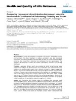
Additionally, 3-mercaptopyruvate sulfurtransferase (3-MST) has recently
been identified as an H2S-generating enzyme in the mitochondria (Figure 1).
Production of H2S from CBS primarily arises from conversion of L-cysteine (Cys)
to L-serine with concomitant release of H2S. Similarly, CSE can also convert
L-cysteine to H2S directly. Alternatively, CSE reacts with L-cystine to generate
thiocysteine, which, upon further reaction with a thiol, generates H2S. CBS and
CSE can also work in concert; for example, CBS-mediated condensation of
homocysteine (Hcy) and L-serine forms L-cystathionine, which is a substrate for
subsequent CSE-mediated H2S production. In addition to CSE and CBS, 3-MST
converts 3-mercaptopyruvate, which is generated from L-cysteine by cysteine
aminotransferase (CAT), to H2S. Non-enzymatic H2S production pathways
include the conversion of thiosulfate to H2S under reducing conditions, typically
by GSH, with concomitant formation of sulfate and oxidation of the thiol reducing
agent to the corresponding disulfide. Once generated, H2S can react with a variety
of organic and inorganic biological targets, including heme irons, thiols, and other
16
In Biochalcogen Chemistry: The Biological Chemistry of Sulfur, Selenium, and Tellurium; Bayse, C., et al.;
ACS Symposium Series; American Chemical Society: Washington, DC, 2013.
Publication Date (Web): December 5, 2013 | doi: 10.1021/bk-2013-1152.ch002
reactive oxygen/nitrogen species. Although the basic H2S-producing pathways
are known, the exact intercellular interplay between these enzymes, as well as
crosstalk with other signaling molecules, remains an emerging arena.
Figure 1. Biosynthetic pathways for H2S formation in mammalian cells.
CBS: cystathionine β-synthase; CSE: cystathionine γ-lyase; 3-MST:
3-mercaptopyruvate sulfurtransferase; CAT: cysteine aminotransferase.
In addition to H2S generation, storage of biological H2S is an important, yet
still poorly understood, aspect of H2S homeostasis. Drawing parallels to other
important bioinorganic analytes that exist in both free and bound pools, such
as NO and Zn(II) (7), different H2S storage mechanisms likely play important
roles in releasing H2S under different physiological conditions. For example,
iron-sulfur clusters can be a source of acid-labile H2S, although release of
H2S is only efficient under acidic conditions. Although these conditions are
significantly removed from normal physiological pH, such acidities are accessible
in different cellular locales, such as lysosomes. A likely more important pool
of stored biological H2S is sulfane-bound sulfur, resulting from reaction of
H2S with a thiol under oxidizing conditions (8, 9). Such sulfane-bound sulfur
sources include hydropersulfides (RS-SH) and polysulfides (RS-S(n>1)-R), which
release H2S under reducing conditions or after transulfurization reactions with
other reduced thiols. A variety of stored sulfur pools are likely required for
ensuring H2S homeostasis, but the release of biologically-stored sulfur from these
sources complicates H2S detection. For example, many classical methods of H2S
measurement require sample acidification or disruption of the pre-established
redox balance prior to analysis (vide infra).
17
In Biochalcogen Chemistry: The Biological Chemistry of Sulfur, Selenium, and Tellurium; Bayse, C., et al.;
ACS Symposium Series; American Chemical Society: Washington, DC, 2013.
Publication Date (Web): December 5, 2013 | doi: 10.1021/bk-2013-1152.ch002
Emerging biological and physiological roles of H2S clearly establish that H2S
plays important and multifaceted roles in the cardiovascular, nervous, endocrine,
and immune systems (6). One major challenge in establishing and understanding
such roles is that the physiological response to H2S is often dependent upon
the method of H2S administration or modulation. Despite these complications,
the role of H2S has been established in numerous biological processes. For
example, H2S functions as a vasorelaxant in the cardiovascular system with EC50
levels for induced vasorelaxation that correlate well with measured plasma H2S
levels (10), although the detection limit of the H2S measurement method used in
these studies has subsequently been revised (11). Additionally, high expression
levels of CBS in the hippocampus and cerebellum, as well as the interaction
with N-methyl-D-aspartate (NMDA) receptors, suggest important roles for H2S
in the central nervous system (CNS) in the modulation of neurotransmission
and long term potentiation (LTP) (12, 13). Furthermore, the existence of H2S
has been implicated in the endocrine system by influencing glucose metabolism
homeostasis in islets through action on KATP channels in beta cells (14). Hydrogen
sulfide plays important function in the immune system, displaying both proand anti-inflammatory effects depending on the mode and concentration of H2S
administration (15–18). Taken together, H2S clearly plays diverse and important
roles in various physiological systems. Although a complete description of the
diverse biological roles of H2S is beyond the scope of this review, the interested
reader is referred to a recent, comprehensive summary of H2S in biology (6).
As the field continues to grow, revision and refinement of many of the current
paradigms is likely as better tools for selectively delivering and measuring
biological H2S levels continue to emerge.
H2S Detection Strategies
Classical instrumental methods of H2S quantification include gas
chromatography (GC), polarography, and sulfide-selective electrodes (19–22).
For these techniques, samples are typically homogenized, and either the resultant
solution or the gaseous headspace is analyzed. Polarographic and GC methods
detect H2S gas released from the solution and therefore require accurate pH
measurements to correct for H2S speciation under physiological conditions. Based
on what is now known about acid-labile endogenous sulfur pools, such sample
acidification may result in releasing bound sulfur, thereby resulting in total, rather
than free, sulfide measurements. Most sulfide-selective electrodes also require
sample homogenization followed by treatment of a sulfide antioxidant buffer
containing sodium salicylate, ascorbic acid, and sodium hydroxide. Because the
electrodes only measure S2–, the least prevalent species in the H2S acid-base
equilibria, sulfide-sensitive electrodes are quite sensitive to small changes in
sample pH. Additionally, commonly-used additives contain redox-active species,
thereby increasing the possibility of perturbing the redox homeostasis of the
sample and allowing for either release of or additional storage by sulfane-bound
sulfur. Driven by the drawbacks of the instrumental methods of H2S detection and
quantification, biocompatible chemical analyses for H2S are beginning to emerge.
18
In Biochalcogen Chemistry: The Biological Chemistry of Sulfur, Selenium, and Tellurium; Bayse, C., et al.;
ACS Symposium Series; American Chemical Society: Washington, DC, 2013.