Advances in lipid research volume 24 skin lipids
Bạn đang xem bản rút gọn của tài liệu. Xem và tải ngay bản đầy đủ của tài liệu tại đây (29.73 MB, 355 trang )
FOUNDING EDITORS
DAVID KRITCHEVSKY
RODOLFO PAOLETTI
EDITORIAL ADVISORY BOARD
G. AlLHAUD
R. M. BELL
P. LAGGNER
I. PASCHER
G. H. ROTHBLAT
FOUNDING EDITORS
DAVID KRITCHEVSKY
RODOLFO PAOLETTI
EDITORIAL ADVISORY BOARD
G. AlLHAUD
R. M. BELL
P. LAGGNER
I. PASCHER
G. H. ROTHBLAT
Advances
in
Lipid
Research
Volume 24
Skin Lipids
Edited by
PETER M . ELIAS
Department of Dermatology
University of California, San Francisco
San Francisco, California
SERIES EDITORS
RICHARD J. HAVEL
Cardiovascular Research Institute
University of California, San Francisco
San Francisco, California
DONALD M. SMALL
Department of Biophysics
Boston University
Boston, Massachusetts
ACADEMIC PRESS, INC.
^
S
Harcourt Brace Jovanovich, Publishers
San Diego New York Boston London Sydney Tokyo Toronto
This book is printed on acid-free paper. @
Copyright © 1991 by ACADEMIC PRESS, INC.
All Rights Reserved.
No part of this publication may be reproduced or transmitted in any form or
by any means, electronic or mechanical, including photocopy, recording, or
any information storage and retrieval system, without permission in writing
from the publisher.
Academic Press, Inc.
San Diego, California 92101
United Kingdom Edition published by
Academic Press Limited
24-28 Oval Road, London NW1 7DX
Library of Congress Catalog Number: 63-22330
International Standard Book Number: 0-12-024924-3
PRINTED IN THE UNITED STATES OF AMERICA
91 92 93 94
9 8 7 6 5 4 3 2 1
PREFACE
This volume of Advances in Lipid Research is intended to provide a unique
resource, with a comprehensive and current overview of the field of skin lipids.
Because of the acknowledged importance of epidermal lipids for cutaneous barrier
function, the first three chapters address structural, biochemical, and metabolic
aspects of the role of lipids in permeability barrier formation and maintenance. In
addition, Chapters Six and Seven describe the lipid biophysics of the intercellular
lipid domains in the stratum corneum, and the regulation of percutaneous absorption
by these domains, respectively. Chapter Four describes the lipid content and
metabolism of cultured keratinocytes, grown under standard immersed conditions
and in various in vitro systems that attempt to produce an epidermal equivalent,
including a competent barrier.
The remaining chapters cover a broad panoply of subjects not directly related to
the epidermal barrier. Chapter Eight describes the role of epidermal lipids in the
pathogenesis of several disorders of cornification and the insights gained from these
"experiments of nature" about the role of specific lipids in normal cohesion and
desquamation. Chapter Five discusses the important new field of lipid signaling
mechanisms in the epidermis, focusing on the emerging potential role of sphingolipid
metabolites in regulating epidermal proliferation and differentiation. A discussion
of eicosanoids is specifically not included, however, since this subject has been
exhaustively covered in several recent reviews. The ninth chapter provides a
comprehensive description of the biochemistry of mammalian sebaceous gland
lipids, including speculations about the function of some of these species in normal
and diseased skin (e.g., acne). Chapter Ten compares the structure, function, and
lipid biochemistry of integumental lipids from plants, invertebrates, and cold
blooded vertebrates to warm-blooded (homeothermic) organisms. Finally, Chapter
Eleven reviews the explosion of new information about vitamin D and the skin,
including new clinical, pathophysiological, and therapeutic applications. Again, we
have specifically chosen not to include chapters on the other major fat-soluble
vitamin known to influence epidermal function (i.e., vitamin A), only because so
many recent, exhaustive reviews of this subject are already available.
In summary, this volume not only provides abundant current and comprehensive
information, but also each of the chapters represents a unique effort in the literature.
PETER M.
IX
ELIAS
ADVANCES IN LIPID RESEARCH, VOL. 24
Structural and Lipid Biochemical Correlates of the Epidermal
Permeability Barrier
PETER M. ELIAS AND GOPINATHAN K. MENON
Dermatology Service
Veterans Administration Medical Center
San Francisco, California 94121
and
Department of Dermatology
University of California School of Medicine
San Francisco, California 94143
I.
II.
III.
IV.
Introduction and Historical Perspective
Stratum Corneum Two-Compartment Model
Cellular Basis for Lipid-Protein Sequestration in the Stratum Corneum of Terrestrial Mammals
Insights from Aves and Marine Mammals (Cetaceans)
A. Aves
B. Marine Mammals (Cetaceans)
V. Intercellular Membrane Structures in Mammalian Stratum Corneum
VI. Structural Alterations in Pathological Stratum Corneum
VII. Structural-Lipid Biochemical Correlates
VIII. Summary
References
I.
Introduction and Historical Perspective
A pivotal point in terrestrial adaptation is prevention of desiccation and main
tenance of internal water homeostasis. Mammals have evolved an impressive
array of adaptive responses for water conservation, among the most remarkable
of which is the development of a cutaneous barrier to water loss. The outermost
integumentary tissue, the epidermis, maintains a reserve of germinal cell layers
whose proliferation, stratification, and differentiation result in production of the
outermost layer, the anucleate stratum corneum. The loose "basketweave" pattern
of the stratum corneum in typical histological preparations delayed appreciation
of its responsibility for cutaneous barrier function. Physical chemists were the
first to show that the stratum corneum is extremely resilient, possessing the per
meability properties of a homogeneous film (for review see Scheuplein and
Blank, 1971). Later studies revealed the stratum corneum to be composed of in
terlocking, vertical columns of foreshortened polyhedral cells, with thickened
membrane envelopes (MacKenzie, 1969; Christophers, 1971; Menton and Eisen,
1971). More recent work has revealed the unique structural organization of this
tissue into a two-compartment system (see below).
1
Copyright © 1991 by Academic Press, Inc.
All rights of reproduction in any form reserved.
PETER M. ELIAS AND GOPINATHAN K. MENON
2
The importance of stratum corneum lipids for barrier integrity has been appre
ciated for several decades. For example, the observation that topical applications
of organic solvents produce profound alterations in barrier function is over 40
years old (for review see Scheuplein and Blank, 1971). More recently, the impor
tance of bulk stratum corneum lipids for the barrier has been demonstrated by (1)
the inverse relationship between the permeability of the stratum corneum to water
and water-soluble molecules at different skin sites (e.g., abdomen versus palms
and soles) and the lipid content of the first site (Elias et al., 1981a; Lampe et al.,
1983a), (2) the observation that organic solvent-induced perturbations in barrier
function occur in direct proportion to the quantities of lipid removed (Grubauer et
al., 1989a), (3) the observation that stratum corneum lipid content is deficient or
defective in pathological states that are accompanied by compromised barrier
function, such as essential fatty acid deficiency (Elias and Brown, 1978), and, fi
nally, (4) that replenishment of stratum corneum lipids, which follows removal by
solvents or detergent, parallels the recovery of barrier function (Menon et al.,
1985a; Grubauer et al, 1989b).
II.
Stratum Corneum Two-Compartment Model
More recently, the concept of the stratum corneum as merely a homogeneous
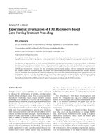
film has been replaced by a model of the stratum corneum consisting of proteinenriched corneocytes embedded in a lipid-enriched, intercellular matrix (Fig. 1)
(Elias, 1983), i.e., a continuous lipid phase surrounding a discontinuous protein
1WO-
Aqueous
Hydrophobie
Aqueous
I Hydrophobie
1 (Membranes)
Hydrcphilic
Hydrophobie
(Membranes)
1
I
Stratum
Corneum
Nucleated
Layers
FIG. 1. Depiction of two-compartment model, illustrating the localization of lipid-enriched do
mains to the stratum comeum interstices. [Modified from Fitzpatrick et al. (1987). "Dermatology in
General Medicine," 3rd Ed. McGraw-Hill, New York, with permission.]
Epidermal Permeability Barrier
3
phase. The evidence for such protein-lipid sequestration is based upon
freeze-fracture replication (Elias and Friend, 1975; Elias et al, 1977a,b), histochemical (Elias et al., 1977b), biochemical (Grayson and Elias, 1982), cell fractionation (Grayson and Elias, 1982), cell separatory (Smith et al., 1982), and
physical-chemical (Elias et al, 1983; White et al, 1988; Rehfeld et al, 1990)
studies (Table I). First, the freeze-fracture method revealed that stacks of inter
cellular bilayers existed in the intercellular spaces (Fig. 2) (Elias and Friend,
1975; Elias et al, 1977a,b); transmission electron microscopy previously had re
vealed only empty spaces (Brody, 1964,1966). Likewise, histochemical stains re
vealed the membrane domains of the stratum corneum to be enriched in neutral
lipids (Fig. 3), but only when these stains were applied to frozen sections (Elias et
al, 1977b). Later, it became possible to isolate the peripheral membrane domains
as a separate subcellular fraction. Analysis of these preparations showed that
(Grayson and Elias, 1982) (1) the bulk of stratum corneum lipids were in these
preparations, (2) the lipid composition of these preparations was virtually identi
cal to that of whole stratum corneum, and (3) the freeze-fracture pattern of mem
brane multilayers, previously described in whole stratum corneum, was dupli
cated in the membrane preparations. More recently, X-ray diffraction and
electron-spin resonance studies have localized all of the bilayer structures, as well
as lipid-based thermal phenomena, to these membrane domains (Elias et al,
1983; White et al, 1988; Rehfeld et al, 1990).
The two-compartment arrangement, which is sometimes simplified to a "bricks
and mortar" analogy (Elias, 1983), also explains both the ability of cells in the outer
stratum corneum to take up water (i.e., lipid-enriched bilayers act as semipermeable
membranes) (Middleton, 1968; Imokawa et al, 1986) as well as the differences in
rates of percutaneous absorption of topically applied lipophilic versus hydrophilic
agents—the latter penetrating at much slower rates, suggesting a separate pathway
(Michaels et al., 1975) (see Potts et al., this volume). But the two-compartment
lipid-versus-protein model also requires further modification (see below), based
upon recent evidence that extracellular proteins, such as desmosomal components
Table I
EVIDENCE FOR LIPID-PROTEIN COMPARTMENTALIZATION IN MAMMALIAN STRATUM CORNEUM (SC)
1. Pulverization destroys osmotically active structures responsible for the water-holding capacity
oftheSC
2. Freeze-fracture reveals lipid lamellae segregated within the SC interstices
3. Histochemistry displays neutral lipids solely in SC interstices
4. Organic solvents disperse the SC into individual cells
5. Isolated SC membrane sandwiches account for most SC lipids
6. X-Ray diffraction shows ordered lipids in isolated SC membranes
7. Catabolic enzymes colocalize with lipids in SC interstices
8. Electron-spin resonance localizes lipid signals to SC membranes
4
PETER M. ELIAS AND GOPINATHAN K. MENON
\
Epidermal Permeability Barrier
5
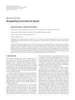
FIG. 3. Frozen sections of neonatal mouse stratum corneum stained with the lipophilic fluorophore, 8-anilino-l-sulfonic acid (A) and oil red O for neutral lipids (B). Note localization of lipid
staining to membrane domains (arrows). [Reprinted from Elias et al. (1979). / . Invest. Dermatol. 73,
339-348, with permission.]
(Allen and Potten, 1975; Haftek et al, 1986), glycoproteins (Brysk et al, 1988),
and abundant enzymatic activity (Menon et al., 1986c) exist within the intercellular
spaces. Indeed, even the intercellular lipids are heterogeneous; in various animal
species and topographic sites, there are different proportions of nonpolar, sebaceous
gland-derived lipids in addition to lipids derived from the epidermis (Nicolaides,
1974) (see Stewart and Downing, this volume).
III.
Cellular Basis for Lipid-Protein Sequestration in the Stratum
Corneum of Terrestrial Mammals
Since its earliest descriptions (for review see Ödland and Holbrook, 1987), hy
potheses have abounded about the function of the epidermal lamellar body. These
ellipsoidal organelles, measuring about lA x lA μιη, appear initially in the first
FIG. 2.
Freeze-fracture replicas of murine epidermis. (A) Note multilamellar stacks (arrows) in
the intercellular spaces (ICS). (B) The initially secreted lamellar body contents cross-fracture (ar
rows), consistent with enrichment in polar lipids (SC, stratum corneum; SG, stratum granulosum). (C)
Finally, note abundant lamellar bodies (arrows) in apical cytoplasm of outer granular cell (SG).
[Reprinted from Elias et al. (1981b). Lab. Invest. 44, 531-540 (A) and Elias et al. (1977a). Anat. Rec.
189, 577-593 (B and C), with permission.]
6
PETER M. ELIAS AND GOPINATHAN K. MENON
suprabasal cell layer, the stratum spinosum, and they continue to accumulate in
the stratum granulosum until they account for up to 25% of the volume of the cytosol (Elias and Friend, 1975). Although the subcellular site of lamellar body gen
eration is not known, cytochemical studies have tentatively traced its origin to el
ements of the smooth endoplasmic reticulum (Wolff-Schreiner, 1977) or the
Golgi apparatus (Wolffand Holubar, 1967; Weinstock and Wilgram, 1970; Chap
man and Walsh, 1989).
Many ultrastructural studies have depicted the internal structure of these mem
brane-enclosed organelles. They are described to contain parallel stacks of lipidcontaining disks enclosed by a limiting trilaminar membrane (for review see Öd
land and Holbrook, 1987). In near-perfect cross-sections, each lamella shows a
major electron-dense band (shared by adjacent lamellae) separated by electronlucent material, divided centrally by a minor electron-dense band (Fig. 4). Yet,
despite published information about the fusion of secreted lamellar body disks, as
well as the behavior of model liposomes made from stratum corneum lipids
(Landmann, 1984, 1988; Abraham et al, 1987), our recent observations suggest
that lamellar body contents may actually be composed of bilayers already con
nected to each other, folded in an accordion-like fashion (Menon et al, 1991b).
When the epidermis is permeabilized with acetone, the limiting membranes of
lamellar bodies are occasionally disrupted and the folded bilayers appear at dif
ferent stages of unfurling (Fig. 4).
In the outer granular layer, lamellar bodies are arrayed at the lateral and apical
surfaces, where they are poised to undergo rapid exocytosis. Although tracer perfusion studies first demonstrated a potential role for these organelles in the initial
formation of the water barrier (Schreiner and Wolff, 1969; Squier, 1973; Elias and
Friend, 1975; Elias et al, 1977a), these electron-dense tracers may not reflect the
actual diffusion pathway for much smaller molecules, such as water.
Cytochemists provided the next clues about the function of this organelle in the
barrier, describing lamellar bodies to be enriched in sugars (Ashrafi and Meyer,
1977) and lipids (Olah and Rohlich, 1966; Breathnach and Wylie, 1966;
Schreiner and Wolff, 1969; Elias et al, 1977b; Landmann, 1980; Squier, 1982),
thereby generating the initial hypothesis that their contents might be important for
epidermal waterproofing (Schreiner and Wolff, 1969). Moreover, tracer perfusion
studies demonstrated the role of the lamellar body secretory process in the initial
formation of the barrier (Schreiner and Wolff, 1969; Squier, 1973; Elias and
Friend, 1975; Elias et al., 1977b). Indeed, the outward egress of water-soluble
tracers through the epidermis is blocked at sites of lamellar body secretion, and
no other membrane specializations, such as tight junctions, are present at these lo
cations to account for barrier formation (Elias and Friend, 1975).
Biochemical studies on partially purified lamellar body preparations have
demonstrated that these organelles are enriched in glycosphingolipids, free
sterols, and phospholipids (Fig. 5) (Grayson et al, 1985; Freinkel and Traczyk,
1985; Wertz et al 1985). These lipids are the putative source of almost all of the
Epidermal Permeability Barrier
1
FIG. 4.
Electron micrograph of epidermal lamellar body (insert), and secreted contents at the
stratum granulosum (SG) and stratum corneum (SC) interface (B). Note that the lamellar body "disks"
(D) actually appear to be a continuous sheet within the organelle, which begins to "unfurl" immedi
ately after secretion (arrows) (A; c.f. Fig. 6). [Fig. 4C reprinted from Grayson et al. (1983). Science
221,962-964. Copyright © 1983 by the American Association for the Advancement of Science, with
permission.]
PETER M. ELIAS AND GOPINATHAN K. MENON
8
Table II
HYDROLYTIC ENZYME CONTENT OF EPIDERMAL LAMELLAR BODIES"
Present
Lipid catabolic
Acid lipase
Phospholipase A
Sphingomyelinase
Glycosidases
Others
Acid phosphatase
Cathepsins
Carboxypeptidase
Absent or not increased
Lipid catabolic
Steroid sulfatase*
Others
Arylsulfatases A and Bc
ß-Glucuronidase
Protease
Plasminogen activator^
"Modified from Grayson et al. (1985).
*Microsomal.
'Typical lysosomal enzyme.
Extracellular.
stratum corneum intercellular lipids (for review see Elias, 1983; Ödland and Holbrook, 1987; Williams and Elias, 1987).
However, others described hydrolytic enzymes in lamellar bodies, suggesting
alternatively that these organelles might instead modulate desquamation (see
below). In addition to lipids, the lamellar body is enriched in certain hydrolytic
enzymes, including acid phosphatase, proteases, lipases, nonspecific esterases,
and presumably glycosidases (Wolff and Holubar, 1967; Squier and Waterhouse,
1970; Weinstock and Wilgram, 1970; Nemanic et al, 1983; Grayson et al, 1985;
Freinkel and Traczyk, 1983, 1985; Wertz et al, 1989). Hence, the lamellar body
has been considered to be a type of lysosome (Waterhouse and Squier, 1966;
Wolff and Holubar, 1967; Lazarus et al, 1975), and a recent report of proton
pumps in its limiting membrane supports this analogy (Chapman and Walsh,
1989). Yet, certain acid hydrolase activities characteristic of lysosomes, such as
arylsulfatases A and B and ß-glucuronidase, are notably absent (Grayson et al.,
1985) (Table II). Moreover, the same enzymes that are concentrated in the outer
epidermis and/or in lamellar bodies have been found in high specific activity in
the stratum corneum (Nemanic et al, 1983; Elias et al, 1984; Menon et al,
1986b) and further localized to intercellular domains both biochemically and cytochemically (Elias et al, 1984; Menon et al, 1986b). Hence, it is likely that the
enzymes present in lamellar bodies fulfill specific functions.
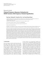
In Fig. 5, we have presented a model that is consistent with current information
about the colocalization of selected lipids and catabolic enzymes within the
lamellar body, suggesting a dual role for these enzymes in both barrier formation
and desquamation. The colocalization of "probarrier" lipids and various lipases
(phospholipase A, sphingomyelinase, and acid lipase) and glucosidases to the
Epidermal Permeability Barrier
"Pro-Barrier" Lipids:
Glycolipids, Cholesterol
Phospholipids
9
Conversion of "Pro-Barrier"
Lipids to Nonpolar Products
( Upases, Glucosidases)
Glycolipids - ►
Phospholipids
Ceramides
►
FFA
►
Cholesterol
Catabolic Enzymes:
Acid Phosphatase,
Proteases, Lipases,
Glycosidases
• Barrier Function
».
>
1) Release of Desmosomes into
Intercellular Space
( Lipases, Proteases )
►
Desquamation
2) Degradation of Desmosomes /
Other Nonlipid Intercellular Species
(Acid Phosphatase, Proteases )
FIG. 5.
Speculative program that links available information about lamellar body lipid and hydrolase content to modulations leading to barrier formation and desquamation. The release of desmo
somes may be facilitated by the detergent action of fatty acids and/or phospholipases or proteases at
sites of desmosomal insertion. [Reprinted from Elias (1987). In "Skin Pharmacokinetics" (B. Shroot
and H. Schaeffer, eds.), pp. 1-9. Karger, Basel, with permission.]
same tissue compartment may mediate the changes in lipid composition and
structure that occur during transit through the stratum corneum (Figs. 5 and 6)
(Nemanic et al, 1983; Menon et α/., 1986b; Elias et al., 1988; Wertz and Down
ing, 1989). However, several features of this model are still speculative; e.g., the
function of acid phosphatase in the cellular interstices has not been investigated.
Moreover, the function of lamellar body-derived proteases is unknown. One pos
sibility would be the activation of other lamellar body-derived enzymes under
conditions present in the intercellular spaces. An acidic environment could result
either from the deposition of acidic lamellar body contents and/or from the inser
tion of proton pumps in the plasma membrane in association with lamellar body
exocytosis, if these pumps continue to be active in that site (Chapman and Walsh,
1989). These conditions may initiate a sequence that begins initially with
SG-SC INTERFACE
LOWER SC
MID-TO-OUTER SC
T7£T^T
IE
ΓΤΤ^Τ
Τ
5WSW533WR?
FIG. 6.
Diagram that illustrates alterations of membrane structures that follow lamellar body se
cretion as a result of degradation of polar species by codeposition of hydrolases (SC, stratum
corneum; SG, stratum granulosum) (c. f. Fig. 5). [Modified from Elias et al. (1988). /. Invest. Dermatol. 91, 3-10, with permission.]
10
PETER M. ELIAS AND GOPINATHAN K. MENON
protease activation, followed by conversion of proenzymes to active forms of the
lamellar body-derived hydrolases, leading ultimately to the compositional and
structural changes known to occur in the intercellular spaces of the lower stratum
corneum (Elias et al., 1988).
A second possible function of lamellar body-derived proteases may relate to
desmosomal degradation. Although desmosomes cannot form a physiological
barrier to water loss (Arnn and Staehelin, 1981), they may contribute to the in
tegrity of this tissue by mediating its cohesiveness. During stratum corneum tran
sit, desmosomes decrease in number (Allen and Potten, 1975), a change that cor
relates spatially with the gradual loss of cohesiveness of this layer (King et ah,
1979). Intercellular proteases appear to mediate desmosomal degradation in plan
tar stratum corneum, because cell shedding requires an acidic environment and is
blocked by serine protease inhibitors (Lundström and Egelrud, 1988, 1990a,b).
Moreover, desmosomal proteins, such as desmoglein, are progressively deleted
during transit through plantar stratum corneum (Egelrud and Lundström, 1989).
However, because plantar stratum corneum is lipid depleted (Elias et al.y 1981a;
Lampe et aL, 1983a), it is possible that in other topographic sites, access of pro
teases to desmosomes is limited by more extensive, lipid-enriched domains. Yet,
very recent studies suggest that proteases may participate in stratum corneum
shedding even in nonvolar sites (Egelrud and Lundström, 1990). Thus, although
it seems likely that lamellar body-derived proteases contribute to stratum
corneum desquamation, and that desmosomes play an important role in stratum
corneum integrity, the regulation of these processes remains unknown.
Another inadequately studied potential consequence of lamellar body secretion
relates to changes in (1) the intercellular volume and (2) the surface area:volume
ratio of the stratum corneum and individual comeocytes, respectively. Massive
exocytosis of lamellar bodies results in the deposition of abundant lipid, enzyme
protein, and undoubtedly other substances into the stratum corneum interstices.
As a result, preliminary studies suggest that this compartment is greatly expanded
(5-15% of total volume) in comparison to the volume of the interstices in other
epithelia (1-5%) (Elias and Leventhal, 1979). Moreover, the intercellular com
partment serves as a selective "sink" for exogenous lipophilic agents, which may
result in further expansion of this compartment (Nemanic and Elias, 1980). Fi
nally, the splicing of the limiting membrane of the lamellar body into the plasma
membrane of the granular cell should result in a massive expansion of the surface
area : volume ratio of individual comeocytes (Elias and Leventhal, 1979). This
change may explain the remarkable capacity of comeocytes to absorb up to four
times of stratum corneum dry weight in water (for review see Scheuplein and
Blank, 1971).
To date, the factors that regulate lamellar body secretion are not known. Recent
studies have shown that acute perturbations of the barrier result in lamellar body
secretion, accompanied by a striking paucity of these organelles in the cytosol
Epidermal Permeability Barrier
11
(Feingold et al., 1990; Menon et al., 1991b). However, by 1-2 hours, abundant
nascent lamellar bodies appear in the cytosol. Clearly, secretion must occur under
both basal and stimulated conditions, and it is possible that separate factors may
regulate each process, as is the case for the surfactant-enriched lamellar bodies of
the alveolar type II cell (Chander and Fisher, 1990). In fact, preliminary correla
tive ultrastructural and confocal microscopic studies suggest that lamellar bodies
are organized into a continuous network by components of the cellular cytoskeleton (Cullander et al., 1990). Unfortunately, little work exists in this area, and the
control of lamellar body secretion remains a ripe area for investigation.
Although the lamellar body accounts for the delivery and sequestration of the
majority of the stratum corneum lipids within the intercellular spaces, other deliv
ery mechanisms may also be operative. For example, cholesterol sulfate, which
accounts for up to 5% of total stratum corneum lipids in humans (Williams and
Elias, 1981; Lampe et al., 1983a), is not concentrated in lamellar bodies (Grayson
et al., 1985). Yet, in the stratum corneum this molecule becomes localized to in
tercellular domains (Elias et al., 1984). Hence, unless cholesterol sulfate is lost
during lamellar body isolation procedures, other mechanisms may account for its
delivery to the interstices; e.g., the amphipathic properties of this compound
could allow it to move freely across the cell membrane without the requirement
of a specific delivery mechanism (Ponec and Williams, 1986). Likewise, steroid
sulfatase, the enzyme responsible for desulfation of cholesterol sulfate (for re
view see Williams and Elias, 1987), is not enriched in lamellar bodies (Table II),
yet it is also localized to membrane domains in the stratum corneum (Elias et al.,
1984). How this microsomal enzyme reaches the cell periphery is a mystery.
Again, it is possible that the enzyme is present in lamellar bodies but is lost or de
stroyed during isolation. But it also is possible that steroid sulfatase may be trans
ferred from microsomes to the limiting membrane of the lamellar body. This
would result in "splicing" of enzyme into the corneocyte periphery during exocytosis, as noted above for the proton pump (Chapman and Walsh, 1989).
IV. Insights from Aves and Marine Mammals (Cetaceans)
A.
AVES
Like terrestrial mammals, aves are warm-blooded organisms that face a dry ex
ternal environment. However, in feathered body regions, avian plumage provides
some degree of impediment to water loss, and as a result the epidermis is less wa
terproof than is epidermis of mammals living under comparable conditions (Web
ster et al., 1985). Yet, nestlings are initially featherless and often must survive at
extremely low ambient humidities (Welty and Baptista, 1988). Hence, avian epi
dermis must be able to adapt to changing ambient temperatures and humidity.
12
PETER M. ELIAS AND GOPINATHAN K. MENON
Like its mammalian counterpart, avian stratum corneum consists of corneocytes embedded in a lipid matrix (Lucas, 1980). However, in comparison to mam
mals, avian corneocytes are wafer-thin, effete structures (Menon et al., 1986b).
Under basal conditions, the mechanism for lipid delivery to the interstices of
avian stratum corneum also differs from that in mammals. Rather than delivery
by lamellar body secretion, an analogous (Menon et al., 1991a) but larger mem
brane bound organelle, the multigranular body (MGB), under the usual environ
mental conditions, does not secrete its lamellar contents, but instead deteriorates
and coalesces with its neighbors to form large neutral lipid droplets within the
corneocyte cytosol (Menon et al., 1981; Purton, 1988). As the corneocytes be
come progressively more attenuated, these droplets normally are extruded into
the interstices through membrane porosities (Menon etal., 1981; Purton, 1988).
Yet, when zebra finches are xerically stressed, i.e., under conditions of waterdeprivation, MGBs appear to be secreted in a manner analogous to mammalian
lamellar bodies (Menon et al, 1988). Moreover, secretion of the disklike contents
of MGBs gives rise to intercellular bilayer structures in the intercellular spaces,
with features similar to those of terrestrial mammals (Menon et al., 1988; cf.
Landmann, 1980) (Fig. 7). Simultaneously, epidermal barrier function improves,
as shown by significantly lower rates of transepidermal water loss (Menon et al.,
1988). With water replenishment or environmental rehumidification, this pattern
reverts to basal conditions (Menon et al., 1989b). Interestingly, though secretion
of MGB contents gives rise to intercellular membrane structures (and a less per
meable barrier), the porous extrusion of MGB-derived lipid droplets, which oc
curs under basal conditions, does not. The inability of MGB-derived lipid
droplets (versus MGB-derived membrane structures) to provide a significant bar
rier may be due to hydrolysis of certain key species (e.g., glycosphingolipids
and/or phospholipids) (Menon et al, 1986b) by cytosolic hydrolases. This would
result in a loss of lamellar structures, with the resultant emergence of a neutral
lipid-enriched mixture that is incapable of forming membrane structures. Thus,
under basal, hydrated conditions, these polar species would be absent and a more
permeable stratum corneum would result (Menon et al., 1988).
B.
MARINE MAMMALS (CETACEANS)
Ocean water is slightly hypertonic; hence, glabrous marine mammals, such as
whales and dolphins, are exposed to less rigorous barrier requirements than are
terrestrial species (Gaskin, 1982; Geraci et al., 1986). Yet, marine mammals must
retain as much metabolic water as possible, because exogenous sources are not
available. Moreover, because the skin lies outside the subcutaneous fat layer,
which effectively insulates the remainder of the organism, cetacean epidermis is
exposed to water temperatures as low as 4°C. Furthermore, marine mammals
have definite but less well-defined requirements for surface lubrication, solar pro-
Epidermal Permeability Barrier
13
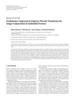
AVIAN
desiccated
MAMMALS
normal
extrusion
lipid
droplets
dissolution
♦
secretion
fusion
" ^ " ^
.catabolism
+
reformation
secretion
lamellar
bodies
multigranular
bodies
FIG. 7. Fate of lipid-secretory organelles in avian and mammalian epidermis. Under basal con
ditions, multigranular bodies are degraded within the cytosol, resulting in the disappearance both of
polar lipids and of hydrolytic enzymes. Hence, the resultant extracted lipid displays no lamellar sub
structure. Under conditions of extreme xeric stress, the fate of multigranular bodies resembles that of
terrestrial mammals, i.e., secretion of lamellae into intercellular domains, as occurs in mammals.
[Reprinted from Elias et al. (1987). Am. J. Anat. 180, 161-177, with permission.]
tection, buoyancy, and antimicrobial activity that may be subserved by epidermal
lipids (Gaskin, 1982; Geraci et al., 1986).
Although there have been very few studies on cetacean skin, the epidermis has
been shown to contain abundant lamellar bodies (Sokolov et al., 1982; Menon et
al., 1986a). Moreover, lamellar body contents are secreted from all suprabasal,
nucleated cell layers of cetacean epidermis (Menon et al., 1986a). But, in contrast
to terrestrial mammals, lamellar body-derived lipids are not reorganized into the
basic unit system of membrane bilayers. Moreover, lamellar body-derived lipids
do not appear to be completely catabolized to a more hydrophobic mixture; e.g.,
glycosphingolipids persist at all levels of the parakeratotic stratum corneum
(Menon et al., 1986a). It is possible that the incomplete transformation and
metabolism of lamellar body contents reflect the less stringent barrier require
ments of cetaceans, and that the partially hydrolyzed, intercellular lipid mixture
serves other functions, e.g., lubrication and streamlining. This interpretation is
consistent with existing information about the structure and composition of oral
mucosal epithelia, which also are exposed to a hydrated environment. These
PETER M. ELIAS AND GOPINATHAN K. MENON
14
Table III
POSSIBLE FUNCTION OF NEUTRAL LIPID DROPLETS IN HOMEOTHERMIC SKIN 0
Animal groups
Cell type
Possible function
Terrestrial mammals
Sebocyte
Natural emollients
Antimicrobial activity
Pheromones
Cetaceans and Sirenia
(Manatees)
Lipokeratinocytes
Thermogenesis
Flotation
Cryoprotectancy
Source of metabolic
water
Avians
Sebocytes
(uropygial gland)
Feather flexibility
Plumage hygiene
Antimicrobial activity
Pheromones
Vitamin D
Avians
Sebokeratinocytes
Permeability barrier
Antimicrobial activity
Emolliency
Ultraviolet filter
"Modified from Elias et al. (1987), Am. J. Anat. 180, 161-177, with permission.
tissues display abundant lamellar bodies (Squier, 1973; Hayward and Hacker
mann, 1973; Lavker, 1976; Elias et al., 1977a), but, as in marine mammals,
lamellar body-derived lipids form a less effective barrier and appear to be incom
pletely hydrolyzed; i.e., glycosphingolipids are much more abundant than in the
epidermis of the same species (Squier et al, 1986). Moreover, in mucosal epithelia, this compositional profile correlates with a less effective permeability barrier
(Squier, 1975).
In contrast to terrestrial mammals, cetaceans also possess large, intracellular
lipid droplets at all levels of the epidermis (Sokolov et al, 1982; Geraci et al.,
1986; Menon et al., 1986a). And, in contrast to avians, these droplets are not ex
pelled into the intercellular spaces in the stratum corneum. Their tinctorial prop
erties, coupled with the known lipid biochemical composition of cetacean epider
mis (Menon et al., 1986a), suggest that they are enriched in neutral lipids, such as
triglycerides. Because of their composition and persistence in the epidermis, it is
likely that these droplets subserve some other functions of cetacean epidermal
lipids described above, e.g., the oxidation of lipid stores to generate calories
(Table III) (Gaskin, 1982; Geraci etal., 1986; Elias etal., 1987).
Epidermal Permeability Barrier
15
V. Intercellular Membrane Structures in Mammalian Stratum Corneum
As noted above, elucidation of membrane structure in mammalian stratum
corneum was impeded by the extensive artifacts produced during processing for
light and/or electron microscopy. Typically, in published studies prior to the mid1970s, the intercellular spaces appear dilated and devoid of membrane structures,
or collapsed and lacking in intervening lamellar bilayers (Brody, 1964, 1966).
Following the application of freeze-fracture replication to the epidermis (Breathnach et aL, 1973), the mid-to-outer stratum corneum interstices in both epidermis
and keratinizing mucosal epithelia later were found to be replete with a multilamellar system of broad membrane bilayers (Fig. 2) (Elias and Friend, 1975;
Elias et aL, 1977a,b). With osmium vapor fixation, these membrane layers com
prised a multilayered system of alternating electron-dense and electron-lucent
lamellae of approximately equal thickness (Elias and Friend, 1975; Elias et aL,
1977a,b). At the stratum granulosum-stratum corneum interface and in the inter
stices of the lowermost layers of the stratum corneum, a transition can be seen
from cross-fractured, lamellar body-derived sheets to successively broader lamel
lae (Fig. 2) (Elias et aL, 1977a; Landmann, 1986).
The sequence of events that leads to the formation of broad intercellular bilay
ers has been studied ultrastructurally (Elias et aL, 1977b, 1988; Landmann, 1984,
1986), biochemically (Elias et aL, 1988), and in model vesicles prepared from
synthetic and naturally occurring stratum corneum lipids (Landmann, 1984;
Wertz et aL, 1986; Abraham et aL, 1987). As described above, immediately fol
lowing extrusion, the lamellar body-derived membranes begin to unfold parallel
to the plasma membrane. Within the first two layers of the stratum corneum, endto-end fusion appears to occur, giving rise to broad, uninterrupted lamellae,
which undergo further changes in substructure (Fig. 6). From the liposome work,
it has been suggested that this fusion process occurs spontaneously, perhaps due
to the high radius of curvature at the edges of the disks (Landmann, 1984) and/or
calcium-mediated aggregation (Abraham et aL, 1987). However, this change in
freeze-fracture characteristics also correlates with a sequence of changes in com
position (Fig. 5) (Table IV); i.e., from the polar lipid-enriched mixture of glycosphingolipids, phospholipids, and free sterols present in lamellar bodies and at
the SG-SC interface to a more nonpolar mixture, enriched in ceramides, free
sterols, and free fatty acids, present in the bulk of the stratum corneum (Gray and
Yardley, 1975; Elias etaL, 1979; Lampe etaL, 1983a,b; Bowser et aL, 1985; Cox
and Squier, 1986). Because the lamellar body also is enriched in proteases, glycosidases, and various types of lipases (Grayson et aL, 1985; Freinkel and
Traczyk, 1985; Menon etaL, 1986c; Elias etaL, 1988), deposition and activation
of these enzymes presumably account for the change both in composition and
structure of the membrane bilayers (Elias et aL, 1988). An explanation for the
structural changes, more consistent with the compositional changes and enzyme
PETER M. ELIAS AND GOPINATHAN K. MENON
16
Table IV
POSSIBLE RELATIONSHIPS OF BIOCHEMICAL MODULATIONS AND OBSERVED CHANGES IN
STRATUM CORNEUM MEMBRANE STRUCTURE0
Step
2
Membrane event
Responsible enzyme(s)
Unfurling of lamellar
body arrays
None
Fusion of lamellar
body arrays
Phospholipases
3
Transformation of
elongated disks
to broad lamellae
4
Breakup of membrane
bilayers ->
desquamation
Biochemical alteration
None known
Diacylphospholipids
—> lysolecithin
Sphingomyelinase
Sphingomyelin
—> ceramides
Glycosidases
Glycolipids
—> ceramides
Phospholipases +
sphingomyelinase +
glycosidases
Degradation of
residual polar
lipids (e.g.,
lysolecithin
->FFA)*
Steroid sulfatase
Cholesterol sulfate
—> cholesterol
Acid lipase
Triglycerides —> FFA
? Ceramidase
Ceramides
-> sphingosine base
+ FFA
-After Elias et al. (1988).
ft
FFA, free fatty acids.
localization data (Table IV), is that the "unfurled" lamellar body-derived sheets
initially fuse end to end (Landmann, 1986; Elias et al, 1988; Menon et al.,
1991b), perhaps through the degradation of phospholipids to free fatty acids
under acidic conditions by phospholipase A, which is present in abundance in
lamellar bodies and in the lower stratum corneum (Berger et al., 1988; Elias et al,
1988). The subsequent transformation of elongated disks into a broad, multilamellar membrane system (see below) may be associated with the further, com
plete hydrolysis of residual phospholipids and glycosphingolipids, leaving only
free fatty acids and ceramides (Lampe et al., 1983b).
Though these membrane bilayers are not seen in routine electron micrographs
of the epidermis, elongated membrane bilayers are readily observed in the stra
tum corneum intercellular spaces in mucosal epithelia (Squier, 1973; Hay ward
Epidermal Permeability Barrier
17
and Hackermann, 1973; Lavker, 1976), in the epidermis of marine mammals
(Menon et al., 1986a), and in murine stratum corneum stained with ruthenium
tetroxide (Menon et al., 1991b). It is likely that the incomplete hydrolysis of
lamellar body-derived lipids, i.e., persistence of relatively polar species such as
glycosphingolipids (see above), accounts for the routine visualization of these
structures in "moist" epithelia.
Recently, much more detailed information about intercellular membrane struc
tures has resulted from the application of ruthenium tetroxide to the study of stra
tum corneum membrane structures (Madison et al., 1987). Despite its extreme
toxicity to structural proteins, which appear etched away, with improvements in
standard fixation procedures this highly reactive and electron-dense substance
has revealed finer details of the structural heterogeneity in both electron-dense
and electron-lucent lamellae (Hou et al., 1991). The electron-lucent lamellae
consist of pairs of continuous bands, alternating with a single fenestrated lamella
(Fig. 8A). Each electron-dense lamella is separated by an electron-dense structure
of comparable width.
The membrane complex has been variously termed the Landmann (Swartzendruber et al., 1989) or basic (Hou et al., 1991) unit. The lamellar spacing or
FIG. 8.
(A) Overview of ruthenium tetroxide-stained intercellular lamellar bilayers in murine
stratum corneum. Note alterations in every third electron-lucent lamella, all of which appear to be fen
estrated (arrows), and regular interruptions of the intercellular domains by electron-dense, lenticular
dilatations (L); x36,800. (B) Higher magnification of the periphery of ruthenium tetroxide-stained,
murine stratum corneum. Note fenestrated, electron-lucent lamellae and attenuation of the numbers of
lamellae at lateral margins of cells (arrows). [Reprinted from Hou et al. (1991). /. Invest. Dermatol.
96, 215-223, with permission.]
18
PETER M. ELIAS AND GOPINATHAN K. MENON
repeat distance of ruthenium tetroxide-fixed lamellae has been analyzed by opti
cal diffraction with computerized reconstruction (Hou et al, 1991). The resultant
center-to-center spacing of 12.9 ± 0.2 nm correlates extremely well with indepen
dent measurements of unfixed samples by X-ray diffraction (13.1 ± 0.2 nm)
(White et al, 1988; Hou et al, 1991), indicating that the ruthenium staining
method provides realistic images of intercellular membrane structures (see Hou
et al, this volume). Because the repeat distance is more than twice the thickness
of typical lipid bilayers, White et al (1988) proposed that each lamellar repeating
unit consists of two opposing bilayers (see Hou et al, this volume). Multiples of
these units (up to three) occur frequently in murine (and less commonly in
human) stratum corneum (Hou et al, 1991). Simplifications of the basic unit,
with deletion of one or more lamellae, occur at the lateral surfaces of corneocytes,
i.e., at three cell junctures (Fig. 8B). Dilatations of the electron-dense lamellae,
corresponding to sites of desmosomal hydrolysis, are visualized with ruthenium
staining at all levels of the stratum corneum (Hou et al, 1991). These data, cou
pled with the known biochemical diversity of these domains, reveal the intercel
lular domains to be quite heterogeneous (Table V). In fact, the "bricks and mor
tar" model no longer does justice to this complex region.
The ruthenium staining technique has also provided further information about
the membrane leaflet immediately exterior to the cornified envelope. This trilaminar structure survives exhaustive solvent extraction (Fig. 9) (Elias et al, 1977b;
Swartzendruber et al, 1987), but is destroyed by saponification (Swartzendruber
et al, 1987), which yields a family of very long-chain, ω-hydroxyacid-containing
ceramides that are believed to be covalently attached to the cornified envelope
(Swartzendruber et al, 1987). Although this leaflet is enriched in ω-hydroxyacidcontaining ceramides, it also contains small amounts of free fatty acids and free
Table V
HETEROGENEITY OF STRATUM CORNEUM
INTERCELLULAR DOMAINS
Membrane bilayers
Nonpolar domains
Polar domains
Covalently bound envelope
Protein-enriched material
Desmosomal breakdown products
Extracellular glycoprotein(s)
Catabolic enzymes
Others
Sebaceous lipids
Eccrine gland salts
Xenobiotes
Water
Epidermal Permeability Barrier
19
FIG. 9.
Electron micrographs of murine stratum corneum after extraction with the organic sol
vent pyridine. (A and B) Note the loss of intercellular membrane bilayers but persistence of trilaminar
structures adjacent to cornified envelope (arrows). This structure presumably correlates with the ceramide-enriched structure, covalently bound to the cornified envelope (Swartzendruber et a/., 1987);
D, desmosomes. [Reprinted from Elias et aL (1977b). / . Invest. Dermatol. 69,535-546, with permis
sion.]
hydroxyacids (Wertz and Downing, 1987). This leaflet also differs in composi
tion from the intercellular bilayers, because it apparently lacks cholesterol (Kitajima et aL, 1985). The persistence of this envelope, after prior solvent extraction
has rendered the stratum corneum porous, suggests that it may mediate functions
other than permeability; e.g., it has been postulated to function as a scaffold for
the deposition and organization of lamellar body-derived, intercellular bilayers
(Wertz et aL, 1989). Based upon selected ultrastructural and biochemical data,
three-dimensional models of these membranes have been proposed, which imply
PETER M. ELIAS AND GOPINATHAN K. MENON
20
that ceramides are the principal constituents of the intercellular bilayers
(Swartzendruber et al, 1987, 1989; Wertz et al, 1989). In light of (1) the demon
strated importance of cholesterol and free fatty acids for barrier homeostasis (see
Feingold, this volume) and (2) the presence of approximately equimolar quanti
ties of ceramides, cholesterol, and free fatty acids in these domains, future models
will need to be modified to include cholesterol and free fatty acids. Correlation of
images obtained with ruthenium tetroxide, biochemical methods, X-ray diffrac
tion methods, and other physical-chemical methods (e.g., ESR and NMR) (see
Hou et al, this volume) ultimately should provide an integrated model of the ar
chitecture of the stratum corneum intercellular membrane system. Likewise, this
correlative approach should yield important new insights about the alterations in
membrane structure responsible for altered permeability states and pathological
desquamation (see Williams and Potts et al, this volume).
VI.
Structural Alterations in Pathological Stratum Corneum
If the intercellular membrane bilayers regulate epidermal barrier function, then
perturbations in barrier function should display altered membrane structures. In
deed, both solvent and detergent treatment of stratum corneum lead to depletion
of stainable neutral lipids (Menon et al, 1985a; Grubauer et al., 1989a) and loss
of intercellular membrane bilayers (Feingold et al., 1990; Menon et al, 1991b).
Such acute perturbations of the barrier are followed by an immediate secretion of
lamellar bodies (Feingold et al., 1990; Menon et al., 1991b), which leads to
restoration of intercellular lipids in 6 to 48 hours (Grubauer et al, 1989a,b). Al
though the chronology of events that follows barrier perturbation is currently
under investigation, available information already indicates that the intercellular
domains can be depleted and repleted rapidly in response to acute perturbations
of the barrier (Menon et al., 1991b; see also Feingold, this volume), and that per
turbations of the normal epidermal calcium gradient are involved (Menon et al.,
1985b; Menon and Elias, 1991).
Chronic models of barrier dysfunction, such as essential fatty acid deficiency
(EFAD), also are characterized both by depletion of intercellular lipid (Elias and
Brown, 1978) and defective intercellular membrane bilayers (Elias and Brown,
1978; Hou et al., 1991). Moreover, the membrane structures in EFAD stratum
corneum display a number of alterations in substructure, as observed with ruthe
nium tetroxide staining (Hou et al., 1991). Finally, the lamellar body secretory
system appears to be defective in EFAD epidermis; these organelles display alter
ations in internal structure and defective secretion into the intercellular spaces
(Menon et al., 1989b).
Similar results are observed with repeated applications of lovastatin, a compet
itive inhibitor of hydroxymethylglutaryl CoA (HMG-CoA) reductase, to intact
murine skin. After several daily applications, a defect in barrier function occurs