Progress in molecular biology and translational science, volume 142
Bạn đang xem bản rút gọn của tài liệu. Xem và tải ngay bản đầy đủ của tài liệu tại đây (14.14 MB, 326 trang )
VOLUME ONE HUNDRED AND FOURTY TWO
PROGRESS IN
MOLECULAR BIOLOGY
AND TRANSLATIONAL
SCIENCE
Host-Microbe Interactions
VOLUME ONE HUNDRED AND FOURTY TWO
PROGRESS IN
MOLECULAR BIOLOGY
AND TRANSLATIONAL
SCIENCE
Host-Microbe Interactions
Edited by
Michael San Francisco
Department of Biological Sciences and Honors College
Texas Tech University, Lubbock, TX, United States
Brian San Francisco
Carl R. Woese Institute for Genomic Biology University
of Illinois, Urbana-Champaign, IL, United States
AMSTERDAM • BOSTON • HEIDELBERG • LONDON
NEW YORK • OXFORD • PARIS • SAN DIEGO
SAN FRANCISCO • SINGAPORE • SYDNEY • TOKYO
Academic Press is an imprint of Elsevier
Academic Press is an imprint of Elsevier
125 London Wall, London EC2Y 5AS, United Kingdom
525 B Street, Suite 1800, San Diego, CA 92101-4495, United States
50 Hampshire Street, 5th Floor, Cambridge, MA 02139, United States
The Boulevard, Langford Lane, Kidlington, Oxford OX5 1GB, United Kingdom
First edition 2016
Copyright © 2016 Elsevier Inc. All Rights Reserved.
No part of this publication may be reproduced or transmitted in any form or by any
means, electronic or mechanical, including photocopying, recording, or any
information storage and retrieval system, without permission in writing from
the publisher. Details on how to seek permission, further information about the
Publisher’s permissions policies and our arrangements with organizations such as
the Copyright Clearance Center and the Copyright Licensing Agency, can be
found at our website: www.elsevier.com/permissions.
This book and the individual contributions contained in it are protected under
copyright by the Publisher (other than as may be noted herein).
Notices
Knowledge and best practice in this field are constantly changing. As new research
and experience broaden our understanding, changes in research methods, professional practices, or medical treatment may become necessary.
Practitioners and researchers must always rely on their own experience and
knowledge in evaluating and using any information, methods, compounds, or
experiments described herein. In using such information or methods they should
be mindful of their own safety and the safety of others, including parties for whom
they have a professional responsibility.
To the fullest extent of the law, neither the Publisher nor the authors, contributors,
or editors, assume any liability for any injury and/or damage to persons or property
as a matter of products liability, negligence or otherwise, or from any use or operation
of any methods, products, instructions, or ideas contained in the material herein.
ISBN: 978-0-12-809385-6
ISSN: 1877-1173
For information on all Academic Press publications
visit our website at />
Publisher: Zoe Kruze
Acquisition Editor: Alex White
Editorial Project Manager: Helene Kabes
Production Project Manager: Magesh Kumar Mahalingam
Designer: Vicky Pearson Esser
Typeset by Thomson Digital
CONTRIBUTORS
D. Bishop
Wound Infections Department, Naval Medical Research Center, Silver Spring, MD,
United States
A.Q. Byrne
Department of Environmental Science Policy and Management, University of California,
Berkeley, CA, United States
D. Carter
PAI Life Sciences, Seattle, WA, United States; Infectious Disease Research Institute, Seattle,
WA, United States; Department of Global Health, University of Washington, Seattle, WA,
United States
R.N. Coler
Infectious Disease Research Institute, Seattle, WA, United States; Department of Global
Health, University of Washington, Seattle, WA, United States
J.A. Colmer-Hamood
Department of Immunology and Molecular Microbiology, Texas Tech University Health
Sciences Center, Lubbock, TX, United States; Department of Medical Education, Texas Tech
University Health Sciences Center, Lubbock, TX, United States
N. Dzvova
Department of Immunology and Molecular Microbiology, Texas Tech University Health
Sciences Center, Lubbock, TX, United States
D. Fleming
Department of Surgery, Texas Tech University Health Sciences Center, Lubbock, TX,
United States; Department of Immunology and Molecular Microbiology, Texas Tech
University Health Sciences Center, Lubbock, TX, United States
N. German
Department of Pharmaceutical Sciences, Texas Tech University Health Sciences Center,
Amarillo, TX, United States
S.A. Gray
PAI Life Sciences, Seattle, WA, United States
A.N. Hamood
Department of Immunology and Molecular Microbiology, Texas Tech University Health
Sciences Center, Lubbock, TX, United States; Department of Surgery, Texas Tech
University Health Sciences Center, Lubbock, TX, United States
ix
x
Contributors
X. Hao
Institute of Urban Environment, Chinese Academy of Sciences, Xiamen, China
N. Hugouvieux-Cotte-Pattat
Microbiology Adaptation and Pathogenesis, CNRS, University of Lyon, University Claude
Bernard Lyon 1, INSA Lyon, Villeurbanne, France
T.E. Kehl-Fie
Department of Microbiology, University of Illinois at Urbana-Champaign, Urbana, IL,
United States
J.L. Kelliher
Department of Microbiology, University of Illinois at Urbana-Champaign, Urbana, IL,
United States
C. Kruczek
Honors College, Texas Tech University, Lubbock, TX, United States
F. Lu¨thje
Department of Plant and Environmental Sciences, University of Copenhagen,
Frederiksberg, Denmark
F. Meyer
Department of Biochemistry & Molecular Biology, Entomology & Plant Pathology,
Mississippi State University, Starkville, MS, USA
G. Muskhelisvili
Department of Biology, University of Lyon, INSA-Lyon, Villeurbanne, Lyon, France
W. Nasser
Department of Biology, University of Lyon, INSA-Lyon, Villeurbanne, Lyon, France
R. Ravirala
Roche Molecular System, Pleasanton, CA, United States
C. Rensing
Department of Plant and Environmental Sciences, University of Copenhagen,
Frederiksberg, Denmark
S. Reverchon
Department of Biology, University of Lyon, INSA-Lyon, Villeurbanne, Lyon, France
G. Rios-Sotelo
Department of Biology, University of Nevada, Reno, NV, United States
R. Rønn
Department of Biology, University of Copenhagen, Copenhagen, Denmark
Contributors
xi
E.B. Rosenblum
Department of Environmental Science Policy and Management, University of California,
Berkeley, CA, United States
K.P. Rumbaugh
Department of Surgery, Texas Tech University Health Sciences Center, Lubbock, TX,
United States; Department of Immunology and Molecular Microbiology, Texas Tech
University Health Sciences Center, Lubbock, TX, United States
M. San Francisco
Department of Biological Sciences, Texas Tech University, Lubbock, TX, United States
A.A. Siddiqui
Department of Internal Medicine, Texas Tech University School of Medicine, Lubbock,
TX, United States; Center of Tropical Medicine and Infectious Diseases, Texas Tech
University School of Medicine, Lubbock, TX, United States
J. Thekkiniath
Department of Medicine, University of Massachusetts Medical School, Worcester, MA,
United States
J. Voyles
Department of Biology, University of Nevada, Reno, NV, United States
C. Watters
Wound Infections Department, Naval Medical Research Center, Silver Spring, MD,
United States
PREFACE
As advances in molecular biology, biochemstry, and genomics have furthered
our understanding of biological systems, we are faced with new questions.
These questions have become even more pressing in the study of cell–cell
interactions, particularly those of pathogens with their hosts. Strategies
utlized by microorganisms to acquire nutrition, evade host defenses, and
gain a foothold in the host are varied and inventive. Host cells have, in turn,
evloved mechanisms to supress pathogen processes, limit nutrient access and
“seek and destroy” microbial invaders.
Nutritional immunity is at the center of the host–pathogen interaction,
particularly with regard to metal acquistion. Two chapters address the
acquisition of transition metals by pathogens. Work from the Kehl-Fie
group discusses strategies for acquisition and sequestration of manganese
by pathogen and host, respectively. Reduction of manganese avaliabilty can
impair microbial spread and make them more susceptible to host defenses.
German et al., describe how some other transition metals influence
bacterial gene expression related to pathogenicity and virulence. They also
highilght an interesting host strategy for pathogen elimination; termed
“Brass Dagger” for its reliance on copper and zinc, the phagolysosomes
of host macrophages accumulate metals to toxic levels to facilitate pathogen
killing.
Bacterial pathogens of plants can cause great losses in agriculture; three
chapters discuss various aspects of the genus Dickeya (formerly Erwinia), an
important plant pathogen globally. Reverchon et al., focus their review on
the complex regulatory networks that modulate early events of host adherence and virulence in the pathogen–plant interaction. The role that chromosomal superhelical density plays in regulating these interactions is of
special note. Hogouvieux-Cotte-Pattat describes the dual role of plant cell
wall-degrading enzymes as both nutritional providers and virulence factors.
Pectate lyases in particular, which degrade the cementing pectin in plant cell
walls, play important roles in modulating different phases of the infection
process. Thekkiniath et al., discuss the role of multidrug efflux pumps in
conferring Dickeya resistance to a powerful and varied arsenal of host-synthesized antimicrobial chemicals.
xiii
xiv
Preface
Pseudomonasaeruginosa is an opportunistic pathogen of plants and animals.
This bacterium is a common of cause infection in the wounds of burn victims
and in the lungs of cystic fibrosis patients. The highly nimble Pseudomonas
expression platform permits facile adaptation to different environments, such
as serum or mucus. Colmer-Hamood et al., describe work to mimic, in vitro,
various host environments to study virulence gene expression in the bacterium. One mechanism employed by many successful pathogens, including
Pseudomonas, is biofilm formation. Biofilms are important in adhesion, drug
and toxin resistance, horizontal gene transfer, and long-term survival.
Watters et al., reflect on the novel contribution of biofilms to immune
evasion and supression of host immune responses.
Splicing of RNA to maximize coding potential in eukaryotic organisms
is well known. RNA splicing in viral pathogens is likely the evloutionary
ancestor of these systems. Meyer discusses mRNA biogenesis and stability in
the context of RNA splicing in viruses and how these systems vary with
different viruses.
Emerging diseases globally have risen many fold over the last decade. One
of the most notable of these is the fungal chytrid pathogen of amphibians,
Batrachochytrium dendrobatidis. Our understanding of this pathogen and its
relationship to the host can be enhanced through effective use of genomic
tools. Byrne et al., discuss the value of genomic tools with evolutionary,
physiological, biochemical, epidemiological, immunological, and epidemiological approaches, to make important advances to guide in the conservation of these fragile hosts.
Ultimately, our understanding of microbes and the mechanisms they use
to cause disease will allow us to devlop novel and useful strategies to prevent,
diagnose, and treat infections. Gray et al., discuss strategies to develop treatments for neglected tropical diseases (NTDs). NTDs impact millions of
individuals worldwide and yet are termed “neglected” in part because they
have limited impact in western nations where funding is typically directed
elsewhere. NTD research requires philanthropic and often multinational
cooperation, hence the outgrowth of the Millinium Development Goals.
CHAPTER ONE
Competition for Manganese
at the Host–Pathogen Interface
J.L. Kelliher, T.E. Kehl-Fie*
Department of Microbiology, University of Illinois at Urbana-Champaign, Urbana, IL,
United States
* Corresponding author. E-mail address:
Contents
1. Introduction
2. Imposition of Manganese Starvation by the Host
3. Bacterial Adaptation to Manganese Limitation
4. Impact of Manganese Limitation on Invading Microbes
5. Conclusions and Broader Impacts
References
2
3
10
15
16
17
Abstract
Transition metals such as manganese are essential nutrients for both pathogen and
host. Vertebrates exploit this necessity to combat invading microbes by restricting
access to these critical nutrients, a defense known as nutritional immunity. During
infection, the host uses several mechanisms to impose manganese limitation. These
include removal of manganese from the phagolysosome, sequestration of extracellular manganese, and utilization of other metals to prevent bacterial acquisition of
manganese. In order to cause disease, pathogens employ a variety of mechanisms
that enable them to adapt to and counter nutritional immunity. These adaptations
include, but are likely not limited to, manganese-sensing regulators and high-affinity
manganese transporters. Even though successful pathogens can overcome hostimposed manganese starvation, this defense inhibits manganese-dependent processes, reducing the ability of these microbes to cause disease. While the full impact
of host-imposed manganese starvation on bacteria is unknown, critical bacterial
virulence factors such as superoxide dismutases are inhibited. This chapter will review
the factors involved in the competition for manganese at the host–pathogen interface
and discuss the impact that limiting the availability of this metal has on invading
bacteria.
Progress in Molecular BiologyandTranslational Science, Volume 142
ISSN 1877-1173
/>
© 2016 Elsevier Inc.
All rights reserved.
1
2
J.L. Kelliher and T.E. Kehl-Fie
1. INTRODUCTION
Transition metals such as iron (Fe), zinc (Zn), and manganese (Mn) are
necessary for the proliferation of all organisms. Their importance is emphasized by analysis of protein databases, which predict that 30% of proteins
utilize a metal cofactor.1 Metals act as catalytic cofactors and structural
components to perform a variety of tasks in the cell; metals including iron,
zinc, and manganese also directly influence regulation of their own cellular
homeostasis. Iron is utilized by almost every form of life and facilitates a
variety of processes, such as respiration, metabolism, and macromolecule
synthesis.2 Iron is a cofactor in multiple types of catalytic centers, including
mononuclear enzymes, such as superoxide dismutases; Fe–S cluster proteins,
such as aconitase; and in heme-containing enzymes, such as cytochrome
c oxidase. Zinc frequently functions as a structural cofactor, such as in the Fur
and zinc-finger families of transcriptional regulators, and as catalytic cofactor. Zinc has a catalytic role in enzymes such as alcohol dehydrogenases,
hydrolases, and kinases.3 Manganese is an essential cofactor for a diverse set
of processes, including in enzymes involved in nucleotide metabolism
(ribonucleotide reductase), carbon metabolism (phosphoglycerate mutase),
phosphorylation (serine/threonine kinase), and oxidative stress response
(superoxide dismutase).4–6
To combat pathogens, vertebrates take advantage of the essential nature of
transition metals by restricting their availability, a defense termed nutritional
immunity. The most well characterized example of nutritional immunity is
the iron-withholding response elaborated by the host. As a first line of
defense, the availability of free iron in the absence of infection is kept very
low throughout the body by multiple mechanisms. First, the majority of iron
in the body is present in the form of heme, which is bound by hemoglobin
within red blood cells.2 Second, extracellular Fe2+ is rapidly oxidized to
Fe3+, which is insoluble at physiological pH.7 Further restricting the availability of extracellular iron, scavenging molecules such as transferrin bind Fe3
+
, and haptoglobin and hemopexin sequester hemoglobin and heme, respectively.7,8 In response to infection, the host activates additional mechanisms
to restrict the availability of iron.2 Serum levels of the iron-oxidizing
enzyme ceruloplasmin increase, presumably to increase the conversion of
Fe2+ to Fe3+, circulating levels of transferrin increase, and immune cells
release lactoferrin, another protein capable of sequestering free iron, at sites
of infection.8–11 Despite the multiple tools used by the host to restrict iron
Competition for Manganese at the Host-Pathogen Interface
3
availability, successful invaders possess mechanisms that enable them to circumvent this defense. To accomplish this task, bacteria utilize a variety of
approaches, including expressing high-affinity iron-uptake systems such as
siderophores.2,12–15 The ongoing struggle for essential transition metals is
highlighted by the observation that to combat bacterial siderophores, the
host expresses siderocalin (lipocalin-2), which can bind enterobactin and
prevent its uptake by bacteria.7 In response, bacteria have evolved modified
siderophores that can resist sequestration by siderocalin.16 In addition to
attempting to acquire free iron, some pathogens express receptors for heme,
hemoglobin, and transferrin, allowing them to hijack iron-bound host
molecules.2,8,15 Additionally, in response to iron limitation, many bacteria
activate an iron-sparing response, which functionally diverts this metal away
from nonessential to essential enzymes.17 Disrupting either the ability of the
host to withhold iron or the ability of a pathogen to obtain this nutrient can
significantly alter the outcome of infection in favor of one or the other
party.2,8,14 This experimental observation is manifested by the increased
susceptibility of people with iron overload to infection by a diverse
collection of pathogens, including Yersinia enterocolitica, Listeria monocytogenes,
Mycobacterium tuberculosis, and Plasmodium falciparum.8,12
It has become apparent that in addition to restricting iron availability, the
host also limits the availability of manganese and zinc. While the timing and
mechanisms employed by the host to restrict the availability of these essential
metals during infection has not been fully elucidated, it is clear that restricting them contributes to the ability of the host to combat invaders.12,13,18,19
This chapter will focus on the mechanisms used by the host to limit the
ability of pathogens to acquire manganese. It will also discuss how bacteria
adapt and respond to host-imposed manganese limitation and the impact of
this host defense on invaders. Finally, current questions in the field and
broader impacts will be highlighted.
2. IMPOSITION OF MANGANESE STARVATION BY THE
HOST
In contrast to iron, which is constantly and globally restricted, manganese availability appears only to be restricted in the presence of invading
microbes and at the site of infection. Manganese can be sequestered from
pathogens both intracellularly and extracellularly (Fig. 1).18,20 A key cellular
factor in preventing intracellular pathogens from obtaining manganese is the
4
(A)
(B)
Neutrophil cytoplasm
Mn
Bacterial cytoplasm
Mn
P
NRAM
Unknown
Mn-dependent
proteins
inhibited
Mn
Phagolysosome
ATP7A
Cu
Cu
−
SOD
MntR
H2O
Zn
mnt
Engulfed
bacterium
Zn
Mn
Zn CP Mn
Mn
tAB
Zn
C
H
CP
Mnt
CP
CP
O2•
Zn CP Mn
Zn CP Zn
Zn CP Mn
Cu
Cu
Zn
Cu
Zn
J.L. Kelliher and T.E. Kehl-Fie
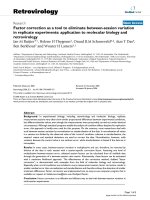
Figure 1 The host and pathogens compete for manganese during infection. (A) Diagram of the mechanisms utilized by the host to limit the
ability of invaders to obtain manganese. In response to microbial invaders, host cells, primarily neutrophils, release calprotectin (CP), which
limits the ability of extracellular pathogens to obtain manganese (and zinc). In the phagolysosomal membrane, NRAMP transporters remove
manganese (Mn) from the phagolysosome. Additionally, zinc (Zn) and copper (Cu) are imported into this compartment, inhibiting the activity of
bacterial manganese importers. (B) Diagram of how bacteria respond to manganese limitation and the processes that are disrupted by hostimposed manganese limitation. In response to manganese starvation, the MntR regulon is derepressed, and the expression of dedicated
manganese importers such as MntH and MntABC increases. Despite the expression of high-affinity manganese importers by invading
pathogens, the host remains capable of imposing manganese starvation, which inactivates manganese-dependent superoxide dismutase
(SOD) and unknown essential enzymes.
Competition for Manganese at the Host-Pathogen Interface
5
divalent cation transporter NRAMP1 (natural resistance-associated macrophage protein-1), also known as DMT-2. The importance of this transporter
to host defense was first revealed by the observation that macrophages lacking
NRAMP1 are more susceptible to intracellular pathogens, including
Mycobacterium bovis and Salmonella enterica Typhimurium.20–25 NRAMP1 is
constitutively expressed by macrophages and lymphocytes, where it associates
with lysosomes, late endosomes, and maturing phagosomes.26–30 In addition
to M. bovis and S. enterica Typhimurium, loss of NRAMP1 in mice leads to
increased susceptibility to a variety of pathogens, including Toxoplasma gondii,
Candida albicans, Mycobacterium lepraemurium, and Leishmania donovani.24,25,31–33
The contribution of NRAMP1 to restricting manganese availability during
infection was revealed by investigations with S.enterica Typhimurium. Analysis
of Salmonella mutants lacking high-affinity manganese importers revealed that
they are less capable than wild type bacteria of surviving in primary peritoneal
macrophages derived from NRAMP1+/+ mice, but not those derived from
NRAMP1À/À mice.34 Consistent with this result, in an oral infection model,
the Salmonella manganese uptake mutants are attenuated in NRAMP1+/+
mice but not NRAMP1À/À mice.34 The importance of NRAMP1 and
intracellular manganese sequestration to host defense is emphasized by the
identification of polymorphisms in humans that are associated with increased
risk of developing tuberculosis, leishmaniasis, meningococcal disease, and
others.20,23,35,36
NRAMP1 belongs to SLC11 family of solute transporters, members of
which are present in all three domains of life.29,37 Humans express two
SLC11 family members, NRAMP1 and DMT-1.29,37 The latter transporter
facilitates absorption of iron, and potentially manganese, in the intestine.29
This family of transporters contains 11–12 transmembrane segments that
form a single channel pore.37,38 NRAMP transporters symport a divalent
cation and a proton, coupling metal transport with the energetically favorable flow of protons out of the phagolysosome.37 In vitro, NRAMP1 can
transport Mn2+, Fe2+, Zn2+, Co2+, Ni2+, and Cd2+, but not alkaline earth
metals.39,40 In vitro fluorescent probe-based assays and infection experiments, however, indicate NRAMP1 is important for the transport of
Mn2+ and Fe2+ out of the phagolysosome.20,29,41,42 Since NRAMP1 is an
integral membrane protein, biophysical studies of the transporter have been
challenging; however, a prokaryotic homolog from Staphylococcus capitis has
been structurally characterized.38 The metal-binding site is located within
two short, unstructured regions, in between two sets of membrane-spanning
alpha helices. The metal is coordinated by the side chains of N52 (both an
6
J.L. Kelliher and T.E. Kehl-Fie
oxygen and nitrogen ligand) and D49 (oxygen), the oxygen atom of the
peptide bond linking residues 223 and 224, and the thioether of M226,
resulting in a planar coordination.38 These residues are conserved among
the NRAMP family, and the human and S. capitis residues are identical.38
The binding site is selective for Mn2+, Fe2+, Co2+, Ni2+, Cd2+, and to some
extent Cu2+ and Zn2+. However, the latter two metals are coordinated by
slightly different residues than Mn2+, Fe2+, Co2+, Ni2+, and Cd2+ and may
not be effectively transported.38 Cumulatively, the biophysical and infection
studies suggest that NRAMP1 contributes to host defense by removing
manganese and iron from the phagolysosome during infection.
In addition to restricting intracellular manganese availability, the extracellular availability of this metal is also limited by the host during infection.43,44 This discovery was made possible by the application of advanced
elemental imaging techniques, such as laser ablation inductively coupled
plasma mass spectrometry (LA-ICP-MS), to the study of infection. LAICP-MS enables the assessment of the spatial distribution of metals within
a tissue.45 The prototypical example of the extracellular manganese-withholding response is the Staphylococcus aureus abscess, which is rendered
virtually devoid of manganese by the host.43,44 Notably, while the staphylococcal abscess is depleted of manganese, total tissue levels of this metal do
not decrease.44 Similar to manganese, zinc is also withheld from the staphylococcal abscess, and total tissue levels of this metal do not change.43,44
These findings highlight both, the ability of the host to locally restrict metal
availability in response to infection and the importance of assessing metal
distribution within a tissue when evaluating the impact of nutritional immunity on invaders. A critical component of the manganese-withholding
response is the manganese- and zinc-binding protein calprotectin (also
known as S100A8/S100A9, calgranulin A/B, and MRP8/14). This innate
immune effector is constitutively expressed by neutrophils, where it accounts
for approximately 50% of the total cytosolic protein.46 In addition to neutrophils, proinflammatory cytokines such as IL-17 and IL-22 can induce the
production of calprotectin in other cell types, most notably epithelial
cells.47,48 At sites of infection where neutrophils release calprotectin, extracellular concentrations can be found in excess of 1 mg/mL.49 Calprotectindeficient (S100A9À/À) mice have defects in manganese sequestration and are
more susceptible to a variety of bacterial and fungal pathogens, including
S. aureus, Klebsiella pneumoniae, Acinetobacter baumanii, Aspergillus nidulans,
Aspergillus fumigatus, and C. albicans.43,44,50–54 In vitro, the sequestration
of transition metals by calprotectin inhibits the growth of a range of
Competition for Manganese at the Host-Pathogen Interface
7
Gram-positive, Gram-negative, and fungal pathogens, including S.aureus, K.
pneumoniae,A.baumanii,C.albicans, and A.nidulans.43,50–53,55 Analysis of metal
distribution during staphylococcal infection revealed that while calprotectin-deficient mice do not remove manganese from staphylococcal liver
abscess, they are still able to deplete kidney abscesses of manganese.43,44
This finding indicates that the host possesses additional unknown mechanisms for restricting manganese availability at sites of infection. The importance of restricting extracellular manganese availability to host defense is
emphasized by the observation that staphylococcal strains lacking highaffinity manganese uptake systems have a virulence defect in the livers of
wild type mice but not calprotectin-deficient mice.44
Calprotectin is a member of the S100 family of calcium-binding EF-hand
proteins. Unlike the other members of this family, which are homodimers,
calprotectin is a heterodimer comprised of S100A8 and S100A9. Similar to
calprotectin, a subset of the S100 family, including S100A7 (psoriasin),
S100A12 (calgranulin C), and S100A15 (koebnerisin), are capable of binding
transition metals.56–59 These three proteins possess two identical transition
metal binding sites located at the dimer interface. The canonical S100 protein
transition metal binding site, possessed by S100A7 and S100A12, is composed
of three histidines and an aspartic acid. Two of the histidines, arranged in an
HXXXH motif, are contributed to the binding site by one of the monomers,
while the third histidine and aspartic acid are contributed by the other monomer.58,60 Unlike the other S100 family members, calprotectin has two nonidentical transition metal binding sites. Based on homology with other S100
proteins, the first transition metal binding site possessed by calprotectin was
originally thought to be comprised of H17 and H27 from S100A8 and H91
and H95 from S100A9.61,62 However, subsequent investigations revealed that
H103 and H105, contributed by a C-terminal extension of S100A9, also
contribute to the ability of this site to bind manganese.55,63 Crystallographic
studies revealed that the six histidines bind manganese with a nearly perfect
octahedral geometry.55 This hexahistidine coordination, which had not previously been observed in proteins, has been confirmed by solution-based
electron paramagnetic resonance.63,64 The critical importance of the C-terminal extension, which only S100A9 possesses, and the observation that
neither S100A7 nor S100A12 are capable of binding manganese suggests that
among the S100 proteins, calprotectin is unique in its ability to bind this
metal.55,63 The second transition metal binding site is identical to the canonical
transition metal binding site found in other S100 proteins, comprised of
H83 and H87 from S100A8 and H20 and D30 from S100A9.61,62
8
J.L. Kelliher and T.E. Kehl-Fie
Analysis of wild type calprotectin and variants lacking the two transition
metal binding sites revealed that the first site is capable of binding both
manganese and zinc tightly (subsequently referred to as the Mn/Zn site),
while the second site is capable of binding only zinc tightly (subsequently
referred to as the Zn site).55,65,66 A combination of isothermal titration
calorimetry (ITC) and dye competition studies revealed that the Mn/Zn
site binds manganese with an affinity (Kd) of approximately 10 nM or less and
zinc with an affinity of less than 240 pM.55,62,65 Weaker affinities for manganese have been reported;66 however, they are not consistent with the
ability of calprotectin to prevent manganese acquisition by bacteria, which
express manganese-importers with low nanomolar affinities.67–69 Dye competition studies revealed that the Zn site binds zinc with an affinity of less than
10 pM.65 Obtaining more precise binding affinities for manganese and zinc
binding has been hampered by the lower limit of resolution for both ITC and
dye-based studies.65,69 Additional studies revealed that H103 and H105,
which are located in the C-terminal extension of S100A9, are essential for
the Mn/Zn site to bind manganese, but not zinc, tightly.55,63 Similar to other
S100 proteins, the ability of calprotectin to bind transition metals is influenced by the presence of calcium. In the absence of calcium, the Kd of
calprotectin for manganese weakens to 5 μM, and the affinity of the Mn/Zn
site and Zn site for Zn increase to 219 nM and 133 pM, respectively.65,66
Modeling suggests that calcium binding to the EF-hand elongates two alpha
helices that contain the transition metal binding sites.70 Relative to the
extracellular space, the cytoplasm is calcium-limited, leading to the suggestion that calcium binding serves as a switch to ensure that calprotectin does
not bind manganese and zinc until released into the extracellular space.65,66
Activity assays utilizing the calprotectin binding site variants revealed that
the Mn/Zn site is necessary for maximal antimicrobial activity against a wide
range of Gram-positive and -negative pathogens, including S. aureus,
Staphylococcus epidermidis, A. baumanii, Escherichia coli, Enterococcus faecalis,
Pseudomonasaeruginosa, and Shigella£exeneri.55 The importance of the Mn/Zn
site suggests that manganese binding is necessary for maximal broadspectrum antimicrobial activity by calprotectin. However, not surprisingly,
given the diversity of microbes, the Mn/Zn site and presumably manganese
binding is not necessary for maximal antimicrobial activity in all cases.71
In addition to Mn2+ and Zn2+, calprotectin has been observed to bind Fe2+
in vitro via the Mn/Zn site in reducing environments.71 Notably, this is in
contrast to several prior studies in which iron binding by calprotectin was not
observed.43,55 In these studies the ionic state of iron was not controlled but
Competition for Manganese at the Host-Pathogen Interface
9
due to the aerobic nature of these experiments, iron was most likely present
as Fe3+.43,55 Based on the observation that calprotectin is capable of binding
Fe2+, it has been suggested that the antimicrobial activity of calprotectin is
due to iron sequestration.71 However, several observations argue against this
proposal. First, manganese-dependent enzymes in S. aureus are inhibited by
calprotectin, both in culture and during infection.55,62 Second, staphylococcal mutants lacking high-affinity manganese importers are more sensitive
to calprotectin in culture and have a virulence defect in wild type but not
calprotectin-deficient mice.44 Third, following growth in the presence
of calprotectin, A. baumanii has reduced intracellular levels of manganese
and zinc, but not iron. In fact, following exposure to calprotectin in A. baumanii iron levels actually increase.51 Additionally, our current understanding
of iron homeostasis suggests that due to the generally oxidizing nature of the
extracellular space, outside of the cytoplasm iron should be present not as
Fe2+ but as Fe3+, a form of iron that calprotectin cannot bind.8,43,55,71 Due to
the oxidative burst of immune cells, sites of infection are likely to be more
oxidizing than healthy tissues. This idea is supported by the observation that
pathogens such as S.enterica Typhimurium have evolved to take advantage of
metabolites that are generated by the oxidative burst as terminal electron
receptors.72 Additionally, Fe3+-binding proteins such as transferrin and lactoferrin are critical to controlling infection.2,7 While in select environments
exceptions may exist, these observations strongly suggest that manganese and
zinc sequestration are primarily responsible for the antimicrobial activity of
calprotectin, both in culture and during infection.
Even though transition metals are necessary for life, they can also be
toxic.12,73 To prevent transition metal toxicity, intracellular levels of these
metals are highly regulated through the coordinated expression of metal
importers and exporters.12,13 Transition metal efflux pumps for zinc and
copper contribute to the ability of organisms such as Streptococcus pyogenes,
M. tuberculosis, Streptococcus pneumoniae, Neisseria meningitidis, Brucella abortus,
and Helicobacterpylori to cause disease,74–80 suggesting that pathogens encounter toxic levels of these metals during infection. Further supporting this idea,
zinc colocalizes with S. pyogenes in neutrophils, and chelation of this metal
reduces the antimicrobial activity of the cells.75 Supporting the use of the
antimicrobial properties of copper by the host is the observation that this
metal is transported into phagolysosome via ATP7A.81
The toxicity of transition metals is thought to be driven by the ability of a
metal to bind inappropriately to (mismetalate) noncognate metalloenzymes
or, in some cases, the ability of the metal to generate reactive oxygen species.
10
J.L. Kelliher and T.E. Kehl-Fie
The general affinity of a metal for organic molecules is described by
the Irving–Williams series, where Mg2+/Ca2+ < Mn2+ < Fe2+ < Co2+
< Ni2+ < Cu2+ > Zn2+.82,83 Functionally, this means that an overabundance of metals such as copper and zinc can inhibit the activity of metalloproteins that use a weaker binding metal, such as manganese, as a cofactor.82
Zinc and copper are capable of inhibiting a variety of intracellular enzymes
and processes. For example, zinc can inhibit glycolytic enzymes such as
phosphofructokinase, and copper can disrupt Fe–S cluster-containing
enzymes.73,84–88 While pathogens can attempt to regulate cytoplasmic levels
of transition metals, they are unable to control their surrounding environment. As such, extracellular metalloproteins such as manganese-specific
transporters (discussed subsequently) are particularly vulnerable to elevated
extracellular concentrations of zinc and copper. In vitro, zinc binds irreversibly to PsaA, the solute-binding protein of the pneumococcal PsaABC
manganese importer, preventing it from binding manganese.67,89–91 In culture, a 30:1 ratio of zinc to manganese prevents S.pneumoniae from importing
manganese and inhibits bacterial growth. Ratios of zinc to manganese in
excess of this can be found in tissues during pneumococcal infection.67
While the ability of copper to inhibit manganese uptake has not been directly
evaluated, it also binds irreversibly to PsaA, suggesting that copper should
also inhibit manganese acquisition.89 Similar results have also been obtained
with the staphylococcal solute-binding protein, suggesting that manganese
specific ABC transporters are generally susceptible to zinc and copper
poisoning.68
3. BACTERIAL ADAPTATION TO MANGANESE
LIMITATION
In order to successfully cause disease, bacteria must adapt and respond
to the ever-changing environment within the host, including the availability
of manganese. Many bacteria sense manganese availability through a DtxRfamily transcriptional repressor usually called MntR (Fig. 1). MntR homologs are present in a variety of Gram-positive and -negative bacteria, including S. aureus, Bacillus subtilis, S. pneumoniae, M. tuberculosis, S. enterica
Typhimurium, E. coli, and Treponema pallidum.92–98 A canonical repressor,
manganese-bound MntR represses gene expression when manganese levels
are sufficient. When manganese becomes scarce, apo-MntR releases from
the DNA and allows transcription of targets to occur. In several species,
Competition for Manganese at the Host-Pathogen Interface
11
including S.aureus,S.pneumoniae,S.enterica Typhimurium, B.subtilis, S.£exneri,
and Corynebacterium diphtheriae, MntR represses the expression of highaffinity manganese importers when manganese is available.92–94,99–101 Loss
of the MntR homolog PsaR results in reduced virulence of S. pneumoniae,102
suggesting that in addition to encountering manganese limitation, pathogens
may be exposed to toxic levels of this metal. This idea is further supported by
the identification of manganese efflux systems that contribute to virulence,
such as MntE of S.pneumoniae and MntX of N.meningitidis.77,78 While it is not
clear when invaders would experience elevated levels of manganese, these
observations highlight the diversity of metal environments encountered
by pathogens within the host. Given the position of manganese in the
Irving–Williams series, it is less apparent why elevated levels of this metal
are toxic. It has been proposed that regulation of intracellular manganese levels
is necessary to maintain an appropriate ratio of manganese to iron within the
cell.82,103 The importance of maintaining an appropriate balance between
manganese and iron is emphasized by the observation that the manganese
exporter MntX of N.meningitidis plays a part in regulating the intracellular ratio
of these two metals.78 Further supporting this idea, in response to manganese
limitation Bradyrhizobium japonicum reduces the accumulation of iron.104 In
addition to MntR, several other metal-responsive/binding transcriptional
regulators have been shown to bind and respond to manganese. These
regulators include Fur, which canonically responds to iron availability, and
the peroxide sensor PerR.69,92,95,96,99,105,106 The full implications of the ability
of manganese to bind these regulators are still being understood.
In addition to increasing the expression of manganese importers, bacteria
frequently respond to manganese limitation by modifying the expression of
numerous other cellular processes.98,102,107,108 One example is the altered
expression of genes involved in glucose utilization in S. pneumoniae when
manganese availability is restricted.109 However, analysis of the PsaR regulon
indicates that this regulator does not control the expression of these genes.102
This observation suggests that there are likely to be additional regulators that
coordinate the bacterial response to host-imposed manganese limitation.
While the holistic response of bacteria to host-imposed manganese limitation and the cellular factors that control this response are still being
elucidated, the expression of high-affinity manganese acquisition systems
has emerged as a common theme (Fig. 1). The vast majority of bacteria
express manganese importers belonging to either the NRAMP or ABC
family of transporters.110 NRAMP homologs are typically referred to as
MntH, while ABC transporters are frequently named MntABC. There
12
J.L. Kelliher and T.E. Kehl-Fie
are, however, a few prominent examples of MntABC homologs with alternative names, including PsaABC in S. pneumoniae and SitABC in Salmonella
species. Many bacteria such as M. tuberculosis, Mycobacterium leprae, and some
strains of E. coli express only MntH, while others such as Yersinia pestis,
Porphyromonas gingivalis, S. pneumoniae, S. pyogenes, C. diphtheriae, Bacillus
anthracis, and E. faecalis express only MntABC.101,111–119 Notably, genetic
redundancy of manganese transport systems is common among pathogens,
with many species, including S. aureus, S. enterica Typhimurium, and
S. £exneri, encoding for homologs of both MntH and MntABC.92,100,113,120
In addition to these conserved manganese importers, other less widely
distributed and less characterized systems have also been identified in several
organisms. Interestingly, the ubiquitous human pathogen H. pylori, which
expresses putatively manganese-dependent enzymes, appears to lack homologs
of any known manganese importers.121 This observation suggests that additional unidentified manganese importers may exist. Emphasizing the general
importance of these systems to bacterial virulence and resisting host-imposed
manganese starvation, both S. enterica Typhimurium and S. aureus mutants
that lack dedicated manganese importers have virulence defects relative to
wild type bacteria in mice that are capable of restricting manganese availability,
but not mice with defects in sequestering this metal.34,44 In addition to
these two species, loss of manganese importers results in reduced virulence
of numerous pathogens, including S. pneumoniae, S. pyogenes, Streptococcus
mutans, Streptococcus suis, B. abortus,Yersinia pseudotuberculosis, Neisseria gonorrhoeae,
and certain strains of E. coli.117,122–129
The bacterial MntH family is evolutionarily related to the NRAMP1
family of transporters used by eukaryotes to remove divalent cations from the
phagolysosome. MntH is essential for virulence of some pathogens, such as
B. abortus and Y. pseudotuberculosis.126,127 To date, the only structurally characterized NRAMP transporter is that of S. capitis, discussed in detail in the
previous section. However, this family of transporters and the residues that
coordinate the transported metal are highly conserved, suggesting that the
structure and metal specificity should be similar across species.38 Similar to
the eukaryotic transporters, MntH has a preference in vitro for Mn2+ and
Fe2+ but is capable of transporting other divalent cations, such as Cd2+ and
Co2+, as well.110 While several metals can be transported by MntH homologs, in bacteria manganese is generally the physiological relevant substrate.
However, in some cases iron import may also be relevant.114 In bacteria, the
expression of MntH is frequently induced by manganese limitation or
increased cellular demand for manganese.92,93,95,98,130 These latter situations
Competition for Manganese at the Host-Pathogen Interface
13
include when iron availability is reduced and the presence of peroxide stress,
which triggers the replacement of iron with manganese to prevent Fenton
chemistry-induced damage.131–133 Additionally, the relative selectivity of
MntH for manganese tends to be greater than for other cognate metals, as
observed with S. enterica Typhimurium and others.113,134 In S. enterica
Typhimurium, MntH has a pseudoaffinity (solute concentration at halfmaximal transport, or K0.5) for Mn2+ of ∼100 nM, whereas its affinity for
Fe2+ is approximately 25 μM.113
ABC transporters, the second primary family of manganese importers
expressed by bacteria, possess a four-domain structure. These domains
include two transmembrane proteins that facilitate substrate translocation
and two nucleotide-binding proteins that power import via ATP hydrolysis.135 In addition to these domains possessed by all ABC transporters,
importers also possess a high-affinity solute-binding protein (SBP).136
ABC-type manganese transporters are widespread among pathogens and
can be found in S. aureus, S. pneumoniae, S. enterica Typhimurium, S. £exneri,
Y. pestis, P. gingivalis, E. faecalis, and more.44,69,110,115,119 In some bacteria,
the MntABC system is critical for pathogenesis, including in S. pneumoniae,
S. pyogenes, S. mutans, S. suis, S. aureus, and N. gonorrhoeae.44,117,122–125,128
Regardless of species, all manganese SBPs belong to the Cluster A-I
group of SBPs of ABC transporters, along with iron- and zinc-specific
SBPs.137 MntABC transporters are capable of remarkably high affinities
for their substrate; the Kd of PsaA for Mn2+ is 3.3 nM, and the SBP of S.
aureus has a Kd for Mn2+ of 8 nM.67,68 The metal-binding site of Cluster
A-I SBPs consists of two nitrogen atoms from two conserved histidine
residues, one carboxylate from either an aspartate or a glutamate, and a
variable fourth ligand, which is thought to dictate specificity for the metal.138
In manganese-specific SBPs, this ligand is another carboxylate group
donated by a glutamate residue,67,68 which, as with the conserved carboxylate, can donate two oxygen ligands. This allows for a total of six coordinating ligands, which is the preferred coordination for manganese. However,
the physical constraints imposed by the protein result in imperfect octahedral
coordination, which facilitates release of manganese to the translocation
domain.89 In addition to their respective cognate metal, manganese SBPs
are capable of binding a range of noncognate divalent transition metals
including zinc and copper.68,89 Differing from manganese, zinc is capable
of binding to PsaA with a near perfect tetrahedral coordination. This coordination results in an extremely stable complex, which prevents the release of
zinc even when extensively dialyzed against strong chelating agents. This
14
J.L. Kelliher and T.E. Kehl-Fie
stability is thought to prevent the release of zinc from PsaA to the translocation
domain, rendering the transporter nonfunctional.67,90 Hence, the specificity of
the ABC transporter is driven by release of the metal rather than initial
binding to the SBP. These findings also provide a mechanistic explanation
for the ability of zinc and copper to poison these transporters.67,89,90
In addition to the canonical NRAMP and ABC-family transporters,
other types of manganese importers have been identified in Borrelia burgdorferi, Lactobacillus plantarum, and Vibrio species.139–141 Notably, B. burgdorferi
and L. plantarum accumulate extremely high levels of manganese (∼30 mM
in L.plantarum, for example) and are thought to have eliminated the need for
iron.142,143 B.burgdorferi lacks homologs of MntH and MntABC, and instead
utilizes a ZIP family homolog named BmtA to acquire manganese. BmtA is
thought to be responsible for the import of both manganese and zinc.139 Loss
of BmtA in B. burgdorferi results in decreased intracellular manganese levels
and abrogates virulence.139 Differing from B. burgdorferi, L. plantarum
expresses homologs MntH and MntABC, as well as a P-type ATPase transporter, MntA, which has been implicated in the import of manganese.140,144
The expression of MntA in L. plantarum is induced in manganese-depleted
media, and deletion of the gene abrogates high-affinity manganese
import.144 A third novel class of putative manganese transporter has also
been identified and is widely conserved among marine bacteria, including
the human pathogenVibriocholerae.141 The transporter, named MntX (unrelated to the manganese efflux system of N. meningitidis), appears to be
repressed in manganese-replete conditions and enhances growth of other
Vibrio species, which have been engineered to lack a manganese importer, in
manganese-poor media.141 Additional studies including metal accumulation
and transport assays are necessary to determine if MntA from L.plantarum and
MntX fromV.cholerae are true manganese importers. In addition to transporting manganese across the inner membrane, Gram-negative bacteria must also
transport this nutrient across the outer membrane. Originally, transition
metals including manganese were thought to pass nonspecifically through
outer membrane porins. However, this assumption has been challenged by
the identification of dedicated outer membrane channels that facilitate
acquisition of divalent cations, specifically by the characterization of a
zinc-specific outer membrane receptor in N. meningitidis.145,146 An analogous protein MnoP in B.japonicum facilitates the passage of manganese across
the outer membrane.147 While similar manganese-specific systems have not
yet been described in other species, MnoP belongs to the OprB superfamily,
many members of which await characterization.147
Competition for Manganese at the Host-Pathogen Interface
15
4. IMPACT OF MANGANESE LIMITATION ON INVADING
MICROBES
The finding that mice with defects in restricting manganese availability are more susceptible to infection indicates that despite expressing
high-affinity manganese acquisition systems, invading pathogens experience manganese starvation during infection.43,50–53 While the breadth of
biological processes to which manganese can contribute is significant,4 the
impact that host-imposed manganese starvation has on invading pathogens
is only just beginning to be elucidated. This task is hampered by an
incomplete understanding of manganese-dependent processes in bacteria
and the observation that metal-dependent enzymes are frequently capable
of using more than one metal as a cofactor. The latter challenge is
highlighted by the observation that in response to oxidative stress, E. coli
replaces iron in mononuclear enzymes with manganese in order to limit
Fenton chemistry-induced damage.131
While the role of manganese in many cellular processes may be uncertain,
it is clear that manganese is a critical contributor to resisting oxidative
stress, serving as a cofactor for manganese-dependent superoxide dismutases.130,148 In addition to enzymatic dismutase activity, manganese in complex with phosphate or cellular metabolites, such as lactate, has dismutase
activity.130,142,149–152 This chemical activity has led to the suggestion that
these complexes may contribute to the ability of bacteria that accumulate
high levels of manganese to resist oxidative stress.150,152 However, relative to
enzymatic dismutation, these complexes are much less efficient, leading to
uncertainty regarding the contribution of manganese complexes to resisting
oxidative stress during infection.130 Given the established link between
manganese and resisting oxidative stress, the ability of pathogens to resist
oxidative stress under conditions of host-imposed manganese starvation has
received significant attention. This area has primarily been investigated using
S. aureus and S. pneumoniae, both of which utilize manganese-dependent
superoxide dismutases. In S. aureus, calprotectin-induced manganese starvation reduces staphylococcal superoxide dismutase activity and increases bacterial sensitivity to paraquat-induced oxidative stress.44,55,62 Calprotectin
also renders S. aureus more sensitive to neutrophil-mediated killing.62
Infection experiments employing staphylococcal superoxide dismutase
mutants and calprotectin-deficient mice revealed that staphylococcal superoxide dismutase activity is inhibited by host-imposed manganese starvation
16
J.L. Kelliher and T.E. Kehl-Fie
during infection.55,62 In S.pneumoniae, elevated zinc levels lead to a reduced
accumulation of manganese and reduced superoxide dismutase activity.67,90
Additionally, elevated zinc levels increase pneumococcal sensitivity to paraquat-induced oxidative stresses and killing by polymorphonuclear leukocytes.67,90 The enhanced killing by immune cells indicates that not only does
restricting manganese availability inhibit growth, but also renders invaders
more susceptible to other immune effectors. As superoxide dismutase activity is not essential for viability of S.aureus or S.pneumoniae, it seems likely that
other cellular factors are also inhibited by host-imposed manganese starvation. The complement of manganese-dependent enzymes in any microbe
and the specific impact of host-imposed manganese starvation are likely to be
as varied as the diversity of lifestyles adopted by pathogens. Potential processes that may be inhibited by manganese limitation during infection
include enzymes involved in energy generation, nucleotide metabolism,
and cell signaling.4,153–159
5. CONCLUSIONS AND BROADER IMPACTS
Nutritional immunity is a powerful defense employed by the host to
control invading pathogens. While canonically associated with restricting
iron from invading microbes, the concept of nutritional immunity has been
expanded to include limiting the availability of other essential metals,
including manganese, during infection.12,18,20,43 Even though significant
progress has been made elucidating how the host imposes manganese
starvation, it is clear that additional unidentified host factors contribute
to this defense. This gap in knowledge is highlighted by the ability of
calprotectin-deficient mice to remove manganese from kidney but not
liver abscesses.43,44 Simultaneously, it has been revealed that the host not
only physically removes manganese from sites of infection but also harnesses the toxic properties of zinc and copper to prevent acquisition of this
metal.13,73 However, LA-ICP-MS and the importance of zinc importers to
bacterial pathogenesis indicate that pathogens also encounter zinc limitation during infection.43,44,51,160–165 These two disparate observations raise
the question of when the host utilizes the toxic properties of zinc to control
infection versus when it restricts the availability of this metal. Adding even
more complexity, more recent investigations utilizing C. albicans suggest
that the host may also restrict copper availability.166 Both the Centers for
Disease Control and World Health Organization have stated that due to the
Competition for Manganese at the Host-Pathogen Interface
17
emergence and spread of antibiotic resistance, there is a critical need for
new approaches to treating infection.167,168 Despite our nascent understanding of how noniron metal levels are manipulated in order to combat
invaders, it is clear that preventing pathogens from acquiring manganese
contributes to host defense. It is equally clear that successful pathogens,
despite expressing high-affinity metal acquisition systems, experience
metal starvation and are able to overcome this host defense.62 However,
the adaptations that enable this success are unknown. Therapeutics that
augment nutritional immunity by manipulating metal levels during infection or prevent bacteria from adapting to this host defense represent a
promising new approach for treating infection. However,our ability to
successfully harness the full potential of these approaches will require a
greater understanding of metal homeostasis during infection, how the host
utilizes transition metals to combat infection, and how invading microbes
circumvent nutritional immunity.
REFERENCES
1. Andreini C, et al. Metal ions in biological catalysis: from enzyme databases to general
principles. J Biol Inorg Chem. 2008;13(8):1205–1218.
2. Cassat JE, Skaar EP. Iron in infection and immunity. Cell Host Microbe. 2013;13
(5):509–519.
3. Cerasi M, Ammendola S, Battistoni A. Competition for zinc binding in the host–
pathogen interaction. Front Cell Infect Microbiol. 2013;3:108.
4. Kehres DG, Maguire ME. Emerging themes in manganese transport, biochemistry and
pathogenesis in bacteria. FEMS Microbiol Rev. 2003;27(2–3):263–290.
5. Donat S, et al. Transcriptome and functional analysis of the eukaryotic-type serine/
threonine kinase PknB in Staphylococcus aureus. J Bacteriol. 2009;191(13):4056–4069.
6. Srinivasan VB, et al. Functional characterization of a novel Mn2+ dependent protein
serine/threonine kinase KpnK, produced by Klebsiella pneumoniae strain MGH78578.
FEBS Lett. 2012;586(21):3778–3786.
7. Ganz T, Nemeth E. Iron homeostasis in host defence and inflammation. Nat Rev
Immunol. 2015;15(8):500–510.
8. Soares MP, Weiss G. The Iron age of host-microbe interactions. EMBORep. 2015;16
(11):1482–1500.
9. Beaumier DL, Caldwell MA, Holbein BE. Inflammation triggers hypoferremia and de
novo synthesis of serum transferrin and ceruloplasmin in mice. Infect Immun. 1984;46
(2):489–494.
10. Cernat RI, et al. Serum trace metal and ceruloplasmin variability in individuals treated
for pulmonary tuberculosis. IntJTuberc Lung Dis. 2011;15(9):1239–1245. i.
11. Letendre ED, Holbein BE. Ceruloplasmin and regulation of transferrin iron during
Neisseria meningitidis infection in mice. Infect Immun. 1984;45(1):133–138.
12. Becker KW, Skaar EP. Metal limitation and toxicity at the interface between host and
pathogen. FEMS Microbiol Rev. 2014;38(6):1235–1249.
13. Hood MI, Skaar EP. Nutritional immunity: transition metals at the pathogen-host
interface. Nat Rev Microbiol. 2012;10(8):525–537.