Skin stress response pathways
Bạn đang xem bản rút gọn của tài liệu. Xem và tải ngay bản đầy đủ của tài liệu tại đây (8.54 MB, 454 trang )
Georg T. Wondrak Editor
Skin Stress
Response
Pathways
Environmental Factors and Molecular
Opportunities
Skin Stress Response Pathways
Georg T. Wondrak
Editor
Skin Stress Response
Pathways
Environmental Factors and Molecular
Opportunities
123
Editor
Georg T. Wondrak
Department of Pharmacology
and Toxicology
College of Pharmacy & The University
of Arizona Cancer Center
University of Arizona
Tucson, AZ
USA
ISBN 978-3-319-43155-0
DOI 10.1007/978-3-319-43157-4
ISBN 978-3-319-43157-4
(eBook)
Library of Congress Control Number: 2016946320
© Springer International Publishing Switzerland 2016
This work is subject to copyright. All rights are reserved by the Publisher, whether the whole or part
of the material is concerned, specifically the rights of translation, reprinting, reuse of illustrations,
recitation, broadcasting, reproduction on microfilms or in any other physical way, and transmission
or information storage and retrieval, electronic adaptation, computer software, or by similar or dissimilar
methodology now known or hereafter developed.
The use of general descriptive names, registered names, trademarks, service marks, etc. in this
publication does not imply, even in the absence of a specific statement, that such names are exempt from
the relevant protective laws and regulations and therefore free for general use.
The publisher, the authors and the editors are safe to assume that the advice and information in this
book are believed to be true and accurate at the date of publication. Neither the publisher nor the
authors or the editors give a warranty, express or implied, with respect to the material contained herein or
for any errors or omissions that may have been made.
Printed on acid-free paper
This Springer imprint is published by Springer Nature
The registered company is Springer International Publishing AG Switzerland
Preface
If the skin were parchment and the blows you gave were ink,
Your own handwriting would tell you what I think.
(William Shakespeare, The Comedy of Errors)
It is now understood that the interplay between environmental exposure and cellular
stress response pathways plays a critical role in skin structure and function, and a
refined mechanistic understanding of this phenomenon at the molecular level
promises to open novel avenues for targeted therapeutic strategies that may benefit
skin health of patients in the near future. The comprehensive coverage of cutaneous
cell stress response pathways as presented for the first time in this book is intended
to provide a state-of-the-art perspective that is of interest to both basic researchers
focusing on fundamental skin biology in the context of environmental exposure as
well as translational biomedical health care professionals.
With the completion of this project, I would like to express my gratitude to those
who were instrumental in its creation. First and foremost, I would like to thank my
co-authors from four continents who have graciously contributed their talent and
time to assemble this first in a kind perspective on skin stress response pathways.
Second, I am indebted to my department head Walt Klimecki for allowing me
to pursue this project. Moreover, I am grateful for this outstanding opportunity
and the expert support provided by Melania Ruiz and Ilse Hensen-Kooijman at
Springer Science+Business Media B.V.
Finally, I would like to thank my family, Claudia, Gil, Philip, and Annie, for
letting me divert precious time and energy from them in pursuit of this book project.
Tucson
June 2016
Georg T. Wondrak
v
Contents
1
2
3
4
The Skin Lipidome Under Environmental
Stress—Technological Platforms, Molecular Pathways
and Translational Opportunities . . . . . . . . . . . . . . . . . . . . . . . . . . . .
Florian Gruber
1
Squalene and Skin Barrier Function: From Molecular Target to
Biomarker of Environmental Exposure . . . . . . . . . . . . . . . . . . . . . . .
Boudiaf Boussouira and Dang Man Pham
29
Sunlight-Induced DNA Damage: Molecular
Mechanisms and Photoprotection Strategies . . . . . . . . . . . . . . . . . . .
Thierry Douki
49
Urocanic Acid and Skin Photodamage:
New Light on an Old Chromophore . . . . . . . . . . . . . . . . . . . . . . . . .
Leopold Eckhart
79
5
The Skin Extracellular Matrix as a Target
of Environmental Exposure: Molecular Mechanisms,
Prevention and Repair . . . . . . . . . . . . . . . . . . . . . . . . . . . . . . . . . . . . 101
Kieran T. Mellody, Mike Bell and Michael J. Sherratt
6
Nitric Oxide Derivatives and Skin Environmental Exposure to
Light: From Molecular Pathways to Therapeutic Opportunities . . . .
Christoph V. Suschek
127
7
Melanocortin 1 Receptor (MC1R) as a Global
Regulator of Cutaneous UV Responses: Molecular Interactions
and Opportunities for Melanoma Prevention . . . . . . . . . . . . . . . . . . 155
Erin M. Wolf Horrell and John A. D’Orazio
8
The Cutaneous Melanocyte as a Target of Environmental
Stressors: Molecular Mechanisms and Opportunities . . . . . . . . . . . . 175
Laurent Marrot
vii
viii
9
Contents
The Role of Epidermal p38 Signaling in Solar
UV Radiation-Induced Inflammation: Molecular Pathways
and Preventive Opportunities . . . . . . . . . . . . . . . . . . . . . . . . . . . . . . . 197
Jin Mo Park and Yasuyo Sano
10 UV-Induced Chemokines as Emerging Targets
for Skin Cancer Photochemoprevention . . . . . . . . . . . . . . . . . . . . . . 211
Scott N. Byrne and Gary M. Halliday
11 TLR3 and Inflammatory Skin Diseases: From Environmental
Factors to Molecular Opportunities . . . . . . . . . . . . . . . . . . . . . . . . . . 235
Risa Tamagawa-Mineoka, Mayumi Ueta and Norito Katoh
12 Sirtuins and Stress Response in Skin Cancer,
Aging, and Barrier Function . . . . . . . . . . . . . . . . . . . . . . . . . . . . . . . 251
Yu-Ying He
13 Cutaneous Opioid Receptors and Stress Responses: Molecular
Interactions and Opportunities for Therapeutic Intervention . . . . . 265
Hanane Chajra
14 Regulation of Cutaneous Stress Response Pathways
by the Circadian Clock: From Molecular Pathways to
Therapeutic Opportunities . . . . . . . . . . . . . . . . . . . . . . . . . . . . . . . . . 281
Elyse van Spyk, Milton Greenberg, Faraj Mourad and Bogi Andersen
15 Endocannabinoids and Skin Barrier Function:
Molecular Pathways and Therapeutic Opportunities . . . . . . . . . . . . 301
Sergio Oddi and Mauro Maccarrone
16 The Aryl Hydrocarbon Receptor (AhR) as an Environmental
Stress Sensor and Regulator of Skin Barrier Function: Molecular
Mechanisms and Therapeutic Opportunities . . . . . . . . . . . . . . . . . . . 325
Rebecca Justiniano and Georg T. Wondrak
17 Biological Cell Protection by Natural Compounds,
a Second Line of Defense Against Solar Radiation . . . . . . . . . . . . . . 361
Ludger Kolbe
18 The Cutaneous Microbiota as a Determinant of Skin Barrier
Function: Molecular Interactions and Therapeutic Opportunities. . . .
Julia J. van Rensburg, Lana Dbeibo and Stanley M. Spinola
379
19 Sensing Environmental Factors: The Emerging Role
of Receptors in Epidermal Homeostasis and Whole-Body Health . . .
Mitsuhiro Denda
403
Contents
ix
20 The Cutaneous Circadian Clock as a Determinant
of Environmental Vulnerability: Molecular Pathways
and Chrono-pharmacological Opportunities . . . . . . . . . . . . . . . . . . . 415
Kyongshin Cho, Rajendra P. Gajula, Kenneth I. Porter
and Shobhan Gaddameedhi
21 Psychological Stress as a Determinant of Skin
Barrier Function: Immunological Pathways and Therapeutic
Opportunities . . . . . . . . . . . . . . . . . . . . . . . . . . . . . . . . . . . . . . . . . . . . 433
Mark E. Mummert
Index . . . . . . . . . . . . . . . . . . . . . . . . . . . . . . . . . . . . . . . . . . . . . . . . . . . . . . 449
Chapter 1
The Skin Lipidome Under Environmental
Stress—Technological Platforms,
Molecular Pathways and Translational
Opportunities
Florian Gruber
Abstract The skin is an organ with a high level of lipid metabolism and is divided
into regions with very differing lipid composition. Skin lipids determine the barrier
function of the skin but are also important signaling mediators. Environmental
stressors can modify lipid composition, reactivity and distribution and thereby
influence skin biology. In the last decade the technology to investigate the lipids
made explosive progress, allowing now for in-depth investigation of the role of
lipids in skin biology. In this chapter the current developments in lipidomic analysis
of environmental skin stress and the translational opportunities of this technology
are discussed.
Á
Á
Á
Á
Á
Keywords Oxidized lipids Lipidomic Mass spectrometry Redox Stress
Eicosanoids Reactive oxygen species Ultraviolet Skin Keratinocyte
Fibroblast Phospholipids
Á
1.1
Á
Á
Á
Á
Á
Introduction
Redox biologists, (bio-) chemists, skin researchers, physicians—we are all usually
not trained to deal with the big data from the inflationary—omics approaches that
pour in over us. As soon as we have accepted that such projects are interdisciplinary, require adequate statistics, that they often are exploratory and hypothesis
generating—then the benefits of such approaches become accessible. We learn to
use the huge potential of lipidomic/metabolomic—postgenomic—data formats that
F. Gruber (&)
Department of Dermatology, Division for Biology and Pathobiology
of the Skin and Christian Doppler Laboratory for the Biotechnology
of Skin Aging, Medical University of Vienna, Währinger Gürtel 18-20,
1090 Vienna, Austria
e-mail:
© Springer International Publishing Switzerland 2016
G.T. Wondrak (ed.), Skin Stress Response Pathways,
DOI 10.1007/978-3-319-43157-4_1
1
2
F. Gruber
do not adhere to the familiar linear information format provided under the central
dogma of molecular biology (DNA-RNAs-protein).
The ultimate aim of lipidomic analyses of the skin, its cells or the subcellular
structures like membrane subdomains is to identify those compounds, networks,
pathways, physical forces, and interactions that keep the skin functioning in a
hostile, changing milieu. In this chapter I will review, from the viewpoint of a
molecular biologist of the skin, what has been learned by lipidomic approaches
about how environmental stress affect the skin’s lipids in their function as signaling
mediators or structural molecules, and what translational potential such technology
may yield.
1.1.1
Lipids of the Skin—Distribution of Stress Accessible
Lipid Classes
The skin displays an active and diverse lipid metabolism, and lipids are essential for
the barrier—and signaling functions of this organ (Feingold and Elias 2014; van
Smeden et al. 2014b; Kendall et al. 2015). Any disturbance of lipid homeostasis by
environmental stressors thus may result in impairment of these functions, and may
cause disease or accelerated aging. The most common stressor the skin is exposed
to and affects the lipids is sunlight, with very distinct effects on skin biology that are
governed by wavelength and penetration depth. But also physical, chemical or
biological stress can affect the cutaneous biology by changing lipid composition or
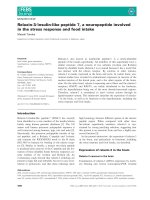
structure (Fig. 1.1).
Fig. 1.1 Lipids of the skin
1 The Skin Lipidome Under Environmental Stress
3
Environmental stressors affect lipids of the skin from its surface down to the
dermis, which houses fibroblasts, epithelial structures of the hair follicle, nerve
cells, microvasculature, dermal resident cells of the immune system and—depending on stimulation—infiltrating immune cells. The cells but also the dermal
matrix gives evidence on stress related changes to lipids, as reactive carbonyls that
originate from lipid peroxidation are detectable throughout the epidermis and can
crosslink collagen and elastic fibers in the dermis (Larroque-Cardoso et al. 2015;
Williams et al. 2014).
The lipid composition of the cells residing in the dermis (excluding the hair
follicle and sebaceous gland) is very rich (75 %) in phosphatidylcholine (PC) and
phosphatidylethanolamine (PE) phospholipids (PL), with low amounts of triglycerides, cholesterol, free fatty acids (FFA) and the other classes. The basement
membrane separates the dermis from the epidermis, made up mostly of keratinocytes (KC) in various grades of terminal differentiation, and epidermal lipid
preparations will mostly reflect KC lipids.
The lipid composition within the differentiating epidermis changes in a highly
dynamic process, as on the one hand KC in the basal layer take up lipids (most
polyunsaturated fatty acids) from the circulation and the microenvironment. On the
other hand lipids inside vesicles and as part of vesicle membranes are transferred
intracellularly for major metabolic conversion, and finally relocate to the extracellular lipid matrix or to the lipid envelope of terminally differentiated keratinocytes (corneocytes). Lipid content of keratinocytes in the basal layer of the
epidermis (and in cultured cells) is made up mostly of phospholipids (70 %),
cholesterol (13 %), triacylglycerides (11 %) (Ponec et al. 1988). Upon terminal
differentiation there are drastic changes to the lipid composition. In the lamellar
bodies (LB) of the granular layer KC glucosylceramides (GlcCERs), phospholipids
and sphingomyelin are stored. These are substrates for the enzymes that catalyze the
final stratum corneum lipids. The LB content is released by exocytose together with
these enzymes beyond the SG and forms the low permeable and flexible connection
(lipid matrix) between the rigid corneocytes which are themselves surrounded by
the “lipid envelope” of hydroxylated ceramides that are esterified to involucrin by
transglutaminase. The composition of stratum corneum lipids is dominated by FFA
(40 %), cholesterol and ceramides (both 30 %) (Thakoersing et al. 2012). The FFA
are mostly saturated in the SC, and are rather long-chained with C18, C24 and C26
being the dominant ones. The composition was determined first by GC-MS and
could recently be confirmed with novel methodology that also detects other lipid
species at the same time (see below, van Smeden et al. 2014a).
The lipids at the skin’s surface have however a second major source, the
sebaceous glands. The lipids synthesized in these glands consist mainly of
triglycerides (45 %, TAG), wax esters, (25 %, WE), squalene (12 %), and FFA
(10 %). How much sebum contributes to the total surface lipids depends on the
body site, the abundance and activity of the sebaceous glands.
4
1.2
F. Gruber
Lipidomic Methods to Analyze One or More Lipid
Classes
Recently, a broad consensus was found by the lipid researchers organized in the
lipid MAPS community, on a common systematic nomenclature of lipids, grouping
them into eight categories (fatty acyls, glycerolipids, glycerophospholipids, sphingolipids, sterol lipids, prenol lipids, saccharolipids, and polyketides) based on
their chemical properties (Fahy et al. 2005). Due to the differences in hydrophobicity, polarity, molecule size, quantitative isolation procedures and other aspects, it
is technically difficult, and until recently regarded too complex to analyze in depth
with lipidomics several lipid classes at one time. However, in the last decade the
field made remarkable progress regarding sensitivity, accuracy and in data processing. The classical thin layer chromatography analysis is still a useful standard to
identify major changes in lipid classes, but the selectivity of mass spectrometry
(MS), and the possibility to couple it with numerous separation methods makes MS
todays gold standard for identification and quantification of lipids, also in skin
research.
MS analysis requires ionization of the analytes, which is usually achieved by
electrospray ionization (ESI), matrix-assisted laser desorption ionization (MALDI)
or atmospheric pressure chemical ionization (APCI). The sample can be injected
without or with prior separation, and the unseparated direct infusion approach,
usually termed “shotgun lipidomics” usually utilizes a triple quadrupole analyzer to
scan precursor ions and neutral loss. The data generated this way usually base on
lipid class specific fragments (e.g. Phosphocholine) but do not permit interpretation
of the individual FA composition but rather the sum of fatty acyl carbons. However,
quantification of analytes can be more robust as direct infusion is not subject to
quantitative bias brought in by chromatographic separation.
Chromatographic separation prior to MS makes the analysis of some complex
lipid classes possible because it adds retention time as a parameter that boosts the
method’s specificity. After the separation and ionization, several detection modes
are applicable. For fingerprinting the intact molecular adduct ions can be scanned
(single ion monitoring—MS), the advent of triple quadrupole devices then allowed
to use one of the quadrupoles to be used as a collision cell between two mass
resolving quadrupole units. After passing the first quadrupole (Q1), the precursor
ions are collided in the second quadrupole with an inert gas and there noncovalent
bonds dissociate (neutral loss, NL) or at higher collision energy the precursors are
fragmented. The fragmented product ions are then detected with the third quadrupole (Q3) as mass analyzer at high frequency, in either of various modes: Product
ion scan, where for a given precursor in Q1 the fragments are scanned in Q3, or the
reverse, precursor ion scan, where for a specific fragment the precursors are
scanned, or in multiple reaction monitoring (MRM) mode, where a number of
preselected m/z pairs (targeted approach) are detected by the quadrupoles.
The sensitivity and mass resolution of MS/MS was multiplied by several technologies that emerged in the last decade. First, hybrid mass spectrometers that
1 The Skin Lipidome Under Environmental Stress
5
combine a quadrupole mass analyzer with a time of flight (TOF) device, which
allow a higher scan rate and mass accuracy. The latest generation detectors are
equipped with orbitrap or linear ion trap devices combined to TOF or Fourier
transform ion cyclotron resonance—MS. These, together with novel separation
techniques like ultra-performance HPLC, and hydrophilic interaction liquid chromatography (HILIC), and ion mobility detection, provide the accuracy needed for
the structural identification in some approaches described below. MALDI-TOF,
which cannot be coupled to chromatography, is not nearly as frequently used for
(skin) lipidomics, but combining it with TLC may be very useful. For in-depth
reviews on the technological platforms, application fields, biological settings and
data processing for lipidomic research and identification strategies the reader is
referred to Murphy and Gaskell (2011), Brügger (2014), Cajka and Fiehn (2014),
Köfeler et al. (2012), Lam and Shui (2013), Serhan et al. (2007)—a list without
claim to completeness.
The various variants of liquid chromatography coupled to MS, followed by
direct infusion MS are the overwhelmingly dominant methods for lipidomic analysis in life sciences over the last years (Cajka and Fiehn 2014), and below I provide
a collection of state of the art technical approaches that have been undertaken in the
skin lipidomic field or at least would be applicable to study stress related changes in
the skin lipidome.
1.2.1
Fatty Acyls (Fatty Acids, Eicosanoids,
Endocannabinoids)
An early lipidomic study of eicosanoids in mouse skin utilized GC/MS of eicosanoid derivatives to identify and quantify the major lipoxygenase products of
arachidonic and linoleic acid in mouse epidermis (Lehmann et al. 1992). Also more
recent studies show that with high-temperature gas chromatography coupled to
electron impact or chemical ionization MS large numbers of compounds including
fatty acids, eicosanoids but also other skin surface lipids can be identified in one
analytical run (Michael-Jubeli et al. 2011). Most of the recent analyses of fatty
acyls, including eicosanoids are performed by ESI-MS/MS coupled to high or
ultra-performance HPLC (rev. in Astarita et al. 2015). For the detection of potentially beneficial resolvins and other DHA derived mediators Hong et al. applied
LC–ultraviolet spectrometer–tandem MS; the UV absorption data added information about conjugated di-ene arrangement in the species (Hong et al. 2005).
A method to identify positional isomers in FA by Yang et al. used charge switching
derivatization and MS/MS (Yang et al. 2013). A current standard method to analyze
endocannabinoids inserts a lipid fractionation step on silica gel before RP-LC
coupled to ESI-MS (Astarita and Piomelli 2009). Chlorinated fatty acids were
analyzed in detail by TLC combined with ESI-MS (Schroter et al. 2015) and
nitrated and nitro-oxidized fatty acids with a targeted high resolution LC-MS/MS
6
F. Gruber
method (Milic et al. 2015). The lipidomic analyses of PUFA derived bioactive
lipids were reviewed by Massey and Nicolaou (2011).
1.2.2
Glycerolipids (Tri- and, Di-Acylglycerols)
Canine skin glycerolipids and ceramides were analyzed with HP-TLC, and partially
bands (could be identified via a TLC-MS interface (Angelbeck-Schulze et al. 2014).
A more comprehensive method to identify and quantify TAG and DAG of murine
skin was recently presented by King et al. (2015) where an ESI-MS/MS—neutral
loss (NL) scan approach was successful in identifying and quantifying most relevant acylglycerol species, and found that dietary restriction and exercise reduces
skin TG species containing 18:1 fatty acid chains.
1.2.3
Glycerophospholipids (PC, PE, PI, PS, PG, PA,
Cardiolipin)
On the lipidomic analysis of the phospholipid species several excellent reviews
have been recently published (Spickett and Pitt 2015; O’Donnell 2011), which
reflect that as broad standard HPLC-ESI-MS/MS is used for identification and
quantification of phospholipid (PL) species, native or oxidized. The high end laboratories introduced high resolution equipment including q-TOF, spin trapping,
orbitrap and FT-IR-MS in combination with high performance columns for PL
analysis (Sala et al. 2015), also very recently for nitrated PL (Melo et al. 2016) and
for glycated and glycoxidized PLs (Simoes et al. 2012). The generated big data are
used for smart scanning approaches that facilitate lipid identification (Simons et al.
2012). Chlorinated phospholipids have been detected by also by MALDI-TOF
approaches (Panasenko et al. 2007) and TLC-ESI-MS (Schroter et al. 2015).
Cardiolipin analysis with HILIC-MC/MS together with phosphatidylglycerol
(PG) and phosphatidic acid (PA) species is described by Scherer et al. (2010), and a
recent review on the topic by Tyurina et al. (2014) is recommended.
1.2.4
Sphingolipids (Sphingomyelin, Sulfatides,
Sphingosine, Ceramides, Gangliosides)
Ceramides (Cer) and sphingomyelin (SM) species have been profiled in mouse
epidermis at various prenatal developmental stages with HPLC-MS/MS using a
triple-quadrupole mass spectrometer operated in positive mode and ultrahighpressure LC coupled with hybrid quadrupole TOF mass spectrometer by
1 The Skin Lipidome Under Environmental Stress
7
Wang et al. (2013). For structural identification of non-hydroxyacyl epidermal
ceramides Shin et al. applied chip-based direct infusion nanoelectrospray-ion trap
mass spectrometry, generating characteristic fragmentation patterns of acyl and
sphingoid units that allow identification of numerous compounds (Shin et al. 2014).
A more complex approach by van Smeden et al. that utilized LC/MS/MS with an ion
trap (IT) system, a Fourier transform-ion cyclotron resonance system, and a triple
quadrupole system, which did not only detect all 11 known subclasses of ceramides
but identified the presence of other lipid subclasses using a 3D multi mass chromatogram [12]. More than 300 Cer species in 11 known and one unknown class
were detected with a NPLC-ESI-MS/MS method (Masukawa et al. 2009). With
reversed-phase LC coupled to high-resolution quadrupole time-of-flight MS operated in both positive and negative ESI mode T’Kindt et al. detected that Cer species
display skeletal isomers due to varying length of the sphingoid and FA components
(t’Kindt et al. 2012). The effect of sphingoid bases on the sphingolipidome of
differentiating KC was investigated using HPLC ESI-MS/MS (Sigruener et al.
2013). To specifically investigate phosphorylated ceramides very recently a method
including phosphate tagging and MALDI-MS was introduced for skin and other
tissues (Yamashita et al. 2016). Gangliosides were quantified in skin fibroblasts from
gangliosidosis patients with RP-LC-ESI-MS/MS in MRM mode (Fuller et al. 2014).
1.2.5
Sterol Lipids
Cholesterol hydroperoxides were quantified and identified with TLC coupled to
GC-MS(SIM) in mouse fibroblasts (Nakamura et al. 2013), and cholesterol esters
(CE) from epidermis of fetal, adult and keloid skin were investigated with silica gel
purification followed by chemical ionization and MS (Tachi and Iwamori 2008).
1.2.6
Prenol Lipids
Today’s standard method to quantify squalene in biological samples is GC-MS
(Hall et al. 2016), also applicable for squalene analysis in hair (Wu et al. 2016). The
in vitro non-volatile ozonolysis products of squalene were analyzed by ESI-high
resolution MS (Fooshee et al. 2015).
As there is yet no literature available to the author on the lipidomic study of
saccharolipids and polyketides as metabolites deriving from skin itself, these classes
will not be dealt with in this chapter.
8
1.2.7
F. Gruber
Methods for Several Lipid Classes
High resolution and mass accuracy detectors allow to cover wide ranges of lipid
classes in single experiments: UPLC-ESI-MS (+ and- mode) analysis of 11 of the
major lipid subclasses [FA, cholesterolsulfates, PA, PE, PS, PC, phosphatidyl
glycerols (PG), Cers, SM, diacylglycerols (DG), triacylglycerols (TG)] was performed. A combination of TOF and triple quadrupole MS following LC was
applied to comprehensively study non-invasively sampled sebaceous lipids
(Camera et al. 2010). Using a non-targeted approach that first creates a data base
with retention time (tR) values, equivalent of carbon numbers (ECN) for each lipid
class, and then a MS/MS fragmentation pattern, Lanzini and colleagues could
perform structure assignments within each analyzed subclass of skin lipids. For that
they utilized LIPID MAPS and METLIN data with high mass accuracy (5 ppm
tolerance), a lipid class specific relation between equivalent number of carbon
(ECN) and retention time, and MS/MS fragmentation patterns. By that they could
identify over 100 lipids of different classes (Lanzini et al. 2015). van Smeden et al.
describe a dual injection method with RPLC–negative ion mode APCI-MS (for
detection of FFA) and NPLC-Positive ion mode APCI-MS (to detect Cer and Chol)
which was also very useful for analysis of a broader range of SC lipids in one step
(van Smeden et al. 2014a).
1.2.8
Lipid Organization
Organization of lipids in the lamellae between the corneocytes (lateral organization)
can be analyzed by Fourier transform infrared spectroscopy (FTIR), electron
diffraction and wide- and small angle X-ray diffraction (WAXD/SAXD). The
localization of ceramides, cholesterol and fatty acids in the lipid matrix was also
studied with neutron diffraction analysis (Mojumdar et al. 2015). Barrier function
impairment can also be caused by changes in the lateral lipid organization (van
Smeden et al. 2014b). A recent review summarizes the methodologies to study lipid
organization in stratum corneum function (Wertz 2013).
1.2.9
Lipid Imaging
Using the specificity of MS together with an imaging technique for tissues or even
cells to determine the distribution of lipids is another technical breakthrough with
potential. The ionization methods applied for that approach include positively
charged fullerenes (C60) with secondary ion MS (SIMS) (Kurczy et al. 2010), and
MALDI which was successfully used to impressively image lipids in total skin
1 The Skin Lipidome Under Environmental Stress
9
(Hart et al. 2011) or cholesterol sulphate (Enthaler et al. 2013) and phospholipids
(Patterson et al. 2014) in sections.
1.3
How Stressors Affect the Lipidome
Extracellular stress can affect the cutaneous lipid composition (and—ordering)
directly or indirectly. The most prominent stressor—ultraviolet light—can directly
generate reactive oxygen species (ROS) within the tissue down to the dermal
compartment, which are capable of modifying lipids, as is contact to topical reactive
chemicals. At the same time, stressors may induce rapid intracellular lipid modifying
cascades, as the enzymatic production of lipid mediators from stored precursors, or
de novo synthesis of variant lipid species, or they set free intracellular reactive
oxygen—or nitrogen—species that again act non-enzymatically on lipids. In the
longer run, as reaction to stress, cells of the immune system can be directed to the
skin, and secrete novel lipid mediator classes or oxidize lipids in their microenvironment by releasing ROS, as in the respiratory burst. And on an even larger time
scale, cells of the skin can photo age or become senescent after chemotherapy, both
resulting in changed cellular redox state, damaged mitochondria and resulting in
changes to intracellular and secreted lipids. Last, when stress leads to cell death, the
dying cell may expose special modified lipid moieties from its membrane that act as
danger signals to the immune system and neighboring cells.
1.3.1
Stress Induced Enzymatic Pathways that Affect
the Lipidome
Most of the research on ultraviolet induced bioactive lipid generation via enzymatic
pathways was done on eicosanoid synthesis by the actions of phospholipases,
cyclooxygenases and lipoxygenases, and a review by Nicolaou and colleagues
systematically deals with synthesis and action of these and other enzymatically
generated mediators (Nicolaou et al. 2011). Below listed are the most prominent
lipid metabolizing enzymes that are UV-regulated.
1.3.1.1
Phospholipase A2 (PLA2) and Other Phospholipases
Phospholipase A2 (PLA2) hydrolyzes fatty acids from the sn-2 position of phospholipids. Phospholipase activity in mammalian epidermis after UV was first
described by Black and Anglin (1971) and later studies detected phospholipase
activation after UVB (DeLeo et al. 1988) and UVA (Hanson and DeLeo 1990)
exposure of keratinocytes. Pentlands group identified that cytosolic PLA2 (cPLA2,
10
F. Gruber
PLA2G4A) induced by secondary oxidative stress after UVB was responsible for
the immediate increase in E type prostaglandins (Gresham et al. 1996; Chen et al.
1996). In epidermis cPLA2 and the secreted sPLA2 are the main enzymes for
eicosanoid production in homeostasis and to maintain barrier function. These
enzymes are regulated in inflammation but also by UV, reviewed in Dan et al.
(2012), Ilic et al. (2014). Lipidomic analyses upon selective PLA2 inhibition
identified novel substrates and metabolites (Duvernay et al. 2015), which has yet to
be done for epidermal PLAs. In addition to PLA2, also PLC and PLD, both generating 1,2 diacylglycerols (DAG) out of phospholipids, were activated by UVR
(broadband) in mouse transformed melanocytes and fibroblasts and human keratinocytes and melanocytes, where these lipids may contribute to pigment production (Carsberg et al. 1995).
1.3.1.2
Cyclooxygenases and Prostaglandin Synthases
The free fatty acids (e.g. those set free by UV induced PLA2) can be metabolized to
further bioactive products by enzymes of which several are also stress regulated. In
keratinocytes, Cyclooxygenase-2 (COX-2, PTGS2) induction by UV was observed
in first in guinea pig (KC differentiation dependent), where it induced five prostaglandin species (Karmali and Safai 1984). COX-2 is also induced by H2O2 and by
PMA in mouse skin (Nakamura et al. 2003). COX-2 catalyzes the oxidation of
PGH2 out of AA, but also utilizes other unsaturated fatty acids as substrates.
Further, COX-2 can oxygenate endocannabinoids like anandamide to prostamides
(PG-EAs). Using PGH2 as substrate, UV induced prostaglandin synthases form
PGD and PGEs which were quantified with lipidomics (Black et al. 2008).
Prostaglandin E synthase is induced in human skin after UV and heat stress
(Weinkauf et al. 2012).
1.3.1.3
Lipoxygenases
Lipoxygenases synthesize oxygenated products from unsaturated fatty acids but also
from complex lipids, and give rise to many important signaling mediators (HETEs
and HODEs) but also structural lipid species of the skin (hydroxyacyl ceramides of
the cornified lipid envelope), reviewed in Krieg and Furstenberger (2014). In HaCaT
keratinocytes a UV mediated switch in the lipoxygenase activities from 12-LOX to
15-LOX. 15-LOX1 metabolizes LA to 13-HODE, whereas 15-Lox2 AA to
15-HETE. 12-LOX was decreased by UVB 100–300 and UVA 10–30. The
UV-induced switch in LOX activity was enhanced by 15-LOX metabolites which
inhibited 12-LOX expression when added to the medium (Yoo et al. 2008). 12-LOX
can produce 12-Hete Glycerol Ester (GE) from the endocannabinoid anandamide
(Kozak et al. 2002).
1 The Skin Lipidome Under Environmental Stress
1.3.1.4
11
Peroxiredoxins
Peroxiredoxins (PRDX1) is UV inducible and have documented photo-protective
activity, and PRDX6 has PLA2 and PL-OOH reductase-, and newly described also
lysophosphocholine acyltransferase (LPCAT) (Fisher et al. 2016; Fisher 2011)
activities that affect OxPL levels and is important in skin chemical stress response,
carcinogenesis and wound healing (Rolfs et al. 2013).
1.3.1.5
PAF Acetyltransferase and PAF Hydrolase
PAF and PAF like lipids are inflammatory mediators important in skin inflammation associated with UV and oxidative stress (Barber et al. 1998; Travers 1999).
Whereas PAF can be generated in keratinocytes nonenzymatically upon UV (see
below), PAF can be formed also by PAF acetyltransferase metabolizing the LysoPC
generated by PLA2. PAF acetylhydrolase, the enzyme degrading PAF translocates
to the cell membrane after UVB irradiation (Marques et al. 2002).
1.3.1.6
Other UV/Chemical Stress Regulated Lipid Metabolizing
Enzymes
Cytochrome oxidases can produce eicosanoids from PUFA and endocannabinoids,
and Cyp1B1 (Villard et al. 2002), CYP4A11 (Villard et al. 2002) are inducible by
UVB. P450 family enzymes can also induce oxysterol generation from cholesterol
(Jusakul et al. 2011). Keratinocyte ATP binding casette transporters important for
epidermal lipid transport were differentially regulated by UVB (Marko et al. 2012).
UV inducible glutathione peroxidases (GPx) detoxify lipid hydroperoxides (Girotti
and Kriska 2004). Leukotriene A4 hydrolase, together with COX-2, mPGES-2,
PGDS, 5-LOX are regulated by sulphur mustard (Black et al. 2010). UVB reduces
ceramidase activity, but also a ceramide mediated pro apoptotic effect was observed
after high doses of UVB (Uchida et al. 2010).
1.3.2
Non-enzymatic Pathways that Affect the Skin
Lipidome
Free radicals (molecules with a single unpaired electron) are produced in normal
cell metabolism. When mitochondria are damaged and the electron transport into
the respiratory chain is impaired, superoxide anion is formed (and not further
processed) by one-electron reduction of oxygen. OÁ−
2 is also physiologically produced by, e.g. NADPH oxidases to load the phagocytic vacuole.
12
F. Gruber
Superoxide anion can promote LDL (and phospholipid-) oxidation in various
cell types including skin fibroblasts (Steinbrecher 1988). Superoxide is rapidly
converted to H2O2 and O2 by abundant superoxide dismutases.
Hydrogen peroxide is not a free radical and quite stable, but rapidly destroyed by
antioxidants like catalase or glutathione peroxidase. It can react with thiols and
reduced transition metals and can influence activity of metal containing enzymes.
Low concentrations of H2O2 activate cyclooxygenases and lipoxygenases (Forman
2010), catalyzing lipid modifications. Catalase reduces H2O2 to O2 and H2O, when
H2O2 levels are high, Glutathione peroxidase (GPx) is more important to detoxify
H2O2 with reduced glutathione, which upon oxidation is reduced back using
NADPH. However, H2O2 can be cleaved hemolytically by UV radiation to give rise
to hydroxyl radicals (ÁOH), and in the presence of redox metal ions it gives rise to
hydroxyl radicals in the Fenton reaction.
Hydroxyl radical is the most reactive species and reacts almost unselectively
with most compounds. Lipid peroxidation chain reaction starts most effectively
with hydrogen abstraction by hydroxyl radical. Lipid peroxidation generates lipid
peroxy radicals (LOOÁ) which abstract hydrogen from another lipid molecule
generating LOOH and another carbon centered lipid radical, and this continues
unless chain terminated by e.g. phenolic antioxidants. Recently a role for hydroxyl
radical in sphingolipid modification was demonstrated using lipidomics (Couto
et al. 2015).
Molecular Oxygen O2 is not a free radical but reacts rapidly with radicals. A
non-radical ROS is the long wave UV (UVA) photo-excited singlet oxygen 1O2
which has two paired electrons in the same orbital, leaving an empty orbital for
reactivity. It reacts rapidly with histidine and cysteinyls in proteins, with lipids
(PUFA phospholipids, cholesterol and others) and nucleic acids. Interestingly 1O2 is
also formed during the UVA irradiation of polyunsaturated fatty acids in free and
esterified form (Baier et al. 2008).
NO radical and superoxide anion can form peroxynitrite (ONOO−) and the much
more reactive protonated peroxinitrous acid can oxidize unsaturated fatty acids in
biological membranes to form nitrated fatty acids that have biological activity and
damages DNA (8OHdG, 8 nitroguanine). Also ÁNO2 can react with unsaturated
lipids yielding e.g. nitrohydroxy derivatives (Rubbo et al. 2009; Trostchansky et al.
2011). ONOOH can be reduced by GPx and PRDX.
Hypochlorous acid (HOCl, bleach) is formed by myeloperoxidase/H2O2
dependent oxidation of Cl− anion. HOCl can form chlorohydrin derivatives of
lipids but obviously also more complex modifications (Schroter et al. 2015).
Chemotherapy using Doxorubicin, cis-platin or bleomycin, quinones and the use
of photosensitizers result in generation of ROS which then may affect skin lipid
oxidation. The more unsaturated the free or esterified fatty acids is, the more prone
to oxidation they are, but also the head groups of e.g. phosphatidylethanolamines
are subject to ROS mediated modification.
1 The Skin Lipidome Under Environmental Stress
1.4
13
Stress Generated Lipid Modifications and Bioactive
Lipid Mediators
1.4.1
Fatty Acyls Derived Bioactive Lipids
1.4.1.1
EICOSANOIDS
In a study combining several analytic approaches, the GC-TOF-MS analysis of mouse
liver lipids revealed significantly increased un-oxidized linoleic and palmitic—but
decreased elaidic acid 6 weeks after UVB irradiation (Park et al. 2014). Black et al.
studied UVB induced changes in prostaglandin levels of mouse keratinocytes with
HPLC of derivatized PGs using a UV detector, and found increased PGE(2) and PGJ
(2) levels, while PGD(2) decreased after 24 h (Black et al. 2008). Using microdialysis
to isolate dermal interstitial fluid Grundmann et al. identified eicosanoids generated in
the first 5 h and 24 h post UVB irradiation with GC-MS (Grundmann et al. 2004) and
found several hydroxyeicosatetraeonic acids (HETEs), 8-isoPGF2a and other prostanoid lipid species increased, several of them non-enzymatically generated lipid
mediators. Rhodes, et al. studied the eicosanoid profiles during the sunburn response
of human skin in detail using LC-ESI MS/MS (Rhodes et al. 2009) and show that early
erythema was accompanied by vasodilatory PGE2, PGF2a, and PGE3, immediate
appearance of 11-, 12-, 8-HETE and a late increase of pro-resolving 15-HETE.
Lipidomic analysis of eicosanoids (HPLC-ESI MS/MS negative mode) in chemically
(DMBA/phorbolester) induced papillomas identified PGE2 and PGF2a as the most
promising candidates and the COX-2 pathway as important factor for tumor progression (Jiao et al. 2014).
The bioactivity of hydroperoxyoctadecadienoic acids, (HODEs; linoleate oxidation products) was investigated with respect to the mechanism of generation by
Akazawa-Ogawa et al., and they differentiated singlet oxygen derived, peroxidation
products and enzymatic products on their ability to induce Nrf2 (Akazawa-Ogawa
et al. 2015). Interestingly, two 1O2 specific HODE isomers (10-HODE and
12-HODE), that had earlier been identified by lipidomic analysis in UVA exposed
mouse skin using GC/MS/SIM (single ion monitoring) of trimethylsilyl HODE
derivatives(Bando et al. 2004), turned out to be very efficient inducers of Nrf2.
9-HODE which is also formed by radical oxidation and 13-ZE-HODE, a
15-lipoxygenase product, both are peroxisome proliferator activated receptor c
(PPARc) agonists (Itoh et al. 2008). These lipids were no efficient Nrf2 inducers,
nicely showing stereo specificity of HODEs and the importance of how they were
generated (Akazawa-Ogawa et al. 2015). The balance between 12-LOX and
15-LOX activity is disturbed in psoriatic keratinocytes (FADS1 and 15-LOX-2 are
downregulated, increased 12-HETE leads to hyperproliferation). UV-induced
15-LOX metabolites (Yoo et al. 2008) detected by lipidomics ameliorate the
inflammation.
14
1.4.1.2
F. Gruber
Endocannabinoids
N-acylethanolamines (NAE) are strongly increased in necrosis and in ischemic
stress, but may function as a cyto-protective response that stabilizes membranes,
especially in preventing the mitochondrial permeability transition (Epps et al. 1982)
Among the NAE are however also agonists for the endocannabinoid receptors like
anandamide, therefore Berdyshev et al. studied generation of NAEs and their
precursor N-acyl phosphatidylethanolamines by GC-MS, and found that 18:1
n-9 N-acyl PE and NAE in general were increased after UVB, but levels recovered
over time, especially when serum containing media was supplemented (Berdyshev
et al. 2000).
1.4.2
Glycerolipids (Tri-, Di-Acylglycerols)
Epidermal samples from UVB irradiated volunteers were analyzed for triacylglycerols TAG (and FFA), and decrease of both observed at 48 and 72 h post
irradiation, however no further identification was performed (Kim et al. 2010). In a
study combining several analytic approaches, the LTQ-MS analysis of mouse liver
lipids revealed significantly decreased triglycerides (56:4) 6 weeks after UVB
irradiation (Park et al. 2014).
1.4.3
Glycerophospholipids Derived Bioactive Mediators
1.4.3.1
Phosphatidylethanolamines
In an elegant study Melo et al. identified many UV induced oxidation and glycoxidation products of phosphatidylethanolamine PLs (Melo et al. 2013). The
chemical reactivity of glycated PEs was additionally investigated with a spin
trapping approach (Simoes et al. 2012) and immuno-modulating activity on
monocyte derived cells was subsequently identified for selected glycoxidized
compounds (Simoes et al. 2013).
1.4.3.2
Phosphatidylcholines
Our group investigated with HPLC MS/MS in cell culture the generation of oxidation specific lipid species immediately after exposure to physiologic fluences of
UVA-1 in fibroblasts. We could identify roughly two hundred fluence dependent
induced lipid species (Gruber et al. 2012). One product tentatively identified as
epoxy isoprostanoid modification of the arachidonoyl residue by us and others was
a strong Nrf2 activator in dermal FB (Gruber et al. 2007). The method was applied
1 The Skin Lipidome Under Environmental Stress
15
to study PL oxidation in autophagy deficient versus wildtype keratinocytes (Zhao
et al. 2013) and melanocytes (Zhang et al. 2015), and we found that autophagy
deficiency seems to result in impaired OxPL degradation. We detected in murine
dendritic cells deficient in 15-lipoxygenase a decrease in PL hydroperoxides (Bluml
et al. 2005), which caused excessive DC maturation. Also in vivo, in the epidermis
of peroxiredoxin reductase 6 (PRDX6) deficient or transgenic mice (Rolfs et al.
2013) changes in oxPL species could be quantified that suggested a role for PRDX6
in the degradation of oxidized lipids. Some of the lipids we detected as
UV-inducible are cytotoxic to melanoma cells (Ramprecht et al. 2015). In a study
combining several analytic approaches, the UPLC-Q-TOF-MS analysis mouse
hepatic lipids revealed significantly decreased PUFA lysophosphocholines 6 weeks
after UVB irradiation (Park et al. 2014). For more advanced, high resolution
methods to detect oxidized PC and PE species a recent review by O’Donnell (2011)
is highly recommended.
1.4.3.3
PAF-Like Lipids
The Travers group found that UVB induces formation of PAF receptor agonists and
PPARc agonists that derive from 1-alkyl-GPC, and the first PPARc ligand they could
identify with HPLC—tandem MS was 1-hexaydecyl-2-AzPC (Zhang et al. 2005) and
C4 PAF analogs with butanoyl and butenoyl moieties esterified to the sn-2 position
(Marathe et al. 2005), subsequently they characterized with HPLC-ESI-tandem MS
in MRM mode several 1-alkyl but also 1-acyl PAFr agonists (Yao et al. 2012). UVB
generated PAF-agonists mediate systemic immunosuppression in a PAF-R and IL10
dependent way, thereby inhibiting antitumor immunity (Sahu et al. 2012)—an
important finding based on initial lipidomic study of stressed skin cells. Cigarette
smoke exposure is associated with a redox imbalance in keratinocytes, and causes
increased formation of carbonyl adducts but also the regulation of keratinocyte lipid
scavenger receptors (Sticozzi et al. 2012), but also less reactive lipid mediators are
produced, as PAF-like lipids that are immunosuppressive (Sahu et al. 2013). A recent
study has highlighted that chemotherapy could limit its own efficacy by generation of
PAF-agonistic lipids that reduce antitumor immunity, but that could be inhibited by
COX-2 inhibition (Sahu et al. 2014). Similarly, photodynamic therapy, where a
photosensitizer is taken up by (pre) malignant cells and makes them susceptible to
directed photo toxicity can lead to production of immunosuppressive PAF agonists
(Ferracini et al. 2015). Aluminium (for US: aluminum) cytotoxicity in skin fibroblasts caused lipid peroxidation, that was however studied by TBARS only (Anane
and Creppy 2001).
16
F. Gruber
1.4.4
Sphingolipid Changes
1.4.4.1
Ceramides
Early lipidomic studies found ceramide III and cholesterol sulphate in stratum
corneum lipid models to be inert to oxidative stress (Trommer et al. 2003), however
a major ceramide transport protein CERT is UV regulated, changing the intracellular ceramide distribution and sphingomyelin production after UVB (Charruyer
et al. 2008).
1.4.4.2
Glycosphingolipids
In vitro photo-oxidation of glycosphingolipids was studied with ESI-MS (QTOF)
and LC-MS/MS, which allowed in combination to identify novel hydroperoxyl
derivatives of galactosyl- and lactosyl ceramides (Santinha et al. 2014). These
modifications (if detectable in vivo) likely affect organization and signaling in lipid
rafts and sphingolipid signaling under redox stress, as observed Parkinson’s
disease.
1.4.5
Bioactive Sterol Lipids
Yamazaki found UV to induce oxidation of Cholesterol in human skin (Yamazaki
et al. 1999), and later study with lipidomic methods has identified three significantly
increased ChOOH isomers after 2 h and their ability to induce MMP9 activity was
confirmed (Nakamura et al. 2013). Oxysterols can act as pro-inflammatory signaling mediators and are linked to carcinogenesis also as some types are highly
reactive, other oxysterols can signal via liver-X-receptor (Jusakul et al. 2011).
1.4.6
Bioactive Prenol Lipids
Squalene as a quantitatively prominent skin surface lipid is long known to be UV
oxidized, and also found increased after cigarette smoke exposure but a recently
developed lipidomic method (QTRAP MS/MS) allowed analyzing the positional
isomers of SQ-OOHs on skin after 3 h of sunlight exposure. As Sq-OOH is so
prominent can further oxidize to carbonylic malondialdehyde, it is believed to play
a role in UV induced lipid peroxidation damage in skin (Nakagawa et al. 2007).
1 The Skin Lipidome Under Environmental Stress
1.5
17
Translational Applications and Therapeutic
Opportunities of Lipidomics
In the last paragraph I will discuss novel ongoing, developing and feasible applications of skin lipidomic technology.
1.5.1
Drug Development Opportunities
COX-2 induction and the metabolites are not only implicated in UV induced
inflammation but also in photo carcinogenesis (Elmets et al. 2014) and chemically
induced skin cancer (Jiao et al. 2014), so it is feasible that lipidomic analysis may
identify carcinogenic metabolites as pharmacological targets for prevention. In a
recent review by Lamaziere et al. discuss how lipidomic analysis could be best used
to screen chemical libraries for substances that inhibit lipogenesis and have thus
potential as anticancer drugs (Lamaziere et al. 2014). Restoration of the 12-HETE
to 15-HETE balance in psoriasis may be a treatment option to develop out of
lipidomic analysis of psoriasis and UV therapy, which identified lipid mediators
that are affected by disease and therapy (Yoo et al. 2008). Novel lipid based
approaches to ameliorate diseases with epidermal barrier defects are being initiated
recently and would be impossible without the knowledge gained by- and surveillance performed with lipidomic analysis (Elias 2014).
1.5.2
Mechanistic Insights into Disease from Lipidomics
Melanoma—Lipidomics revealed that melanoma cells take up palmitic acid,
probably from adjacent adipocytes, ant that may promote melanoma cell growth
(Kwan et al. 2014) and autophagy (Jia et al. 2016).
Alopecia—The Foxn1 gene, responsible for the nude phenotype of mice, is
another example where lipidomic analysis complemented and widened the classical
phenotypical investigations. The alopecia and the epidermal changes (irregular
corneocytes, abnormal differentiation, barrier defect) observed in nude mice made
can be explained in part by major changes in lipids classes. Cholesterol sulfate is
synthesized in the stratum granulosum by cholesterol sulfotransferase and hydrolyzed to cholesterol in the SC by steroid sulfatase. This process is necessary for
desquamation, and thus changes in CS levels affect proper corneocyte shedding but
also influence expression of differentiation associated proteins, signaling function
via RORa- and CS may affect the Nude phenotype of the Foxn1 deficient mouse
(Lanzini et al. 2015).