Pain control
Bạn đang xem bản rút gọn của tài liệu. Xem và tải ngay bản đầy đủ của tài liệu tại đây (5.48 MB, 313 trang )
Handbook of Experimental Pharmacology 227
Hans-Georg Schaible Editor
Pain
Control
Handbook of Experimental Pharmacology
Volume 227
Editor-in-Chief
W. Rosenthal, Jena
Editorial Board
J.E. Barrett, Philadelphia
V. Flockerzi, Homburg
M.A. Frohman, Stony Brook, NY
P. Geppetti, Florence
F.B. Hofmann, Mu¨nchen
M.C. Michel, Ingelheim
P. Moore, Singapore
C.P. Page, London
A.M. Thorburn, Aurora, CO
K. Wang, Beijing
More information about this series at
/>
Hans-Georg Schaible
Editor
Pain Control
Editor
Hans-Georg Schaible
Jena University Hospital
Institute of Physiology/Neurophysiology
Jena
Thu¨ringen
Germany
ISSN 0171-2004
ISSN 1865-0325 (electronic)
Handbook of Experimental Pharmacology
ISBN 978-3-662-46449-6
ISBN 978-3-662-46450-2 (eBook)
DOI 10.1007/978-3-662-46450-2
Library of Congress Control Number: 2015936907
Springer Heidelberg New York Dordrecht London
# Springer-Verlag Berlin Heidelberg 2015
This work is subject to copyright. All rights are reserved by the Publisher, whether the whole or part
of the material is concerned, specifically the rights of translation, reprinting, reuse of illustrations,
recitation, broadcasting, reproduction on microfilms or in any other physical way, and transmission
or information storage and retrieval, electronic adaptation, computer software, or by similar or
dissimilar methodology now known or hereafter developed.
The use of general descriptive names, registered names, trademarks, service marks, etc. in this
publication does not imply, even in the absence of a specific statement, that such names are exempt
from the relevant protective laws and regulations and therefore free for general use.
The publisher, the authors and the editors are safe to assume that the advice and information in this
book are believed to be true and accurate at the date of publication. Neither the publisher nor the
authors or the editors give a warranty, express or implied, with respect to the material contained
herein or for any errors or omissions that may have been made.
Printed on acid-free paper
Springer-Verlag GmbH Berlin Heidelberg is part of Springer Science+Business Media
(www.springer.com)
Preface
Current pain treatment is successful in many patients, but nevertheless numerous
problems have to be solved because still about 20 % of the people in the population
suffer from chronic pain. A major aim of pain research is, therefore, to clarify the
neuronal mechanisms which are involved in the generation and maintenance of
different pain states, and to identify the mechanisms which can be targeted for pain
treatment. This volume on pain control addresses neuronal pain mechanisms at the
peripheral, spinal, and supraspinal level which are thought to significantly contribute to pain and which may be the basis for the development of new treatment
principles. Chapters on nociceptive mechanisms in the peripheral nociceptive
system address the concept of hyperalgesic priming, the role of voltage-gated
sodium channels in different inflammatory and neuropathic pain states, the
hyperalgesic effects of NGF in different tissues and in inflammatory and neuropathic pain states, and the contribution of proteinase-activated receptors (PAR) to
the development of pain in several chronic pain conditions. Chapters on nociceptive
mechanisms in the spinal cord address the particular role of NO and of glial cell
activation in the generation and maintenance of inflammatory and neuropathic pain,
and they discuss the potential role of local inhibitory interneurons, of the endogenous endocannabinoid system, and the importance of non-neuronal immune
mechanisms in opioid signaling in the control of pain. Furthermore, it is presented
how spinal mechanisms contribute to the expression of peripheral inflammation. At
the supraspinal level, the role of the amygdala and their connections to the medial
prefrontal cortex in pain states are addressed. A particular chapter discusses the
experimental methods to test central sensitization of the nociceptive system in
humans. Finally, differences and similarities of the neuronal systems of pain and
itch are reported. Altogether, the chapters demonstrate that both the concentration
on single key molecules of nociception and the interference with disease-related
mediators may provide novel approaches of pain treatment.
Jena, Germany
Hans-Georg Schaible
v
ThiS is a FM Blank Page
Contents
Emerging Concepts of Pain Therapy Based on Neuronal Mechanisms . . . .
Hans-Georg Schaible
1
The Pharmacology of Nociceptor Priming . . . . . . . . . . . . . . . . . . . . . . . . 15
Ram Kandasamy and Theodore J. Price
Sodium Channels and Pain . . . . . . . . . . . . . . . . . . . . . . . . . . . . . . . . . . . 39
Abdella M. Habib, John N. Wood, and James J. Cox
Role of Nerve Growth Factor in Pain . . . . . . . . . . . . . . . . . . . . . . . . . . . . 57
Kazue Mizumura and Shiori Murase
Central Sensitization in Humans: Assessment and Pharmacology . . . . . . 79
Lars Arendt-Nielsen
Nitric Oxide-Mediated Pain Processing in the Spinal Cord . . . . . . . . . . . 103
Achim Schmidtko
The Role of the Endocannabinoid System in Pain . . . . . . . . . . . . . . . . . . 119
Stephen G. Woodhams, Devi Rani Sagar, James J. Burston,
and Victoria Chapman
The Role of Glia in the Spinal Cord in Neuropathic
and Inflammatory Pain . . . . . . . . . . . . . . . . . . . . . . . . . . . . . . . . . . . . . . 145
Elizabeth Amy Old, Anna K. Clark, and Marzia Malcangio
Plasticity of Inhibition in the Spinal Cord . . . . . . . . . . . . . . . . . . . . . . . . 171
Andrew J. Todd
Modulation of Peripheral Inflammation by the Spinal Cord . . . . . . . . . . 191
Linda S. Sorkin
The Relationship Between Opioids and Immune Signalling
in the Spinal Cord . . . . . . . . . . . . . . . . . . . . . . . . . . . . . . . . . . . . . . . . . . 207
Jacob Thomas, Sanam Mustafa, Jacinta Johnson, Lauren Nicotra,
and Mark Hutchinson
The Role of Proteases in Pain . . . . . . . . . . . . . . . . . . . . . . . . . . . . . . . . . . 239
Jason J. McDougall and Milind M. Muley
vii
viii
Contents
Amygdala Pain Mechanisms . . . . . . . . . . . . . . . . . . . . . . . . . . . . . . . . . . 261
Volker Neugebauer
Itch and Pain Differences and Commonalities . . . . . . . . . . . . . . . . . . . . . 285
Martin Schmelz
Index . . . . . . . . . . . . . . . . . . . . . . . . . . . . . . . . . . . . . . . . . . . . . . . . . . . . 303
Emerging Concepts of Pain Therapy Based
on Neuronal Mechanisms
Hans-Georg Schaible
Contents
1
Pathophysiological Background . . . . . . . . . . . . . . . . . . . . . . . . . . . . . . . . . . . . . . . . . . . . . . . . . . . . . . . . . . . . . 3
1.1 Types of Pain . . . . . . . . . . . . . . . . . . . . . . . . . . . . . . . . . . . . . . . . . . . . . . . . . . . . . . . . . . . . . . . . . . . . . . . . . . . 3
1.2 The Nociceptive System . . . . . . . . . . . . . . . . . . . . . . . . . . . . . . . . . . . . . . . . . . . . . . . . . . . . . . . . . . . . . . . 4
1.3 Neuronal Mechanisms of Pathophysiologic Nociceptive and Neuropathic Pain . . . . 5
1.4 Molecular Mechanisms of Pain . . . . . . . . . . . . . . . . . . . . . . . . . . . . . . . . . . . . . . . . . . . . . . . . . . . . . . . . 8
2 Conclusion . . . . . . . . . . . . . . . . . . . . . . . . . . . . . . . . . . . . . . . . . . . . . . . . . . . . . . . . . . . . . . . . . . . . . . . . . . . . . . . . . . . . 11
References . . . . . . . . . . . . . . . . . . . . . . . . . . . . . . . . . . . . . . . . . . . . . . . . . . . . . . . . . . . . . . . . . . . . . . . . . . . . . . . . . . . . . . . . 11
Abstract
Current pain treatment is successful in many patients, but nevertheless numerous
problems have to be solved because still about 20 % of the people in the
population suffer from chronic pain. A major aim of pain research is, therefore,
to clarify the neuronal mechanisms which are involved in the generation and
maintenance of different pain states and to identify the mechanisms which can
be targeted for pain treatment. This volume on pain control addresses neuronal
pain mechanisms at the peripheral, spinal, and supraspinal level which are
thought to significantly contribute to pain and which may be the basis for the
development of new treatment principles. This introductory chapter addresses
the types of pain which are currently defined based on the etiopathologic
considerations, namely physiologic nociceptive pain, pathophysiologic nociceptive pain, and neuropathic pain. It briefly describes the structures and neurons of
the nociceptive system, and it addresses molecular mechanisms of nociception
which may become targets for pharmaceutical intervention. It will provide a
frame for the chapters which address a number of important topics. Such topics
H.-G. Schaible (*)
Institute of Physiology 1/Neurophysiology, Jena University Hospital, Friedrich Schiller University
of Jena, Teichgraben 8, Jena 07740, Germany
e-mail:
# Springer-Verlag Berlin Heidelberg 2015
H.-G. Schaible (ed.), Pain Control, Handbook of Experimental Pharmacology 227,
DOI 10.1007/978-3-662-46450-2_1
1
2
H.-G. Schaible
are the concept of hyperalgesic priming, the role of voltage-gated sodium
channels and nerve growth factor (NGF) in different inflammatory and neuropathic pain states, the hyperalgesic effects of NGF in different tissues, the
contribution of proteinase-activated receptors (PARs) to the development of
pain in several chronic pain conditions, the role of spinal NO and of glial cell
activation in the generation and maintenance of inflammatory and neuropathic
pain, the potential role of spinal inhibitory interneurons, the endogenous
endocannabinoid system, and the importance of nonneuronal immune
mechanisms in opioid signaling in the control of pain, the influence of spinal
mechanisms on the expression of peripheral inflammation, the role of the
amygdala and their connections to the medial prefrontal cortex in pain states,
the experimental methods to test central sensitization of the nociceptive system
in humans, and differences and similarities of the neuronal systems of pain and
itch. Finally it will be discussed that both the concentration on single key
molecules of nociception and the interference with disease-related mediators
may provide novel approaches of pain treatment.
Keywords
Nociceptive pain • Neuropathic pain • Nociceptive system • Peripheral
sensitization • Central sensitization • Nociceptor • Pain mechanisms
Pain therapy is an important need in most fields of medicine because numerous
diseases are associated with significant pain. Although pain treatment is successful
in many patients, numerous problems still have to be solved. An impressive fact is
that about 20 % of the people in the population suffer from chronic pain. According
to epidemiological studies, chronic pain is most frequent in the musculoskeletal
system, and osteoarthritis pain and low back pain are the leading causes (Breivik
et al. 2006).
There are numerous reasons for the existence of chronic pain and the failure of
pain therapy. Concerning drug therapy, we have only a limited spectrum of drugs
which are available for pain treatment. It is largely based on the use of nonsteroidal
anti-inflammatory drugs (NSAIDs) which inhibit prostaglandin synthesis and on
the use of opioids. In addition, for the treatment of neuropathic pain, drugs are used
which reduce the neuronal excitability. The available drugs may not be sufficient to
achieve long-lasting pain relief. Furthermore, they have side effects which limit
their use in the long term. Intense pain research is, therefore, necessary to improve
the situation.
Pain research has several aims. A major first aim is to clarify the neuronal
mechanisms which are involved in the generation and maintenance of pain.
Based on the numerous studies in different disciplines, it is quite clear that pain is
the result of complex mechanisms which interact in many ways. Pain is determined
by neurophysiological mechanisms in the nociceptive system as well as by other
components such as psychological and social factors. The key to better pain therapy
Emerging Concepts of Pain Therapy Based on Neuronal Mechanisms
3
is an advanced understanding of the processes which are integrated in order to
produce the clinical symptom pain. The second major aim is to identify the
mechanisms which can be targeted for pain treatment. However, due to the complexity of factors contributing to pain, pain treatment is not limited to the use of
drugs. For the treatment of chronic pain, rather interdisciplinary approaches are
suitable which include drug therapy, physiotherapy, psychotherapy, and others.
This volume on pain control addresses neuronal pain mechanisms at the peripheral, spinal, and supraspinal level which are thought to significantly contribute to
pain and which may be the basis for the development of new treatment principles.
Naturally it will not be possible to cover all relevant areas in this volume.
Related to the sensation of pain is the sensation of itch (see Schmelz 2015). Pain
and itch are generally regarded antagonistic as painful stimuli such as scratching
suppress itch. Several findings are in agreement with the specificity theory for itch,
but there are also considerable overlaps of mechanisms of pain and itch, and
therefore, research concepts should address the common mechanisms.
1
Pathophysiological Background
1.1
Types of Pain
From the etiopathological point of view, currently three types of pain are distinguished (Schaible and Richter 2004). If noxious stimuli threaten normal tissue,
physiologic nociceptive pain is elicited. Usually intense mechanical (noxious
pressure, noxious movements, etc.) or thermal stimuli (noxious heat, noxious
cold) are necessary to activate the nociceptive system. This type of pain protects
the body from being damaged.
In the course of inflammation or tissue injury, pathophysiologic nociceptive
pain is evoked. It is characterized by mechanical and/or thermal allodynia and
hyperalgesia. The threshold for elicitation of pain is lowered into the normally
innocuous range, with the consequence that normally non-painful stimuli elicit
pain. Pathophysiologic nociceptive pain is the most frequent cause for seeking
medical treatment. The nociceptive system undergoes significant changes, but
overall, its functions are intact. This pain is often dependent on stimulation, i.e.,
evoked by load. Pathophysiologic nociceptive pain has the purpose to prevent the
tissue from further damage and to support healing processes. Under suitable
conditions, it disappears after successful healing.
The third type of pain results from damage or disease of neurons of the
nociceptive system. In this case nerve fibers themselves are afflicted, and therefore,
this type of pain is called neuropathic pain. This form of pain is abnormal, often
aberrant, because it does not signal tissue inflammation or tissue injury, and it may
be combined with loss of the normal nerve fiber function. Neuropathic pain is
useless because it does not serve as a warning signal for body protection. Damage or
diseases of the peripheral as well as of the central nociceptive system can elicit
neuropathic pain.
4
1.2
H.-G. Schaible
The Nociceptive System
Pain is produced by the activation of the nociceptive system, the part of the nervous
system which is specialized for the detection and processing of noxious stimuli. In
the brain the nociceptive system cooperates with other systems allowing bidirectional interactions between the nociceptive and other systems. The peripheral
nociceptive system provides the sensors for noxious stimuli; the central nervous
system processes the nociceptive input and produces the conscious sensation
of pain.
The peripheral nociceptive system is composed of the nociceptive nerve fibers
which innervate the tissue. Peripheral nociceptors are either C-fibers or A∂-fibers,
and their sensory endings in the tissue are free nerve endings. Most nociceptive
sensory fibers are polymodal and respond to noxious mechanical and thermal
stimuli as well as to a variety of chemical stimuli. The excitation threshold of
these nerve fibers is near or at the noxious (tissue damaging) range, and the fibers
encode noxious stimuli of different intensities by their discharge frequencies. In
order to sense noxious stimuli, nociceptive sensory endings are equipped with ion
channels which open upon the application of noxious stimuli. Some of these
transduction molecules were identified, but there are still numerous gaps in knowledge (see Sect. 1.4). By opening such ion channels, noxious stimuli depolarize the
sensory neurons. If the so-evoked depolarizing sensor potentials reach a sufficiently
high amplitude, they trigger the opening of sodium channels and elicit action
potentials which propagate along the nerve fiber and cause synaptic activation of
nociceptive neurons in the spinal cord (or of the brain stem for nociceptive input
from the head) (Schaible and Richter 2004).
The central nociceptive system consists of the nociceptive neurons in the spinal
cord and in different supraspinal structures which are activated by noxious stimuli.
Nociceptive neurons in the spinal cord form either ascending tracts which transmit
the nociceptive information to the thalamus and the brain stem, or they are local
interneurons which activate neurons within the same or adjacent segments. The
spinothalamic tract ascends to the ventrobasal complex of the thalamus which is a
relay nucleus on the way to the sensory cortex. Branches of the spinothalamic tract
or other ascending tracts project to the brain stem, e.g., to the parabrachial nucleus
which forms a pathway to the amygdala (Bushnell et al. 2013). They form also
connections to brain stem nuclei which are the origin of the descending inhibitory
and excitatory systems (Ossipov et al. 2010).
Nociceptive neurons in the thalamocortical system generate the conscious pain
experience. Currently a distinction is made between the lateral system and the
medial system. The lateral system consists of neurons in the ventrobasal nucleus of
the thalamus and in the cortical areas S1 and S2, i.e., the somatosensory cortex. The
activation of these neurons is thought to generate the sensory discriminative
component of pain, i.e., the sensory analysis of the noxious stimulus. The medial
system consists of neurons in the medial part of the thalamus and projections to the
insula, the anterior cingulate cortex, and the forebrain. These pathways generate the
affective emotional component of pain, the unpleasantness and the suffering, and
Emerging Concepts of Pain Therapy Based on Neuronal Mechanisms
5
they are involved in the generation of behavioral responses to pain (Treede
et al. 1999; Vogt 2005). Nociceptive stimuli also activate the amygdala which is
a major site for the generation of fear (Duvarci and Pare 2014). The thalamocortical
nociceptive system interacts with numerous other systems which are involved in
brain functions, e.g., neuronal circuits which are involved in the generation of
emotions and others (Bushnell et al. 2013). A well-known consequence of such
interactions is the occurrence of depression during pain states.
The brain stem forms a descending system which generates descending inhibition and descending excitation. The nucleus of origin of descending inhibition is
the periaqueductal gray which projects to the rostroventral medulla. From there
tracts descend to the spinal cord where they influence the spinal nociceptive
processing. The descending inhibitory system serves as an endogenous pain control
system which keeps the nociceptive system under control. It can be activated from
the brain and is, e.g., active during placebo responses (Ossipov et al. 2010).
In the chapter on itch, Schmelz addresses the differences and similarities of the
neuronal systems of pain and itch. Separate specific pathways for itch and pain
processing have been uncovered, and several molecular markers at the primary
afferent and spinal level have been established in mice that identify neurons
involved in the processing of histaminergic and non-histaminergic itch. However,
in addition to broadly overlapping mediators of itch and pain, there is also an
evidence for overlapping functions in primary afferents. Nociceptive primary
afferents can provoke itch when activated very locally in the epidermis, and
sensitization of both nociceptors and pruriceptors has been found following local
nerve growth factor (NGF) application in volunteers. Thus, the mechanisms that
underlie the development of chronic itch and pain including spontaneous activity
and sensitization of primary afferents as well as spinal cord sensitization may well
overlap to a great extent (Schmelz 2015).
1.3
Neuronal Mechanisms of Pathophysiologic Nociceptive
and Neuropathic Pain
In clinically relevant pain states, the nociceptive system undergoes significant
changes at the peripheral as well as the central level. Pathophysiologic nociceptive
pain and neuropathic pain involve different as well as common mechanisms.
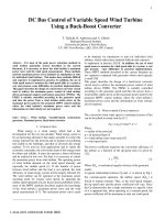
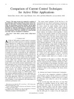
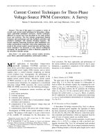
Figure 1 displays major changes which are observed in chronic pain states.
At the peripheral level distinct processes were observed which characterize
pathophysiologic nociceptive and neuropathic pain. The hallmark of pathophysiologic nociceptive pain, e.g., pain during inflammation or after tissue injury, is
peripheral sensitization. Nociceptive nerve fibers exhibit a lowering of their
excitation threshold for the response to mechanical and/or thermal stimuli and
increased firing frequencies during the application of stimuli of noxious intensities.
Such processes were characterized in the skin, muscle, joint, and visceral organs
(Schaible and Richter 2004). Molecular mechanisms of peripheral sensitization are
addressed in Sect. 1.4 (see below). More recently, the concept of hyperalgesic
6
H.-G. Schaible
Activation of the thalamocortical
system (conscious pain)
Activation of the amygdala (fear)
Atrophy of „pain areas“
Cortical reorganization
Changes in brain stem:
Reduction of descending
inhibition
Increase of descending
excitation
Spinal sensitization
(involvement of neurons and glial cells)
Peripheral sensitization
and hyperalgesic priming
Inflammation
Tissue injury
Ectopic discharges
in sensory fibres
Injury or disease
of nerve fibres
Fig. 1 Changes in the nociceptive system during pathophysiologic nociceptive pain and neuropathic pain. Spinal sensitization and increased hyperexcitability at the supraspinal level form the
process of central sensitization
priming was introduced (see Kandasamy and Price 2015). Priming arises from an
initial injury and results in the development of a remarkable susceptibility to
normally subthreshold noxious inputs causing a prolonged pain state in primed
animals. Priming increases the sensitization process which is evoked by sensitizing
mediators. As an example, application of prostaglandin E2 to normal tissue causes a
short-lasting sensitization of nociceptors if applied before injury or priming. However, if the neurons were primed, e.g., by interleukin-6, NGF, and other priming
stimuli, prostaglandin E2 will cause a long-lasting sensitization (see Kandasamy
and Price 2015).
A frequent process of neuropathic pain at the peripheral level is the generation of
ectopic discharges. These action potentials can be elicited at the lesion site of the
nerve fibers, but they can also be generated in the soma of the lesioned neurons
(Devor 2009). Underlying mechanisms are changes in the expression of ion
channels, actions of inflammatory mediators on lesioned fibers, and effects of the
sympathetic nervous system on lesioned nerve fibers. In the latter case the neuropathic pain may be sympathetically maintained (Schaible and Richter 2004).
Peripheral nociceptive processes often trigger changes in the spinal cord which
are called central sensitization. The changes in the spinal cord provide a gain of
the nociceptive processing at the spinal site (Cervero 2009; Woolf and Salter 2000).
Nociceptive spinal cord neurons which receive increased input from inflamed
regions show the following phenomena: a lowering of threshold, increased
responses to innocuous and noxious stimuli, and an expansion of the receptive
Emerging Concepts of Pain Therapy Based on Neuronal Mechanisms
7
fields (Schaible et al. 2009). In the sensitized state, more spinal cord neurons show
responses to a stimulus applied to a specific peripheral site. These changes reflect an
increase of the synaptic processing including the suprathreshold activation of
synapses which may be too weak in the normal state to depolarize the neuron
sufficiently. In many aspects these processes are similar to the long-term potentiation which was characterized as a major process of memory formation in the
hippocampus (Sandku¨hler 2000). Central sensitization is also thought to occur in
neuropathic pain states.
Several cell types may contribute to the spinal sensitization. First, the sensitization of peripheral nociceptors increases the sensory input into the spinal cord, thus
providing a stronger presynaptic component of synaptic activation. Second, postsynaptic spinal cord neurons are rendered hyperexcitable by the activation of
NMDA and other receptors (Sandku¨hler 2000; Woolf and Salter 2000). Third,
glial cells may be activated and produce cytokines and other mediators which
facilitate the spinal processing. Glial cells are strongly activated in neuropathic
pain states, but they may also contribute to inflammatory pain (McMahon and
Malcangio 2009). The involvement of glial cells in pain states is addressed by
Old et al. (2015). Fourth, the activity of inhibitory interneurons may be reduced.
The inhibitory neurons in the spinal cord and the mechanisms by which the
inhibitory control is decreased or lost are addressed by Todd (2015). The spinal
sensitization and the resulting thalamocortical processing are thought to underlie
the observation that in many pain states the pain becomes widespread (Phillips and
Clauw 2013). The significance of central sensitization in humans under clinically
relevant conditions, and the experimental methods to test central sensitization in
humans, will be addressed by Arendt-Nielsen (2015).
Ascending nociceptive information activates the thalamocortical system. During
chronic pain states significant changes of this system were observed in patients
using fMRI. Remarkably, many chronic pain states such as chronic osteoarthritic
pain are associated with a so-called atrophy of the regions in which pain is
processed. The underlying cellular mechanisms have not been identified. Interestingly, this atrophy seems to be reversible because after successful treatment of pain,
the brain structures show a normalization (Bushnell et al. 2013; Gwilym et al. 2010;
Rodriguez-Raecke et al. 2009). Under neuropathic conditions the cortex may show
a reorganization with significant changes in the cortical maps. Such changes were,
e.g., observed during phantom limb pain.
As already mentioned, ascending tracts not only activate the thalamocortical
system. They also activate the amygdala via the parabrachial nucleus. Further input
to the amygdala is provided by the nerve fibers from the thalamus and from the
cortex (Duvarci and Pare 2014). The amygdala is key nuclei in the generation of
fear, and they can be activated in pain conditions (Kulkarni et al. 2007). In this
volume, the role of the amygdala and their connections to the medial prefrontal
cortex (mPFC) in pain states will be addressed by Neugebauer (2015). Pain-related
mPFC deactivation results in cognitive deficits and the failure of inhibitory control
of amygdala processing. Impaired cortical control allows the uncontrolled persistence of amygdala pain mechanisms.
8
H.-G. Schaible
Neural pathways descending from the brain stem mediate inhibition and facilitation of nociceptive spinal cord neurons (Ossipov et al. 2010; Vanegas and
Schaible 2004). During severe chronic pain, a reduction of descending inhibition,
in particular the diffuse inhibitory noxious control (DNIC), was reported (Kosek
and Ordeberg 2000; Lewis et al. 2012). In addition, descending facilitation may
contribute to pain, in particular during neuropathic pain (Vanegas and Schaible
2004). Thus, descending inhibitory systems from the brain stem may be less
effective and/or descending excitatory systems from the brain stem may be overactive during chronic pain. These changes may be (partly) reversible after successful
pain treatment (Kosek and Ordeberg 2000).
Effects of the nervous system on inflammation. It must be noted that the
importance of the nociceptive nervous system extends beyond the generation of
pain. The nervous system is able to influence inflammatory processes in the organs.
Such influences are mediated by the efferent effects of nociceptive sensory
afferents which produce neurogenic inflammation, by fibers of the sympathetic
and parasympathetic nervous system, and by neuroendocrine influences (Schaible
and Straub 2014). Spinal hyperexcitability is not only important for pain generation
(see above). It plays also a role in the regulation of joint inflammation (Waldburger
and Firestein 2010). In this volume this topic will be addressed by Sorkin (2015).
Both pro- and anti-inflammatory feedback loops can involve just the peripheral
nerves and the spinal cord or can also include more complex, supraspinal structures
such as the vagal nuclei and the hypothalamic–pituitary axis.
1.4
Molecular Mechanisms of Pain
Molecular mechanisms of nociception are of considerable interest for pharmacologic approaches, and therefore, they are particularly addressed in this volume. The
peripheral nociceptor as well as the spinal cord and the amygdala are in the focus.
Nociception in the periphery consists of two elementary processes, the transduction of stimuli (the generation of a sensor potential by the impact of a noxious
stimulus) and the transformation of the sensor potential into a series of action
potentials. Noxious stimuli are mechanical or thermal (heat and cold), and also
some chemical mediators (e.g., bradykinin or H+) cause pain. The chemosensitivity
of nociceptors is particularly important for the process of sensitization (and
priming).
For the transduction of thermal stimuli into sensor potentials, ion channels of the
transient receptor potential (TRP) family are responsible. While the involvement of
TRPV1, TRPV2, and TRPM8 in the sensation of noxious heat (TRPV1 and
TRPV2) and innocuous cold (TRPM8) has been established, the significance of
other TRP channels in thermo(noci)ception is not that clear. For two TRP channels
(TRPA1 and TRPV4), a role in mechanical hyperalgesia is being discussed (Kwan
et al. 2009; Levine and Alessandri-Haber 2007; Malsch et al. 2014; Segond von
Banchet et al. 2013) although these channels may not be the transduction molecules
involved in the “normal mechanonociception.” The current knowledge on the
Emerging Concepts of Pain Therapy Based on Neuronal Mechanisms
9
involvement of TRP ion channels in the sensation of noxious heat and noxious cold
and of the involvement of these ion channels in the generation of thermal
hyperalgesia has been summarized (Basbaum et al. 2009; Julius 2013; Stein
et al. 2009) and is not the topic of this volume.
Some chemicals can also open ion channels. For example, H+ triggers the
opening of acid-sensing ion channels (ASICs), and capsaicin opens TRPV1. Most
mediators, however, activate membrane receptors and are thereby involved in the
sensitization of nociceptive neurons (see below).
The sensor potential triggers the generation of action potentials. For action
potentials voltage-gated sodium channels are essential. In nociceptive neurons,
mainly the sodium channels Nav1.7, Nav1.8, and Nav1.9, and under neuropathic
conditions Nav1.3, are expressed (Waxman and Zamponi 2014). Nav1.7 is activated
by slow, subtle depolarization close to the resting potential, and it thus sets the gain
on nociceptors. Nav1.8, which shows depolarized voltage dependence, produces
most of the current responsible for the action potential upstroke, and it supports
repetitive firing. Nav1.9 does not contribute to the action potential upstroke but
depolarizes the cells and prolongs and enhances small depolarization thus enhancing excitability (Waxman and Zamponi 2014). In this volume Habib et al. (2015)
will address the role of these ion channels in different inflammatory and neuropathic pain states. They show that particular Nav ion channels are involved in
different pathophysiologic states. Because Na+ channel blockers are thought to be
promising targets for new analgesics (Gold 2008), such knowledge is important for
the understanding of which blocker might be suitable under the particular
conditions.
When neurons are sensitized both the channels of transduction and the voltagegated ion channels, in particular the Na+ channels, show changes such that the
excitability is enhanced (Linley et al. 2010; Schaible et al. 2011). Some mediators
such as prostaglandin E2 change the opening properties of TRPV1 and of sodium
channels such that weaker stimuli are sufficient to open the ion channels. The effect
of prostaglandin E2 is mediated by G protein-coupled receptors which activate
second messengers in the nociceptors (Hucho and Levine 2007), and these second
messenger systems change the opening properties of the ion channels.
While prostaglandins are known for a long time as sensitizing molecules, more
recent research revealed a number of other receptor types in nociceptive sensory
neurons which are of great importance for the sensitization. It was shown that
proinflammatory cytokines such as TNF-α, interleukin-6, and interleukin-17 induce
a persistent state of sensitization in C-fibers (Brenn et al. 2007; Richter et al. 2010,
2012). Cytokines are thought to play a significant role in the generation of inflammatory and neuropathic role (Schaible et al. 2010; Sommer and Kress 2004;
¨ ceyler et al. 2009). Interleukin-6 is thought to be an important molecule of
U
hyperalgesic priming (see Kandasamy and Price 2015).
NGF and its receptor trkA were discovered as suitable targets for pain treatment.
A single application of an antibody to NGF was shown to provide significant pain
relief in osteoarthritis for several weeks (Lane et al. 2010). NGF has a variety of
actions on nonneuronal cells and sensory neurons which regulate the excitability in
10
H.-G. Schaible
the long term (Bennett 2007). In this volume Mizumura and Murase (2015) address
the hyperalgesic effects of NGF in different tissues and in inflammatory and
neuropathic pain states, and they address the mechanisms involved.
Proteinase-activated receptors (PARs) are a family of G protein-coupled receptor that is activated by extracellular cleavage of the receptor in the N-terminal
domain. This slicing of the receptor exposes a tethered ligand which binds to a
specific docking point on the receptor surface to initiate intracellular signaling.
McDougall and Muley summarize how serine proteinases activate PARs leading to
the development of pain in several chronic pain conditions. The potential of PARs
as a drug target for pain relief is discussed (McDougall and Muley 2015).
Excitatory synaptic transmission in the spinal cord under basal conditions is
mediated by the transmitter glutamate, the transmitter of nociceptive sensory
neurons. Central sensitization is also dependent on glutamate, in particular acting
on NMDA receptors. However, numerous other transmitters and mediators are
involved in the complex signaling in the spinal cord (e.g., NK1 receptors for
substance and CGRP receptors) (Woolf and Salter 2000). Other mediators such as
spinal prostaglandins contribute to spinal sensitization (Ba¨r et al. 2004). The
particular role of NO to nociceptive spinal cord signaling will be addressed by
Schmidtko (2015). The role of mediators involved in glial cell activation and
functions will be addressed by Old et al. (2015).
Under normal conditions, excitatory and inhibitory synaptic mechanisms are
presumably in a balanced activity state. Such inhibition is provided by specific local
inhibitory interneurons (see Todd 2015), but it may also be provided by mediators
which act in a feedback manner from activated neurons. Such inhibitory control is,
e.g., provided by endocannabinoids which are addressed in this volume by
Woodhams et al. (2015). Cannabinoid 1 (CB1) receptors are found at presynaptic
sites throughout the peripheral and central nervous systems, while the CB2 receptor
is found principally (but not exclusively) on immune cells. The endocannabinoid
(EC) system is now known to be one of the key endogenous systems regulating pain
sensation, with modulatory actions at all stages of pain processing pathways. As
already discussed, pain states may involve a reduction of inhibitory mechanisms.
A particular interesting aspect is that some mediators may exert excitatory as
well as inhibitory actions, depending on the functional context. An example is the
change of GABAergic inhibitory mechanisms in neuropathic pain states (see Todd
2015). However, even mediators such as prostaglandin E2 which are usually
considered excitatory may provide antinociception when pain pathways are
activated, by the activation of receptor subtypes which are coupled to inhibitory
signaling pathways (Natura et al. 2013). In this volume Schmidtko (2015) reports
about both the pro- and antinociceptive effects of NO signaling resulting from a
different downstream signaling.
Spinal cord mechanisms may even alter the antinociceptive effect of potent
analgesic drugs. Opioids are considered the gold standard for the treatment of
moderate to severe pain. However, heterogeneity in analgesic efficacy, poor
potency, and side effects are associated with opioid use. Traditionally opioids are
thought to exhibit their analgesic actions via the activation of the neuronal G
Emerging Concepts of Pain Therapy Based on Neuronal Mechanisms
11
protein-coupled opioid receptors. However, neuronal activity of opioids cannot
fully explain the initiation and maintenance of opioid tolerance, hyperalgesia, and
allodynia. In this volume Thomas et al. (2015) report the importance of
nonneuronal mechanisms in opioid signaling, paying particular attention to the
relationship of opioids and immune signaling.
Abnormally enhanced output from the CeLC of the amygdala is also the
consequence of an imbalance between excitatory and inhibitory mechanisms (see
Neugebauer 2015). Impaired inhibitory control mediated by a cluster of
GABAergic interneurons in the intercalated cell masses (ITC) allows the development of glutamate- and neuropeptide-driven synaptic plasticity of excitatory inputs
from the brain stem (parabrachial area) and from the lateral–basolateral amygdala
network (LA-BLA, site of integration of polymodal sensory information).
2
Conclusion
It is increasingly evident how many different neuronal and molecular mechanisms
contribute to the expression of pain, in particular in clinically relevant pain states.
We begin to understand some mechanisms of pain vulnerability (Denk et al. 2014).
The complexity of pain processing and related neuronal events puts a considerable
challenge to the development of new therapeutic strategies. Is the focus on single
key molecules such as a particular sodium channel an appropriate therapeutical
approach or should one aim to interfere with disease-related mediators such as NGF
or cytokines? The answer to this crucial question is not straightforward. Both types
of drugs have been proven useful in medical therapy. Local anesthetics targeting
specifically sodium channels can interrupt pain (usually for a short time only), but
on the other hand, the use of antibodies to particular cytokines which have numerous actions is extremely potent in the therapy of rheumatic diseases such as
rheumatoid arthritis. Thus, future pain therapy should provide effective treatments
using either specific drugs with the aim of interfering with specific nociceptive
processes or using drugs which have the potency of long-term modification of pain
mechanisms.
References
Arendt-Nielsen L (2015) Central sensitization in humans: assessment and pharmacology. In:
Schaible H-G (ed) Pain control. Springer, Berlin, pp 79–102
Ba¨r K-J, Natura G, Telleria-Diaz A, Teschner P, Vogel R, Vasquez E, Schaible H-G, Ebersberger
A (2004) Changes in the effect of spinal prostaglandin E2 during inflammation—prostaglandin
E (EP1-EP4) receptors in spinal nociceptive processing of input from the normal or inflamed
knee joint. J Neurosci 24:642–651
Basbaum AI, Bautista DM, Scherrer G, Julius D (2009) Cellular and molecular mechanisms of
pain. Cell 139:267–284
Bennett D (2007) NGF, sensitization of nociceptors. In: Schmidt RF, Willis WD (eds) Encyclopedia of pain, vol 2. Springer, Berlin, pp 1338–1342
12
H.-G. Schaible
Breivik H, Beverly C, Ventafridda V, Cohen R, Gallacher D (2006) Survey of chronic pain in
Europe: prevalence, impact on daily life, and treatment. Eur J Pain 10:287–333
Brenn D, Richter F, Schaible H-G (2007) Sensitization of unmyelinated sensory fibres of the joint
nerve to mechanical stimuli by interleukin-6 in the rat. An inflammatory mechanism of joint
pain. Arthritis Rheum 56:351–359
Bushnell MC, Ceko M, Low LA (2013) Cognitive and emotional control of pain and its disruption
in chronic pain. Nat Rev Neurosci 14:502–511
Cervero F (2009) Spinal cord hyperexcitability and its role in pain and hyperalgesia. Exp Brain
Res 196:129–137
Denk F, McMahon SB, Tracey I (2014) Pain vulnerability: a neurobiological perspective. Nat
Neurosci 17:192–200
Devor M (2009) Ectopic discharge in Aβ afferents as a source of neuropathic pain. Exp Brain Res
196:115–128
Duvarci S, Pare D (2014) Amygdala microcircuits controlling learned fear. Neuron 82:966–980
Gold MS (2008) Na+ channel blockers for the treatment of pain: context is everything, almost. Exp
Neurol 210:1–6
Gwilym SE, Filippini N, Douaud G, Carr AJ, Tracey I (2010) Thalamic atrophy associated with
painful osteoarthritis of the hip is reversible after arthroplasty. Arthritis Rheum 62:2930–2940
Habib AM, Wood JN, Cox JJ (2015) Sodium channels and pain. In: Schaible H-G (ed) Pain
control. Springer, Berlin, pp 39–56
Hucho T, Levine JD (2007) Signaling pathways in sensitization: toward a nociceptor cell biology.
Neuron 55:365–376
Julius D (2013) TRP channels and pain. Annu Rev Cell Dev Biol 29:355–384
Kandasamy R, Price TJ (2015) The pharmacology of nociceptor priming. In: Schaible H-G
(ed) Pain control. Springer, Berlin, pp 15–37
Kosek E, Ordeberg G (2000) Lack of pressure pain modulation by heterotopic noxious conditioning stimulation in patients with painful osteoarthritis before, but not following surgical pain
relief. Pain 88:69–78
Kulkarni B, Bentley DE, Elliott R, Julyan PJ, Boger E, Watson A, Boyle Y, El-Deredy W, Jones
AKP (2007) Arthritic pain is processed in brain areas concerned with emotions and fear.
Arthritis Rheum 56:1345–1354
Kwan KY, Glazer JM, Corey DP, Rice FL, Stucky CL (2009) TRPA1 modulates
mechanotransduction in cutaneous sensory neurons. J Neurosci 29:4808–4819
Lane NE, Schnitzer TJ, Birbara CA, Mokhtarani M, Shelton DL, Smith MD, Brown MT (2010)
Tanezumab for the treatment of pain from osteoarthritis of the knee. N Engl J Med
363:1521–1531
Levine JD, Alessandri-Haber N (2007) TRP channels: targets for the relief of pain. Biochim
Biophys Acta 1772:989–1003
Lewis GN, Rice DA, McNair PJ (2012) Conditioned pain modulation in populations with chronic
pain: a systematic review and meta-analysis. J Pain 13:936–944
Linley JE, Rose K, Ooi L, Gamper N (2010) Understanding inflammatory pain: ion channels
contributing to acute and chronic nociception. Pflugers Arch 459:657–669
Malsch P, Andratsch M, Vogl C, Link AS, Alzheimer C, Brierley SM, Hughes PA, Kress M (2014)
Deletion of interleukin-6 signal transducer gp130 in small sensory neurons attenuates
mechanonociception and down-regulates mechanotransducer ion channel TRPA1. J Neurosci
34:9845–9856
McDougall JJ, Muley MM (2015) The role of proteases in pain. In: Schaible H-G (ed) Pain control.
Springer, Berlin, pp 239–260
McMahon SB, Malcangio M (2009) Current challenges in glia-pain biology. Neuron 64:46–54
Mizumura K, Murase S (2015) Role of nerve growth factor in pain. In: Schaible H-G (ed) Pain
control. Springer, Berlin, pp 57–77
Natura G, Ba¨r K-J, Eitner A, B€
ottger M, Richter F, Hensellek S, Ebersberger A, Leuchtweis J,
Maruyama T, Hofmann GO, Halbhuber K-J, Schaible H-G (2013) Neuronal prostaglandin E2
Emerging Concepts of Pain Therapy Based on Neuronal Mechanisms
13
receptor subtype EP3 mediates antinociception during inflammation. Proc Natl Acad Sci U S A
110:13648–13653
Neugebauer V (2015) Amygdala pain mechanisms. In: Schaible H-G (ed) Pain control. Springer,
Berlin, pp 261–284
Old EA, Clark AK, Malcangio M (2015) The role of glia in the spinal cord in neuropathic and
inflammatory pain. In: Schaible H-G (ed) Pain control. Springer, Berlin, pp 145–170
Ossipov MH, Dussor GO, Porreca F (2010) Central modulation of pain. J Clin Invest
120:3779–3787
Phillips K, Clauw DJ (2013) Central pain mechanisms in the rheumatic diseases. Arthritis Rheum
65:291–302
Richter F, Natura G, Loeser S, Schmidt K, Viisanen H, Schaible H-G (2010) Tumor necrosis
factor-α (TNF-α) causes persistent sensitization of joint nociceptors for mechanical stimuli.
Arthritis Rheum 62:3806–3814
Richter F, Natura G, Ebbinghaus M, Segond von Banchet G, Hensellek S, K€
onig C, Bra¨uer R,
Schaible H-G (2012) Interleukin-17 sensitizes joint nociceptors for mechanical stimuli and
contributes to arthritic pain through neuronal IL-17 receptors in rodents. Arthritis Rheum
64:4125–4134
Rodriguez-Raecke R, Niemeier A, Ihle K, Ruether W, May A (2009) Brain gray matter decrease in
chronic pain is the consequence and not the cause of pain. J Neurosci 29:13746–13750
Sandku¨hler J (2000) Learning and memory in pain pathways. Pain 88:113–118
Schaible H-G, Richter F (2004) Pathophysiology of pain. Langenbecks Arch Surg 389:237–243
Schaible H-G, Straub RH (2014) Function of the sympathetic supply in acute and chronic
experimental joint inflammation. Auton Neurosci 182:55–64
Schaible H-G, Richter F, Ebersberger A, Boettger MK, Vanegas H, Natura G, Vazquez E, Segond
von Banchet G (2009) Joint pain. Exp Brain Res 196:153–162
Schaible H-G, Segond von Banchet G, Boettger MK, Bra¨uer R, Gajda M, Richter F, Hensellek S,
Brenn D, Natura G (2010) The role of proinflammatory cytokines in the generation and
maintenance of joint pain. Ann N Y Acad Sci 1193:60–69
Schaible H-G, Ebersberger A, Natura G (2011) Update on peripheral mechanisms of pain: beyond
prostaglandins and cytokines. Arthritis Res Ther 13:21
Schmelz M (2015) Itch and pain differences and commonalities. In: Schaible H-G (ed) Pain
control. Springer, Berlin, pp 285–300
Schmidtko A (2015) Nitric oxide mediated pain processing in the spinal cord. In: Schaible H-G
(ed) Pain control. Springer, Berlin, pp 103–117
Segond von Banchet G, Boettger MK, K€
onig C, Iwakura Y, Bra¨uer R, Schaible H-G (2013)
Neuronal IL-17 receptor upregulates TRPV4 but not TRPV1 receptors in DRG neurons and
mediates mechanical but not thermal hyperalgesia. Mol Cell Neurosci 52:152–160
Sommer C, Kress M (2004) Recent findings on how proinflammatory cytokines cause pain:
peripheral mechanisms in inflammatory and neuropathic hyperalgesia. Neurosci Lett
361:184–187
Sorkin LS (2015) Modulation of peripheral inflammation by the spinal cord. In: Schaible H-G
(ed) Pain control. Springer, Berlin, pp 191–206
Stein C, Clark JD, Oh U, Vasko MR, Wilcox GL, Overland AC, Vanderah TW, Spencer RH
(2009) Peripheral mechanisms of pain and analgesia. Brain Res Rev 60:90–113
Thomas J, Mustafa S, Johnson J, Nicotra L, Hutchinson M (2015) The relationship between
opioids and immune signalling in the spinal cord. In: Schaible H-G (ed) Pain control. Springer,
Berlin, pp 207–238
Todd AJ (2015) Plasticity of inhibition in the spinal cord. In: Schaible H-G (ed) Pain control.
Springer, Berlin, pp 171–190
Treede RD, Kenshalo DR, Gracely RH, Jones A (1999) The cortical representation of pain. Pain
79:105–111
¨ ceyler N, Scha¨fers M, Sommer C (2009) Mode of action of cytokines on nociceptive neurons.
U
Exp Brain Res 196:67–78
14
H.-G. Schaible
Vanegas H, Schaible H-G (2004) Descending control of persistent pain: inhibitory or facilitatory?
Brain Res Rev 46:295–309
Vogt BA (2005) Pain and emotion. Interactions in subregions of the cingulate cortex. Nat Rev
Neurosci 6:533–544
Waldburger JM, Firestein GS (2010) Regulation of peripheral inflammation by the central nervous
system. Curr Rheumatol Rep 12:370–378
Waxman SG, Zamponi GW (2014) Regulating excitability of peripheral afferents: emerging ion
channel targets. Nat Neurosci 17:153–163
Woodhams SG, Sagar DR, Burston JJ, Chapman V (2015) The role of the endocannabinoid system
in pain. In: Schaible H-G (ed) Pain control. Springer, Berlin, pp 119–143
Woolf CJ, Salter MW (2000) Neuronal plasticity: increasing the gain in pain. Science
288:1765–1768
The Pharmacology of Nociceptor Priming
Ram Kandasamy and Theodore J. Price
Contents
1
2
3
Introduction . . . . . . . . . . . . . . . . . . . . . . . . . . . . . . . . . . . . . . . . . . . . . . . . . . . . . . . . . . . . . . . . . . . . . . . . . . . . . . . . . . .
Why Use Hyperalgesic Priming Models? . . . . . . . . . . . . . . . . . . . . . . . . . . . . . . . . . . . . . . . . . . . . . . . . . . .
Mechanisms of Priming in the Periphery: A Model for Sustained Nociceptor Plasticity . .
3.1 PKCε as a Crucial Mechanism of Nociceptor Priming . . . . . . . . . . . . . . . . . . . . . . . . . . . . . . .
3.2 Local Translation Is a Key Mediator of Nociceptor Priming . . . . . . . . . . . . . . . . . . . . . . . .
4 CNS Regulation of Hyperalgesic Priming . . . . . . . . . . . . . . . . . . . . . . . . . . . . . . . . . . . . . . . . . . . . . . . . . .
4.1 Atypical PKCs and Brain-Derived Neurotropic Factor . . . . . . . . . . . . . . . . . . . . . . . . . . . . . . .
4.2 Endogenous Opioids, μ-Opioid Receptor Constitutive Activity, and Hyperalgesic
Priming . . . . . . . . . . . . . . . . . . . . . . . . . . . . . . . . . . . . . . . . . . . . . . . . . . . . . . . . . . . . . . . . . . . . . . . . . . . . . . . . .
4.3 Surgery as a Priming Stimulus and the Effects of Opioids . . . . . . . . . . . . . . . . . . . . . . . . . . .
5 Therapeutic Opportunities and Conclusions . . . . . . . . . . . . . . . . . . . . . . . . . . . . . . . . . . . . . . . . . . . . . . . .
References . . . . . . . . . . . . . . . . . . . . . . . . . . . . . . . . . . . . . . . . . . . . . . . . . . . . . . . . . . . . . . . . . . . . . . . . . . . . . . . . . . . . . . . .
16
17
18
19
20
24
24
27
29
31
33
Abstract
Nociceptors and neurons in the central nervous system (CNS) that receive
nociceptive input show remarkable plasticity in response to injury. This plasticity is thought to underlie the development of chronic pain states. Hence, further
understanding of the molecular mechanisms driving and maintaining this
R. Kandasamy
Department of Pharmacology, The University of Arizona, Tucson, AZ 85721, USA
T.J. Price (*)
Department of Pharmacology, The University of Arizona, Tucson, AZ 85721, USA
Bio5 Institute, The University of Arizona, Tucson, AZ 85721, USA
Graduate Interdisciplinary Program in Neuroscience, The University of Arizona, Tucson, AZ
85721, USA
School of Brain and Behavioral Sciences, The University of Texas at Dallas, Richardson, TX
75080, USA
e-mail:
# Springer-Verlag Berlin Heidelberg 2015
H.-G. Schaible (ed.), Pain Control, Handbook of Experimental Pharmacology 227,
DOI 10.1007/978-3-662-46450-2_2
15
16
R. Kandasamy and T.J. Price
plasticity has the potential to lead to novel therapeutic approaches for the
treatment of chronic pain states. An important concept in pain plasticity is the
presence and persistence of “hyperalgesic priming.” This priming arises from an
initial injury and results in a remarkable susceptibility to normally subthreshold
noxious inputs causing a prolonged pain state in primed animals. Here we
describe our current understanding of how this priming is manifested through
changes in signaling in the primary nociceptor as well as through memory like
alterations at CNS synapses. Moreover, we discuss how commonly utilized
analgesics, such as opioids, enhance priming therefore potentially contributing
to the development of persistent pain states. Finally we highlight where these
priming models draw parallels to common human chronic pain conditions.
Collectively, these advances in our understanding of pain plasticity reveal a
variety of targets for therapeutic intervention with the potential to reverse rather
than palliate chronic pain states.
Keywords
Atypical PKC • AMPA • NMDA • mTORC1 • PKC • Epac • Hyperalgesic
priming • Prostaglandins • NGF • Interleukin 6
1
Introduction
A fundamental principle underlying our current understanding of pathological pain
states is plasticity in the nociceptive system. While research into pathological pain
states has long recognized this idea, it is only relatively recently that we have started
to gain insight into mechanisms that cause this plasticity. On the most general level,
plasticity in the pain system occurs at two locations, at the primary afferent
nociceptor and at synapses receiving nociceptive input throughout the central
nervous system (CNS). Preclinical models of acute and chronic inflammatory
pain as well as models of neuropathic pain have revealed a plethora of molecular
targets that have developed our understanding of how chronic pain develops as well
as revealing important potential therapeutic intervention points. In the late 1990s
and early 2000s, Jon Levine and colleagues developed “hyperalgesic priming”
models (for review see Reichling and Levine 2009). These models provide unique
insight into plasticity in the nociceptive system because they allow for molecular
dissection of pain states in two distinct phases. These models involve a priming
stimulus, aimed at causing an acute sensitization of peripheral nociceptors and their
central inputs, albeit with some notable exceptions which will be discussed later.
Next, in opposition to most other preclinical models, the initial sensitization is
allowed to resolve and a second, normally subthreshold, stimulus is delivered.
Importantly, this second stimulus, which has only a transient effect in naı¨ve
animals, leads to a prolonged state of pain hypersensitivity that allows for investigation of molecular mechanisms that define the primed nociceptor and/or the