Progress in molecular biology and translational science, volume 130
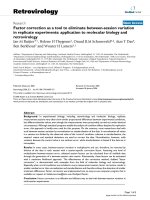
Bạn đang xem bản rút gọn của tài liệu. Xem và tải ngay bản đầy đủ của tài liệu tại đây (4.2 MB, 137 trang )
Academic Press is an imprint of Elsevier
225 Wyman Street, Waltham, MA 02451, USA
525 B Street, Suite 1800, San Diego, CA 92101-4495, USA
125 London Wall, London, EC2Y 5AS, UK
The Boulevard, Langford Lane, Kidlington, Oxford OX5 1GB, UK
First edition 2015
Copyright © 2015, Elsevier Inc. All Rights Reserved.
No part of this publication may be reproduced or transmitted in any form or by any means,
electronic or mechanical, including photocopying, recording, or any information storage and
retrieval system, without permission in writing from the publisher. Details on how to seek
permission, further information about the Publisher’s permissions policies and our
arrangements with organizations such as the Copyright Clearance Center and the Copyright
Licensing Agency, can be found at our website: www.elsevier.com/permissions.
This book and the individual contributions contained in it are protected under copyright by
the Publisher (other than as may be noted herein).
Notices
Knowledge and best practice in this field are constantly changing. As new research and
experience broaden our understanding, changes in research methods, professional practices,
or medical treatment may become necessary.
Practitioners and researchers must always rely on their own experience and knowledge in
evaluating and using any information, methods, compounds, or experiments described
herein. In using such information or methods they should be mindful of their own safety and
the safety of others, including parties for whom they have a professional responsibility.
To the fullest extent of the law, neither the Publisher nor the authors, contributors, or editors,
assume any liability for any injury and/or damage to persons or property as a matter of
products liability, negligence or otherwise, or from any use or operation of any methods,
products, instructions, or ideas contained in the material herein.
ISBN: 978-0-12-802912-1
ISSN: 1877-1173
For information on all Academic Press publications
visit our website at store.elsevier.com
CONTRIBUTORS
Jonathan D. Bohbot
United States Department of Agriculture, Beltsville Agricultural Research Center, Invasive
Insect Biocontrol and Behavior Laboratory, Beltsville, Maryland, USA
Arthur de Fouchier
Institute of Ecology & Environmental Sciences of Paris, INRA, Versailles, France
Joseph C. Dickens
United States Department of Agriculture, Beltsville Agricultural Research Center, Invasive
Insect Biocontrol and Behavior Laboratory, Beltsville, Maryland, USA
Jean-Franc¸ois Gibrat
INRA UR1077 Mathe´matique Informatique et Ge´nome, Domaine de Vilvert,
Jouy-en-Josas, France
Emmanuelle Jacquin-Joly
Institute of Ecology & Environmental Sciences of Paris, INRA, Versailles, France
Nicolas Montagne´
Institute of Ecology & Environmental Sciences of Paris, UPMC-Sorbonne Universite´, Paris,
France
Richard D. Newcomb
School of Biological Sciences, University of Auckland, and The New Zealand Institute for
Plant & Food Research Limited, Auckland, New Zealand
Edith Pajot-Augy
INRA UR 1197 NeuroBiologie de l’Olfaction, Domaine de Vilvert, Jouy-en-Josas, France
Marie-Annick Persuy
INRA UR 1197 NeuroBiologie de l’Olfaction, Domaine de Vilvert, Jouy-en-Josas, France
Guenhae¨l Sanz
INRA UR 1197 NeuroBiologie de l’Olfaction, Domaine de Vilvert, Jouy-en-Josas, France
Jackson T. Sparks
United States Department of Agriculture, Beltsville Agricultural Research Center, Invasive
Insect Biocontrol and Behavior Laboratory, Beltsville, Maryland, USA
Thierry Thomas-Danguin
INRA UMR 1129 Flaveur, Vision et Comportement du Consommateur, Dijon, France
Anne Tromelin
INRA UMR 1129 Flaveur, Vision et Comportement du Consommateur, Dijon, France
William B. Walker
Chemical Ecology Research Group, Department of Plant Protection Biology, Swedish
University of Agricultural Sciences, Alnarp, Sweden
vii
viii
Contributors
Guirong Wang
State Key Laboratory for Biology of Plant Diseases and Insect Pests, Institute of Plant
Protection, Chinese Academy of Agricultural Sciences, Beijing, PR China
Dieter Wicher
Max Planck Institute for Chemical Ecology, Department Evolutionary Neuroethology, Jena,
Germany
Jin Zhang
State Key Laboratory for Biology of Plant Diseases and Insect Pests, Institute of Plant
Protection, Chinese Academy of Agricultural Sciences, Beijing, PR China
PREFACE
Smell is a potent wizard that transports you across thousands of miles and all the
years you have lived.
Helen Keller
This poignant quotation by Helen Keller speaks to the evocative nature of
olfaction for humans. Beyond being simply an important diagnostic mechanism for interpreting the environment, olfaction can often recall old memories or stir complex emotions. In my home country of Australia, there are
stories of soldiers returning from battle in World War II by ship and realizing
that they were nearing their homeland prior to sighting it, simply from the
characteristic smell of the oil-laden Eucalyptus trees that dominate much of
the Australian landscape. These weary combatants were not just detecting
trees but imbibing their loved ones, their childhoods, their hopes, and
their loss.
Coming from Helen Keller, this quote also subtly hints at the key role
olfaction plays when sight is not the primary sense used for navigation. This
is actually the case for most of the animals on earth; huge numbers of species
of invertebrates use olfaction as their key method of assessing their environment and detecting food, mates, hosts, predators, etc. In creatures such as
insects, olfaction-related cognition is much simpler than for humans; however, it is known to be important in individual learning, in parasitic wasps for
example. Olfaction is so important to insects that they have evolved
extremely sensitive olfactory receptors (ORs) to detect low concentrations
(sometimes nanomolar and below) of volatile compounds; these receptors
largely reside in their antennae but do occur elsewhere. The olfactory
sensitivity of insects helps make them formidable evolutionary competitors
but is also exploited by humans to disrupt insect behavior (e.g., pheromone
disruption of moth pests and pheromone trapping).
Olfaction has attracted significant scientific interest for many years. In
1937, Japanese researchers utilized electrodes to measure the negative electrical potential generated across olfactory epithelium of dogs, caused by
olfactory stimulation. This technique was adapted for study of frogs and rabbits in 1956 and given the name electro-olfactography; it has since been
widely utilized for study of olfaction in mammals. In 1957, the technique
was adapted to insects and named electroantennography, and in 1959 the
first insect pheromones were characterized from the silk moth, Bombyx mori.
ix
x
Preface
While electrophysiological techniques such as these were used successfully
for decades and could be used to detect the presence and degree of olfactory
stimulation by various compounds, they were unable to decipher the molecular basis of olfaction.
However, around the same time in 1953, Watson and Crick published
the structure of DNA. This was a seminal moment in science and was built
on by others to produce great advances in our understanding of molecular
biology and in the power of the techniques available to study it. Then in
1991, Richard Axel and Linda Buck discovered that vertebrate ORs were
a subclass of the well-known G protein-coupled receptor (GPCR) family of
proteins. This discovery (which was subsequently recognized with a Nobel
Prize in 2004) combined with advances in DNA/RNA sequencing technologies and bioinformatics led to the elucidation of OR repertoires of a range
of vertebrate species and of associated molecular signaling processes.
The first vertebrate receptor to be deorphaned (have its cognate ligands
characterized) was OR17 from the rat in 1998, which was shown to react
to C7–C10 saturated aldehydes.
Because insects also express many GPCRs including homologs of human
proteins (e.g., serotonin and histamine receptors), it was expected that invertebrate ORs would be readily isolated through homology searches. While
this was true for the nematode Caenorhabditis elegans, it took until 1999
for the first insect OR to be identified from the vinegar fly (Drosophila melanogaster) using unbiased approaches. This is because insect ORs are not
GPCRs but an unrelated group of receptor proteins with a similar tertiary
structure. Being different to classic GPCRs, the signaling mechanisms have
also proven to be different in insects, such as the existence of a highly conserved universal chaperone protein and the activation of both metabotropic
and ionotropic signaling cascades (first reported in 2008).
The purpose of this volume is to summarize the latest understanding of
molecular mechanisms of olfaction in vertebrates and insects. I have chosen
to focus most chapters on insects for several reasons. First, molecular biology
of insect olfaction is still an evolving paradigm compared to that of vertebrate
olfaction which is relatively well characterized. Second, insects are a
megadiverse group that interact with varying levels of specificity, with
virtually all other land organisms and therefore as a group have a huge array
of ORs that detect countless volatile compounds, many important to
humans. This is of great interest in terms of studying general biology but
insect ORs also show huge promise in many applications such as pest/disease
management and biosensing. Lastly, a lean toward insects gives a point of
Preface
xi
differentiation with other works on olfaction that have traditionally focused
on mammals, of which there are relatively few species.
This first edition of Molecular Basis of Olfaction is designed to provide
insight into key areas of olfaction research and is intended for use by
researchers, teachers, students, molecular biologists, and biologists in general.
Leading researchers from China, United States, France, Germany, Sweden,
and New Zealand have contributed the chapters presented here, and I take
this opportunity to sincerely thank all authors for their effort and expertise.
The chapter “Mammalian Olfactory Receptors: Molecular Mechanisms
of Odorant Detection, 3D-Modeling, and Structure–Activity Relationships”
by Persuy and coworkers from France summarizes our knowledge of molecular mechanisms of odorant detection in mammals and includes 3D modeling
of mammalian ORs, and relationships between receptor structure and activity. In chapter “Olfactory Signaling in Insects,” Dieter Wicher (Max Planck
Institute for Chemical Ecology) discusses cellular signaling in various types of
olfactory neurons in insects. The chapter “Advances in the Identification and
Characterization of Olfactory Receptors in Insects” by Montagne´ et al. provides an insight into the latest advances in isolating and characterizing insect
ORs, including the use of transcriptomics. The final two chapters focus on
specific areas of insect olfaction research of importance to humans. The chapter “Olfactory Disruption: Toward Controlling Important Insect Vectors of
Disease” by Sparks et al. (U.S. Department of Agriculture) discusses disruption of olfaction in insect vectors of human disease such as mosquitoes
and tsetse flies. The last chapter (“Pheromone Reception in Moths: From
Molecules to Behaviors” by Zhang and colleagues) summarizes knowledge
of one of the great olfactory phenomena in biology, pheromone detection
by moths, and the events leading from antennal detection of a pheromone
to neural processing and resultant behaviors.
I anticipate that future editions of this volume will update these summaries as well as expanding the focus of the current edition.
RICHARD GLATZ
19 November 2014
Kangaroo Island, Australia
CHAPTER ONE
Mammalian Olfactory Receptors:
Molecular Mechanisms of Odorant
Detection, 3D-Modeling, and
Structure–Activity Relationships
Marie-Annick Persuy*, Guenhaël Sanz*, Anne Tromelin†,
Thierry Thomas-Danguin†, Jean-François Gibrat{, Edith Pajot-Augy*,1
*INRA UR 1197 NeuroBiologie de l’Olfaction, Domaine de Vilvert, Jouy-en-Josas, France
†
INRA UMR 1129 Flaveur, Vision et Comportement du Consommateur, Dijon, France
{
INRA UR1077 Mathe´matique Informatique et Ge´nome, Domaine de Vilvert, Jouy-en-Josas, France
1
Corresponding author: e-mail address:
Contents
1. Mammalian Olfactory Receptors: From Genes to Proteins
1.1 Genes and pseudogenes
1.2 OR protein expression
1.3 Olfactory signal transduction
2. Olfactory Receptor Activity Regulation: Homodimerization, Binding Cooperativity,
and Allostery
3. Olfactory Receptor 3D Modeling and Use for Virtual Screening
3.1 Model building
3.2 Ligand virtual screening
3.3 GPCR inverse agonist, antagonist, and agonist ligands
4. Odorant Ligands Structure–Activity Relationships
References
2
2
5
7
9
12
18
20
21
23
25
Abstract
This chapter describes the main characteristics of olfactory receptor (OR) genes of vertebrates, including generation of this large multigenic family and pseudogenization. OR
genes are compared in relation to evolution and among species. OR gene structure and
selection of a given gene for expression in an olfactory sensory neuron (OSN) are tackled. The specificities of OR proteins, their expression, and their function are presented.
The expression of OR proteins in locations other than the nasal cavity is regulated by
different mechanisms, and ORs display various additional functions.
A conventional olfactory signal transduction cascade is observed in OSNs, but individual ORs can also mediate different signaling pathways, through the involvement of
other molecular partners and depending on the odorant ligand encountered. ORs are
engaged in constitutive dimers. Ligand binding induces conformational changes in the
Progress in Molecular Biology and Translational Science, Volume 130
ISSN 1877-1173
/>
#
2015 Elsevier Inc.
All rights reserved.
1
2
Marie-Annick Persuy et al.
ORs that regulate their level of activity depending on odorant dose. When present,
odorant binding proteins induce an allosteric modulation of OR activity.
Since no 3D structure of an OR has been yet resolved, modeling has to be performed using the closest G-protein-coupled receptor 3D structures available, to facilitate
virtual ligand screening using the models. The study of odorant binding modes and
affinities may infer best-bet OR ligands, to be subsequently checked experimentally.
The relationship between spatial and steric features of odorants and their activity in
terms of perceived odor quality are also fields of research that development of
computing tools may enhance.
1. MAMMALIAN OLFACTORY RECEPTORS: FROM GENES
TO PROTEINS
Olfactory receptors are predominantly expressed in the main olfactory
epithelium located in the nasal cavity. They are the gateways, located across
the plasma membranes of olfactory sensory neurons (OSN) cilia, through
which the message conveyed by the odorant molecules in the ambient air
transit, before being transduced into an electrical signal.
1.1. Genes and pseudogenes
In mammals, there exist several hundred (up to several thousand) OR genes
accounting for 1–3% of estimated mammalian gene repertoire,1,2 and
representing the largest gene superfamily.
The number of OR genes exceeds 1700 in the rat and is around 860 in
humans.3 This abundance is justified by the number of physiological functions in which olfaction is involved (food intake and preferences, search for
prey, predator avoidance, social behaviors, mother–young relationships,
spatial orientation, stress, etc.), even though this chemical sense was for a
while considered to be a minor sense relative to vision. ORs being GPCRs
are characterized by seven-transmembrane helices (TMHs), participating in
the transmission of the olfactory message carried by the volatile odorant
compounds of the environment.4–6 Because ORs are involved in the detection of chemical messages from the environment of animals, their genes have
undergone selection pressure, inducing the evolution of the olfactory
repertoires of the various species. Some OR genes evolved to nonfunctional
pseudogenes7 in varying proportions depending on the species, from
%20% in the mouse and dog8,9 to %50–60% in primates and humans1,3,10
(for review, see Ref. 11). Indeed, if the number of OR genes differs from
Mammalian Olfactory Receptors
3
species to species (133 ORs in zebrafish to 1300 in pigs,12 2129 in cows,
4200 in African elephants13) the amount of pseudogenes is also variable.
Some primates have less than 400 types of functional ORs (humans and
chimpanzees, orangutans, and macaques even less14,15) compared to over
1000 for pigs, rodents and dogs,12,16,17 and 1948 in African elephants.13
However, the cognitive power of these species, i.e., the ability to process
olfactory data, allows them to integrate information from complex olfactory
environments, beyond simply the number of functional ORs that can be
activated.18
Mammalian OR genes are organized in a large number of clusters distributed on many chromosomes e.g., 9 chromosomes for mice,19 all chromosomes except 20, and Y for humans.7 Potentially, coding sequences may
predominate on some chromosomes (7, 16, and 17 in humans, for
instance7). OR pseudogenes are interspersed with full-length OR genes.
Closely located OR genes within a cluster tend to be closely related evolutionarily, while duplication of whole OR gene clusters appears to be rare.20
Generation of this large and diverse multigenic family involved in a key biological function may result from successive duplications of large genomic
regions during evolution,11,21 followed by an accumulation of mutations.
Moreover, evolutionarily distantly related genes may be found in a given
OR gene cluster, and OR genes with a close evolutionary relationship
may be located at different clusters or chromosomes,20 suggesting additional
chromosomal rearrangements within OR gene clusters and shuffling of the
genes from different clusters.
In different species, a number of OR genes exhibit sequence identities
above 90%, for instance in dogs and humans,22 humans and other
primates,7,14,23–25 rats and mice.25 Man et al.26 showed that orthologs (coded
by genes deriving from the same ancestor by speciation) were more similar
than paralogs (coded by genes deriving from the same ancestor gene by
duplication) when measuring amino acid similarity, using either the whole
coding sequence or the 22 amino acids predicted to be involved in ligand
binding. In closely related species, orthologs tend to present similar ligand
selectivity but important differences in receptor potency (EC50) to a given
ligand. However, while paralogous ORs within the same species respond to
a common ligand only 33% of the time, orthologous ORs respond to a common ligand 82% of the time on average (from 93% for human–chimpanzee
orthologs to 83% for human–mouse orthologs).25 Moreover, the genetic
variation in the coding region of OR genes may contribute to the variation
in odor perception among individuals.
4
Marie-Annick Persuy et al.
Mammalian OR genes are divided into two classes. Class I was initially
ascribed to fish OR genes for which OR proteins mostly bind hydrophilic
odorants (amino acids), while Class II was related to mammalian OR genes
with OR proteins binding hydrophobic odorants. In fact, recent studies
show that Class I ORs can be subdivided into several groups, among which
the α group is proposed to encode ORs specific to airborne odorants, while
the δ, ε, ζ, and η group genes appear to primarily detect water-soluble
odorants. Only the α group of Class I is present in mammals, together with
the Class II genes (which consists only of γ group genes).27 Fishes encode only
Class I genes, of groups δ, ε, ζ, and η, and in amphibians OR genes are found
from both Classes (Fig. 1). Interestingly, both in the human and mouse
genomes, all Class I OR genes (thus of the α group) are encoded in a single
genomic cluster, contrary to Class II genes.11,28 Pseudogenes are present in a
lower proportion among human Class I ORs (52%) than Class II ORs
(77%),1 suggesting that “fish” OR genes still have a functional significance.
OR genes exhibit a relatively well-conserved structure including one or
several small untranslated exons at their 50 termini, followed by a large
3–10 kb intron preceding a single coding exon of about 1 kb and a polyadenylation signal.30 Cloning OR coding sequences from genomic DNA
is therefore quite straightforward. The generation of the repertoire of
OR genes exhibiting a single coding exon may partly arise from
retroposition of OR mRNA in an early evolutionary process.31 OR gene
clusters could have resulted from duplication of these ancestral retrogenes.
Zebrafish
Fugu
Xenopus
Chicken
Human
α β
(Air)
γ
(Air)
δ
ε
ζ
η θκ
(Water)
Figure 1 Evolutionary dynamics of OR genes: a phylogenetic tree of OR genes from five
vertebrate species. The genes that belong to different groups are represented by different colored triangles. The size of each triangle is approximately proportional to
the number of OR genes from each species. The α and γ group genes are proposed
to primarily detect airborne odorants because they exist in tetrapods, whereas the δ,
ε, ζ, and η group genes that exist in fishes and Xenopus appear to primarily detect
water-soluble odorants. The functions of the group β, θ, and κ genes are unclear.
Adapted by permission from Macmillan Publishers Ltd. Nature Reviews Genetics, Ref. 29
copyright 2008.
Mammalian Olfactory Receptors
5
Promoter sequences present a low homology, even for closely related
ORs.2,32,33 An extremely high level of single nucleotide polymorphism is
reported in OR promoters, which may be related to personalized odor coding.34 TATA boxes are found in at least a subset of OR promoters,35,36 contrary to suggestions from previous studies.31,32,37 The OR gene transcription
efficacy also depends on transcription start sites, which are investigated by
large-scale mapping technologies.38 There does not seem to be a consensus
on their location, which still needs to be confirmed. Ongoing studies have
shown, on OR gene promoters, an enrichment of binding sites for transcription factors of the O/E family, or for homeodomain factors.35,39 In addition
to a minimal promoter, long-range elements like the so-called core-H (noncoding) region have been shown to regulate expression of all OR genes in
the same cluster.40
1.2. OR protein expression
OR genes encode integral membrane proteins belonging to the seventransmembrane domain, GPCR superfamily, participating in the cellular
response to environmental chemosensory signals.4 According to the
GRAFS (Glutamate, Rhodopsin, Adhesion, Frizzled/Taste2, Secretin) classification, GPCRs are divided into five families,41 and all ORs belong to the
“rhodopsin-like” receptors or “Class R” family. ORs account for more than
half the GPCRs in mammalian species. However, they often exhibit very
low sequence identity between each other, except for some characteristic
consensus sequences.6 ORs seem to carry no signal peptide sequence. Their
N-terminal end is extracellular and short, while the C-terminal part is intracellular and interacts with the G-proteins.
OR expression was first discovered in the olfactory epithelium by Buck
and Axel,4 who were later awarded the Nobel prize for this. ORs are located
at the membrane of the dendrites of OSNs, and each OSN expresses a single
allele of a single OR gene. The spatial organization of these genes in the
chromatin of a given neuron is likely to be important for both the
monoallelic and monogenic character of their expression.42 The OR choice
seems to involve an escape from silencing, in a model in which all OR genes
in olfactory neuron progenitors initially reside in inactive heterochromatin,
and derepression of a given gene by demethylation of the repressive histone
marker H3K9me3, allows its expression.43–45 The two homologous alleles
of a given OR gene are associated with different heterochromatin domains;
one with deeply repressed constitutive heterochromatin and thus
6
Marie-Annick Persuy et al.
permanently repressed, the other one with the more plastic facultative
heterochromatin, thus available for transcription.46 A Locus Control
Region located upstream of OR genes (the so-called core-H region), to
which chromatin-remodeling/transcription-activating factors can bind,
physically interacts with one promoter site through random collision,
thereby remodeling the chromatin structure, and activating one particular
OR gene within the cluster47,48 (for review, see Ref.49). The facultative heterochromatin domains could themselves result from the negative feedback
signal elicited by an expressed OR gene to prevent the expression of additional ORs, thereby contributing to the stability of OSN OR gene
choice.50,51 In fact, once an OR gene is activated, its expression may inhibit
further activation of other OR genes by downregulating a histone
demethylase required for the removal of the repressive histone marker
H3K9me3 on OR genes, which would allow their expression.52 However,
the presence of transcripts for two different ORs was reported in a subset of
OSNs,53 possibly resulting in the coexpression of these two ORs.
The expression of most OR genes of Class I appears to be confined to the
dorsal region in the mouse olfactory epithelium.5,54,55 This is in line with the
presence of common sequences in their promoters that may restrict their
expression to specific regions.39 As for Class II OR genes, their expression
in OSNs is scattered in partially overlapping regions of the epithelium.55
This suggests that their expression pattern may arise from gene-specific
promoters.39
The OR role in the olfactory epithelium is to detect and discriminate
odorant molecules according to a combinatorial code in which an OR
can detect various odorant molecules and an odorant can activate various
ORs. Thus, a mixture of odorants activates a specific group of ORs and
there may be some overlapping between the groups of ORs stimulated
by different odorants.
Besides their well-known role in odorant detection from the air inspired
through the nose, ORs appear to exhibit additional functions when
expressed in locations other than the ciliae of the OSNs (for review, see
Ref.56–58). ORs may be locally synthesized in OSN axons emerging from
the olfactory epithelium59 and contribute to axon sorting by favoring and
stabilizing fascicles of axons expressing the same OR,60 in a model where
both homo- and heterotypic dynamic axon–axon interactions may mediate
adhesion.61 Pronin et al.62 reported the expression of an OR in arterioles
of the eye, suggesting a role in sensing chemicals in its environment.
Some ORs are involved in sperm chemotaxis and migration,63–66 and in
Mammalian Olfactory Receptors
7
cell migration and adhesion in the skeletal muscle.67 ORs expressed in the
kidney may modulate renin secretion and regulate blood pressure,68,69 and
ORs in enterochromaffin cells induce serotonin secretion in the gut.70,71
Several ORs were also reported in duodenal enterocytes, some of them
being upregulated by a high-fat diet in obesity-prone rats. These receptors
may thus be involved in the regulation of dietary fat, and in individual
susceptibility to obesity.72 Eleven ORs of Class II were also found in rat
placenta.73 However, most studies only demonstrate OR transcript
presence,58 with no evidence of protein expression.
A specific OR expression was detected in both primary small intestine
neuroendocrine carcinoma and metastases, and could thus constitute a
potential novel clinical tissue biomarker.74 Other ORs are also reported
to be overexpressed in tumor cells where they constitute tumor markers.
This upregulation should be explored more extensively, since ORs could
be involved in tumor progression.75–78 ORs are reported to participate in
early cytokinesis by exerting a regulatory role on the actin cystoskeleton,
and particularly in cancer cell lines.79
The regulation of OR gene expression seems to be different in OSNs
compared to other cells. Indeed, it was reported that sperm cells and enterochromaffin cells coexpress various ORs contrary to OSNs.63,78 Eight OR
transcripts were detected in pulmonary macrophages (and OR protein
presence was confirmed for one of them), with a potential role in the
response to microbial infection which seems to be mediated by bacteriareleased odorants promoting macrophage migration and accumulation at
the site of infection.80
1.3. Olfactory signal transduction
Events resulting from odorant binding on ORs and subsequent triggering of
the olfactory signal remain poorly known. Indeed in vitro expression of functional ORs at a significant level is still a challenge for investigating the
mechanisms involved. This results from a poor trafficking of the receptors
to the plasma membrane in heterologous systems, although expression
was performed in various systems, including bacteria, yeasts, insect cells,
Xenopus oocytes, and mammalian cells, some possibly derived from olfactory
epithelium. This also partly explains the still high percentage of orphan ORs:
only 8% and 10% of the mouse and human ORs repertoires, respectively,
have been deorphaned, i.e., at least some of their ligands have been identified, as of the beginning of 201481,82 (for review, see Ref.83). It has been
8
Marie-Annick Persuy et al.
shown that some GPCRs require dimerization or association to chaperone
proteins for adequate folding and membrane targeting. Similarly, a
number of studies have shown that ORs exist as dimers with other GPCRs
(adrenergic, purinergic, or adenosine receptors),84,85 or are associated with
other membrane proteins (receptor expression enhancing protein and
receptor transporting protein).86 However, this cannot yet be extended
to all ORs.
Although ORs mediate various functions depending on their expression
site, the signal transduction cascade is mainly described in the OSNs of the
olfactory epithelium. ORs are expressed at the surface of the ciliae that
emerge from the dendritic knob of the OSNs into the nasal cavity and
are bathed by the olfactory mucus. Olfactory transduction covers all the biochemical (production of second messengers) and electrical (opening of ionic
channels) steps from odorant-ligand binding on the OR until the emission of
action potentials by the OSN. In mammals, the majority of OSNs of the
olfactory epithelium share the same signaling pathway in the olfactory ciliae,
where all proteic actors are present.87–91 Binding of a ligand to an OR activates a heterotrimeric G-protein composed of a GTP-binding Gαolf protein
subunit and of a βγ dimeric complex.92 Gαolf dissociates from the βγ complex upon GTP binding, and selectively stimulates the adenylate cyclase III
enzyme, responsible for cAMP (cyclic adenosine 30 ,50 -monophosphate)
synthesis. In mouse OSNs, Gβ1 and Gγ13 seem to be the exclusive βγ partners of Gαolf.93 cAMP acts as second messenger, by activating the opening of
cyclic nucleotide-gated channels, which results in the inward flow of the
mainly extracellular Na+ and Ca2+ cations.94 In turn, the increase of Ca2+
concentration in the olfactory ciliae opens the ClÀ channels, inducing an
outward flow of ClÀ, which further depolarizes the neuron locally and transiently, resulting in the generation of a receptor potential. The amplitude of
this depolarization depends on the nature and amount of the odorant
molecules detected by the ORs. The receptor potential triggers an action
potential, which is emitted with a frequency depending upon the intensity
and duration of the olfactory message. Trains of action potentials (spike trains)
are transmitted along the axons of the OSNs toward the olfactory bulb,
which is the first integration relay of the olfactory message.
However, other studies reveal that in some OSNs, ORs can mediate different signaling pathways, even when activated by structurally similar
ligands.95 This might be due to the different conformations of the intracellular regions of ORs induced by the binding of different odorants, which
have an impact on the selectivity of coupling to the Gα proteins.
Mammalian Olfactory Receptors
9
Depending on the combinations of cellular partners present and on the
odorant considered, the stimulation of an OR may orient the response of the
OSN toward different signaling pathways, due to the type of Gα protein that
is coupled, the effector involved, and the second messengers. Individual
ORs can use pathways other than cAMP production to increase intracellular
calcium concentration, providing another mode for odorant signaling in the
olfactory system.96 Indeed, the phospholipase C-β2 (PLC-β2) pathway may
be activated instead of the adenylate cyclase pathway.53,97–99 Some studies
provide evidence that these pathways do not work independently in rat
olfactory neurons, but rather show a functional antagonism.100 Although
the PLC-β2 pathway and its second messenger product IP3 were implicated
in odor transduction in fish,101,102 amphibians,103 and lobster,104 the activation of PLC in response to odors may be indirect and constitute a modulation of the odor transduction.105,106
Some of the other cell types expressing ORs also express part of the
canonical signaling pathway (Gαolf, possibly adenylate cyclase
III).62,68,70,73 This suggests that the olfactory machinery may be involved
in additional functions in other tissues. Moreover, odorant mixtures can
induce unpredictable responses, due to possible competitive or additive
effects between odorants or signaling pathways.107–109
2. OLFACTORY RECEPTOR ACTIVITY REGULATION:
HOMODIMERIZATION, BINDING COOPERATIVITY,
AND ALLOSTERY
The functional response of some ORs expressed in heterologous systems, such as mammalian cells (e.g., HEK293) or yeasts (e.g., Saccharomyces
cerevisiae), displays a bell-shaped dose–response curve with increasing
odorant doses.110,111 This appears in apparent contradiction with the sigmoid
curves observed by stimulating ORs in natural tissues.112,113 Yet, a decreased
response of ORs at high odorant doses can be explained by a model involving
allosteric modulation of OR activity by OBPs114 (Fig. 2) and ligand binding
cooperativity within an OR homodimer.115 On the one hand, it was
described that OBPs can bind ORs116 and restore OR activity at high
odorant doses.114 OBP modified the functional OR-1740 dose–response
to helional, from a bell-shaped to a saturation curve, thus preserving OR
activity at high ligand concentration. This unravels an active role for OBPs
in olfaction, in addition to a passive transport or scavenger role. It is also consistent with a physiological effect, in which olfactive sensing is kept upon
10
Marie-Annick Persuy et al.
Figure 2 Effect of OBP-1F on helional detection by OR17-40 assayed by surface
plasmon resonance (SPR). Each curve is plotted as the difference in response to helional
relative to controls obtained by replacing the odorant with water. The SPR shift amplitude is shown as a function of the helional concentration, without or with OBP-1F. The
OBP restores OR activity at high odorant doses, changing the response curve from bellshaped to sigmoidal. Adapted from Ref. 114 with permission from the Royal Society of
Chemistry.
approaching the source of an odorant plume, to maintain an animal’s
behavioral response toward food or predators for instance. On the other
hand, ORs were shown to exist as constitutive homodimers using bioluminescence resonance energy transfer (BRET).115 Thus, it was assumed that
OBPs could regulate OR activity by exerting an allosteric control within
OR dimers.
Furthermore, OR dimers were demonstrated to display different conformational changes upon stimulation with various odorant doses, corresponding to different levels of activity115 (Fig. 3). At low doses, odorants
induce a first conformational change in the OR dimers (shown as an increase
of the initial BRET level, that is due to the presence of constitutive OR
dimers) and are able to activate ORs, whereas at higher doses, odorants
induce another conformation of the OR dimers (shown by a smaller increase
of the initial BRET level) and are less efficient in activating the receptors. It
was thus proposed that at low odorant doses, only one odorant molecule
could bind to the OR dimer on one protomer, this binding inducing a conformational change of the second protomer that reduces its affinity for the
odorant. OR dimers binding only one odorant molecule would be in an
active form. On the contrary, at high ligand doses, the free and low affinity
protomer of the OR dimer could bind a second odorant molecule, leading
to an inactive conformation of the receptors. Yet, in the presence of OBPs
and at high odorant doses, OBPs binding to the OR dimer at an allosteric
site would prevent the binding of a second odorant molecule and would thus
preserve OR activity. Such a “multistate” model in which the receptor
Mammalian Olfactory Receptors
11
Figure 3 (A) Surface plasmon resonance (SPR) response (RU: relative units) obtained
from the stimulation of the OR17-40 receptor with helional (agonist) or vanillin (negative control odorant) at different concentrations. A schematic representation of the proposed molecular mechanism for odorant interaction with the OR is shown. At low and
moderate odorant doses, the receptor dimer binds only one odorant molecule and is
active, while at high odorant doses it binds two odorant molecules and is in an inactive
state. (B) Bioluminescence resonance energy transfer (BRET) level variation upon OR1740 stimulation with various helional or vanillin concentrations. BRET levels are expressed
relative to that measured in the absence of odorant. OR17-40 receptor dimer conformational changes, induced upon stimulation with various odorant doses, elicit an evolution
of the BRET level that correlates with the different levels of activity shown in (A). Panel
(A) This figure was originally published in Ref. 115 © the American Society for Biochemistry
and Molecular Biology.
12
Marie-Annick Persuy et al.
activity depends on the occupation rate of the various sites on the dimers has
already been reported for other GPCRs.117
Since there is increasing evidence that ORs can display pathophysiological functions outside the olfactory epithelium58,67,69,71,77,79,118 and in
particular they can be tumor markers and involved in tumor cell invasion
and metastasis emergence;76,78,119 this negative modulation of OR activity
by odorants themselves must be taken into account when aiming to control
OR activity in a therapeutic context.
3. OLFACTORY RECEPTOR 3D MODELING AND USE FOR
VIRTUAL SCREENING
With the advent of powerful high-throughput sequencing technologies, the so-called next-generation sequencing technologies, the genomes of
many organisms have been sequenced and analyzed. Using in silico homology search techniques, these analyses have revealed the existence of many
OR genes and pseudogenes.120 Contrasting with this wealth of data available
in silico, very few OR proteins have been studied experimentally. In particular, the ligands of most ORs are unknown (they are termed orphan ORs).
As mentioned previously, ORs can be activated by several ligands (odorants
are usually low molecular weight, airborne molecules) and a ligand can activate several ORs. This leads to a combinatorial mechanism that endows
organisms with the capability of potentially recognizing ten of thousands
of odorants. To help explore the extremely wide range of potential odorant
ligands, researchers can rely on computer-aided molecular design techniques
that have proven useful in drug design.121 Two approaches are available:
ligand-based techniques, which will be described in the next section, and
structure-based techniques. The former requires knowledge of a validated
set of ligands with known properties (e.g., agonists, antagonists, inverse agonists). Unfortunately, they are ill adapted to orphan receptors for which, by
definition, this information is missing. However, such a ligand-based
approach was successfully applied for a human OR for which agonists
and antagonists were known.122 The latter is based on the knowledge of
the receptor three-dimensional (3D) structure. This 3D structure is used
to perform virtual screening (VS) in which large libraries of chemical compounds are computationally docked to the 3D structure to predict their
binding modes and affinities.123 The 3D structure of receptors can be
obtained by biophysical methods (X-ray crystallography or NMR
Mammalian Olfactory Receptors
13
spectroscopy) or by molecular modeling techniques if the 3D structure of a
sufficiently close homolog is known.
Until now, no OR 3D structure has been experimentally determined.
However, as mentioned above, ORs belong to the large GPCR superfamily. According to the GRAFS classification,41 ORs belong to the δ-subclass
of the R (rhodopsin-like) class, with which they form a monophyletic cluster in phylogenetic analyses. During the last 4 years, an increasing number of
3D structures of the R class have been solved. Table 1 displays the 21 different R class receptors for which 3D structures have been solved so far (and
the corresponding literature), including two receptors of the δ-subclass that
should be the closest relatives of the ORs (the P2Y purinoreceptor 12 and
the human protease activated receptor). As shown in Table 1, GPCRs have
been crystallized when bound to different ligand types (inverse agonists,
antagonists, partial agonists, agonists—including some endogenous ones,
biased agonists) resulting in the resolution of different conformational states
for the receptors. Receptors crystallized with inverse agonists, antagonists, or
partial agonists are in an inactive conformational state, and those crystallized
with agonists are in a partially active conformation. Only ternary complexes
composed of the receptors, agonists, and the whole G-protein
heterotrimer134 (or a camelid nanobody, which mimics the behavior of
the α-subunit of the G-protein, in other structures) have been successfully
utilised to produce activated conformations for structural resolution. Indeed,
experimental evidence shows that G-proteins are necessary to fully stabilize
the GPCR activated conformation,164 with the exception of rhodopsin
whose covalently bound ligand (the retinal that switches from a cis to a trans
conformation upon being hit by a photon) appears sufficient to stabilize the
activated conformation.165 The knowledge of these structures exhibiting
activated receptor conformation combined with biophysical techniques
such as NMR spectroscopy,166 helped researchers to gain more insight into
the molecular basis of the signal transduction mechanism.167,168
This wealth of experimental data has allowed a better sampling of the
GPCR families, subfamilies and subtypes; besides R class receptors, structures
of GPCRs from the S class (Secretin-like),169 F class (Frizzled-like),170 and
G class (Glutamate-like)171 have also been solved recently. It has also provided
a structural framework to understand GPCR activation: large-scale
rearrangement of TMHs, and identification of residues acting as liganddependent “triggers” and conserved microswitches in these helices.172
All this information can be advantageously harnessed to discover new
OR ligands using VS techniques.173–176 Since no 3D structure of ORs
Table 1 Experimental structures of complexes of GPCRs with different types of ligand
PDB Resolution
Class/
Ligand
Receptor
subclassa Year code (Å)
Type
Conformational
state
References
2000 1F88 2.8
11-cis Retinal
Inverse
agonist
Inactive
Palczewski et al.124
2004 1U19 2.2
11-cis Retinal
Inverse
agonist
Inactive
Okada et al.125
2011 3PQR 2.8
All-trans retinal
Agonist
Activated1
Choe et al.126
2011 3OXA 2.6
Beta-ionone
Allosteric siteb Inactive
Makino et al.127
R/α
2008 2Z73 2.5
11-cis Retinal
Inverse
agonist
Inactive
Murakami and
Kouyama128
Human β2 adrenergic R/α
2007 2RH1 2.4
Carazolol
Inverse
agonist
Inactive
Rasmussen et al.129
2008 3D4S 2.8
Timolol
Inverse
agonist
Inactive
Hanson et al.130
2010 3NY8 2.8
ICl118551
Inverse
agonist
Inactive
Wacker et al.131
2010 3NY
3.2
Alprenolol
Antagonist
Inactive
Wacker et al.131
2011 3P0G 3.5
BI-167107
Agonist
Activated2
Rasmussen et al.132
2011 3PDS 3.5
FAUC50
Irreversible
agonist
Intermediate
Rosenbaum
et al.133
2011 3SN6 3.2
BI-167107
Agonist
Activated3
Rasmussen et al.134
2013 4LDL 3.1
Hydroxybenzylisoproterenol Agonist
Activated2
(high affinity)
Bovine rhodopsin
Squid rhodopsin
R/α
Ring et al.135
Turkey β1 adrenergic
Human A2A
adenosine
R/α
R/α
2013 4LDO 3.2
Adrenaline
Agonist
Activated2
(endogenous)
Ring et al.135
2013 4LDE 2.8
BI-167107
Agonist
(ultrahigh
affinity)
Activated2
Ring et al.135
2008 2VT4 2.7
Cyanopindolol
Antagonist
Inactive
Warne et al.136
2011 2Y00 2.5
Dobutamine
Partial agonist Inactive
Warne et al.137
2011 2Y04 3.0
Salbutamol
Partial agonist Inactive
Warne et al.137
2011 2Y02 2.6
Carmoterol
Agonist
Inactive
Warne et al.137
2011 2Y03 2.8
Isoprenaline
Agonist
Inactive
Warne et al.137
2011 2YCW 3.0
Carazolol
Inverse
agonist
Inactive
Moukhametzianov
et al.138
2012 4AMI 3.2
Bucindolol
Biased
agonist
Inactive
Warne et al.139
2012 4AMJ 2.3
Carvedilol
Biased
agonist
Inactive
Warne et al.139
2013 3ZPR 2.7
Quinoline
Possibly an
antagonist
Inactive
Christopher et al.140
2008 3EML 2.6
ZM241385
Inverse
agonist
Inactive
Jaakola et al.141
2011 3QAK 2.7
UK-432097
Agonist
Intermediate
Xu et al.142
Continued
Table 1 Experimental structures of complexes of GPCRs with different types of ligand—cont'd
Class/
PDB Resolution
Receptor
subclass Year code (Å)
Ligand
Type
Conformational
state
References
2011 2YDO 3.0
Adenosine
Agonist
Intermediate
Lebon et al.143
2011 2YDV 2.6
NECA
Agonist
Intermediate
Lebon et al.143
2011 3REY 3.3
XAC
Antagonist
Inactive
Dore et al.144
2011 3RFM 3.6
Caffeine
Antagonist
Inactive
Dore et al.144
2010 3ODU 2.5
IT1t
Antagonist
Inactive
Wu et al.145
2010 2OE0 2.9
CVX15
Antagonist
Inactive
Wu et al.145
Human dopamine D3 R/α
2010 3PBL 2.9
Eticlopride
Antagonist
Inactive
Chien et al.146
Human histamine H1 R/α
2011 3RZE 3.1
Doxepin
Inverse
agonist
Inactive
Shimamura et al.147
Human sphingosine
1-phosphate
R/α
2012 3V2Y 2.8
ML056
Antagonist
Inactive
Hanson et al.148
Human M2 muscarinic R/α
acetylcholine
2012 3UON 3.0
QNB
Antagonist
Inactive
Haga et al.149
2013 4MQS 3.5
Iperoxo
Agonist
Activated2
Kruse et al.150
Human chemokine
CXCR4
R/γ
Rat M3 muscarinic
acetylcholine
R/α
2012 4DAJ 3.4
Tiotropium
Inverse
agonist
Inactive
Kruse et al.151
Mouse μ-opioid
R/γ
2012 4DKL 2.8
β-FNA
Irreversible
antagonist
Inactive
Manglik et al.152
Human κ-opioid
R/γ
2012 4DJH 2.9
JDTic
Selective
antagonist
Inactive
Wu et al.153
Mouse δ-opioid
R/γ
2012 4EJ4 3.4
Naltrindole
Selective
antagonist
Inactive
Granier et al.154
Human nociceptin/
orphanin FQ
R/γ
2012 4EA3 3.0
C-24
Antagonist
Inactive
Thompson et al.155
Rat neurotensin
R/β
2012 4GRV 2.8
Neurotensin
Agonist
Intermediate
White et al.156
Human protease
activated receptor 1
R/δ
2012 3VW7 2.2
Vorapaxar
Antagonist
Inactive
Zhang et al.157
Human chemokine
CXCR1
R/γ
2012 2LNL NMR
–
–
Inactive
Park et al.158
5-Hydroxytryptamine R/α
(5-HT) receptor
2013 4IBA 2.7
Ergotamine
Biased
agonist
Intermediate
Wacker et al.159
CCR5 chemokine
receptor
R/γ
2013 4MBS 2.7
Maraviroc
Inverse
agonist
Inactive
Tan et al.160
P2Y purinoceptor 12
R/δ
2014 4PXZ 2.5
2MeSADP
Agonist
Inactive
Zhang et al.161
2014 4NTJ 2.6
AZD1283
Reversible
antagonist
Inactive
Zhang et al.162
2014 4PHU 2.3
TAK-875
Partial agonist Inactive
Srivastava s.163
Free fatty acid
receptor 1
a
R/?
GRAFS classification.41
Beta-ionone binds to an allosteric site; ternary complex with: 111 amino-acid C-terminal fragment of Gt α-subunit, 2camelid nanobody, 3Gs protein.
b
18
Marie-Annick Persuy et al.
has been elucidated yet, the first step of an alternative approach is to build a
model of the 3D structure of the ORs of interest using molecular homology
modeling techniques.177 The second step consists of docking, in silico,
a library of small molecules with these 3D structures to find those that exhibit
the best predicted affinity with the corresponding receptors.123
3.1. Model building
Before the first high-resolution GPCR structure was available, that of rhodopsin in 2000,124 GPCR models, including some early OR models, were
based on de novo modeling of the GPCR characteristic 7-TMHs.178 With
the availability of an increasing number of new, high-resolution GPCR
3D structures, it is becoming increasingly beneficial to use homology
modeling techniques based on these structures, rather than de novo modeling.
Homology modeling is based on the notion of homology, which is a central
concept in biology. Two genes are homologous if they descend from a common ancestor. The product of this ancestor (the ancestor protein) had a particular sequence, 3D structure, and function. Its modern descendants may
have retained similar sequences, have kept the same global 3D structure,
and often exhibit closely related functions. The point that concerns us here
is the fact that homologous proteins have kept very similar global 3D structures. Therefore, it is possible to build a model of the structure of a protein
from the knowledge of the 3D structure of one of its homologs. There is a
correlation between the sequence similarity of the two homologous proteins
and the resemblance of their 3D structures. As a rule of thumb, when the
sequence identity (computed after having aligned the two sequences) is
above 50%, the two corresponding structures are very similar (the difference
is within experimental errors). Below this value, even though the two proteins retain the same fold (global 3D structure), details of the 3D structure
start to differ increasingly (for instance the secondary structure elements
move a few Angstroms relative to each other). The smaller the sequence
identity, the larger the 3D structure differences. The largest structural
differences are observed in the loop regions that are usually less conserved.
Homology modeling techniques consist of four steps: (i) search for a
template (the protein to be modeled is called the query and the homologous
protein whose 3D structure is known is called the template), (ii) alignment of
the query and template sequences, (iii) construction of the query 3D model
based on this alignment, and (iv) validation of the query model. ORs belong
to the GPCR superfamily, therefore, step (i) is a formality. Step (ii) is
Mammalian Olfactory Receptors
19
absolutely crucial. If the alignment is faulty, the resulting model will be irremediably erroneous. For instance, for GPCRs, a shift of two residues in the
TMHs of the model will cause residues that ought to be in the lumen of the
binding pocket to face the membrane on the opposite face of the helix with,
quite obviously, disastrous consequences in the subsequent docking stage.
The alignment of ORs with the 21 GPCRs whose structure has been solved
is a tricky point since the sequence identity between OR and other GPCRs
is often below 20%. In many GPCR sequence alignments, this difficulty is
mitigated by the good conservation, within the TMHs, of a number of
sequence motifs or residues (for instance, the ones that are at the basis of
the Ballesteros–Weinstein numbering scheme179). These motifs and residues
help anchoring the alignments of the TMHs. Unfortunately, Ballesteros–
Weinstein N50 residues of TMH5 and TMH6 are not conserved in most
OR sequences. N50 is the most conserved residue in each TMH as observed
in GPCR multiple sequence alignments. However, notice that Ballesteros
and Weinstein did not include ORs in their alignments.179 Likewise, the
CWxP microswitch motif of TMH6 is not well conserved. Therefore, accurately aligning TMH6 can be challenging. Regarding step (iii), the most
structurally conserved region in GPCRs is the 7-TMH domain (7TMD).
Comparisons of crystal structures listed in Table 1 show that 7TMDs are sufficiently similar in known GPCRs to form a good basis for building their
counterparts in query proteins. It is much more challenging to accurately
model the three extracellular and three intracellular loops. Loops are often
the most variable regions in proteins and, indeed, this is what is observed
when analyzing the known GPCR structures. One must thus resort to
de novo or knowledge-based loop modeling.180 Even with these techniques,
accurately modeling loops (i.e., with a root-mean-square deviation of the
˚ ) longer than 12 residues still remains a difficult
Cα atoms less than 2 A
181
task. The second extracellular loop (ECL2) has been shown to be important for ligand binding in some of the GPCRs listed in Table 1. Its length can
be up to 30 residues in many GPCRs (this is the case for ORs), and it is thus
difficult to model precisely. Possibly, the resulting model can be optimized
using different techniques described in Ref.182 Finally, the fourth and last
step is model validation, whereby both theoretical evaluations (employing
tools for estimating the correctness of crystallographic structures) and
available experimental validations (known mutations, cysteine accessibility,
structure–activity relationship information) can be used. To give maximum
confidence in the resulting models, it is essential to incorporate all the
experimental pieces of information at hand about the receptor of interest,