SEROLOGICAL STATUS OF NEWCASTLE DISEASE IN COMMERCIAL CHICKENS IN PROVINCES OF SOUTHERN VIETNAM
Bạn đang xem bản rút gọn của tài liệu. Xem và tải ngay bản đầy đủ của tài liệu tại đây (1.72 MB, 45 trang )
MINISTRY OF EDUCATION AND TRAINING
NONG LAM UNIVERSITY
DEPARTMENT OF BIOTECHNOLOGY
BACHELOR THESIS
SEROLOGICAL STATUS OF NEWCASTLE DISEASE IN
COMMERCIAL CHICKENS IN PROVINCES
OF SOUTHERN VIETNAM
Major
: Biotechnology
Student
: Nguyen Thi Thoi
Course
: 2007 - 2011
July, 2011
MINISTRY OF EDUCATION AND TRAINING
NONG LAM UNIVERSITY
DEPARTMENT OF BIOTECHNOLOGY
BACHELOR THESIS
SEROLOGICAL STATUS OF NEWCASTLE DISEASE IN
COMMERCIAL CHICKENS IN PROVINCES
OF SOUTHERN VIETNAM
Supervisor
Student
Dr. Karnam Shiv Shankar
Nguyễn Thị Thời
July, 2011
ACKNOWLEDGEMENTS
This chapter of acknowledgements has given me a golden occasion to convey my
hearty thankfulness to all of them who have directly or indirectly contributed to this
thesis and stretched their helping hands for successful execution of my research
project.
It gives me immense pleasure to express my sincere and heartiest sense of
gratitude, abstruse indebtedness and best regards to Respected Managing Director,
Uttara Feeds & Foods Pvt. Ltd. for giving me permission to do my research and thesis
under his lab.
I take the privilege in expressing my deep sense of gratitude and indebtedness to
Dr. Karnam Shiv Shankar, Manager of Venky’s Poultry Disease Diagnostic and
Nutrition Laboratory for noble guidance, close supervision, constructive criticism and
constant encouragement with relevant suggestions during entire period of my study
and research work.
I take the opportunity of recording my heartfelt thanks to Dr. Le Dinh Don, Head
of Biotechnology Department of Nong Lam University for his awe - inspiring support,
constant encouragement, cooperation, precious help and charming loom during entire
study period.
It is my amusing duty to pronounce my heartily thanks to All staff of Venky’s
Vietnam Co., Ltd. for their support, cooperation and timely help during research work.
I would like to extend my sincere thanks to all my teachers and friends of
Biotechnology Department of Nong Lam University for their untiring help, never
ending support and active cooperation whenever required during the entire study.
I found my vocabulary to be exhausted to render my most sincere, respected and
deepest sense of reverence to my beloved parents for their eternal blessing and
affections and innumerable sacrifice, which has brought me up to this stage.
Nguyễn Thị Thời
July, 2011
i
SUMMARY
The thesis title “Serological Status of Newcastle Disease in commercial chickens
in some provinces of southern Vietnam” was conducted in Venky’s Poultry Disease
Diagnostic and Nutrition Laboratory at Research Institute for Biotechnology and
Environment, Nong Lam University from February to June, 2011.
A survey on serological status of Newcastle Disease was carried out in broiler,
layers and colour chickens in Vung Tau, Binh Duong, Dong Nai province of southern
Vietnam. A total of 701 samples were collected from broiler, layers and colour
chickens farms were screened for presence of antibody titers against NDV by using
haemaglutination inhibition test. Of these, 596 samples (85.0%) were positive for
NDV antibody titer (HI titers ≥ log23) while 105 samples (15.0%) were negative.
Positive percentage of ND - HI titers of broiler, colour chickens and layers were 77.2,
80.0 and 95.5 respectively. Statistical analysis by Chi - square test revealed that there
was significant difference (p < 0.05) in the prevalence of positive NDV antisera from
layers in 6 - 18 weeks group (91.7%) and 19 - 40 weeks group (100%). There was
significant difference (p < 0.05) in rate of sero - positive colour chickens of different
age groups. 71.8%, 76.4% and 86.6% colour chickens had protective HI titers in group
of 1 - 13 days, 14 - 27 days and 28 - 63 days respectively. Seroprevalence rate of NDV
antibodies of broilers were 69.6% in group of 1 - 13 days, 71.9% in group of 14 - 27
days and 84.8% in group of 28 - 42 days but the difference was not significant
(p > 0.05). There was no significant difference (p > 0.05) in positive for specific
immunity of serum samples between different regions. The result showed that the level
of protection in layers was more satisfactory than that of broiler and colour chickens.
Keywords: Newcastle disease virus, Haemagglutination, Haemagglutination inhibition
ii
TABLE OF CONTENTS
Page
ACKNOWLEDGEMENTS .........................................................................................1
SUMMARY ................................................................................................................ii
TABLE OF CONTENTS........................................................................................... iii
LIST OF ABBREVIATIONS ......................................................................................v
LIST OF FIGURES, TABLES ...................................................................................vi
Chapter 1 INTRODUCTION....................................................................................1
1.1. Introduction ..........................................................................................................1
1.2. Objectives .............................................................................................................2
1.3. Performances ........................................................................................................2
Chapter 2 LITERATURE REVIEWS......................................................................3
2.1. Newcastle disease .................................................................................................3
2.1.1. History ...............................................................................................................3
2.1.2. Etiology .............................................................................................................3
2.1.2.1 Genome of NDV ..............................................................................................3
2.1.2.2 Important viral proteins and biologic properties ...............................................3
2.1.3. Epidemiology.....................................................................................................6
2.1.3.1. Hosts...............................................................................................................6
2.1.3.2. Pathogenesis ...................................................................................................6
2.1.3.3. Sources of NDV..............................................................................................7
2.1.3.4. Spread of NDV ...............................................................................................7
2.1.4. Clinical signs .....................................................................................................7
2.1.5. Lesions ..............................................................................................................8
2.1.6. Immunity ...........................................................................................................8
2.1.6.1. Cell - mediated immunity................................................................................8
2.1.6.2. Humoral immunity ..........................................................................................9
2.1.6.3. Local immunity...............................................................................................9
2.1.6.4. Passive immunity ............................................................................................9
2.1.6.5. Immunosuppression ........................................................................................9
2.1.7. Diagnostics ........................................................................................................9
2.1.7.1. Serology..........................................................................................................9
2.1.7.2. Virus isolation............................................................................................... 10
iii
2.1.7.3. Pathogenicity tests ........................................................................................ 10
2.1.7.4. Molecular techniques ....................................................................................11
2.1.8. Prevention........................................................................................................11
2.1.8.1. Vaccination ...................................................................................................11
2.1.8.2. Biosecurity and hygiene ................................................................................ 12
2.1.9. Zoonosis ..........................................................................................................12
2.2. Haemagglutination and hsaemagglutination inhibition test..................................13
2.2.1. Haemagglutination ........................................................................................... 13
2.2.2. The haemagglutination inhibition test .............................................................. 14
2.3. Studies about prevalence of antibody against NDV in vaccinated chickens.........15
Chapter 3 MATERIALS AND METHODS ........................................................... 17
3.1. Time and place to do thesis ................................................................................. 17
3.2. Materials............................................................................................................. 17
3.2.1. Subject ............................................................................................................17
3.2.2. Equipment........................................................................................................17
3.2.3. Reagents ..........................................................................................................17
3.3. Method of study ..................................................................................................18
3.4. Technical ............................................................................................................ 18
3.4.1. Preparation of sera samples .............................................................................. 18
3.4.2. Preparation of 1% CRBC ................................................................................. 18
3.4.3. HA and HI test .................................................................................................18
3.5. Statistical analysis............................................................................................... 20
Chapter 4 RESULTS AND DISCUSSIONS........................................................... 21
4.1. Prevalence of antibodies against NDV in commercial chickens .......................... 22
4.2. Prevalence of antibodies against NDV in broiler chickens ..................................22
4.3. Prevalence of antibodies against NDV in colour chickens ...................................25
4.4. Prevalence of antibodies against NDV in layers .................................................. 26
4.5. Prevalence of antibodies against NDV in commercial chickens in relation .........29
Chapter 5 CONCLUSIONS AND FURTHER STUDIES .....................................30
5.1. Conclusions ........................................................................................................30
5.2. Further studies ....................................................................................................30
REFERENCES ..........................................................................................................31
APPENDIX
iv
LIST OF ABBREVIATIONS
AIV
: Avian Influenza virus
APMV
: Avian paramyxovirus
C
: Control
CRBC
: Chicken red blood cells
ELISA
: Enzyme - linked immunosorbent assay
F
: Fusion
HA
: Haemagglutination
HAU
: Haemagglutinin units
HI
: Haemaglutination Inhibition
HN
: Haemagglutinin - neuraminidase
IB
: Infectious Bronchitis
IBD
: Infectious Bursal Disease
ICPI
: Intracerebral pathogenicity index
IVPI
: Intravenous pathogenicity index
MDA
: Maternally derived antibody
MDL
: Maternal antibody level
MDT
: Mean death time
NC
: Negative control
ND
: Newcastle Disease
NDV
: Newcastle Disease Virus
PBS
: Phosphate buffered saline
PC
: Positive control
RBC
: Red blood cells
RT-PCR
: Reverse - transcription polymerase chain reaction
SD
: Standard Deviation
v
LIST OF FIGURES, TABLES
Page
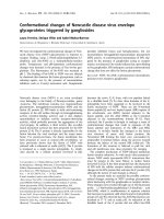
Figure 2.1 Diagrammatic representation of Newcastle disease virus ...................... 4
Figure 3.1 Diagrammatic presentation of procedure of method of study............... 17
Figure 3.2 Photograph showing haemagglutination test ........................................21
Figure 3.3 Photograph showing haemagglutination inhibition test ........................21
Figure 4.1 Percentage of HI titers in different broiler ...........................................24
Figure 4.2 Percentage of HI titers in different colour ...........................................26
Figure 4.3 Percentage of HI titers in different layers ............................................28
Table 3.1 Protocol of the haemagglutination test ...................................................19
Table 3.2 Protocol of the haemagglutination inhibition test ...................................20
Table 4.1 Distribution of NDV antibodies in commercial chickens ......................22
Table 4.2 Comparison of prevalence of antibody to NDV .....................................23
Table 4.3 Distribution of HI titers against NDV in serum samples of broiler .........23
Table 4.4 Comparison of prevalence of antibody to NDV .....................................25
Table 4.5 Distribution of HI titers against NDV in serum samples of colour ........25
Table 4.6 Comparison of prevalence of antibody to NDV .....................................27
Table 4.7 Distribution of HI titers against NDV in serum samples of layers .........27
Table 4.8 Comparison of prevalence of antibody to NDV in ................................ 29
Table 4.9 Comparison of prevalence of antibody to NDV in .................................29
vi
Chapter 1 INTRODUCTION
1.1. Introduction
Poultry industry is an emerging agri - business and has established its position as
fastest growing segment in the agriculture sector of world. Poultry industry is
emerging in Vietnam from seventies. Many small and large scale chicken farms are
rapidly growing in Vietnam. Vietnam is an agriculture country with 70% of population
living in rural and 90% of household keep poultry. The growth of this profitable
subsector is interrupted by a number of infection diseases. Such diseases include
Newcastle disease (ND), Infectious bronchitis (IB) andInfectious bursal disease (IBD) etc.
Newcastle disease is a highly contagious and commonly fatal viral poultry
disease. Newcastle Disease was discovered in Newcastle upon Tyne, England in 1926.
It was widespread throughout the rest of the world. Its spread is normally either via
newly introduced birds, selling or giving away sick and carrier birds. ND is enzootic in
most countries in Africa, Asia and South America, where it is a major constraint
against the development of both industrial and village poultry production
(Aldous and Alexander, 2001). In Vietnam the ND is the most important cause
mortality in chicken (Nguyen, 1992). Every year, ND occurs in many localities and is
responsible for economic losses in poultry industry of Vietnam. These losses will be
due to losses in productivity and death of poultry. Vaccination is practiced widely and
is the recommended method for prevention. Farmers or persons in charge of
vaccination are likely to believe that the chicken flocks will be protected after
vaccination. But apparent ideal ND vaccination programs do not always guarantee
protection of chickens flocks against ND due to incautious handling of vaccines and so
on. So, seromonitoring of humoral immune response in vaccinated chicken flocks is
necessary for controlling the ND (Rahman et al., 2002). Arising from above, I
conducted a research on “Serological Status of Newcastle Disease in commercial
chickens in some provinces of southern Vietnam” with the following objective in
commercial poultry.
1
1.2. Objectives
To determine the serological status of ND in vaccinated chickens.
To investigate the influence of different factors (age, type of chickens, regions)
on NDV antibody prevalence in commercial chickens.
1.3. Performance
Collection of samples from different poultry farms in some regions of southern
Vietnam to perform Haemagglutination test (HA)/ Haemagglutination Inhibition test
(HI) to determine antibody titers against NDV.
2
Chapter 2 LITERATURE REVIEWS
2.1. Newcastle disease
Newcastle disease has been one of the most important diseases of poultry
worldwide ever since the advent of high - density, confinement husbandry systems
(Murphy et al, 1999). It is a highly contagious, generalized virus disease of domestic
poultry and wild birds characterized by gastro - intestinal, respiratory and nervous
signs.
2.1.1. History
ND was the name given to a highly pathogenic disease seen in chickens in
England in 1926 by Doyle. Doyle reported the first outbreak to have occurred, in the
spring of 1926, on the farm near Newcastle - upon - Tyne and hence the name. The
disease had also emerged in March 1926 on the island of Java, Indonesia. Generally it
has been considered that the presence of the virus in England resulted from
transportation to the port of Newcastle - upon - Type from South East Asia by ship,
either in frozen meat or as a result of the practice of keeping live chickens on board for
eggs and meat. The disease also appears to have been present in Korea in 1926. An
outbreak also occurred in Ranikhet in India in July, 1927. What cannot be excluded is
that outbreaks may have been occurred earlier elsewhere but gone unnoticed due to
lack of available expertise in recognizing an apparently new disease. Wherever the
first outbreak occurred it is obvious from the literature that a highly virulent disease of
poultry appeared within a very short time in England, Java, Philippines, India, Ceylon,
Korea and Japan and that the disease was sufficiently different from other highly
virulent disease to be recorded as distinct and recognized as the same disease.
Wherever it began and however it was spread, in 1926 a new disease emerged or was
recognized and within a few years had spread throughout the world (Alexander, 1988).
In Vietnam
In 1956, Nguyen Luong and Tran Quang Nhien had confirmed the presence of
ND in the northern provinces of Viet Nam. Since then, outbreaks have been occurred
yearly causing heavy losses to poultry production.
3
2.1.2. Etiology
Classification of virus
Family
: Paramyxoviridae
Subfamily
: Paramyxovirinae
Genus
: Rubulavirus
Figure 2.1 Diagrammatic representation of Newcastle disease virus
( />Paramyxovirus virions are pleomorphic in shape (spherical as well as
filamentous forms occur), 100 - 300 nm in diameter. Virions are enveloped, covered
with large peplomers, and contain a herringbone - shaped helically symmetrical
nucleocapsid. The genome consists of a single linear molecule of negative - sense,
single - stranded RNA, 15 - 16 kb in size. The envelope contains two glycoproteins,
a haemagglutinin (in most species with neuraminidase activity also) and a fusion
protein. Replication takes place in the cytoplasm, and assembly occurs via budding on
plasma membranes. The viruses have narrow host ranges and have only been
identified in vertebrates, primarily in mammals and birds. Transmission is mainly by
aerosol and droplets (Murphy et al, 1999).
Newcastle disease virus is a member of the family paramyxoviridae in the genus
Rubulavirus. There are nine serotypes of avian paramyxoviruses designated APMV-1
to APMV-9 and ND virus has been designated APMV-1. NDV has also been
4
categorized into five pathotypes based on clinical signs in infected chickens,
designated: a) viscerotropic velogenic, b) neurotropic velogenic, c) mesogenic, d)
lentogenic or respiratory and e) subclinical enteric. Pathotype groupings are rarely
clear - cut.
The virus is inactivated by 56 oC in 3 hours or 60oC in 30 minutes. The virus is
known to be destroyed by 1:5000 potassium permanganate, 1:1000 cresol, 3%
formalin and 1:5 ethanol. It also is inactivated by acid, pH lesser or equal 2. It survives
for long periods at ambient temperature, especially in faces (Chauha and Roy. 2007).
2.1.2.1 Genome of NDV
The genome of NDV codes for six proteins, which have been described by
Samson (cited by Saif et al, 2003) include L protein is the RNA - directed RNA
polymerase associated with the nucleocapsid; HN is responsible for the
haemagglutinin and neuraminidase activities, forming the larger of two types of
projections seen on surface of paramyxovirus particals; F, fusion protein, forms the
smaller of the surface projections; NP, nucleocapsid protein; P, phosphorylated,
nucleocapsid - associated; and M, matrix. The order of the genes for these proteins in
the virus genome is
3’N – P – M – F – HN – L 5’.
2.1.2.2 Important viral proteins and biologic properties
Two proteins of NDV are inserted in the envelope. They are the
haemagglutinin/neuraminidase protein and the fusion protein. These two proteins are
important in determining the virulence of the virus and how the virus infects host cells.
Haemagglutination activity: the ability of NDV and other avian paramyxoviruses
to
agglutinate
red
blood
cells
(RBCs) is
due
to the
binding
of the
haemagglutinin - neuraminidase (HN) protein to receptor on the surface of the RBCs.
This property and the specific inhibition of agglutination by antisera have proven to be
powerful tools in the diagnosis of the disease. Chicken RBCs usually are used in HA
tests, but NDV will cause agglutination of all amphibian, reptilian, and avian cells.
Neuraminidase activity: the enzyme neuraminidase is also part of the HN
molecule and presented in all members of the Rubulavirus genus. An obvious
consequence of the possession of this enzyme is the gradual elution of agglutinated
RBCs. The exact functions of the neuraminidase in virus replication is unknown, but it
5
seems likely that neuraminidase removes virus receptors from the host cell which
prevents the reattachment of released virus particles and virus clumping.
Cell fusion and hemolysis: NDV and other paramyxoviruses may bring about
hemolysis of RBCs or fusion of other cells by essentially the same mechanism.
Attachment at the receptor site during replication is followed by fusion of the virus
membrane with the cell membrane, which may result the fusion of two or more cells.
The rigid membrane of the RBCs usually results in lysis from the virus membrane
fusion. In order for fusion to occur, the shape of the native fusion protein must be
changed. This change happens when a host cell protease cleaves or cuts the protein at a
specific cleavage site. After this has happened, the fusion protein is activated and can
now fuse to the membrane of the cell. The sequence of the amino acids around the
cleavage site determines the range of proteases that can activate cleavage of the
protein. This sequence therefore determines the virulence.
2.1.3. Epidemiology
2.1.3.1. Hosts
The virulence of NDV strains varies greatly with the host. Chickens are highly
susceptible, but ducks and geese may be infected and so few or no clinical signs, even
with strains lethal for chickens.
2.1.3.2. Pathogenesis
In chickens, the pathogenicity of ND is determined chiefly by the strain of virus,
dose, route of administration, age of the chicken, and environmental conditions all
have an effect. In general, the younger the chicken is, the more acute the disease is.
With virulent viruses in the field, young chicken may experience sudden deaths
without major clinical signs; however, in older birds the disease may be more
protracted and with characteristic clinical signs. Breed or genetic stock does not appear
to have a significant effect on the susceptibility of chickens to the disease. Natural
routes of infection (nasal, oral, and ocular) appear to emphasize the respiratory nature
of disease, and intramuscular, intravenous, and intracerebral routes appear to enhance
the neurologic signs.
Initially the virus replicates in the mucosal epithelium of the upper respiratory
and intestinal tracts, shortly after infection, virus spreads via the blood to the spleen
6
and bone marrow, producing a secondary viremia. This leads to infection of other
target organs, lung, intestine, and central nervous system.
2.1.3.3. Sources of NDV
Sources of NDV are respiratory discharge, feces of infected birds and all parts of
the carcass. Virus is shed during the incubation period, during clinical stages and for a
limited period during convalescence. Wild birds and waterfowl may act as reservoir
hosts for lentogenic pathotypes of ND; subsequently, these viruses could become
virulent following mutation upon establishment in domestic poultry. Some psittacine
birds have been demonstrated to shed NDV intermittently for over 1 year and been
associated with introduction into poultry.
2.1.3.4. Spread of NDV
The following virus sources or methods have been implicated in various
epizootics: 1) movement of live birds, racing pigeons, and commercial poultry; 2)
contact with other animals; 3) movement of people and equipment; 4) movement of
poultry products; 5) airborne spread; 6) contaminated poultry feed; 7) contaminated
water; and 8) vaccines. The importance of any of these factors will depend on the
situation in which the epizootic occurs. In countries where poultry are kept exclusively
in bird proof housing, the ability of feral birds to invade affected flocks and transfer
the disease will be minimal, whereas birds kept an open range are more likely to be
infected with strains carried by feral birds.
2.1.4. Clinical signs
The incubation period of ND after natural exposure has been reported to vary
from 2 - 15 days (average 5 – 6 days). The speed with which signs appear, if at all, is
variable depending on the infecting virus, the host species and its age and immune
status, infection with other organisms, environmental conditions, the route of exposure
and the dose.
With extremely virulent viruses, the disease may appear suddenly, with high
mortality occurring in the absence of other clinical signs. In outbreaks in chickens due
to the virulent NDV pathotype, clinical signs often begin with listlessness, increased
respiration, weakness, ending with prostration and death. Green diarrhea is frequently
seen in birds that do not die early in infection, and prior to death, muscular tremors,
7
torticollis, paralysis of legs and wings. Mortality frequently reaches 100% in flocks of
fully susceptible chickens.
The neurotropic velogenic: sudden onset of severe respiratory disease followed a
day or two later by neurologic signs. Egg production falls dramatically, but diarrhea is
usually absent. Morbidity may reach 100%. Mortality is generally considerably lower,
although up to 50% in adult birds and 90% in young chickens have been recorded.
Mesogenic strains of NDV usually cause respiratory disease in field infections.
In adult birds, there may be a marked drop in egg production that may last for several
weeks. Nervous signs may occur but are not common. Mortality in fowl is usually low,
except in very young and susceptible bird, but may be considerably affected by
exacerbating conditions.
Lentogenic viruses do not usually cause disease in adults. In young, fully
susceptible bird, serious respiratory problems can be seen, often resulting in mortality.
Lentogenic strains usually cause mild respiratory disease with coughing, gasping,
sneezing and rales. It can produce more severe symptoms if the flock is co - infected
with other pathogens.
2.1.5. Lesions
There are no pathognomonic gross lesions, several birds must be examined to
determine a tentative diagnosis and final diagnosis must await virus isolation and
identification. Only velogenic strains produce significant gross lesions. Lesions that
may be found include: swelling of periorbital area or entire head, oedema of the
interstitial or peritracheal tissue of the neck. Congestion and sometimes haemorrhages
in the caudal pharynx and tracheal mucosa. Petechiae and small ecchymoses on the
mucosa of the proventriculus, concentrated around the orifices of the mucous glands.
There have oedema, haemorrhages or degeneration of ovaries. Spleen may appear
enlarged,
friable
and
dark
red
or
mottled.
( />WCASTLE_DISEASE__FINAL.pdf)
2.1.6. Immunity
2.1.6.1. Cell - mediated immunity
The initial immune response to infection with NDV is cell mediated and may be
detectable as early as 2 - 3 days after infection with live vaccine strains. This has been
8
thought to explain the early protection against challenge that has been recorded in
vaccinated birds before a measurable antibody response is seen. However, a later study
concluded that the cell mediated immune response to NDV by itself is not protective
against challenge with virulent NDV. The importance of cell mediated immunity in
protection conferred by vaccines is, therefore, not clear, and a strong secondary
response to challenge similar to the antibody response does not seem to occur.
2.1.6.2. Humoral immunity
When chickens survive NDV infection long enough, antibodies usually are
detectable in the serum within 6 - 10 days. The levels largely depend on the infecting
strain, but generally, peak response is at about 3 - 4 weeks. Decline in antibody titer
varies with the titer achieved but is much slower than their development.
Haemagglutination inhibition antibodies may remain detectable for up to one year in
birds recovered from infection with mesogenic viruses.
2.1.6.3. Local immunity
Antibodies appear in secretions of the upper respiratory tract and intestinal tract
of chickens at about the time humoral antibodies can be first detected. In the upper
respiratory tract, the immunoglubulins appear to be chiefly IgA with some IgG.
2.1.6.4. Passive immunity
Hens with antibodies to NDV will pass these on to their progeny via the egg
yolk. Levels of antibody in day - old chicks will be directly related to titers in the
parent. Maternal immunity is protective and, thus, must be taken into account when
timing the primary vaccination of chicks.
2.1.6.5. Immunosuppression
Suppression of the immunoresponse has important effects on both the
pathogenicity of infecting NDV strains and protection levels achieved by vaccination.
Under natural conditions, immunosuppression may occur due to infection with other
viruses such as IBDV. The subsequent immune deficiency may result in a more severe
disease caused by some NDV strains and a failure to respond adequately to
vaccination.
2.1.7. Diagnostics
2.1.7.1. Serology
9
The presence of specific antibody to NDV in the serum of a bird give little
information on the infecting strain of NDV and, therefore, has limited diagnostic
value. Nevertheless, in certain circumstances the demonstration that infection has
taken place is sufficient for the needs of the diagnostician. Postvaccinal serology can
be used to confirm successful application of vaccine and an adequate immune response
by bird. In the absence of vaccination, the presence of specific antibodies against the
NDV indicates that the bird has been infected by the virus at some time, but not
necessarily that it was suffering from the disease at the time of sampling. In practice, a
high antibody titer is indicative of a recent infection. HI is the most commonly used
serological test. Other tests include virus neutralization, ELISA.
2.1.7.2. Virus isolation
Newcastle disease can be diagnosed by isolating APMV-1 from affected birds.
This virus is usually recovered by inoculating samples into 9 – 11 day old
embryonated chicken eggs. Choriolaallantonic fluid from the eggs is tested for
haemagglutinating activity, and any agents that haemagglutinate are examined for HI
with a monospecific antisera to APMV-1.
2.1.7.3. Pathogenicity tests
The pathogenicity of the isolate can be quantified by 1) the mean death time
(MDT) in chicken embryos, 2) the intracerebral pathogenicity index (ICPI) in
one - day chicks, or 3) the intravenous pathogenicity index (IVPI) in six - week old
chickens. In the MDT assay, velogenic isolates have an MDV of less than 60 hours,
mesogenic strains have an MDV of 60 – 89 hours, and lentogenic viruses have an
MDT greater than 90 hours. The ICPI and IVPI tests are scoring systems that evaluate
illness or death in chickens. The values in the ICPI test range from 0 to 2.0, the most
virulent viruses approach 2.0, while lentogenic strains are usually close to 0.0. The
values in the IVPI test are from 0 to 3.0, the velogenic strains approach 3.0, while
lentogenic strains and some mesogenic strains have IVPI values of 0.0. However,
some viruses that can produce severe disease have IVPI values of 0.0, the ICPI test is
generally preferred for this reason.
2.1.7.4. Molecular techniques
10
Reverse - transcription polymerase chain reaction (RT-PCR), gene sequencing,
restriction enzyme analysis and other molecular techniques are also used to identify
APMV-1 in eggs or clinical specimens.
2.1.8. Prevention
2.1.8.1. Vaccination
Inactivated vaccines are produced by growing a NDV in eggs, and then treating
the infective allantoic fluid with an inactivating agent, such as formalin or
betapropiolactone. An adjuvant, such as mineral oil, is usually then added to make the
inactivated virus more immunogenic. Since the vaccine is no longer capable of
replication or spread, it has to be injected individually into every bird needing
vaccination. It is normally injected into the back of the thigh muscle (sometimes the
breast muscle is used), using 0.3 or 0.5 ml per bird. Inactivated vaccines produce very
high levels of antibodies against NDV, and provide good protection against the
virulent virus.
In intensive poultry production, inactivated vaccines are usually applied after an
initial priming vaccination with a live vaccine. Although inactivated vaccine gives
good protection, it is relatively expensive to produce. It also carries a slight risk to the
user of accidental self - injection. While inactivated vaccines are, to some extent, heat
sensitive, they are much less so than conventional live vaccines which makes
transporting them to villages more feasible.
Live vaccines differ from inactivated vaccines in that they can replicate in the
host. This is both an advantage and a disadvantage. It is an advantage in that it is not
necessary to vaccinate every bird individually; the vaccinal virus can spread on its own
from one bird to another. It is, however, a disadvantage in that, since an infection with
a live virus is involved, this may result in clinical signs because of the innate virulence
of the vaccine virus or by exacerbating other organisms that may be present, especially
in the respiratory tract. The severity of this reaction depends therefore on the particular
vaccinal strain used and the presence or otherwise of concurrent infection with other
pathogens. Another advantage of live vaccines compared to inactivated vaccines, is
their ease of application as they can be applied to the drinking water or with an
eye - dropper.
11
Although NDV has essentially only one serotype, there is a wide difference in
the pathogenicity of different strains, ranging from those that cause virtually no signs
to those that kill within a few days. The majority of live vaccines are derived from
asymptomatic enteric or lentogenic strains, although some vaccines derived from
mesogenic strains are still in use. Live lentogenic vaccines are usually derived from
field viruses that have been shown to have low pathogenicity for poultry but produce
an adequate immune response. Typical vaccine strains are HB1, LaSota and F strain
and some viruses from the asymptomatic enteric pathotype, which are usually based
on the V4 or Ulster 2C viruses. And mesogenic vaccine such as Roakin, Mukteswar
and Komarov. However, these viruses have been frequently subjected to selection
pressures by manufacturers in order to improve their immunogenicity or to enable their
use by a particular method of application.
2.1.8.2. Biosecurity and hygiene
On commercial farms, control measures should attempt to prevent viruses from
infecting the flock. Biosecurity aimed at preventing disease should begin at the
planning stage of commercial poultry farms. Farms and flocks should be well
separated, hatcheries should be isolated from poultry farms, different species should be
reared on different sites, and there should be an adequate fresh water supply,
preferably one that does not draw on surface water. On the farms the following points
should be observed:
• Houses, food stores and water tanks should be bird - proofed.
• Movements on and off the farm should be kept to a minimum.
• All equipment, especially vehicles, should be disinfected before access to the
site is permitted.
• Movements between different farms for egg collection, carcass collection, food
delivery and the like should be confined to a specified collection and delivery point
away from the poultry flocks.
Visits by personnel such as bleeding or vaccination teams, inseminators and
veterinarians are the most likely method of introduction of ND and if such visits are
unavoidable, regimens of clothing change, equipment disinfection and other basic
hygiene controls must be enforced.
2.1.9. Zoonosis
12
NDV
is
a
human
pathogen.
Reported
infections
have
been
non - life - threatening and usually not debilitating for more than a day or two. The
most frequently reported and best substantiated clinical signs in human infections have
been eye infections, usually consisting of unilateral or bilateral reddening, excessive
lacrimation, oedema of eyelids, conjunctivitis and subconjunctival haemorrhage.
Although the effect on the eye may be quite severe, infections are usually transient and
the cornea is not affected. Reports of other clinical symptoms in humans infected with
NDV are less well substantiated but suggest that a more generalized infection may
sometimes occur resulting in chills, headaches and fever, with or without
conjunctivitis. There is evidence that both live vaccine and virulent (for poultry)
strains of NDV may infect and cause clinical signs in human. Human infections with
NDV have usually resulted from direct contact with the virus, infected birds or
carcasses of diseased birds. There have been no reports of human to human spread,
although spread by contagion is theoretically possible. The types of person known to
have been infected with NDV include: laboratory workers (usually as a result of
accidental splashing of infective material into the eye), veterinarians in diagnostic
laboratories (presumably as a result of contact with infective material during
post - mortem examinations), workers in broiler processing plants and vaccination
crews when live vaccines are given as aerosols or fine dust (Mark Pattison, 2008).
2.2. Hemagglutination and haemagglutination inhibition test
2.2.1. Haemagglutination
The ability of suspensions of virus to agglutinate erythrocytes was first observed
with influenza virus, and the reaction involved has been extensively studied with this
virus and other members of myxovirus group. In the myxoviruses the reaction with the
erythrocyte differs from that of some other viruses because most members of the group
possess the enzyme neuraminidase, and the virion can be eluted, by mean of it, from
the receptors on the erythrocyte surface. Besides being of important from the purely
practical point of view, the phenomenon allows the study of the interaction of virus
with the surface of the cell in which it does not multiply, and can also be used for
serological identification of viruses and for titration of antibody by means of the
haemagglutination - inhibition phenomenon. The condition for haemagglutination,
13
e.g., pH and temperature, may be very critical, and the species of the red blood cell is
important.
After adsorption and elution of the virus, the red blood cells have a reduced
electrophoretic mobility and decreased negative charge on the surface. They are
inagglutinable by the same myxovirus as before and usually by some others. By
heating at 56oC under various conditions, the enzyme can be inactivated without
greatly affecting the ability to hemagglutinate. Such virus does not elute from
erythrocytes
and
is
known
as
indicator
virus.
Inhibitors
of
myxovirus
haemagglutination are mostly themselves mucoproteins and behave like the
mucoprotein on the surface of erythrocytes, they adsorb onto the virus surface, and
elute from it by the action of viral neuraminidase. There inhibators are nonspecific in
that they can act on a much wider variety of myxoviruses than do antibody, which are
highly strain - specific, even though some cross - reactions occur. This specificity is
the basis of haemaglutination - inhibition test (Betts and York, 1967).
2.2.2. The haemagglutination inhibition test
The HI test is based on the principle that the haemagglutinin on the viral
envelope can bring about the agglutination of chicken red blood cells and that this can
be inhibited by specific antibodies. U - bottomed microtitration plates are used. The
serum samples are diluted in serial twofold dilutions in phosphate buffered saline and
then a fixed quantity of viral antigen is added to each well. Following incubation, a
suspension of red blood cells is added to each well and the plate is incubated again. In
the absence of any antibody against the virus, haemagglutination occurs, appearing as
a diffuse red colour at the bottom of the well. In the wells where the antibody against
the virus is of a sufficient level, haemagglutination is inhibited and the red blood cells
sediment and appear as a small pellet at the bottom of the well. The presence or
absence of agglutination is accurately assessed by tilting the plates. Only those wells in
which the RBCs stream at the same rate as the control wells (containing RBCs and
PBS only) should be considered to show inhibition. The HI titer is the reciprocal of the
highest dilution of serum which completely inhibits haemagglutination and is usually
and most conveniently expressed as the logarithm to the base 2. Although the test is
difficult to standardize between laboratories, the HI titer gives an indication of the
immune status of the bird. A titer of log23 is indicative of protection and a titer of
14
log26 or more suggests a recent infection by the virus. If no vaccination has taken
place, diagnosis of the infection can be made on this basis, although it cannot be
determined exactly when it took place.
For most avian sera a positive HI titer may be regarded as 1/16 if 4 HAU of
antigen are used and 1/8 if 8 HAU are used (titers are more usually expressed as the
reciprocal of the end - point dilution in several forms of notation, i.e. 8, 23, log23). In
unvaccinated birds, positive serology and clinical signs may be considered as strong
diagnostic evidence of the disease. HI and other tests may be used to measure the
immune status of vaccinated birds. Mean levels expected following vaccination range
from 24 - 26 after a single live vaccine to 2 9 - 211 with multidose programmes, including
oil - emulsion inactivated vaccines (Mark Pattison, 2008).
2.3. Studies about prevalence of antibody against NDV in vaccinated chickens
According to Mozaffor et al., 2010, in a serological survey on the prevalence of
antibody against NDV was carried out in broiler and layer chickens in Rajshahi and
surrounding districts of Bangladesh, seroprevalence of antibody against NDV was
found significantly higher in young than in adults.
Banu et al., 2009 conducted a experiment for the detection of persistence of
Maternally derived antibody (MDA) as well as the comparative evaluation of antibody
production of nine different NDV vaccines in layer chickens and from his research it
was concluded that LaSota strain produced higher immune response than Clone 30 and
B1 strain, Fortdose® and Avipro® vaccine produced higher immune response than all
other vaccines and vaccination with lentogenic strains followed by mesogenic strain
produced higher antibody titers used in this study.
Al-Zubeedy, 2009 reviewed in one day old broiler chicks from four groups of
broiler breeder chickens were immunized by different routes of vaccination and
concluded that humoral immunity to NDV is a key component in the protection
against ND. Therefore, vaccination programs should be directed toward eliciting and
maintaining high antibody level to NDV in flocks of birds.
Shuaib et al., 2006 conducted a research to study humoral immune response to
Newcastle disease vaccine (Lasota strain) in broilers and investigated that significant
increase in HI titer against ND virus was observed in broilers from single to boostered
vaccination group.
15
A serological survey on the prevalence of antibodies to NDV was carried out in
broilers and layers in Faisalabad and surrounding districts by Numan et al., 2005. A
total of 803 serum samples were collected form broiler farm, layer farms and slaughter
shops. HI test was performed to determine the serum antibodies against NDV. The
results showed that the level of protection in vaccinated birds was unsatisfactory in
broilers whereas it was satisfactory in layers.
Vui et al., 2002 carried out a serological survey on the prevalence of antibody to
NDV, IBDV, AIV in two communes of the Hue District, middle in Vietnam. The
results showed that both ND vaccinated and unvaccinated birds in the 2 - 6 month - old
age group had a significantly lower NDV antibody titer than the greater 6 month - old
age group.
Poultry sector has strong presence in Southern Viet Nam. Commercial and
backyard poultry are in two major sectors. The commercial poultry sector has layer
and broiler which are growing enterprise. New castle disease is an important disease of
chickens. During the last few decades, the disease has increased in severity, and the
virus has evolved to greater virulence. The ND was a major disease problem and
source of great economic losses in poultry. Commercial poultry farms in different
districts of Southern Vietnam were examined for to study the prevalence of antibody
titer against Newcastle Disease during the period of February to June 2011.
16
Chapter 3 MATERIALS AND METHODS
3.1. Time and place to do thesis
Time: From February to June, 2011
Place: In Venky’s Poultry Disease Diagnostic and Nutrition Laboratory at
Research Institute for Biotechnology and Environment, Nong Lam university.
3.2. Materials
3.2.1. Subject
Blood samples were collected from vaccinated chickens in poultry farms Vung
Tau, Dong Nai, Binh Duong province of southern Vietnam.
3.2.2. Equipment
U bottom microtiter plate (96 well), measuring cylinder, centrifuge tube,
centrifuge machine, syringe, eppendorf tube, single and multichannel pipettes and tips,
glass Pasteur pipette, autoclave, refrigerator, distilled water machine and weighing
balance.
3.2.3. Reagents
Phosphate buffered saline (PBS)
Sodium chloride (NaCl)
:8g
Potassium chloride (KCl)
: 0.2 g
Disodium hydrogen orthophosphate anhydrous (Na2HPO4) : 1.076 g
Potassium dihydrogen orthophosphate (KH 2PO4)
: 0.2 g
Distilled water
: 1000 ml
Autoclave at 121oC for 15 minutes.
Normal Saline
Sodium chloride (NaCl)
: 8.5 g
Distilled water
: 1000 ml
Adjust pH at 7.2
Autoclave at 121oC for 15 minutes.
Alsever’s Solution (Anticoagulent)
Dextrose
: 20.50 g
Citric acid
: 0.550 g
17