Molecular sieves vol 1 5 karge weitkamp vol 2 structure and structure determination 1999
Bạn đang xem bản rút gọn của tài liệu. Xem và tải ngay bản đầy đủ của tài liệu tại đây (2.01 MB, 157 trang )
Preface to Volume 2
Once a new natural zeolite is found or a new molecular sieve synthezised, via one
or the other of the methods described in Volume 1 for example, the researchers
face the task of confirming that a novel structure has come into their hands.
However, beyond this basic problem, questions soon arise concerning rather
detailed and subtle structural features.
The classical method of determining crystal structures is X-ray diffraction.
Thus, in Chapter 1 of the present volume, H. van Koningsveld and M. Bennett
provide the reader with information about the enormous progress which has
been made in X-ray structure analysis of zeolites. To a large extent, this is due to
outstanding developments in both experimental techniques and methods of
data evaluation, such as the application of synchrotron radiation and Rietveld
analysis. New methods now enable crystallographers to study very small single
crystals or crystallite powders. This is extremely important with respect to most
of the synthetic micro- and mesoporous materials since the size of primary particles is usually in the µm range. The authors stress that, in the context of
reliable structure analysis, the determination of the unit cell and space group is
of paramount importance. Modern tools now allow researchers to study subtle
effects on zeolite structures such as those caused by framework distortions,
dealumination, isomorphous substitution or cation and sorbate location.
The study of structures containing light atoms is the particular domain of
neutron scattering, even though this is not its only advantage. The authors of
Chapter 2, A.N. Fitch and H. Jobic demonstrate the way in which neutron scattering is able to complement structure analysis by X-ray diffraction. In particular,
neutron scattering techniques reveal their strong potential in probing details of
structural arrangements involving hydrogen-containing species (such as water
and hydroxyl groups) as well as determining hydrogen bonds, cation positions,
and the location of adsorbed molecules. Frequently these techniques are successfully used for further refinement of X-ray diffraction data.
Chapter 3, written by O. Terasaki, is devoted to the use of the various kinds of
electron microscopy in the investigation of zeolites and related porous solids.
The author’s contribution focuses on the potential of electron microscopy in
studying crystallite morphologies as well as features of the fine structure, e.g.,
bulk and surface defects; details of the crystal surface (edges and kinks), and, as
such, related to crystal growth; and modification of frameworks. Moreover, the
valuable assistance of electron microscopy in solving new structures is illustrated by a number of examples.
VIII
Preface to Volume 2
Chapter 4 is contributed by W. Depmeier, and it concerns particular phenomena of the structures of zeolites and related solids which are attracting more
and more interest. Such phenomena are, inter alia, phase transitions as well as
mechanisms of reduction in symmetry and volume as a consequence of tilting,
distortion of the whole framework or framework units, modulations of the
framework, and partial amorphization. These are demonstrated by a variety
of instructive examples, and their importance is pointed out in view of, for
example, catalytic, shape selective and separation properties of zeolite materials.
General problems of zeolite structures are dealt with in Chapter 5 which is
jointly authored by W.M. Meier and C. Baerlocher. It includes basic aspects of
zeolite crystallography such as topology, configuration, and conformation of
framework structures. Similarly, the idea of distinguishing zeolites on the basis
of framework densities is presented. The attempts at classification of zeolite
structure types are critically discussed. The authors then describe the interesting concepts of structural characterization via loop configurations and coordination sequences and also reconsider the long-standing question of whether
zeolite framework structures are predictable.
This volume concludes with Chapter 6, a review devoted to industrial synthesis. Contributed by A. Pfenninger and entitled “Manufacture and Use of Zeolites
for Adsorption Processes”, this chapter provides an extremely useful adjunct to
Volume 1 of this series. Important aspects of industrial synthesis are described
and, simultaneously, the characterization and use of zeolites for separation processes are discussed. In these respects, Chapter 6 is something of an introduction
to matters which will be extensively dealt with in Volume 5 (Characterization II)
and Volume 7 (Sorption and Diffusion) of this series.
The originally planned final chapter on the role played by solid state NMR
spectroscopy in the elucidation of structural features of microporous and mesoporous materials was unfortunately not available at the time of going to press.
However, given the importance of this topic, an appropriate treatment of this
area is intended to appear in Volume 4 (Characterization I).
Thus,Volume 2 presents an extended overview over most of the relevant techniques currently employed for investigations into structural properties of
micro- and mesoporous materials and offers in its last contribution a valuable
addition to the topics treated in Volume 1. From this volume it becomes evident
that the various techniques for structure determination are, to a large extent,
complementary and that evaluation of the experimental data, on the other hand,
is profiting much from recent developments in theory and modeling. It is the
Editors’ hope that Volume 2 of the series “Molecular Sieves – Science and
Technology” will provide the researchers in the field of zeolites and related
materials with the necessary awareness of the great potential in modern
methods for structure analysis.
Hellmut G. Karge
Jens Weitkamp
Contents
H. van Koningsveld and J.M. Bennett:
Zeolite Structure Determination from X-Ray Diffraction
. . . . . . . .
1
. . . . . . . . . . . .
31
O. Terasaki:
Electron Microscopy Studies in Molecular Sieve Science . . . . . . . . .
71
W. Depmeier:
Structural Distortions and Modulations in Microporous Materials . . . .
113
W.M. Meier and C. Baerlocher:
Zeolite Type Frameworks: Connectivities, Configurations
and Conformations . . . . . . . . . . . . . . . . . . . . . . . . . . . . . .
141
A. Pfenninger:
Manufacture and Use of Zeolites for Adsorption Processes . . . . . . . .
163
Subject Index . . . . . . . . . . . . . . . . . . . . . . . . . . . . . . . . .
199
Author Index Vols. 1 and 2 . . . . . . . . . . . . . . . . . . . . . . . . . .
215
A.N. Fitch and H. Jobic:
Structural Information from Neutron Diffraction
Zeolite Structure Determination from X-Ray Diffraction
H. van Koningsveld 1 and J. M. Bennett 2
1
2
Laboratory of Organic Chemistry and Catalysis, Delft University of Technology,
Julianalaan 136, 2628 BL Delft, The Netherlands; e-mail:
661 Weadley Road, Radnor, PA 19087, USA; e-mail:
1 Introduction . . . . . . . . . . . . . . . . . . . . . . . . . . . . . . . . .
1
. . . . . . . . . . . . . .
3
3 Incorrect Determination of the Space Group . . . . . . . . . . . . . . .
5
4 Effect of Framework Flexibility . . . . . . . . . . . . . . . . . . . . . . .
8
2 Severe Overlap of Reflections in Powder Data
5 Disorder of Non-Framework Species
. . . . . . . . . . . . . . . . . . . 16
6 Faulting within the Framework . . . . . . . . . . . . . . . . . . . . . . . 23
7 Isomorphous Replacement of Framework Atoms . . . . . . . . . . . . . 24
8 Crystal Size Limitations . . . . . . . . . . . . . . . . . . . . . . . . . . . 25
9 Conclusions
. . . . . . . . . . . . . . . . . . . . . . . . . . . . . . . . . 25
References . . . . . . . . . . . . . . . . . . . . . . . . . . . . . . . . . . . . 26
1
Introduction
Zeolites and related microporous materials are a class of materials with an ever
widening range of compositions, structures and uses. Since the earliest days of
zeolite science X-ray diffraction has been one of the basic and most useful tools
for characterization.
Initially X-ray diffraction was used to answer simple questions such as: “have
I made a new material?” or:“has the crystallization process gone to completion?”
Now the questions encompass everything that a researcher might want to know
about the structure of a material. Early attempts at determining crystal structures using X-ray diffraction were often unsuccessful because many of these early synthetic materials were available only as powder samples. Fortunately many
of these first synthetic materials had natural counterparts with large single
crystals, and data from these were used to determine the framework structures
Molecular Sieves, Vol. 2
© Springer-Verlag Berlin Heidelberg 1999
2
H. van Koningsveld · J.M. Bennett
of their synthetic counterparts. Today, the framework of a new material can be
often determined from powder samples. In addition, single crystal techniques
have improved considerably leading to increased accuracy in the bond angles
and bond distances and to the ability to study crystals of much smaller size. It is
now possible for a single crystal study to reveal details of the structure that show
the interaction of a sorbed material with the framework or movement of cations
within the framework and any ensuing distortions of the framework. Structural
data from powder samples are beginning to reveal similar changes in the crystal
structure with temperature, with sorbed materials and even under catalytic
conditions. Even though the technique of X-ray powder diffraction has improved greatly since the early days of zeolite science, it is still more accurate to
determine the crystal structure of a new material from single crystal data rather
than from powder data.
Many of the advances in the structural information derived for zeolitic materials are a direct result of major improvements in powder and single crystal
X-ray equipment available, in the development of new structure determination
methods and in the use of new characterization tools including magic angle
spinning NMR, neutron diffraction and electron microscopy, which are described in subsequent chapters. Two excellent review papers [1, 2] discuss the
use of X-ray diffraction techniques to study zeolites and the problems encountered, and it is recommended that they be used in combination with this
chapter.
The stages in determining the crystal structure of a material have been
described as: (i) obtain a suitable sample, (ii) collect the data, (iii) determine a
trial structure using ab initio methods, and (iv) refine the data.
However, with zeolites it is not as simple as the above infers since subtle
changes in the zeolite framework can influence, to a greater or lesser extent,
both the observed intensities and the symmetry. These subtle changes in the
observed intensities and the symmetry can cause serious problems for crystallographers performing a zeolite structure analysis. The crystallographic problems
include:
– Severe overlap of reflections in powder data leading to problems with the
techniques used to decompose the peaks into individual reflections
– Incorrect determination of the space group especially when the true symmetry is masked by pseudo-symmetry
– The effect of framework flexibility on the structure analysis
– Disorder of the non-framework species and its effect on the structure
solution
– Faulting within the framework
– Problems caused by isomorphous replacement of framework atoms
– The effects due to small crystal size and the limits on the crystal size that can
be used
In order to help those in the zeolite community to better appreciate the beauty
of an excellent crystallographic study while learning to evaluate the pitfalls that
are present in an incorrect study, several structures, published in the last decade
and that are examples of the problems listed above, will be reviewed.
Zeolite Structure Determination from X-Ray Diffraction
3
2
Severe Overlap of Reflections in Powder Data
For a single crystal structure determination one crystal is chosen from the
sample and it is assumed that the chosen crystal is both suitable for the study
and typical of the bulk material. Often several crystals have to be evaluated before a “good” crystal for the study is found. In contrast, it is relatively easy to
obtain a sample for a powder study and to use a synchrotron source to obtain the
best data. Synchrotron X-ray data are high intensity and high resolution data
and, as such, are far superior to in-house data. The improvements in the quality
of the data obtained from the synchrotron have reduced the magnitude of the
problems that plagued early attempts at structure determination. However,
there is still only one dimensional intensity information in the powder pattern
and it is not a trivial task to determine the correct three-dimensional unit cell
dimensions especially if a few weak peaks from an unknown impurity phase are
present.
A successful structure determination starts with a set of accurately determined peak positions. Unfortunately, this task is often left to the computer with
disastrous results. With carefully deconvoluted data the currently used indexing
programs [3, 4] often yield a number of equally probable answers. When combined with even partial unit cell information from electron diffraction, it is
usually possible to reduce this number to one or two unit cell sets. If no other
data are available then the wrong choice between two equally probable unit cells
may prevent the structure from being accurately determined. Even when a unit
cell is derived it may later prove to be “incorrect” (too highly symmetric) once
the structure has been refined. Unfortunately, the only way to know that a
chosen unit cell is correct is to solve the crystal structure.
Table 1 lists part of the data obtained from a new material. It was known from
TEM/SEM studies that the synthesis product was impure and that the impurity
was an offretite material based on observed d-spacings and a knowledge of the
synthesis conditions. These offretite peaks were removed from the data before
using the indexing programs. However, the best unit cell obtained did not index
all the reflections suggesting that there might be three phases present in the
sample which seemed unlikely. The final solution used several common reflections (such as that at 2 q = 9.958∞) that came from both the offretite impurity and
the new phase and indexed all 60 observed reflections, out to a d spacing of
3.04 Å. The only difference between the first and final unit cell solutions was the
value for the c dimension. The number of un-indexed reflections now became
zero (see Table 1). Thus it is very important to account for all observed peaks in
a pattern even those assigned to other phases and to review even small differences between the observed and calculated 2q values, in order to be sure that
the calculated unit cell dimensions are reasonable.
In order to determine the crystal structure, the intensity of the exactly or partially overlapping reflections are usually separated by a number of simple techniques such as splitting them fifty-fifty. However, these structure determinations were often unsuccessful and more sophisticated methods were developed
to partition the intensity of the overlapping reflections.
4
H. van Koningsveld · J.M. Bennett
Table 1. A partial list of the observed and calculated 2q values for a new phase with an
offretite impurity a
2q Values
Observed
2.402
4.142
4.784
6.606
6.338
7.189
8.302
9.595
9.958
10.244
10.463
10.793
11.451
11.815
12.002
12.295
12.478
12.704
12.980
13.369
13.843
Calculated
Solution 1b
hkl
Final solution c
hkl
100
110
200
1 0 0 (Off.)
210
300
220
400
0 0 1 (Off.)
001
320
U
1 1 0 (Off.)
U
500
U
330
420
U
510
U
100
110
200
1 0 0 (Off.)
210
300
220
400
0 0 1 (both)
101
320
111
1 1 0 (Off.)
211
500
301
330
420
221
510
401
Difference
2.396
4.150
4.792
0.007
0.008
0.009
6.341
7.191
8.305
9.593
9.957
10.243
10.456
10.791
0.003
0.002
0.003
–0.002
–0.001
–0.001
–0.007
–0.002
11.813
11.999
12.293
12.471
12.701
12.980
13.367
13.843
–0.002
–0.003
–0.002
–0.007
–0.003
0.000
–0.002
0.000
Off. indicates an offretite reflection and U an unindexed reflection
Personal communication, Smith W, Bennett JM.
b Solution 1 had a = 36.147(3) and c = 7.329(1) Å.
c Final correct solution had a = 36.150(2) and c = 7.541(1) Å.
a
The use of Direct Methods in determining a weighting scheme for partitioning the intensities was developed by Jansen, Peschar and Schenk [5, 6]. The
method was tested on a structure containing 22 atoms in the asymmetric unit
cell; of the 527 observed reflections, 317 overlapped within half of the peak full
width at half the peak maximum (FWHM) as determined in the fitting process
[7]. Estermann et al. [8] described the structure determination of SAPO-40
(AFR) 1 using a different method for partitioning the intensities of the overlapping reflections. This Fast Iterative Patterson Squaring (FIPS) method indicated
how to partition the intensity and only after this redistribution did an ab initio
structure determination become possible.
Yet another method was applied to the structure determination of VPI-9
(VNI; [9]). This method uses a set of random starting phases for the intensities
1
The arrangement of the tetrahedral atoms in most of the zeolite structures is indicated by a
three letter code. This code is independent of the composition of the zeolite, the space group
and symmetry. A full list of all currently assigned codes can be found in the ‘Atlas of Zeolite
Structure Types’ by W.M. Meier, D.H. Olson and Ch. Baerlocher, Fourth Revised Edition,
published on behalf of the Structure Commission of the International Zeolite Association by
Elsevier, London, Boston, 1996.
Zeolite Structure Determination from X-Ray Diffraction
5
obtained from the powder pattern and is then combined with a topological
search routine in the Fourier recycling procedure. With this method both chemical and structural information are incorporated into the partitioning procedure used for the powder diffraction profile. With seven crystallographically
unique tetrahedral sites, VPI-9 is the most complex framework arrangement
currently solved from powder diffraction without manual intervention.
Since one-dimensional intensity data from powders is resolved into threedimensional intensity data for single crystals, the problem with obtaining individual intensity data is not present with single crystal data. Therefore, the determination of the unit cell and symmetry is less difficult. Using the correct unit
cell dimensions the intensities of all the single crystal reflections can be measured without serious overlap in most cases.
The lack of individually measured reflections with powder data also has a
detrimental effect on the structure determination and refinement procedure. In
powder diffraction the ratio between the number of observations and the number of parameters to be refined is very often less than or equal to one. However,
with single crystal data this ratio usually ranges from three to ten. This over
abundance of data allows an incomplete, or even partly wrong starting model to
be used to yield a successful solution and final refinement of the structure. A
recent example, illustrating the difference between powder and single crystal
data, is the structure determination of GaPO4(OH)0.25 (–CLO; [10]). Even with
high-resolution synchrotron powder data, 552 of the first 617 reflections have
exact 2q overlaps. This extreme example of the overlap of the individual intensity data could not be overcome until a large single crystal became available for
conventional analysis. Then 2776 independent reflections were measured and
the refinement converged smoothly.
3
Incorrect Determination of the Space Group
Space groups are determined from a list of hkl reflections that are not observed.
This is very difficult with powder data because of the occurrence of overlapping
reflections. Without a space group no crystal structure solution can be completed. However, in many cases it is not necessary to determine the space group that
will result from a successful structure refinement. It is often only necessary to
determine the starting space group that defines the maximum symmetry of the
topology (maximum topological symmetry). For example, it is not necessary to
differentiate between the tetrahedral aluminum and phosphorus atoms in a
microporous aluminophosphate material in order to determine the correct framework topology. Fortunately, there have been found to be only a small number
of maximum topology space groups that are applicable; some of them are C2/m,
Cmcm, I41/amd and P63/mmc. Since the choice of unit cell dimensions will affect
the systematic absences and ultimately the space group, this knowledge of applicable space groups can be helpful when choosing between two different, but
equally possible, unit cells. However, it must be remembered that the space
group chosen must account for all of the low hkl systematic absences.
6
H. van Koningsveld · J.M. Bennett
There are many different techniques used by crystallographers to arrive at the
starting topology of a new material. All techniques, except model building, require that the space group be correctly determined. However, this very important
step of determining the starting topology is often not adequately reported, possibly because it is the most time consuming step of a powder structure determination. It is possible to spend months to years determining the correct topology
which, when determined, can lead to spending only days to weeks on the final
refinement. The powder pattern of the proposed topology can be simulated after
refinement of the interatomic distances using a Distance Least Squares (DLS)
refinement [11] procedure and can then be compared to the experimental pattern of the material. Even when there is a passable match between the observed
and simulated powder patterns it does not mean that the proposed framework
arrangement is correct. Probably, any partially incorrect topology can be refined
with the Rietveld technique [12] to yield an apparently acceptable solution.
ZSM-18 (MEI; [13]) is the only aluminosilicate zeolite that has been reported
to contain a three tetrahedral atom ring (a T3-ring)2. However, similar framework structures, such as MAPSO–46 (AFS; [14]), CoAPO–50 (AFY; [14]) and
beryllophosphate–H (BPH; [15]), do not support this novel arrangement. An
examination of the reported framework topology shows that the three ring
arrangements can be replaced by a vertical SiOSi unit with practically no change
in the positions of the remainder of the framework atoms. Lowering the symmetry by removing the six-fold axes and changing to orthorhombic symmetry
allows the framework to rotate off the original six-fold axis thereby reducing the
vertical SiOSi bond angles of 180∞, which are undesirable but observed in the
proposed structure. Unfortunately, any DLS refinement of an orthorhombic
arrangement always refines back to a pseudo six-fold axis. The final answer to
the question of whether ZSM-18 contains three rings will require a complete
structure determination using powder data and consideration of the possibility
that the original space group used to determine the structure was incorrect.
A postulated framework arrangement based on a DLS refinement should
always be treated with suspicion because very few DLS refinements use the full
symmetry of the chosen space group since the only symmetry operations needed are those that generate bonds that lie across the asymmetric unit cell boundaries. In addition, there is always the possibility that the space group chosen is
incorrect and that therefore the final structure is incorrect as well. Several correct structures have been refined in two or more space groups and illustrate that
there are subtle changes in the framework topology depending on the choice of
space group [16].
An example showing that the observed distortions of the framework are
dependent on the choice of the space group is given by the refinement of SAPO40 (AFR; [17, 18]). The ordering of aluminum and phosphorus in the structure
required that the c-axis be doubled and the space group be changed from
orthorhombic Pmmm to monoclinic P112/n. Subsequently, it was realized that
2
The standard method used to describe the number of atoms in a ring of a zeolite structure
is to only count the tetrahedral (T) atoms. Thus a three ring opening would have three silicon atoms and the interconnecting three oxygen atoms for a total of six atoms.
Zeolite Structure Determination from X-Ray Diffraction
7
this doubling generated c-glide planes and that the correct space group was
actually orthorhombic Pccn. This change reduced the number of variables from
186 (for P112/n) to 95 (for Pccn) without affecting the quality of the profile fit. In
addition many of the distances and angles, which were different in the P112/n
refinement, become equivalent in Pccn. From a practical point of view it is very
difficult to say which refinement yields a truer picture of the material and what
effect the framework distortions will have on the material properties.
Sometimes the question of how material properties are affected by changes in
the framework can be answered. In the case of VPI-5 (VFI; [19]) the recognition
that octahedral aluminum is present in the structure of VPI-5 required that the
symmetry be lowered from P63cm to P63. Only after this symmetry change did
the refinement of the structure proceed smoothly. The presence of a triple helix
of occluded water molecules became evident because these water molecules
were required to complete the octahedral coordination of half of the aluminum
atoms in the fused 4-rings. The same octahedral coordination of aluminum was
postulated for AlPO4H2 (AHT; [20]), since both structures contain a triple
crankshaft chain with fused 4-rings. Once the similar octahedral configuration
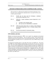
was shown to be present, it was suggested that these octahedral distortions on
the aluminum sites promote the reconstructive phase transition of VPI-5 to
AlPO4-8 (AET) above room temperature and of AlPO4H2 to tridymite (Fig. 1) at
higher temperatures. The phase transition of AlPO4H2 to tridymite is irrever-
a
b
Fig. 1a, b. Schematic illustration of the framework transformation of a VPI-5 to AlPO4-8 and
b AlPO4-H2 to AlPO4-tridymite. Large dots indicate Al positions. Reproduced by permission
of the Royal Society of Chemistry from [20]
8
H. van Koningsveld · J.M. Bennett
sible; conflicting reports exist as to whether the transition of VPI-5 to AlPO4-8 is
reversible or not [21–24].
4
Effect of Framework Flexibility
The difficulty in determining zeolite structures from diffraction data is increased when there are changes in cell dimensions and/or symmetry caused by the
framework flexion in response to having different cations or other non-framework species present. The TO4 tetrahedra are rigid but interconnected through
oxygen atoms which act as flexible hinges [25]. In collapsible frameworks, such
as ABW, GIS, NAT, RHO and SOD, all angles around the TO4 tetrahedra co-rotate
in the same sense when cell dimensions and volumes change. The frameworks
wrap themselves around occluded non-framework species or collapse until the
smallest angle of the TOT hinges (~126∞) is reached [26]. MFI and MEL are also
collapsible frameworks but do it through a shearing of the pentasil layers parallel to the crystallographic c axis [27]. In non-collapsible frameworks, such as
LTA, FAU and KFI, the TOT hinges rotate in opposite directions when the cell
dimensions and volume change. The frameworks are very flexible at intermediate values of the cell dimensions [28].
The distortions observed in the collapsible framework structures MAlSiO4
(ABW) depend on the exchangeable cation M (where M is Li, Na, K, Rb, Cs, Tl or
Ag) [29]. Even though it is theoretically possible to double the unit cell volume
(Fig. 2), the space group usually remains Pna21 even with structures such as
LiGaAlSiO4 · H2O [31], LiBePO4 · H2O, LiZnPO4 · H2O, LiZnAsO4 · H2O [32] and
LiBeAsO4 · H2O [33]. Because there is no change in the observed space group
these framework structures are relatively easily determined from powder diffraction data, and, more importantly, they are relatively easily recognized from
their powder patterns.
Powder structure determination becomes more complicated when changes in
both cell dimension and symmetry occur and in these cases similar arrangements of the framework atoms can go unrecognized. An example is the case of
zeolites with the gismondine-type framework arrangement (GIS). This framework is extremely flexible and changes in both the framework and non-framework atoms cause structural changes. Several minerals, with different compositions, but with the GIS framework arrangement with topological symmetry
I41/amd, have been refined using single crystal data in different space groups
(such as I112/b, Fddd, P21c, P21, P212121, Pnma, I2, I4¯, Pmn21 and P21/a) [34]. The
framework deformation in dehydrated gismondine from Montalto di Castro,
Italy (Ca3.91Al7.77Si8.22O32 · 17.57H2O) was determined by single crystal X-ray diffraction (Fig. 3; [36]). On dehydration the symmetry of this material changes
from P21/c to P212121 with a doubling of the unit cell volume combined with an
observed shrinkage of the (doubled) unit cell volume by 17%. These subtle symmetry changes, that are easily observed using single crystal data, are much more
difficult to observe from powder data. It is much more difficult to recognize
from powder data that two framework connectivities are identical when there
are significant changes in the space group and cell dimensions.
9
Zeolite Structure Determination from X-Ray Diffraction
b
c
a
max
min
Fig. 2 a – c. The ABW framework. a The maximum volume unit cell projected down [001]:
a = 8.96, b =9.50, c=5.49 Å. Approximately the same unit cell is observed in anhydrous CsABW. b The minimum volume unit cell projected down [001]: a = 9.50, b = 5.49, c = 4.48 Å.
Approximately the same unit cell is observed in anhydrous Li-ABW. c Six-ring in the maximum (max) and minimum (min) volume unit cell seen along [100] with [001] vertical. Reproduced by permission of Elsevier Science from [29]
a
b
Fig. 3a, b. Framework deformation in gismondine: a non-dehydrated (space group: P21 /c),
b after dehydration in vacuum at room temperature for 24 h (space group: P212121). Reproduced by permission of Elsevier Science from [36]
10
H. van Koningsveld · J.M. Bennett
For zeolites with the RHO topology [32, 39] the cubic unit cell dimension
varies from a = 13.100 Å in the dehydrated beryllophosphate mineral pahasapaite [44] to a = 15.098 Å in dehydrated deuterium exchanged rho [49]. If a is
larger than 14.95 Å the centrosymmetric space group Im3m is observed and if a
is smaller than this the acentric space groups I4¯3m or I23 are observed. In the
centrosymmetric form the double 8-rings are essentially circular, while in the
acentric forms the 8-rings are elliptical (Fig. 4). The ellipticity parameter (EL) is
a measure of the difference between the major and minor axes of the elliptical
8-ring [42] and is a function of both the cation type present and the degree of
hydration. A regression analysis of the acentric aluminosilicate framework
structures gave EL = 13.265 – 0.798x (unit cell dimension) [48]. A similar linear
variation of the EL parameter against the cubic unit cell dimension is observed
for the beryllophosphates but with an offset from the trend observed for the
aluminosilicates due to the differences in radii of beryllium and phosphorus as
compared to those of aluminum and silicon [46]. All the structural details were
obtained from powder diffraction data, except for those for the mineral pahasapaite. The datum for pahasapaite, determined from a single crystal structure
determination, lays on the regression line determined from the powder data and
so strengthens the significance of these data.
Extensive studies on zeolites with NAT [25, 50] or SOD [54] topology have
shown that, in order to accommodate different sizes of non-framework species,
the frameworks collapse by tilting, shearing and/or deformation of the TO4
tetrahedra. In the NAT frameworks this flexibility leads to a variety of space
groups (e.g., I4¯2d, Fdd2, F1d1, Fd11, C112 and F2), and most of these structures
have been determined by single crystal diffraction. In contrast, nearly all SOD
frameworks exhibit the same (P4¯3n) space group symmetry and nearly all these
structures have been determined by powder diffraction. Therefore, it seems reasonable to state that, when only powder samples are available, a powder pattern
refinement is more successful when the choice of the possible space groups is
limited and the number of refinable parameters is small.
a
b
Fig. 4a, b. The centric (a) and acentric (b) form of the RHO framework projected down [001].
Reproduced by permission of Elsevier Science from [45]
Zeolite Structure Determination from X-Ray Diffraction
11
Subtle symmetry changes are frequently deduced from diffraction data.
Lowering of symmetry usually increases the number of variable parameters and
also increases the number of reflections which, especially with powder data, can
complicate the analysis of the symmetry changes enormously because of overlapping reflections. A successful explanation of such a subtle symmetry change,
caused by shearing of TO4 layers, has been given from the single crystal X-ray
diffraction data of high-silica zeolite ZSM-5 (MFI). The structure of as-synthesized ZSM-5, containing the tetrapropylammonium (TPA) ion, was described
using the orthorhombic space group Pnma [71]. The empty, calcined framework, H-ZSM-5, shows a reversible displacive phase transition at about 340 K.
The precise transition temperature is dependent on the number and type of
atoms substituting for the framework silicon atoms [72, 73]. H-ZSM-5 exhibits
monoclinic symmetry below and orthorhombic symmetry above this transition
temperature. The high-temperature H-ZSM-5 phase is a single crystal with the
same orthorhombic Pnma symmetry and geometry as the as-synthesized ZSM5 crystal (containing TPA) (Fig. 5a; [74]) and it is concluded that the template
does not deform the framework significantly. Upon cooling, the empty orthorhombic Pnma crystal changes into an aggregate of twin domains with monoclinic P21/n11 symmetry.
Rotation photographs from a H-ZSM-5 crystal at different temperatures
(Fig. 6) illustrate this phase transition. At 295 K splitting of the reflection spots
is observed. From these photographs and the framework topology it can be concluded that the twin formation can be ascribed to a mutual shift (a shear) of successive (010) pentasil layers along the + c or – c axis with equal probability
(010) layer
a
Fig. 5. a (100) pentasil layer in H-ZSM-5 with orthorhombic Pnma symmetry. b (100) Pentasil
layer in monoclinic H-ZSM-5 at room temperature. Random (exaggerated) shift of (010)
layers along +c and –c, leading to a twinned crystal with P21/n11 symmetry. The size of the
twin domains in the actual crystal is at least about 50 unit cells (~1000 Å). c Monoclinic
H-ZSM-5 after application of mechanical stress. A perfect monoclinic single crystal is shown.
d (100) Pentasil layer showing the strictly alternating shift of successive (010) layers along c,
leading to orthorhombic P212121 symmetry
12
H. van Koningsveld · J.M. Bennett
b
c
d
Fig. 5 (continued)
Zeolite Structure Determination from X-Ray Diffraction
13
Fig. 6. Details of rotation photographs of a H-ZSM-5 crystal around [100], [010] and [001]
(left to right) at 400 K (top) and 295 K (bottom). The rotation axis runs vertical in the plane
of the paper
(Fig. 5b; [27]). H-ZSM-5 appears to be ferroelastic: application of an appropriate uniaxial mechanical stress during the orthorhombic/monoclinic transition
changes the population of the monoclinic twin domains and a monoclinic
(nearly) single crystal can be produced (Fig. 5c; [76]). From Fig. 7 it can be seen
that the ratio of the intensities in the 0kl doublets change drastically upon application of a uniaxial mechanical stress. The volume fraction of one of the twin
14
H. van Koningsveld · J.M. Bennett
a
b
Fig. 7a, b. 0kl-Weissenberg photographs before (a) and after (b) application of an appropriate
uniaxial mechanical stress on a H-ZSM-5 crystal
domains changes from 0.5 to 0.06 after application of this mechanical stress to
the crystal used for structure determination [77]. At room temperature, the
monoclinic/orthorhombic symmetry change can be reversibly induced by sorption/desorption of various organic molecules (e.g., p-xylene, p-dichlorobenzene, p-nitroaniline and naphthalene [78]). The sorbate loaded and sorbate free
H-ZSM-5 shows orthorhombic and monoclinic symmetry, respectively. At low
sorbate loading, when there are (sufficient) sorbate molecules in the straight
channels only, H-ZSM-5 exhibits the orthorhombic space group Pnma [78, 80,
86]. High sorbate loadings, when there are additional sorbate molecules in the
sinusoidal channels, bring about yet another symmetry change. The shift of
adjacent (010) pentasil layers along c now strictly alternates and the H-ZSM-5
Zeolite Structure Determination from X-Ray Diffraction
15
framework transforms to orthorhombic symmetry with space group P212121
(Fig. 5 d; [78, 79, 82, 84]). All these symmetry changes were studied using single
crystal data. It would be nearly impossible to determine these changes using
powder diffraction data. Moreover, since the ratio between the number of observations and the number of unknowns is dangerously smaller than one, it would
be even more difficult to refine the data.
Zeolite A (LTA) is an example of a non-collapsible framework. The flexibility
of this framework has been effectively summarized by Baur (Fig. 8). Single
crystal and powder structural data from 108 determinations were extracted
from ZeoBase [87]. In one extreme configuration, the T–O1–T angle is almost
180∞ with the corresponding T–O2–T angle of almost 128∞, and in the opposite
configuration the angles are reversed. The size and shape of the 8-ring is therefore almost the same in the two extreme configurations but rotated 45∞ with respect to each other (Fig. 9). For a circular ring opening both T–O–T angles
should be close to 155∞; the framework is very flexible at these intermediate
Fig. 8. Plots of T–O–T angles against the unit cell constant a0 in 108 zeolites with LTA topology.As T–O2–T increases, T–O1–T tends to decrease. The T–O3–T angles (not plotted) increase
in the same sense as the T–O2–T angles, but their increase with a0 is much less. Reproduced
by permission of Academic Press from [28]
16
H. van Koningsveld · J.M. Bennett
a
b
Fig. 9a, b. The two extremes of possible distortions in LTA: a Dehydrated K-exchanged zeolite
A; a0 = 12.31 Å; T–O1–T = 128.5∞, T–O2–T = 178.4∞, T–O3–T = 153.7∞. b Dehydrated Li-exchanged zeolite A; a0 = 11.96 Å; T–O1–T = 171.6∞, T–O2–T = 140.4∞, T–O3–T = 133.4∞. Reproduced by permission of Academic Press from [28]
angles. Furthermore the effective size of the pore openings in Zeolite A can be
modified by the appropriate choice of exchangeable cations which partially
block the pore windows. In such a way pore cross sections of 3 Å (K+ exchanged
form), 4 Å (Na+ exchanged form), or 5 Å (Ca2+/Na+ form) can be produced. The
symmetry changes from Pm3m to approximately Fm3c with a corresponding
eight fold increase in unit cell volume. In most cases only a few very weak reflections are available to support refinement in Fm3c. Therefore, many single crystal
structure refinements were carried out in the higher symmetry space group
Pm3m [88]. However, because the higher symmetry space group constrains the
framework atoms on more special positions, many of the calculated interatomic
distances are in error. A successful refinement in Fm3c has been reported for a
fluoride containing GaPO4-LTA using powder data [91] illustrating the potential
of current powder diffraction methods.
5
Disorder of Non-Framework Species
Another crystallographic problem inherent to zeolite structure analysis is the
localization of non-framework species. The often high symmetry of the framework is rarely obeyed by the guests such as templates, adsorbed molecules or
cations leading to partial occupancies, disorder and pseudo-symmetry. In nearly all zeolite structures presently studied, the occluded material is disordered.
When the point group symmetry of the site where the guest molecule resides is
much higher than the symmetry of the guest molecule itself, the induced disorder is many fold and an accurate determination of the position and geometry
of the extra-framework molecules becomes very difficult. If, in addition, the
occluded material partially occupies two or more positions not related by a symmetry operation of the space group, the electron density of the atoms is spread
Zeolite Structure Determination from X-Ray Diffraction
17
over many sites within the zeolite and the localization of the material becomes
nearly impossible.
Very highly disordered template molecules can sometimes be successfully
modeled as molecules with spherical electron density such as the guest molecules quinuclidine in AlPO4-16 (AST; [92]) and 1-aminoadamantane in dodecasil-1H (DOH; [93]).A novel chiral zincophosphate, (CZP; [94, 95]), very probably
contains a (disordered) infinite helix built up of sodium cations and water molecules. The structural role of the non-framework species is important here because the framework, which is stable under ambient conditions, irreversibly
collapses to a condensed structure on dehydration.
In some cases fully ordered positions of the template molecules have been
observed and the structure analysis provided valuable information as to the
host-guest interaction. For example, the triple helix of water molecules in VPI-5
(VFI; [19]) probably plays an important role as structure directing agent and
exhibits the same symmetry as the framework (Fig. 10). In this structure there is
no disorder and a very accurate description of the structure was obtained, even
though the organic molecule (di-n-propylamine), present in the synthesis mixture, could not be found in the refined structure. This observation raises an
Fig. 10. The triple water helix, represented by the three grey tubes, within the 18-ring channel
in VPI-5. Reproduced by permission of Elsevier Science from [19]
18
H. van Koningsveld · J.M. Bennett
important question, that cannot be answered here, as to the role of the di-n-propylamine in the synthesis of VPI-5.
Another example of template ordering was observed for cobalticinium nonasil (NON; [96]). The point group symmetry of the cobalticinium template cation,
Co(C5H5)2+, is 2/m and its center of symmetry coincides with the center of
–
symmetry of the large cage (point group P1 ) and the template ion is perfectly
ordered. In the final example of template ordering the framework symmetry of
–
AlPO4-34 (CHA; [97]) is very low (P1 ) and the morpholinium template resides
in a fixed general position. The template cations tetraethylammonium in
AlPO4-18 (AEI; [98]), tetrapropylammonium in SAPO-40 (AFR; [17, 18]),
18-crown-6-Na+ in EMC-2 (EMT; [99]) and DABCO in CoGaPO-5 (CGF; [100]),
are only two-fold disordered because of the small difference in symmetry between the templates and the frameworks at the position of the templates. The
results for NON, CHA and CGF were obtained from single crystal data and
those for AEI, AFR and EMT from high-resolution powder data. Thus with
either technique, accurate information on the localization of the template can
be obtained as long as their positions are completely ordered or only slightly
disordered.
The same order/disorder problems arise when (organic) molecules are adsorbed into the zeolites and only a few examples of a successful localization of the
adsorbate within the framework have been reported. The study on the structure
of disordered m-xylene sorbed on barium exchanged X (FAU; [101]) at different
loadings shows that with careful attention to detail excellent results can be obtained from powder data. The study revealed that when the loading increases,
different molecular orientations are adopted by the m-xylene molecules in order
to maximize methyl–methyl distances and minimize the intermolecular repulsion. Other examples which illustrate the potential of accurate powder diffraction are the studies of various organic molecules adsorbed into type Y (FAU)
zeolites. The sites of aniline and m-dinitrobenzene, simultaneously adsorbed
in NaY using selective deuterated organic molecules, were studied by neutron
powder diffraction [102]. UV spectroscopy gives evidence of a charge transfer
interaction between aniline and dinitrobenzene. In the rare earth exchanged
Na,YbY/1,3,5- trimethylbenzene system [103], the mesitylene molecules occupy
two distinct sites. The molecules on site I are two-fold disordered, while site II is
only singly occupied. In contrast, the other Na,YbY/sorbate systems [104, 105]
show highly disordered organic molecules. At the present time these differences
in results cannot be satisfactorily explained.
No serious order/disorder problems are involved in several H-ZSM-5/sorbate
systems. Single crystals of H-ZSM-5 (MFI) have been successfully loaded with
several organic molecules (see also Sect. 4). The structure of a single crystal of
low-loaded H-ZSM-5, containing about three molecules of p-dichlorobenzene
(pdcb) per unit cell, has been determined in the orthorhombic space group
Pnma [83]. The sorbed pdcb molecules prefer the position at the intersection of
channels (Fig. 11a, b). Although the symmetry of the pdcb molecule is compatible with the site symmetry of the framework it turns out that the molecular
mirror plane perpendicular to the Cl–Cl axis does not coincide with the crystallographic mirror plane and a 2-fold positional disorder around the mirror plane
19
Zeolite Structure Determination from X-Ray Diffraction
a
b
c
d
Fig. 11a – d. ORTEP drawings [106] of the position and orientation of adsorbed molecules at
the intersection of channels in low-loaded H-ZSM-5. Open bonds connect framework atoms
and solid bonds connect atoms in adsorbed molecules. a p-dichlorobenzene molecules in
H-ZSM–5/2.6 p-dichlorobenzene, viewed down the straight channel axis. b as in a but viewed
down an axis inclined 20∞ with the straight channel axis. c naphthalene molecules in H-ZSM5/3.7 naphthalene, viewed as in b. d p-nitroaniline molecules in H-ZSM-5/4.0 p-nitroaniline,
viewed as in b
20
H. van Koningsveld · J.M. Bennett
Table 2. Orientation of adsorbates at the intersection of channels in H-ZSM-5
Code a
PDCB1
NAPH
PNAN b
PXYL
PDCB2
x
y
z
ac
0.4860
0.2400
–0.0188
47.1
0.4860
0.2366
–0.0352
40.5
0.4858
0.2332
–0.0260
44.3
0.4894
0.2379
–0.0180
–31.1
0.4824
0.2439
–0.0175
–26.2
a
Codes are as follows:
PDCB1: H-ZSM-5 containing 2.6 molecules p-dichlorobenzene/u.c.
NAPH: H-ZSM-5 containing 3.7 molecules naphthalene/u.c.
PNAN: H-ZSM-5 containing 4.0 molecules p-nitroaniline/u.c.
PXYL: H-ZSM-5 containing 8.0 molecules p-xylene/u.c.
PDCB2: H-ZSM-5 containing 8.0 molecules p-dichlorobenzene/u.c.
b Molecular center calculated disregarding the oxygen atoms.
c The angle a is defined in the text and illustrated in Fig. 11a.
occurs. The location and rotational orientation of the sorbate at the intersection
can, in a first approximation, be described by the fractional coordinates (x,y,z)
of its molecular center and the angle a between the positive a axis and the vector normal to the aromatic ring plane (Fig. 11a; Table 2).
In single crystals of H-ZSM-5 loaded with four molecules of naphthalene [81]
or four molecules of p-nitroaniline [86] per unit cell, both exhibiting orthorhombic Pnma symmetry, the organic molecules at the intersection are in an
analogous orientation as in the low-loaded H-ZSM-5/pdcb system (Fig. 11b, d;
Table 2). In H-ZSM-5, fully loaded with eight molecules p-xylene per unit cell,
the adsorbate has been found to be ordered in the orthorhombic space group
P212121, allowing its packing determination (Fig. 12a; [79]). One of the p-xylene
molecules lies at the intersection of the straight and sinusoidal channels with its
long molecular axis nearly parallel to (100) and deviating about eight degrees
from the straight channel axis.
The second p-xylene molecule is in the sinusoidal channel. Its long molecular
axis is practically parallel to (010) and deviates almost six degrees from [100].
The structural aspects of H-ZSM-5 loaded with eight p-dichlorobenzene (pdcb)
molecules per unit cell [84] are in all details comparable to those in the highloaded H-ZSM-5/8 p-xylene system (compare Figs. 12a and 12b).
The phase transition from Pnma to P212121 can be connected to a sudden
increase in ordering of the sorbed phase with increasing coverage [79, 107, 108].
This commensurate crystallization of molecules within the H-ZSM-5 framework is assumed to be stabilized by establishing contacts at the channel intersection between adjacent molecules (See Fig. 12; [109]). However, the methyl(H)–aromatic ring interactions in H-ZSM-5/8 p-xylene are replaced by
Cl-ring (C,H) interactions in H-ZSM-5/8 pdcb, which are substantially weaker.
The importance of these interactions in stabilizing the guest structure within
the zeolite host framework might therefore need reconsideration. The ring(C,H)–framework (O) contacts, which are the same in both structures, might be
important in stabilizing the actually observed packing arrangement.
Zeolite Structure Determination from X-Ray Diffraction
21
a
b
Fig. 12a, b. Position and orientation of adsorbed molecules in high-loaded H-ZSM-5. a p-xy-
lene molecules at the intersection of channels and in the sinusoidal channel in the high-loaded
H-ZSM-5/8 p-xylene system. The angle between the view direction and the straight channel
axis is 15∞. b As in a with p-xylene replaced by p-dichlorobenzene and viewed down the
straight channel axis
The longest dimension of the adsorbed molecule, being similar to the length
of the straight channel axis, very probably determines the commensurate
crystallization by a flexible response of the channel pores in the zeolite host
framework. The rotational orientation of pdcb in the low-loaded system differs
73.3∞ (= 47.1 + 26.2) and 78.2∞ (= 47.1+31.1) from the rotational orientation of
the pdcb and p-xylene molecules trapped at the intersection in the high-loaded
H-ZSM-5/sorbate systems (see Table 2). These two rotational orientations corre-
22
H. van Koningsveld · J.M. Bennett
spond to the directions of the two maximal pore dimensions observed in the
clover-like window in the empty H-ZSM-5 framework at 350 K [74].
From Table 2 it can be seen that the molecular center (disregarding the oxygen atoms) is approximately the same “off-intersection center” position in all
structures. The same type of disorder is observed in all low-loaded systems.
Refinements of the low-loaded systems show that the starting orientation of the
sorbate molecule may be rather far away from its minimized orientation as long
as its geometry is reasonable and its molecular center is not too far away from
the minimized value. The examples illustrate that sorbed molecules can be located within the zeolite framework by single crystal X-ray diffraction methods
when the symmetry of the adsorption site is compatible with the symmetry of
the sorbed molecule and also in some cases when these symmetries do not coincide and disorder occurs.
Cations often occupy special positions in the structure and their (assumed)
spherical symmetry often coincides with the local framework symmetry. The
positions of these cations have been accurately determined in many zeolites
(particularly in the zeolites with either the FAU, LTA, NAT, RHO or SOD topology). One example is the single crystal structure of zeolite A (LTA) exchanged
with Pb2+ at pH = 6.0 and evacuated at 300 K [89] which contains, in each sodalite cage, a distorted Pb4O4 cube from which one Pb2+ has been removed. The
four oxygen atoms that were connected to the missing Pb2+ ion, are now coordinated to four additional Pb2+ ions thus stabilizing the [Pb7O(OH)3]9+ ion (Fig.
13). The structure determination illustrates the ability of zeolites to capture
solute structures.
It is not possible to cover even a small number of the papers describing many
different zeolite/cation systems. The reader should review a compilation of
Fig. 13. The [Pb7O(OH)3]9+ cluster within and extending out of the sodalite unit. Reproduced
by permission of Elsevier Science from [89]