TÓM tắt TIẾNG ANH nghiên cứu một số đặc tính sinh học của vi rút gây hoại tử thần kinh và tạo kháng nguyên tái tổ hợp làm nguyên liệu sản xuất vắc xin phòng bệnh ch
Bạn đang xem bản rút gọn của tài liệu. Xem và tải ngay bản đầy đủ của tài liệu tại đây (1.06 MB, 27 trang )
MINISTRY OF EDUCATION
MINISTRY OF AGRICULTURAL
AND TRAINING
AND RURAL DEVELOPMENT
VIET NAM ACADEMY OF AGRICULTURAL SCIENCES
---------------
NGUYEN THI THANH
RESEARCH ON SOME BIOLOGICAL CHARACTERISTICS OF NERVOUS
NECROSIS VIRUS AND CREATION OF RECOMBINANT ANTIGENS USING
AS MATERIALS IN THE PRODUCTION OF VACCINES AGAINST DISEASES
IN GROUPERS (Epinephelus spp.)
SUMMARY OF AGRICULTURAL DOCTORAL THESIS
Ha Noi -2018
The study was completed at
VIET NAM ACADEMY OF AGRICULTURAL SCIENCES
Supervisors
1. Ass.Prof.Dr. Pham Cong Hoat
2. Ass.Prof.Dr. Le Van Nam
Reviewer 1:...................................................................
Reviewer 2:...................................................................
Reviewer 3:...................................................................
The thesis will be defended at the Institute Council in
meeting room of Vietnam Acadermy of Agricultural Sciences,
at:............ hr............ date............month year 2018
The dissertation is available in Libraries :
1. National Library of Vietnam
2. Vietnam Acadermy or Agricultural Sciences
3. Instutute for Agricultural Genetic
INTRODUCTION
In recent years, fisheries have been growing rapidly and become one of the important
economic sectors of Vietnam. Marine fish farming is evaluated as a high economic
efficiency of aquaculture and the grouper is considered one of the key species. Groupers
(Epinephelus spp.) have a high economic value because of high nutrient contents, delicious
meat, thus they are popular in both dometic and international markets.
However, when grouper farming grows, farmers face many difficulties because of
fish diseases. Several studies have shown that major causive pathogens of groupers are
viruses, fungi and bacteria. Of those, the most serious one is Betanodavirus, which causes
viral nervous necrosis (VNN) or encephalopathy (Viral Encephalopathy and RetinopathyVER).
The Betanodavirus can cause VNN in groupers at all developmental stages from
larvae, fingerlings to commercial sizes. Common symtoms of infected fish are
manifestations of nervous system disorders, which can be seen as fish swimming
unbalanced, spinning, swimming upside down or hanging on water surface or at the bottom
of culture tanks and cages. Infected fish can be died after 3-5 days with a high mortality rate
of 80-100% (Do Thi Hoa et al., 2004).
The outbreak of the VNN in groupers has increasing and awaisting effectively
preventive measures. Groupers are capable of stimulating an immune response when
exposed to antigens, therefore, it is necessary to research into the production of vaccines for
these species. Based on the practical needs we carried out the project:
" Research on some biological characteristics of nervous necrosis virus and
creation of recombinant antigens using as materials in the production of vaccines
against diseases in groupers (Epinephelus spp.)"
The research aims:
- To identifify the nervous necrosis virus (NNV) in groupers in Viet Nam perspectives and
their biological characteristics;
- To create recombinant antigens and evaluate their immunity stimulating capacity using as
materials in the production of vacinces against the VNN in groupers.
The scientific and practical contribution of thesis:
- The scientific contribution: The thesis identified some pathogenic viruses and some
of their biological characteristics. The thesis also produced the recombinant T4 protein of
the pathogenic virus, evaluated its capacity to stimulate immunity as a basis for the
production of vaccines against VNN in groupers.
- The scientific databases of the thesis will provide more materials for teaching and
researching on fish diseases and orientatiate the production of vaccines to against grouper
diseases.
- The practical contribution: The thesis created a recombinant antigen of T4 protein
and evaluated its ability to stimulate immunity using as materials to produce vaccines
against VNN in groupers. This will contribute to reduce infectious diseases, increase fish
production and sustainable development of grouper culture in general.
1
New contributions of thesis:
- This is the first thesis studying comprehensively on NNV in groupers in Vietnam. It
identified 26 viral strains and their biological characteristics.
- This is also the first research in Vietnam to successfully create the recombinant
antigen, T4 protein, which can stimulate immune responses for the groupers until 90 postvaccinated days. Thereby, it is the scientific basis for using the recombinant T4 protein
antigens as the materials in the production of vaccines against VNN in groupers.
The structure of the dissertation:
The main thesis consists of 105 pages with 16 tables, 32 figures. It is divided into
five chapters as follows:
Introduction: 3 pages,
Chapter 1. Literature overview: 34 pages,
Chapter 2. Research methodology: 19 pages,
Chapter 3. Results and discussion: 47 pages
Conclusions and recommendations: 2 pages.
The thesis includes 71 references, in which there are 15 Vietnamese and 56 English
documents.
CHAPTER 1. LITERATURE OVERVIEW
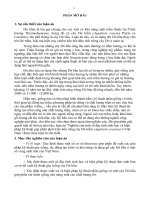
1.1.
Biological characteristics of groupers:
Groupers belong to the Serranidae family,
Epinephelus genus. According to the Institute
of Oceanography Nha Trang, Vietnam has
about 30 species of groupers (Le Anh Tuan,
2004) [11]. Groupers live in warm waters, the
temperature suitable for grouper development
is from 22-32ºC, the most suitable is 25-30ºC. Figure 1.1 Morphology of brownspots grouper (Epinephelus
Fish tolerate salinity is from 11 to 41 ‰.
coioides)
Groupers have mechanical barriers such as oily fluid, skin and gills which
protecting fish body against the intrusion of pathogens (Kim Van Van and Le Thanh Hoa,
2009) [12]. Groupers have a specific immune system, so when antigens enter the body, they
are capable of producing specific antibodies against antigens, protecting them from harmful
effects of pathogens.
1.2. Economic significance and current status of grouper culture
Groupers are of high economic value. For example, black and brown- spots
groupers with body weight between 800 and 1000g have been sold 200,000-300,000 VND/
kg, respectively. Red-spots groupers have prices ranging from 400,000 to 500,000 VND /
kg [73] [74]. Grouper aquaculture in Vietnam is mainly in coastal areas of Quang Ninh, Hai
Phong, Nghe An, Nha Trang, Ninh Thuan, Binh Thuan, Vung Tau and Kien Giang (Le Anh
Tuan, 2004 [11], Vo Van Quang et al, 2013 [5]). In recent years, the country has 500
hectares of coastal areas built into ponds for grouper farming. Annual production of
groupers reaches over 3000 tons of products. However, groupers are susceptible to several
2
diseases such as red spot, muscle necrosis, intestinal diseases which caused by bacteria and
NNV. Up to date, there is no vaccine to prevent these diseases and the main preventive
measure is to ensure hygiene within culture systems and isolate sources of viral infection
from fish.
1.3.
Current status of VNN in groupers in the World and Vietnam
1.3.1. Characteristics of VNN in marine fish
1.3.2. Current status of VNN in groupers in the World
1.3.3. Current status of VNN in groupers in Vietnam
1.3.4. Diagnostic methods for VNN in fish
Now a day, diagnostic methods used for detecting VNN are histopathology, viral
isolation in cells, molecular biology, electron microscopy and immunology (OIE, 2005)
[15].
1.4. Overview of nervous necrosis virus
The causative agents of VNN in groupers are NNV (belong to Betanodavirus),
which have RNA nucleus, spherical shapes and a diameter of 25-30 nm. Their genomes
have fragment structure, single-stranded RNA with two subdivisions. The large subspecies
contains RNA1 (3.1 kb) which encodes a 100 kDa protein that functions as an RNA
polymerase. The small subspecies contains RNA2 which possesses two high conservation
areas, T2 (870 bp) and T4 (420 bp) (Nishizawa et al., 1997).
1.5. Some preventation methods of VNN in groupers
Now a day, there is no vaccine to efficiently prevent VNN in groupers. Therefore,
main preventation methods are to ensure hygiene and to avoid infection sources from fish.
1.5.1. Recombinant antigens and vaccine use for fish disease prevention
1.5.2. Recombinant antigens of pathogens
A recombinant antigen is an antigen (immune protein) produced by genetic technology. In
the world, the use of recombinant antigens to produce vaccines that control some serious
fish diseases has been studied since the 1980s. The application of biotechnology (e.g, the
recombinant method) to produce large quantities of antigens has helped to save production
costs and increase safety for cultured fish. Recombinant DNA technology, which used in
antigen production to against viral diseases in fish, has been developing dramatically.
1.5.3. The current status of vaccine use for fish disease prevention in the world
There are few inactivated vaccines used to prevent viral diseases in fish such as
pancreatic necrosis (IPNV), haematopoietic necrolysis (IHNV), hemorrhagic septicemia
(VHSV) Spring carp blood virus (SVCV) in the world. However, the cost of cell culture in
viral diagnosis is relative high and the cell purification is difficult. Recently, the use of
recombinant DNA technology to produce vaccines from viral proteins using for fish disease
preventation is very usefull, highly economical and effective (Christie et al., 1997; Lorenzen
et al., 2005) [26] [43].
1.5.4. The current status of vaccine use for fish disease prevention in Viet Nam
Since 1996 - 1998, Bui Quang Te et al. produced vaccines from Aeromonas
hydrophila to prevent diseases for grass carps, with an efficiency of 90 to 100% (Bui
Quang Te et al, 2006) [7]. In 2003, Vu Dung Tien developed vaccines from the combination
of intracellular and extracellular antigens of Aeromonas hydrophila bacteria to prevent
3
diseases for grass carps, with the efficiency of 100% (Vu Dung Tien et al., 2003) [9]. Since
2003-2005, the KC-06-20NN project developed a vaccine against viscera necrotic
hemorrhage for pangasius and basa catfish. Vaccines are safe in experimental fish, with an
efficiency of 90% to 100% (Bui Quang Te et al., 2006).
Since 2006- 2007, the Research Institute for Aquaculture No.2 carried out a project
about research on the production of vaccines to prevent Edwardsiella ictaluri for pangasius
(Pangasianodon hypothalmus) comercially cultured in Cuu Long River Delta. The results
showed that the ability of immune response of pangasius to E. ictaluri bacteria through the
blood antibody is relatively high.
Since 2011, Pham Thi Tam et al. conducted research on the production of vaccines
against VNN in comercial farming goupers. The project outcome is the product of
inactivated vaccine by formalin 0.03% at 4°C for 5 days; the adjuvants is the ISA70
Montanique oil which is capable of stimulating the production of immune response in
laboratory conditions. Vaccines have been shown to protect groupers from diseases with an
efficiency over 83% in fingerlings, a 100% safe and an absolute asepsis (Pham Thi Tam et
al., 2015) [6].
CHAPTER 2. RESEARCH METHODOLOGY
2.1. Object and materials research
2.1.1. Object research:
- Nervous necrosis virus (NNV)
- T4 protein recombinant antigen
2.1.2. Materials research
- GS1 cell (Sigma, Germany)
- Vector pGEM-T and vector pET32a+ (Novagen, USA).
- E. coliJM109 and E. coliBL21(DE3) (HV Biotek).
- Enzyme EcoRI (Invitrogen), GS1
- RT-PCR one step kit (Quiagen), DNA Quick Gel Extraction kit (Invitrogen), DNA
and protein ladders (Invitrogen).
- Nikel chelating Resin column (Invitrogen)
- Orange-spotted groupers (Epinephelus coioides), tiger groupers (Epinephelus
fuscoguttatus) và humpback groupers (Cromileptes altivelis) at different sizes;
- Orange-spotted groupers (Epinephelus coioides), fingerling, juvenile stages (body
length 1,5 – 2 cm)
- Medium, equipment for cell culture: Leibovit, MEM, HANK's, FCS, antibiotics
- Pure chemicals for histopathological test; The chemicals used in the molecular
biology are of Sigma, Bio-Basic, Invitrogen, Biological: IPTG, ampicillin, X-gal, agarose,
EDTA, SDS, yeast, peptone, Ethidium Bromide, Ethanol, Isopropanol, Sodium Acetate,
Acetic Acid, Glycine, Methanol, TBST, BSA, TBS, Tris HCl, EDTA, TAE, NaOH, T4
ligase ....
2.2. Research contents:
- Identification of grouper NNV in Viet Nam;
4
- Investigation of some biological characteristics of grouper NNV;
- Creation of the recombinant antigens using as materials to produce vaccines agianst
VNN for groupers.
2.3. Research methodology
2.3.1. Identification of grouper NNV
- Sample collection and processing:
51 grouper samples that doubted of VNN in Quang Ninh (QN1-QN8), Hai Phong
(HP1-HP11), Nam Dinh (ND1-ND11), Khanh Hoa (KH1-KH9), Binh Thuan -B12) were
collected and refrigerated. Disease infected fish samples were then used for collecting brain
and eyes.
- Histopathological method: according to OIE, FAO (2005).
- Viral identification by cell culture according to Q.W.Qin et al (2006)
- Total RNA extraction of the VNN infective samples by using Qiagen Kit RNeasy
Qiagen.
- RT-PCR (Reverse Transcriptase PCR) technology.
- Electrophoresis on agarose gel: according to Sambrook et al., 2001.
- Gene isolation: according to Sambrook et al., 2001.
- Methods of creating T4 recombinant plasmid.
+ pGEM-T plasmid was cut by limited enzyme EcoRI, warm up at 37°C for 2.5 to 3
hours. Electrolytes on 1% agarose gel to test the results.
+ Linked T4 gene to pGEM-T:
- Methods to insert the recombinant vector into cells with host cell is E.coliJM109 by
temperature shock.
- Methods for extracting plasmid from E.coli bacteria:
- Methods for testing recombinant plasmid:
- DNA purification using agarose gel: according to the PureLink® Quick Gel
Extraction Kit included in the TOPO® TA Cloning Kit from Invitrogen.
- Sequencing of T4 gene by ABI 3100 automated machines (Applied Biosystems).
Blast software was used to determine the similarity of T4 gene sequences with GenBank.
2.3.2. Determination of some biological characteristic of grouper NNV
- Determine the virulence of virus NNV on cells: the virus was diluted from 10-1 to 109
with Leibovitz's 15 (10% FCS). The implanted tray was kept at 28oC to absorb the virus on
the cell. The virulence of NNV was assessed by TCID50.
- Determine the virulence of NNV in groupers: the virus was diluted from 10-1 to 10-9,
each one is injected in 30 groupers (1,5-2 cm) at a dose of 0.1 ml/fish. Clinical
manifestations, mortality and gene encoding T4 antigens were observed after 5 days. The
pathogenicity ability of the virus was assessed by LD50.
- Experiment on the effect of temperature on the ability of viral infection on GS1 cells:
QN4 strain with high TCID50 (10-6,8) was used to infect the cells. Each NNV strain was
cultured at 4 temperature levels: 17, 22, 27 and 32oC. The infection dose was TCID50 = 106,8
. Testing and evaluating of CPE were performed after 5 days of experiment.
- Experiment on the effect of temperature on the ability of viral infection in groupers:
QN4 strain with high LD50(10-7,5) was infected in groupers. Fish was injected with
5
Leibovit"z 15 used as controls. Fish were raised at 28oC (all day and night) and 28oC
(daytime)/ 24oC (at night). Mortality of experimental fish and T4 gene were assessed and
identified after 15 days.
2.3.3. Expression of antigen-coding genes and evaluation of the antibody
producing ability of recombinant antigens
- Preparation of pET32a+ vector to perform a T4 gene linked reaction (pET32a+-T4).
Using the temperature shock to put pET32a+-T4 vector into E. coliBL21 cell.
- Extraction of plasmids from colonies after transformation
- Test PCR and restricted enzyme to determine T4 gene in plasmids.
- E. coliBL21 culture that containing recombinant pET32a+-T4 plasmid
- SDS-PAGE electrophoresis.
- Western blot: to identify T4 recombinant protein that is specific for NNV.
- Assessing ability to produce antibody of recombinant antigens
+ Assessing ability to produce neutralizing antibody of recombinant antigen in rabbit:
Positive control: rabbit serum without specific antibody was incubated with QN2
4
(10 TCID50) and adsorbed onto GS1 cells or infected with small groupers (≤ 5 g) by
injection (dose of 0,05 ml of poisonous NNV QN2 with titres 103 LD50)
Negative controls: rabbit serum was immunized with E.coliBL21-pET32a+-T4 and
recombinant T4 protein, diluted with a factor of 10 then adsorbed onto GS1 cells or infected
into juvenile groupers (≤ 5 g) by injection.
Experiment plot: rabbit serum was immunized with E.coliBL21-pET32a+-T4 and
recombinant T4 protein, diluted with a factor of 10, incubated with QN2 at 104 TCID50 and
adsorbed onto GS1 cells or infected into juvenile groupers (≤ 5 g) by injection (0,5 ml dose
of QN2 virus titrated 103 LD50).
+ Evaluate ability producing the neutralizing antibody of recombinant antigens on grouper.
Positive control: specific pathogen free fingerling groupers were milled, treated
with antibiotics, filtrated and diluted with a factor of 10, incubated with QN2 with 10 4
TCID50 and infected into GS1 cells.
Negative controls: specific pathogen-free fingerling groupers were immunized with
E. coliBL21-pET32a+-T4 and recombinant T4 protein antigen with 2 doses (0.2 mg/ dose)
for every 15 days within a period 90days. Samples were periodically collected every 15
days, then milled, antibiotic treatmented, filtered, dilutted with a factor of 10 and infected
into GS1 cells.
Experiment plot: specific pathogen free groupers were immunized with E. coliBL21pET32a+-T4 and recombinant T4 protein with 2 doses (0.2 mg / dose) for every15 days
within a period 90 days. Samples were periodically collected, milled, antibiotic treatmented,
filtrated, dilutted and incubated with QN2 strain (104 TCID50) and infected into GS1 cells,
evaluated CPE for 7 days.
CHAPTER 3. RESULTS AND DISCUSSION
3.1. Identification of NNV in groupers in Viet Nam
3.1.1. Screening NNV infected fish samples by histopathology
6
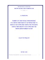
The screening results of 51
grouper samples infected NNV
by histopathology (Figure 3.1
and Table 3.1) show that there
were 26 specimens had vacuole
in the eyes and brain tissues.
The vacuoles in the tested
samples are either round or
elliptical, with sizes range from
5-10 μm.
Figure 3.1 Cytopathogenicity in brain and eyes tissues
(a) infected groupers, (b) vacuole in the brain, (c)
vacuole in the eyes.
Table 3.1. Detection of NNV by histopathology method
Cytopathogenicity in the brain tissue (26 Cytopathogenicity in the eyes tissue
samples)
(17 samples)
Sample ID
Ratio (%)
Sample ID
Ratio (%)
QN2, QN4, QN7
37,50
HP2, HP4, HP5, HP8, HP10
45,45
ND1, ND3, ND4, ND5,
72,72
ND7, ND8, ND10, ND11
KH4, KH5, KH6, KH9
44,44
BT1, BT2, BT4, BT7, BT9,
50,00
BT12
NNV was detected in the neuvous syndrome of
QN2, QN4, QN7
HP4, HP5, HP8
ND1, ND4, ND5,
ND10, ND11
KH4, KH5
BT2, BT4, BT9,
BT12
37,50
27,27
45,45
22,22
33,33
the 26 samples collected. The remaining
samples had no vacuole, however, we could not rule out the possibility that fish were in the
pre-infection. Therefore, the molecular biology method was employed to identify antigenencoding genes of NNV.
3.1.2. Identification of fish samples infected with NNV by RT-PCR method
Bảng 3.2. Identification of NNV by RT-PCR
Samples were positive for T4 gene of NNV
Ratio (%)
(27 samples)
QN2, QN4
HP2, HP4, HP6, HP7, HP8, HP10, HP11
ND1, ND3, ND4, ND5, ND7, ND8, ND10, ND11
KH4, KH5, KH9
BT2, BT3, BT4, BT7, BT9, BT10, BT12
Average
7
25,00
63,63
72,72
33,33
58,33
52,94
The results show that 27/51 were positive with T4 gene, accounting for 52.94%. The PCR
products of HPV, HP2, ND3, ND2, QN2, QN4, KH4, KH5, BT2 and BT3 are shown in the
figure 3.2.
Samples of HP2, ND3,
QN2, QN4, KH4, KH5,
BT2, BT3 appeared to
have a band with a size of
approximately 420 bp,
which is similar to the size
of T4 gene. There was no
band on the samples of
HP1, ND2.
Hình 3.2. RT-PCR products of some samples infected with NNV on the
electrophoresic gel (well 1-10: HP1, HP2, ND3, ND2, QN2, QN4, KH4, KH5, BT2,
BT3, Well 11: marker)
3.1.2. Identification of NNV in cells
In this study, GS1 cell lines were used to assess the capacity of NNV infection. In
the total of 27 samples positive for T4 gene, we identified 26 virus strains (Figure 3.3, Table
3.3). HP11 did not show cytopathogenicity in the brain or eye tissues.
Figure 3.3. Cytopathogenicity of GS1 cells after NNV infection
A: The cells after 2 days NNV infection, granulocyte and rounded cells,
B: The cells after 7 days NNV infection,
C: Vacuolation in GS1 cells after 2 days NNV infection.
From day 2, the cells appeared to have a granular, rounded, multi-particle
acuolation shape; after 5 days, the cells were died throughout the culture plates and all cells
were destroyed at day 7 (Figure 3.3).
8
Table 3.3. Identification of NNV infected in GS1 cells
No
Sample
ID
CPE time (hours)
24
48
72
96
120
144
168
1
QN2
_
_
+
+
+
++
+++
2
QN4
_
+
+
+
++
+++
++++
3
HP2
_
_
+
+
+
++
+++
4
HP4
_
+
+
+
++
+++
++++
5
HP6
_
_
+
+
+
++
+++
6
HP7
_
+
+
+
++
+++
++++
7
HP8
_
+
+
+
++
+++
++++
8
HP10
_
_
+
+
+
++
+++
9
HP11
_
_
_
_
_
_
_
10
ND1
_
+
+
+
++
+++
++++
11
ND3
_
_
+
+
+
++
+++
12
ND4
_
+
+
+
++
+++
++++
13
ND5
_
+
+
+
++
+++
++++
14
ND7
_
_
+
+
+
++
+++
15
ND8
_
+
+
+
++
+++
++++
16
ND10
_
_
+
+
+
++
+++
17
ND11
_
_
+
+
+
++
+++
18
KH4
_
_
+
+
+
++
+++
19
KH 5
_
+
+
+
++
+++
++++
20
KH9
_
_
+
+
+
++
++
21
BT2
_
_
+
+
+
++
++++
22
BT3
_
+
+
+
++
+++
++++
23
BT4
+
+
+
++
+++
++++
24
BT7
_
+
+
+
++
+++
++++
25
BT9
+
+
+
++
++
+++
26
BT10
+
+
+
++
++
++
27
BT12
+
+
+
++
++++
Note: ++++: CPE > 98%; +++: CPE > 85%; ++ : CPE > 50%; +: CPE >
20%; +: suspected CPE > 10%; -: no CPE
Identified viral strains had the T4 gene and could cause cytopathogenicity in cells.
Therefore, they were thought NNV. In order to prove this hypothesis, we sequenced the T4
gene and determined them at the species level.
3.1.4. Sequencing T4 antigen- encoding genes of NNV
T4 gene of representative strains of QN2, HP7, ND1, KH5, BT12 were purified to
isolate and sequence. The results below show the steps of T4 gene sequencing of strain
KH5.
9
Figure 3.5. Design of the
pGEM-T-T4 recombinant
vector
A: Electrophoresis of the
purified T4 gene product
(well 1: purified T4 gene
product, M: 1kb Plus
Ladder well); B: Diagram
of T4 gene isolation vector
design
The purified product was approximately 420bp, which is similar to the size of T4
gene (Figure 3.5A). Splitting by pGEM-T Easy vector, recombinant vector were designed as
Figure 3.5B. Vector recombinant pGEM-T-T4 was transformed into E.coliJM109. White
colonies were selected and cultured in the liquid LB enviroment (ampicillin 100μg / ml) to
separate recombinant plasmids and cut, purify the T4 gene. Plasmid DNA was extracted
from the white colonies and tested on a 1% agarose gel and showed in th Figure 3.6 (A), the
T4 gene amplification product was examined and showed in the Figure 3.6 (B) the produce
was cut by the limited enzyme showed in the figure 3.6 (C).
Figure 3.6. Electrophoresis of recombinant T4 products
(A): DNA plasmid is isolated from 3 white colonies (well 1,2,3);
(B): PCR testing of T4 gene from 3 recombinant plasmids (well 1,2,3)
(C): DNA testing of T4 gene inserted into DNA of 3 recombinant plasmids cut
with EcoRI (well 1,2,3)
Well M: Plus Ladder 1kb scale;
The results in the figure 3.6 (C) show that wells 1-3 appeared a band of 420bp in
size, which is equivalent to T4 gene size, thus there is a possiblity that we were successful
to isolate the T4 gene. After T4 gene sequencing, the Blast software was used to determine
the similarity of T4 gene sequence. Results show that T4 gene sequence (deposited in
GenBank with accession number: HM017077.1) had a similarity of 100% compared to the
antigen-encoding gene of NNV.
10
Figure 3.7. Comparison of the similarity of sequenced genes with T4 gene of
Betanodavirus (HM017077.1)
The results of T4 gene sequencing led us to conclude that the virus strain identified as
Nervous Necrosis Virus belongs to Betanodavirus family.
3.2. Investigation of some biological characteristics of NNV
3.2.1. Investigation of pathogenic characteristics of NNV in cells
Bảng 3.5. Pathogenic characteristics of NNV in GS1 cells
Tissue culture infectious dose of 50% tế bào (TCID50)
No
Sample ID
1st
2nd
3rd
4th
Avarage
10-6,9
10-6,9
10-7
10-7
10-6,9
10-6,8
10-6,8
10-6,8
10-6,9
10-6,8
10-4,3
10-5
10-4,8
10-4,4
10-4,9
10-4,8
10-4,3
10-4,8
10-4,7
10-4,5
10-4,9
10-4,9
10-4,3
10-4,9
10-4,8
HP10
10-6,8
10-6
10-3,9
10-6,9
10-5,8
10-4
10-6,8
10-5,7
10-3,9
10-7
10-6,1
10-3,8
10-6,8
10-5,9
10-3,9
ND1
ND3
ND4
10-6,8
10-3
10-4,9
10-6,8
10-3,1
10-4,9
10-7
10-3
10-4,8
10-6,9
10-3
10-4,9
10-6,8
10-3
10-4,9
1
2
QN2
3
4
5
HP2
HP4
6
7
8
HP7
HP8
9
10
11
QN4
HP6
11
No
Tissue culture infectious dose of 50% tế bào (TCID50)
Sample ID
1st
2nd
3rd
4th
Avarage
12
ND5
10-5,9
10-5,8
10-6
10-5,9
10-5,9
13
ND7
ND8
10-4,3
10-4,3
10-4,2
10-4,3
10-4,3
10-4,8
10-3
10-4,9
10-3
10-4,9
10-3
10-4,9
10-2,9
10-4,9
10-3
10-3
10-2,7
10-3,1
10-2,8
10-3
10-2,7
10-3
10-2,6
10-3
10-2,7
10-6,8
10-3
10-6,8
10-3
10-6,8
10-3,1
10-6,9
10-3
10-6,8
10-3
10-4,8
10-4,9
10-4,9
10-5
10-4,9
10-4,8
10-5,9
10-4,8
10-5,9
10-4,7
10-5,9
10-4,8
10-5,8
10-4,8
10-5,9
10-6,8
10-6,9
10-6,8
10-6,7
10-6,8
10-3,9
10-2,5
10-4,8
10-4
10-2,7
10-4,9
10-3,9
10-2,9
10-4,9
10-3,9
10-2,8
10-4,9
10-3,9
10-2,7
10-4,9
14
15
16
17
18
19
20
21
22
23
24
25
26
ND10
ND11
KH4
KH5
KH9
BT2
BT3
BT4
BT7
BT9
BT10
BT12
The experiments show that 6/26 strains are highly virulent with TCID50 dose of 10-6,8 to 106,9
; 10/26 strains are mildly virulent with a TCID50 dose of 10-4,8-10-5,9 at a dilution of 10-8
and the remaining 10 strains are less virulent with TCID50 doses of 10-2,7-10-4,3.
3.2.2. Investigation of pathogenic characteristics of NNV in groupers
The study choosed 6 high virulent strains (QN2, QN4, HP7, ND1, KH5 and BT7)
and 2 low virulent strains (KH4, BT10) to infect in groupers. Fish were checked for 5 days
to determine mortality rates and T4 gene presence. The results are presented in the table 3.6
Table 3.6. Pathogenic characteristics of NNV in groupers
50% last dead dose of fish (LD50)
No
Sample ID
1st
2nd
3rd
Avarage
-6,2
-6,2
-6,1
1
10
10
10
10-6,2
QN2
2
10-7,5
10-7,6
10-7,5
10-7,5
QN4
3
10-7,5
10-7,5
10-7,4
10-7,5
HP7
4
10-5,4
10-5,6
10-5,5
10-5,5
ND1
5
10-6,3
10-6,2
10-6,4
10-6,2
KH5
6
10-6,5
10-6,5
10-6,4
10-6,5
BT7
7
10-3,7
10-3,6
10-3,6
10-3,6
KH4
8
10-2,5
10-2,7
10-2,5
10-2,6
BT10
9
ND
ND
ND
ND
Control
(Note: ND Not done)
Results show that: 2 strains of QN4 and HP7 have a high virulence with LD50 of
-7,5
10 , 2 low virulent strains with LD50 from 10-2,6-10-3,6 are KH4 and BT10.
3.2.3. Effects of temperature on pathogenicity of NNV in cells
12
GS1 cells were infected with the QN4 strain of NNV with a titre of 10-6,8 TCID50 /
ml and cultured for 5 days to determine survival of the cells.
Figure 3.8. Servival of cells after NNV infection
The results presented in the figure 3.8 show that 22oC is the most suitable for virus
reproduction and GS1 cell infection.
3.2.4. Influence of temperature on pathogenicity of NNV in groupers
The pathogenicity of NNV at 28oC (day and night) is shown in the table 3.8: The
group of groupers infected with QN4 had a mortality rate of 82.1% after 3 days, 100% after
6 days . The mortality rate of control group was 10% after 15 days. It can be seen from the
table that the cause of fish death is QN4 of NNV with the participation of T4 gene.
Table 3.8. The pathogenicity of the NNV in groupers at 28°C of day and night
.Time
Mortality rate of groupers (%)
T4 gene determination
(day)
1st
2nd
3rd
Average
Control Treatments Control
0
0
0
0
0
0
-
-
3
86,6
83,3
76,6
82,1
3,3
+
-
6
100
100
100
100
6,6
+
-
9
6,6
12
10,0
15
10,0
-
Note: +: positive with T4 gene
-: Negative with T4 gene
The pathogenicity of NNV in groupers at temperature water of 28°C (daytime) and 24°C
(night time) was shown in the table 3.9.
13
Table 3.9. The pathogenicity of NNV in groupers at temperature water of 28°C
(daytime) and 24°C (night time)
Time
Mortality rate of groupers (%)
T4 gene determination
(day)
1st
2nd
3rd
Average Control
Treatment
Control
0
3
6
9
12
15
0
80,0
100
0
86,6
100
0
90,0
100
0
85,5
100
0
0
+
+
3,3
6,6
10,0
10,0
Note: +: positive with T4 gene
-: Negative with T4 gene
o
o
At 28 C (day) and 24 C (night), mortality rate of fish was 85.5% after 3 days and
100% after 6 days. In the control group, the mortality rate was 10% after 15 days. The
causive pathogen containing the T4 gene was the QN4 of NNV infection
3.3. Research on creating recombinant antigens using as materials to produce vaccine
against VNN for groupers
3.3.3. pET32a+- T4 recombinant vector design.
The study used the pET32a+ vector to
express T4 gene, E. coliJM109 strain
to test recombinant vector and E.
coliBL21(DE3) strain to express T4
gene.
The process of creating recombinant
vector containing T4 gene (pET32a+T4) is shown in the figure 3.9.
pGEM-T-T4 and pET32a+ plasmid
cutting was performed at 37oC for 4
+
hours,
the
product
was Figure 3.9. Diagram of pET32a -T4 recombinant vector
creation
electrophoresed on 1% agarose gel.
The results of cutting pGEM-T-T4 vector and pET32a+ vector are shown in the figure 3.10
Figure 3.10. pGEM-T-T4 vector (A) and pET32a+ vector (B) were cut by EcoRI enzyme.
Well 1,2,3,4 (A): cutting products of pGEM-T-T4 plasmid ;
Well 1,2,3 (B): cutting products of pET32a+ plasmid ;
Well M: Marker 1kb plus
14
The results in the figure 3.10 (A) show that there were two bands in wells 1, 2, 3,
and 4, of which one band has 420bp in size (T4 gene size), and another band has a size of
approximately 3015bp (similar to pGEM -T Easy vector size). Figure 3.10 (B) shows that in
wells 1, 2, 3, only a single band with the size is equivalent to the vector pET32a + which is
5900bp. Thus, it is demonstrated that the recombination plasmid pGEM-T-T4 and pET32a+
plasmid were successfully cut to produce compatible sites for inserting T4 gene into the
pET32a+ vector. The bands of 420bp (containing T4 gene) and 5900bp (containing
pET32a+ vector) were cut and purified. The cutting products of pGEM-T-T4 plasmid and
pET32a+ vector were electrophoresed on agarose gel 1%, the results are shown in the Figure
3.11.
Figure 3.11. Purification of cutting products of pGEM-T-T4 plasmid and pET32a+
vector
Well 1: T4 gene after purification, Well 2: pET32a+ plasmid after purification
M: 1kb Plus Ladder
Gene linked reaction was performed at 4oC and incubated for 14 to 16h. To test the
formation of the pET32a+-T4 recombinant vector, the gene-linking product was transformed
into E. coliJM109. Bacteria carry the recombinant vector on LB environment supplemented
with Ampicillin (100μg / ml) were screened and cultured at 37oC for 16h, results shown the
Figure 3.12. Six colonies were chosen and cultured in 3 ml of liquid LB environment
supplemented with ampicillin (100μg/ ml) and shaked with a speed of 150 times/minute at
37oC for 16h. The plasmid was then isolated, checked by electrophoresis on 1% agarose gel
and the result is shown in the figure 3.13:
15
Figure 3.12. a colony plate after transfering
Figure 3.13. Cheking the formation of
+
pET32a -T4 gene into E. coliJM109
pET32a+-T4 vector in E. coliJM109 (Well 1-6:
plasmid lines 1 to 6; Well M: 1kb plus ladder)
To test six plasmid samples which
carry the pET32a+- T4 recombinant
vector and pET32a+ vector
(control), a PCR reaction was run
using the pair of primer P1 and R3.
The results in the figure 3.14 show
that six selected bacterial strains
carried T4 gene, while the control
samples did not carry the gene.
Thus, it is possible to confirm that
the recombinant pET32a+- T4
vector has been successfully
designed.
Figure 3.14. Electrophoresis of PCR products to test
the presence of T4 gene in recombinant vector
Well 1-6: PCR products of pET32a+-T4 plasmids,
Well M: 1kb Plus ladder; Well DC: PCR product of
plasmid pET32a+
3.3.2. Expression of T4 antigen-encoding gene of NNV
3.3.2.1. Creation of the E. coliBL21(DE3) strain carrying antigen-encoding gene of NNV.
After successfully creating the
pET32a+-T4 recombinant vector,
four of the six bacterial strains
were selected to transform into the
E. coli BL21 (DE3) strain.
Transgenic bacteria were cultured
on
agar
LB
medium
(supplemented Ampicillin with
100μg / ml) at 37°C for 12 to 16 h,
Figure 3.15. A colony plate after transforming
results shown in the figure 3.15.
pET32a+-T4 into E. coliBL21
16
To test the transformation ability of pET32a+-T4 plasmid into the E. coliBL21(DE3), a
sceening was performed by isolating plasmid then running PCR. The isolated plasmid
product was checked by electrophoresis on 1% agarose gel, the results are shown in the
figure 3.16
Figure 3.16. Electrophoresis of the
Figure 3.17. Checking the transformation ability
recombinant plasmid isolated from E.
of the pET32a+-T4 recombinant vector into E.
coliBL21(DE3) strain carry pET32a+-T4
coliBL21(DE3) strain.
vector
Well 1-4: Plasmid PCR products
Well 1-4: Plasmids;
Well M: GeneRulerTM 1kb DNA Ladder
Well M: GeneRulerTM 1kb DNA Ladder
A PCR reaction was perfomed to test plasmids’ capacity carying pET32a+-T4
vector. The results of electrophoresis on 1% agarose gel in the figure 3.17 show that there
was a band with a similar size of T4 gene, 420 bp on the wells 1 to 4. It is therefore possible
to conclude the E.coliBL21(DE3) strain carrying T4 gene of NNV has been successfully
created.
3.3.2.2. Expression of T4 gene in recombinant bacteria cells
Four selected colonies were innoculated until the OD600nm reaching 0.5 to 0.6, then IPTG
was added. The recombinant bacteria were sampled before and after adding IPTG 2h, 4h, 6h
for expression analysis of the antigen-encoding gene by SDS-PAGE. The results are shown
in the figure 3.18 and figure 3.19.
17
Figure 3.18. SDS-PAGE gel polyacrylamide Figure
3.19.
SDS-PAGE
gel
gel 15% of line 1 and 2
polyacrylamide gel 15% of line 3 and 4
Well 1,5: proteins of 1 and 2 lines before Well 1.5: protein of 3 and 4 lines before
adding IPTG; Well 2,3,4: protein of line 1 adding IPTG; Well 2,3,4: protein of line 3
after adding IPTG for 2h, 4h, 6h; Well 6,7,8: after adding IPTG for 2h, 4h, 6h; Well
protein of line 2 after adding IPTG for 2h, 4h, 6,7,8: protein of line 4 after adding IPTG
6h. Well M: Broad-way Multi prestained for 2h, 4h, 6h. Well M: Broad-way Multi
protein marker.
prestained protein marker
The results in the figure 3.18 show that there was no band of strange proteins on
wells 1 and 5, but a band with a size of 17 kDa appeared on Well 2,3,4 and a bold band of
approximately 25 kDa, which is similar with the theoritical size of T4 protein on well 6,7,8 .
Thus, it is possible to concluded that T4 gene has been successfully expressed in the line 2.
The results in the figure 3.19 show that there was no band of strange protein in wells 1 and
5, however, a bold band with a size of approximatley 25 kDa, which is similar to the
theoritical size of T4 protein antigen, appears in well 2,3,4,6,7,8.
The above findings revealved that T4 gene has been successfully expressed in E.
coliBL21(DE3) bacteria of BL21-pET32a+-T4 lines 2, 3, 4; the recombinant protein had a
size of approximately 25 kDa, which is similar to the theoritical size of the T4 protein
antigen.
Protein expression of the
T4
gene
was
then
confirmed by Western blot
hybridization method with
specific antibody againsts
NNV. The results show
that the reaction site
between T4 recombinant
protein
and
standard
specific antibody was at
the 25 kDa electrophoresis
line (Figure 2.20). It is
therefore can be concluded
that T4 protein antigen of Figure 2.20. Western blot result of recombinant T4 protein
NNV was successfully
M: standard; Well 1: T4 recombinant protein; Well 2:
expressed
in
E.
standard T4 protein of USA
coliBL21(DE3) cells.
18
3.3.2.3. Assessment of expression conditions of T4 antigen-encoding gene of NNV
- Determination of sample collection time
The E. coli cells
carrying recombinant
plasmids were cultured
and collected after
induction of 3, 4 and 12
h. The results (Fig.
3.21) show that the
samples collected after
3h and 12h give lower a
Figure 3.21. T4 recombinant protein was synthesized by the protein content, while
time (Well M: standard protein; Well 1, 2, 3: the the samples collected
recombinant protein was synthesized after 3, 4, 12h)
after 4h give the highest
protein content.
- Determination of culture temperature
E. coliBL21-pET32a+-T4
strain was cultured at
28oC, 30oC and 37o and
sampled
after
4h
induction. The results in
the figure 3.22 show that
there was no protein band
of 25 kDa size observed
at culture temperature
conditions of 28°C and
30°C, however, at 37°C,
the recombinant protein
T4 was seen to be Figure. 3.22. Effects of temperature on T4 recombinant protein
expression (wells 1, 2, 3: protein contents when cultured at
expressed
28oC, 30oC and 37oC, Well M: standard protein)
19
- Determination of the concentration of IPTG induction
IPTG was added to the
culture
medium
at
concentrations from 1 to
6 mM. The results in the
figure 3.23 show that,
after IPTG addition, the
recombinant
strain
synthesized
a
large
amount
of
protein,
approximately 25 kDa
(the theoritical size of the
recombinant T4 protein).
In experiments using
different
IPTG Figure 3.23. Effect of IPTG concentrations on recombinant
concentrations, the T4 T4 protein expression
Well M: standard, wells 1-6: IPTG concentrations from 1
gene was best expressed
mM to 6 mM
when the strain was
inducted at 1 mM IPTG.
3.3.3. Purification of recombinant antigen
The results in the figure
3.24 show that all
purified strains appeared
only one protein band of
25 kDa size, indicating
that the T4 protein was
successfully purified.
The study used this T4
protein antigen to test the
ability of neutralizing
antibodies to produce
vaccines
for
diease Figure 3.24. Electrophoresis of purificated T4 protein on SDSPAGE gel (Well M: standard protein, wells 1-5: T4 antigen in
prevention in grouper
phase 1, 2, 3, 4, 5)
3.3.4. Evaluation of ability to create antibodies of the recombinant antigen
3.3.4.1. Evaluation of ability to create antibodies of the recombinant antigen in
rabbits
The ability to create a neutralizing antibody of E. coliBL21-pET32a+-T4 antigen and the
recombinant T4 protein in rabbits are shown in the table 3.10 and table 3.11.
20
Table 3.10. The titer of neutralizing antibody of NNV in vitro
Time to collect
serum in rabit
after
immunization
(day)
Positive
control
(CPE %)
(CPE %)
E.coli BL21- Recombinant
pET32a+-T4* T4 protein*
5
98
0
15
98
25
Negative control
Dilutions of rabbit serum were
immunizated to neutralize the dose
104 TCID50
E.coli BL21pET32a+-T4*
Recombinant T4
protein*
0
ND
ND
0
0
1: 5
ND
98
0
0
1: 200
1: 50
35
98
0
0
1: 800
1: 200
45
98
0
0
1: 800
1: 200
60
98
0
0
1: 400
1:100
Note: ND (Not done)
Table 3.10 shows that in the negative controls, samples did not cause
cytopathogenic effects, while in positive controls, normal rabbit serum were not able to
neutralize the virulence of QN2 strain, therefore, all cells in culture wells had
cytopathogenic effects.
On 15th day, only the treatment of rabbit serum immunizated E. coli BL21pET32a+-T4 produced antibody at a dilution 1:5. In both treatments, antibodies were
produced from 25th day after immunization. The rabbit serum injected with E.coliBL21pET32a+-T4 had a higher neutralization (at dilution of 1: 200) compared to the serum
injected with recombinant protein (at dilution: 1:50). The neutralized antibody titer reached
the highest in both treatments from 35th to 45th days, at a dilution of 1: 800 for the serum
injected with E. coliBL21-pET32a+-T4 and 1:200 for the serum injected with T4 protein .
On the last day of experiment (60th day) the neutralized antibody titers of two treatments
were decreased. The serum injected with E.coliBL21-pET32a+-T4 had a neutralized titer at
a dilution of 1: 400, while the other, which injected with recombinant T4 protein, had a
neutralized antibody titer at a dilution of 1: 100.
Results of the determination of neutralized antibody titers of NNV in vivo are
shown in the table 3.11.
21
Table 3.11. Neutralization antibody titers of NNV in vivo
Time to collect Positive control
serum in rabit
after
immunization
Infection Death
(day)
(%)
(%)
Negative control
(infection rates
%)
Serum rabbit sera were immunized
E.coli
recombinant T4
E.coli
Recombin
+
BL21- pET32a -T4
protein
BL21ant T4
pET32a+-T4 protein Neutralization T4 Neutralizati T4 gene
titer
gene
on titer
5
100
100
0
0
ND
+
ND
+
15
100
100
0
0
ND
+
ND
+
25
100
100
0
0
1: 100
-
1: 50
-
35
100
100
0
0
1: 100
-
1: 50
-
45
100
100
0
0
1: 100
-
1: 50
+
60
100
100
0
0
1: 50
+
1: 10
+
(Note: ND: not done; (+): have T4 gene, (-): have T4 gene)
Table 3.11 shows that in both two experiments of rabbit serum immunized with E.
coliBL21-pET32a+-T4 and recombinant T4 protein, the serum did not produce antibody in
the first 15 days. On 25th to 45th days, only the rabbit serum immunized with E. coliBL21pET32a+-T4 had an ability to produce a neutralizing antibody against NNV virulence at a
dilution of 1: 100. From 25th to 35th days, the T4 antigen-encoding gene was not detected.
On 45th day, although a neutralization titer of the serum was found at a dilution of 1:50, all
groupers were not killed and T4 gene was detected. Although rabbit serum still produced
antibodies to neutralize virulence, the virus was no longer able to cause diseases in two
experiments, the neutralization titers ranged from 1:10 to 1:50 and the T4 gene was still
detected in experimental fish.
Above findings indicate that recombinant the T4 protein is abble to produce
antibody that neutralize pathogenic viruses, although the antibody is lower than that of the
E. coliBL21-pET32a+-T4.
3.3.4.2. Evaluation of ability to create antibodies of recombinant antigens in
groupers
The results presented in the table 3.12 show that in negative control samples, fish
immunized with E. coliBL21-pET32a+-T4 antigen and recombinant T4 protein produced a
specific antibody of NNV and did not cause cytopathogenic effects.
22
Table 3.12. The titer of creating immune responses of groupers against NNV in vitro
Dilutions of extracted fish fluid immunized to neutralize
NNV of QN2 strain (104 TCID50)
Experiments
30th
45th
60th
75th
90th
15th day
day
day
day
day
day
Positive controls
+
+
+
+
+
+
The fish extracted fluid was
immunizated with E.
+
coliBL21-pET32a -T4
The fish extracted fluid was
immunizated with recombinant T4 protein
The fish extracted fluid was
immunizated with E.
1:64
1:256
1:128
1:128
1:64
1:8
coliBL21-pET32a+ -T4+ NNV
virulence
The fish extracted fluid was
immunizated with recombinant 1:16
1:128
1:128
1:64
1:64
1:8
T4 protein + NNV virulence
(Note: ND: not done; (+): have CPE, (-): no CPE)
In the positive control samples, NNV QN2 strain 104 TCID50 dose was infected into GS1
cells and all cells had cytopathogenic effects.
In both experiments, fish extracted fluid immunizated with E. coliBL21-pET32a+T4 and recombinant T4 protein produced antibodies from 15th day with titers of 1:64 and
1:16, respectively. In the experiment of grouper extracted fluid immunizated with the E.
coliBL21-pET32a+-T4, the antibody concentration was highest on 30th to 60th days with
titers of 1:128 -1:256. By 75th day, the antibody concentrations were decreased with a titer
of 1:64. In the experiment of grouper extracted fluid immunizated with T4 recombinant
protein, the antibody was highest on 30th to 45th day with a titer of 1:128. From 60th to 75th
day, the antibody was gradually decreaed and the titer was only 1:64. The protection lasted
up to 90 days in both treatments. This is the scientific basis to produce vaccines against
VNN in groupers.
Up to date, only few vaccines have been used to prevent dieases for high economic
value fish speices such as Salmon, Sea bass, Grouper, Seabream, Plaice and most of these
vacines are inactive.
In Japan, Yamashita et al. (2009) tested on inactivated vaccines to prevent VNN for
7-dotted groupers (average weight of 25.4 g/ fish). Antibodies were produced after 20 days
of vaccination, while those in the control groups did not produce antibodies [69].
Pakingking et al (2010) used inactivated vaccines of RGNNV strain to prevent VNN for
tiger groupers in commercial culture. The results showed that fish immunized with a
neutralizing antibody produced antibodies from 15th day (a titre of 1: 800) to 190 th days (a
titre of 1: 400) [51].
23